| Dai X, Ruan J, Guo Y et al. Enhanced Radiotherapy Efficacy and Induced Anti-tumor Immunity in HCC by Improving Hypoxia Microenvironment using Oxygen Microcapsules Chemical Engineering Journal 2021-04-01 (IF/IHC, ICC/IF) |
IF/IHC, ICC/IF |
|
| |
|
|
| Reichert E Hypoxia-Inducible Factor 1-alpha Upregulated during Sepsis, Involved in Inflammation Thesis 2019-01-01 (WB, Rat) |
WB |
Rat |
| Tran MGB, Bibby BAS, Yang L et al. Independence of HIF1a and androgen signaling pathways in prostate cancer bioRxiv (IHC, Human) |
IHC |
Human |
| Geiger F, Heigl T, Merolla L et al. HIF1 activity in photoreceptors drives type 3 neovascularization and retinal atrophy in a new mouse model of age-related macular degeneration Cell death & disease 2025-10-06 [PMID: 41053106] |
|
|
| Liu Y, Gu Y, Ng S et al. Circulating levels of hydroxylated bradykinin function as an indicator of tissue HIF-1alpha expression Science Bulletin 2020-01-01 (IHC-P, Human) |
IHC-P |
Human |
| Menendez-Montes I, Escobar B, Gomez MJ Activation of amino acid metabolic program in response to impaired glycolysis in cardiac HIF1 deficient mice bioRxiv 2020-01-01 (IF/IHC, Mouse) |
IF/IHC |
Mouse |
| Fu Y, Cen D, Zhang T et al. Implantable fibrous scaffold with hierarchical microstructure for the ‘on-site’ synergistic cancer therapy Chemical Engineering Journal 2020-12-01 (WB, Mouse) |
WB |
Mouse |
| Gamal-Eldeen A, Baghdadi H, Afifi N, et al. Gum arabic-encapsulated gold nanoparticles modulate hypoxamiRs expression in tongue squamous cell carcinoma Molecular & Cellular Toxicology 2021-01-26 |
|
|
| Tamamura Y, Terai K, Yamaguchi A Notch signaling modulates Fgf23 expression through crosstalk with hypoxia and PTH pathways in osteogenic cells. Molecular and cellular endocrinology 2025-09-22 [PMID: 40992566] |
|
|
| Wang Q, Roh E, Khan A et al. Deletion of p53-Related Protein Kinase Suppresses Solar UV-Induced Photocarcinogenesis by Inhibiting PD-L1 Expression and Enhancing CD8 T-Cell Infiltration. The Journal of investigative dermatology 2025-08-12 [PMID: 40812468] |
|
|
| Grie beta hammer, A;de la Cuesta-Zuluaga, J;Müller, P;Gekeler, C;Homolak, J;Chang, H;Schmitt, K;Planker, C;Schmidtchen, V;Gallage, S;Bohn, E;Nguyen, TH;Hetzer, J;Heikenwälder, M;Huang, KC;Zahir, T;Maier, L; Non-antibiotics disrupt colonization resistance against enteropathogens Nature 2025-07-16 [PMID: 40670795] |
|
|
| Bellissimo C, Ribeiro T, Yeo E et al. Maternal high-fat, high-sucrose diet-induced excess adiposity is linked to placental hypoxia and disruption of fetoplacental immune homeostasis in late gestation. Biology of Reproduction 2025-07-07 [PMID: 40621987] |
|
|
| Kurkela M, Dvořáková L, Koivisto H et al. Targeting HIF-P4H-2 in APP/PS1 Alzheimer's mouse model improves glucose metabolism, reduces dystrophic neurites and maintains exploratory activity. The Journal of Biological Chemistry 2025-07-01 [PMID: 40609789] |
|
|
| Li M, Cao Y, Jin H et al. The Stage-Specific Roles of HIF-1 alpha in Regulating mESCs Pluripotency During Oxygen Transition. The Journal of Biological Chemistry 2025-06-06 [PMID: 40484376] |
|
|
| Hertzog N, Duman M, Bochud M et Al. Hypoxia-induced conversion of sensory Schwann cells into repair cells is regulated by HDAC8 Nat Commun 2025-01-09 [PMID: 39779705] |
|
|
| Song C, Sawall JK, Ji X et Al. Tgf? signaling stimulates glycolysis to promote the genesis of synovial joint interzone in developing mouse embryonic limbs Sci Adv 2025-01-10 [PMID: 39772668] |
|
|
| Ahn JH, da Silva Pedrosa M, Lopez LR, Tibbs TN et Al. Intestinal E. coli-produced yersiniabactin promotes profibrotic macrophages in Crohn's disease Cell Host Microbe 2024-12-19 [PMID: 39701098] |
|
|
| Benoit A, Lequeux A, Harter P et al. Atypical chemokine receptor 2 expression is directly regulated by hypoxia inducible factor-1 alpha in cancer cells under hypoxia Scientific Reports 2024-11-04 [PMID: 39496762] |
|
|
| Yang G, Xiang J, Yang X et al. Nuclear translocation of SIRT4 mediates deacetylation of U2AF2 to modulate renal fibrosis through alternative splicing-mediated upregulation of CCN2 eLife 2024-11-04 [PMID: 39495216] |
|
|
| Soler-Agesta R, Moreno-Loshuertos R, Yim CY et al. Cancer cell-selective induction of mitochondrial stress and immunogenic cell death by PT-112 in human prostate cell lines. Journal of translational medicine 2024-10-12 [PMID: 39394618] |
|
|
| Li M, Jin H, Zhao Y et Al. PHD2 safeguards modest mesendoderm development Commun Biol 2024-09-07 [PMID: 39244636] |
|
|
| Qiao, Z;Nguyen, LC;Yang, D;Dann, C;Thomas, DM;Henn, M;Valdespino, A;Swenson, CS;Oakes, SA;Rosner, MR;Moellering, RE; Direct inhibition of tumor hypoxia response with synthetic transcriptional repressors Nature chemical biology 2024-08-30 [PMID: 39215099] |
|
|
| Ohgaki R, Hirase Y, Xu M et Al. LAT1 expression in colorectal cancer cells is unresponsive to HIF-1/2? accumulation under experimental hypoxia Sci Rep 2024-08-23 [PMID: 39179631] |
|
|
| Ming Chen, Yekai Wang, Roopa Dalal, Jianhai Du, Douglas Vollrath Alternative oxidase blunts pseudohypoxia and photoreceptor degeneration due to RPE mitochondrial dysfunction Proceedings of the National Academy of Sciences of the United States of America 2024-06-12 [PMID: 38865272] |
|
|
| Chung YJ, Hoare Z, Baark F et Al. Elevated Na is a dynamic and reversible modulator of mitochondrial metabolism in the heart Nat Commun 2024-05-20 [PMID: 38769288] |
|
|
| Bernice Woon Li Lee, You Heng Chuah, Jeehyun Yoon, Oleg V. Grinchuk, Yajing Liang, Jayshree L. Hirpara, Yating Shen, Loo Chien Wang, Yan Ting Lim, Tianyun Zhao, Radoslaw M. Sobota, Tseng Tsai Yeo, Andrea Li Ann Wong, Kejia Teo, Vincent Diong Weng Nga, Bryce Wei Quan Tan, Toshio Suda, Tan Boon Toh, Shazib Pervaiz, Zhewang Lin, Derrick Sek Tong Ong METTL8 links mt-tRNA m 3 C modification to the HIF1 alpha /RTK/Akt axis to sustain GBM stemness and tumorigenicity Cell Death & Disease 2024-05-14 [PMID: 38744809] |
|
|
| Yao F, Chu M, Xi G et Al. Single-embryo transcriptomic atlas of oxygen response reveals the critical role of HIF-1? in prompting embryonic zygotic genome activation Redox Biol 2024-05-05 [PMID: 38593632] |
|
|
| Lyu Y, Yang Y, Talwar V et al Hypoxia-inducible factor 1 recruits FACT and RNF20/40 to mediate histone ubiquitination and transcriptional activation of target genes Cell Rep 2024-03-22 [PMID: 38517892] (ChIP, Human) |
ChIP |
Human |
| Ahmed A. Elbassiouny, Leslie T. Buck, Luis E. Abatti, Jennifer A. Mitchell, William G.R. Crampton, Nathan R. Lovejoy, Belinda S.W. Chang Evolution of a novel regulatory mechanism of hypoxia inducible factor in hypoxia-tolerant electric fishes The Journal of Biological Chemistry 2024-02-05 [PMID: 38325739] |
|
|
| Jin, Y;Peng, Y;Xu, J;Yuan, Y;Yang, N;Zhang, Z;Xu, L;Li, L;Xiong, Y;Sun, D;Pan, Y;Wu, R;Fu, J; LUBAC promotes angiogenesis and lung tumorigenesis by ubiquitinating and antagonizing autophagic degradation of HIF1? Oncogenesis 2024-01-25 [PMID: 38272870] |
|
|
| Yong Hwan Kim, Na Yeon Park, Doo Sin Jo, Ji-Eun Bae, Joon Bum Kim, Kyuhee Park, Kwiwan Jeong, Pansoo Kim, Eunbyul Yeom, Dong-Hyung Cho Inhibition of VHL by VH298 Accelerates Pexophagy by Activation of HIF-1 alpha in HeLa Cells. Molecules (Basel, Switzerland) 2024-01-24 [PMID: 38257395] |
|
|
Correa-Gallegos D, Ye H, Dasgupta B et al. CD201+ fascia progenitors choreograph injury repair Nature 2023-11-01 [PMID: 37968392] (IHC-P, Mouse)
Details:
1:100 IHC-P dilution |
IHC-P |
Mouse |
| Wang Y, Halawani D, Estill M et al. Aryl hydrocarbon receptor restricts axon regeneration of DRG neurons in response to injury bioRxiv : the preprint server for biology 2023-11-05 [PMID: 37961567] (WB, Mouse) |
WB |
Mouse |
| Germanier C. Imaging HIF activity and pyruvate metabolism in colon carcinoma xenografts in the context of cancer-driving mutations. Thesis. 1905-07-09 (ICC/IF, WB, Mouse) |
ICC/IF, WB |
Mouse |
| Kearon J. Selective damage to and protection of enteric neurons in vitro in models of ischemia and reperfusion. Thesis. 2014-01-01 (ICC/IF, IHC-P) |
ICC/IF, IHC-P |
|
| Gille AS, Givelet M, Pehlic D et Al. Impact of the hypoxic microenvironment on spermatogonial stem cells in culture Front Cell Dev Biol 2024-01-18 [PMID: 38304612] |
|
|
| Lam G. Exploring the Role of Hypoxia-Related Parameters in the Vascularization of Modular Tissues. Thesis 2013-01-01 (IHC-P, Human) |
IHC-P |
Human |
| Duc-Vinh Pham, Thi-Kem Nguyen, Bao-Loc Nguyen, Jong-Oh Kim, Jee-Heon Jeong, Inho Choi, Pil-Hoon Park Adiponectin restores the obesity-induced impaired immunomodulatory function of mesenchymal stromal cells via glycolytic reprogramming Acta Pharmaceutica Sinica. B 2023-10-28 [PMID: 38261813] |
|
|
Wang N, Su X, Sams D et al. P300/CBP Regulates HIF-1 Dependent Sympathetic Activation and Hypertension by Intermittent Hypoxia American journal of respiratory cell and molecular biology 2023-10-24 [PMID: 37874694]
Details:
DNA immunoprecipitation |
|
|
| Zhu X, Chen J, Li W et al. Hypoxia-responsive CAR-T cells exhibit reduced exhaustion and enhanced efficacy in solid tumors Cancer research 2023-10-24 [PMID: 37874330] |
|
|
| Yamazaki T, Mimura I, Kurata Y et al. Dznep, a histone modification inhibitor, inhibits HIF1? binding to TIMP2 gene and suppresses TIMP2 expression under hypoxia Physiol Rep 2023-09-14 [PMID: 37710084] (Western Blot, Mouse) |
Western Blot |
Mouse |
| Wang D, Zhu Z, Fu Y et al. Bromodomain-containing protein 4 activates androgen receptor transcription and promotes ovarian fibrosis in PCOS Cell reports 2023-09-04 [PMID: 37669164] (IP, Rat) |
IP |
Rat |
| Maduabuchi, WO;Tansi, FL;Heller, R;Hilger, I; Hyperthermia Influences the Secretion Signature of Tumor Cells and Affects Endothelial Cell Sprouting Biomedicines 2023-08-12 [PMID: 37626752] (Western Blot, Mouse) |
Western Blot |
Mouse |
| Kunke, M;Knöfler, H;Dahlke, E;Zanon Rodriguez, L;Böttner, M;Larionov, A;Saudenova, M;Ohrenschall, GM;Westermann, M;Porubsky, S;Bernardes, JP;Häsler, R;Magnin, JL;Koepsell, H;Jouret, F;Theilig, F; Targeted deletion of von-Hippel-Lindau in the proximal tubule conditions the kidney against early diabetic kidney disease Cell death & disease 2023-08-26 [PMID: 37626062] (Western Blot, Mouse) |
Western Blot |
Mouse |
Vellama H, Eskla KL, Eichelmann H et al. VHL-deficiency leads to reductive stress in renal cells Free radical biology & medicine 2023-07-26 [PMID: 37506952] (WB, Human)
Details:
1:1000 WB dilution |
WB |
Human |
| Ouhaddi Y, Dalisson B, Rastinfard A et al. Necrosis reduction efficacy of subdermal biomaterial mediated oxygen delivery in ischemic skin flaps Biomaterials advances 2023-06-16 [PMID: 37392519] (IHC, Rat) |
IHC |
Rat |
| Mawambo G, Oubaha M, Ichiyama Y et al. HIF1?-dependent hypoxia response in myeloid cells requires IRE1? Journal of neuroinflammation 2023-06-21 [PMID: 37344842] (WB, IP, Mouse) |
WB, IP |
Mouse |
Samuel T, Rapic S, O'Brien C et al. Quantitative intravital imaging for real-time monitoring of pancreatic tumor cell hypoxia and stroma in an orthotopic mouse model Science advances 2023-06-09 [PMID: 37285434] (WB, Human)
Details:
1:50 WB dilution |
WB |
Human |
| Pathmanapan, S;Poon, R;De Renshaw, TB;Nadesan, P;Nakagawa, M;Seesankar, GA;Ho Loe, AK;Zhang, HH;Guinovart, JJ;Duran, J;Newgard, CB;Wunder, JS;Alman, BA; Mutant IDH regulates glycogen metabolism from early cartilage development to malignant chondrosarcoma formation Cell reports 2023-06-01 [PMID: 37267108] (IP, ChIP, IHC, WB, Human, Mouse) |
IP, ChIP, IHC, WB |
Human, Mouse |
| Zhang H, Wang J, Prakash J et al. Three-way Cell-based Screening of Antistress Compounds: Identification, Validation, and Relevance to Old-age Related Pathologies The journals of gerontology. Series A, Biological sciences and medical sciences 2023-04-15 [PMID: 37061830] |
|
|
| Cheng Y, Li J, Zhang X et al. Protective Effect of Qingchang Wenzhong Decoction on Colitis and Colitis-Related Carcinogenesis by Regulating Inflammation and Intestinal Fibrosis Journal of inflammation research 2023-04-07 [PMID: 37056910] (IHC-P, Mouse) |
IHC-P |
Mouse |
| Shin J, Nunomiya A, Gonda K, Nagatomi R Specification of skeletal muscle fiber-type is determined by the calcineurin/NFATc1 signaling pathway during muscle regeneration Biochemical and Biophysical Research Communications 2023-03-01 [PMID: 37031590] (Western Blot, Mouse) |
Western Blot |
Mouse |
| Okuda C, Ueda Y, Muroi M et al. Allantopyrone A interferes with the degradation of hypoxia-inducible factor 1? protein by reducing proteasome activity in human fibrosarcoma HT-1080 cells The Journal of antibiotics 2023-03-30 [PMID: 36997727] |
|
|
| Lerma Clavero A, Boqvist PL, Ingelshed K et al. MDM2 inhibitors, nutlin-3a and navtemadelin, retain efficacy in human and mouse cancer cells cultured in hypoxia Scientific Reports 2023-03-20 [PMID: 36941277] (Western Blot, Mouse) |
Western Blot |
Mouse |
| Zuo Q, Yang Y, Lyu Y, Yang C, Chen C, Salman S, Huang T, Wicks E, Jackson W, Datan E, Qin W, Semenza G Plexin-B3 expression stimulates MET signaling, breast cancer stem cell specification, and lung metastasis. Cell Rep, 2023-02-28;42(3):112164. 2023-02-28 [PMID: 36857181] (Chromatin Immunoprecipitation, Immunoprecipitation) |
Chromatin Immunoprecipitation, Immunoprecipitation |
|
| Liu Y, Gu Y, Ng S Et Al. Circulating levels of hydroxylated bradykinin function as an indicator of tissue HIF-1 alpha expression Science Bulletin 2020-01-01 [PMID: 36738075] (EA, WB, Human) |
EA, WB |
Human |
Seike K, Kiledal A, Fujiwara H et al. Ambient oxygen levels regulate intestinal dysbiosis and GVHD severity after allogeneic stem cell transplantation Immunity 2023-02-01 [PMID: 36736321] (IHC-P, Mouse)
Details:
Dilution used 1:1000 |
IHC-P |
Mouse |
Maki T, Sawahata M, Uta D et al. Chronic Treatment with A?42 with a Toxic Conformer and LPS Induces Inflammatory Responses in BV-2 Microglia with Dysregulation of Hypoxia-Inducible Factor Expression Biological & pharmaceutical bulletin 2023-02-01 [PMID: 36724966] (WB, Mouse)
Details:
Dilution used in WB 1:1000 |
WB |
Mouse |
Silwal P, Kim YJ, Lee YJ et al. Chemical mimetics of the N-degron pathway alleviate systemic inflammation by activating mitophagy and immunometabolic remodeling Experimental & molecular medicine 2023-02-01 [PMID: 36720915] (WB, Mouse)
Details:
Dilution used in WB 1:1000 |
WB |
Mouse |
| Karreman MA, Bauer AT, Solecki G et al. Active Remodeling of Capillary Endothelium via Cancer Cell-Derived MMP9 Promotes Metastatic Brain Colonization Cancer research 2023-04-14 [PMID: 36652557] |
|
|
| Chen Z, Bordieanu B, Kesavan R et al. Lactate metabolism is essential in early-onset mitochondrial myopathy Science Advances 2023-01-06 [PMID: 36598990] (Western Blot, Mouse) |
Western Blot |
Mouse |
| Kostyuk SV, Ershova ES, Martynov AV et al. In Vitro Analysis of Biological Activity of Circulating Cell-Free DNA Isolated from Blood Plasma of Schizophrenic Patients and Healthy Controls-Part 2: Adaptive Response Genes 2022-12-04 [PMID: 36553550] (FLOW) |
FLOW |
|
| Stobart JL, Erlebach E, Gl�ck C et al. Altered hemodynamics and vascular reactivity in a mouse model with severe pericyte deficiency Journal of Cerebral Blood Flow & Metabolism 2023-05-01 [PMID: 36545806] (Western Blot, Mouse) |
Western Blot |
Mouse |
| Chen Y, Zhang S, Ye L et al. An Acute Bout of Exercise Suppresses Appetite via Central Lactate Metabolism Neuroscience 2022-12-15 [PMID: 36529295] (Western Blot, Mouse) |
Western Blot |
Mouse |
Yang Y, Chen C, Zuo Q Et al. NARF is a hypoxia-induced coactivator for OCT4-mediated breast cancer stem cell specification Sci Adv 2022-12-09 [PMID: 36490339] (Chemotaxis, Human)
Details:
Citation using the DyLight 405 version of this antibody. |
Chemotaxis |
Human |
| Chidlow G, Chan WO, Wood JPM, Casson RJ. Investigations into photoreceptor energy metabolism during experimental retinal detachment Frontiers in Cellular Neuroscience 2022-11-18 [PMID: 36467607] (Western Blot, Mouse) |
Western Blot |
Mouse |
| Kim D, Khin PP, Lim OK, Jun HS LPA/LPAR1 signaling induces PGAM1 expression via AKT/mTOR/HIF-1 alpha pathway and increases aerobic glycolysis, contributing to keratinocyte proliferation Life sciences 2022-12-15 [PMID: 36400203] (IHC-P, WB, Mouse, Human) |
IHC-P, WB |
Mouse, Human |
| Wang H, Wang X, Ma L et al. PGC-1 alpha regulates mitochondrial biogenesis to ameliorate hypoxia-inhibited cementoblast mineralization Annals of the New York Academy of Sciences 2022-08-02 [PMID: 35917205] |
|
|
Lee E, Jung Y, Park Y et al. A distinct astrocyte subtype in the aging mouse brain characterized by impaired protein homeostasis Nature Aging Aug 1 2022 (IHC-Fr, Mouse)
Details:
Dilution used for IHC 1:300 |
IHC-Fr |
Mouse |
| Bao S, Chen Z, Qin D et al. Single-cell profiling reveals mechanisms of uncontrolled inflammation and glycolysis in decidual stromal cell subtypes in recurrent miscarriage Human reproduction (Oxford, England) 2022-11-10 [PMID: 36355621] |
|
|
| Martinez CA, Jiramongkol Y, Bal N et al. Intermittent hypoxia enhances the expression of hypoxia inducible factor HIF1A through histone demethylation Journal of Biological Chemistry 2022-11-01 [PMID: 36174675] (Western Blot, Mouse) |
Western Blot |
Mouse |
| Chen J, Tang TT, Cao JY et al. KIM-1 augments hypoxia-induced tubulointerstitial inflammation through uptake of small extracellular vesicles by tubular epithelial cells Molecular therapy : the journal of the American Society of Gene Therapy 2022-08-18 [PMID: 35982620] (IHC-P, Mouse) |
IHC-P |
Mouse |
| Li G, Li D, Wu C et al. Homocysteine-targeting compounds as a new treatment strategy for diabetic wounds via inhibition of the histone methyltransferase SET7/9 Experimental & molecular medicine 2022-07-20 [PMID: 35859119] (IP, Human) |
IP |
Human |
| Inna Gertsenshteyn, Boris Epel, Amandeep Ahluwalia, Heejong Kim, Xiaobing Fan, Eugene Barth, Marta Zamora, Erica Markiewicz, Hsiu-Ming Tsai, Subramanian Sundramoorthy, Lara Leoni, John Lukens, Mohammed Bhuiyan, Richard Freifelder, Anna Kucharski, Mihai Giurcanu, Brian B Roman, Gregory Karczmar, Chien-Min Kao, Howard Halpern, Chin-Tu Chen The optimal 18 F-fluoromisonidazole PET threshold to define tumor hypoxia in preclinical squamous cell carcinomas using pO 2 electron paramagnetic resonance imaging as reference truth. European journal of nuclear medicine and molecular imaging 2022-10-04 [PMID: 35792927] |
|
|
| Tapio J, Halmetoja R, Dimova E et al. Contribution of HIF-P4H isoenzyme inhibition to metabolism indicates major beneficial effects being conveyed by HIF-P4H-2 antagonism Journal of Biological Chemistry 2022-07-01 [PMID: 35787374] (WB, Mouse) |
WB |
Mouse |
| Chen M, Hu R, Cavinato C et al. Fibronectin-integrin alpha 5 signaling in vascular complications of type 1 diabetes Diabetes 2022-06-30 [PMID: 35771994] |
|
|
| Li S, Li W, Yuan J et al. Impaired oxygen-sensitive regulation of mitochondrial biogenesis within the von Hippel-Lindau syndrome Nature metabolism 2022-06-01 [PMID: 35760869] (WB, Human) |
WB |
Human |
| Zhang W, Gong P, Tian Q et al. The eIF4A Inhibitor Silvestrol Blocks the Growth of Human Glioblastoma Cells by Inhibiting AKT/mTOR and ERK1/2 Signaling Pathway Journal of Oncology 2022-05-30 [PMID: 35677890] (Western Blot, Mouse) |
Western Blot |
Mouse |
| Han JS, Jeon YG, Oh M et al. Adipocyte HIF2 alpha functions as a thermostat via PKA C alpha regulation in beige adipocytes Nature communications 2022-06-07 [PMID: 35672324] (Western Blot, Mouse) |
Western Blot |
Mouse |
Yamazaki T, Mimura I, Miura R et al. Dznep, a histone modification inhibitor, inhibits HIF1 alpha binding to TIMP2 gene, by reducing open chromatin area Research Square 2022-08-25 (Chemotaxis)
Details:
Dilutions used for ChIP 1:50 |
Chemotaxis |
|
| Maki T, Sawahata M, Akutsu I et al. APP Knock-In Mice Produce E22P-AB Exhibiting an Alzheimer’s Disease-like Phenotype with Dysregulation of Hypoxia-Inducible Factor Expression International Journal of Molecular Sciences 2022-10-31 [PMID: 36362046] (WB, Mouse) |
WB |
Mouse |
| Yegen CH, Haine L, Da Costa Ferreira K et al. A New Model of Acute Exacerbation of Experimental Pulmonary Fibrosis in Mice Cells 2022-10-26 [PMID: 36359778] (Western Blot, Mouse) |
Western Blot |
Mouse |
| Qian L, Rawashdeh O, Kasas L et al. Cholinergic basal forebrain degeneration due to sleep-disordered breathing exacerbates pathology in a mouse model of Alzheimer's disease Nature communications 2022-11-02 [PMID: 36323689] (IHC-FrFl, Mouse) |
IHC-FrFl |
Mouse |
| Yerlikaya EI, Toro AL, Sunilkumar S et Al. Spleen Tyrosine Kinase Contributes to Müller Glial Expression of Proangiogenic Cytokines in Diabetes Invest Ophthalmol Vis Sci 2022-10-28 [PMID: 36306144] (Western Blot) |
Western Blot |
|
| Yin L, Zhang J, Sun Y Early growth response-1 is a new substrate of the GSK3beta-FBXW7 axis Neoplasia (New York, N.Y.) 2022-10-11 [PMID: 36240645] |
|
|
| Xun Wang, Yuemeng Jia, Jiawei Zhao, Nicholas P. Lesner, Cameron J. Menezes, Spencer D. Shelton, Siva Sai Krishna Venigalla, Jian Xu, Chunyu Cai, Prashant Mishra A mitofusin 2/HIF1 alpha axis sets a maturation checkpoint in regenerating skeletal muscle The Journal of Clinical Investigation 2022-12-01 [PMID: 36125902] |
|
|
| Depanwita Saha, Debarpan Mitra, Neyaz Alam, Sagar Sen, Saunak Mitra Mustafi, Pradip K Majumder, Biswanath Majumder, Nabendu Murmu Lupeol and Paclitaxel cooperate in hindering hypoxia induced vasculogenic mimicry via suppression of HIF-1 alpha -EphA2-Laminin-5 gamma 2 network in human oral cancer. Journal of cell communication and signaling 2023-09-01 [PMID: 36063341] |
|
|
| Soler-Agesta R, Marco-Brualla J, MinjArez-SAenz M et al. PT-112 Induces Mitochondrial Stress and Immunogenic Cell Death, Targeting Tumor Cells with Mitochondrial Deficiencies Cancers 2022-08-09 [PMID: 36010843] (WB, Mouse) |
WB |
Mouse |
| Simmler P, Cortijo C, Koch LM et al. SF3B1 facilitates HIF1-signaling and promotes malignancy in pancreatic cancer Cell reports 2022-08-23 [PMID: 36001976] (WB, Chemotaxis) |
WB, Chemotaxis |
|
| Chang YH, Vuong CK, Ngo NH et al. Extracellular vesicles derived from Wharton's Jelly mesenchymal stem cells inhibit the tumor environment via the miR-125b/HIF1 alpha signaling pathway Scientific reports 2022-08-08 [PMID: 35941273] (WB, Human) |
WB |
Human |
| Takiyama T, Sera T, Nakamura M et al. A maternal high-fat diet induces fetal origins of NASH-HCC in mice Scientific reports 2022-07-30 [PMID: 35907977] (IHC-P, Mouse) |
IHC-P |
Mouse |
Ollonen T, Kurkela M, Laitakari A et al. Activation of the hypoxia response protects mice from amyloid-beta accumulation Cellular and molecular life sciences : CMLS 2022-07-19 [PMID: 35852609] (WB, Mouse)
Details:
1:5000 Dilution |
WB |
Mouse |
| Xiaoyu Tang, Kaixuan Cui, Xi Lu, Peiqi Wu, Shanshan Yu, Boyu Yang, Yue Xu, Xiaoling Liang A Novel Hypoxia-inducible Factor 1 alpha Inhibitor KC7F2 Attenuates Oxygen-induced Retinal Neovascularization Investigative Ophthalmology & Visual Science 2022-06-13 [PMID: 35695808] |
|
|
| Mirchandani AS, Jenkins SJ, Bain CC et al. Hypoxia shapes the immune landscape in lung injury and promotes the persistence of inflammation Nature Immunology 2022-06-01 [PMID: 35624205] (Western Blot, Mouse) |
Western Blot |
Mouse |
| Burda JE, O'Shea TM, Ao Y et al. Divergent transcriptional regulation of astrocyte reactivity across disorders Nature 2022-05-25 [PMID: 35614216] (IHC-Fr, Mouse) |
IHC-Fr |
Mouse |
| Zhang RM, Tiedemann K, Muthu ML et al. Fibrillin-1-regulated miR-122 has a critical role in thoracic aortic aneurysm formation Cellular and molecular life sciences : CMLS 2022-05-23 [PMID: 35606547] |
|
|
| Baran N, Lodi A, Dhungana Y et al. Inhibition of mitochondrial complex I reverses NOTCH1-driven metabolic reprogramming in T-cell acute lymphoblastic leukemia Nature communications 2022-05-19 [PMID: 35589701] (CyTof, Human) |
CyTof |
Human |
| Mart�nez-Torres I, Tepale-Segura A, Castro-Escamilla O et al. The Protective Role of pVHL in Imiquimod-Induced Psoriasis-like Skin Inflammation International Journal of Molecular Sciences 2022-05-07 [PMID: 35563616] (Western Blot, Mouse) |
Western Blot |
Mouse |
| S A F, K S C, L D et al. Engineering Vascularized Islet Macroencapsulation Devices: An in vitro Platform to Study Oxygen Transport in Perfused Immobilized Pancreatic Beta Cell Cultures Frontiers in Bioengineering and Biotechnology 2022-04-19 [PMID: 35519615] (Western Blot, Mouse) |
Western Blot |
Mouse |
Salman S, Meyers DJ, Wicks EE Et al. HIF inhibitor 32-134D eradicates murine hepatocellular carcinoma in combination with anti-PD1 therapy J Clin Invest 2022-05-02 [PMID: 35499076] (WB, Chemotaxis, Mouse, Human)
Details:
Citation using the Alexa Fluor 488 version of this antibody. |
WB, Chemotaxis |
Mouse, Human |
| Leigh T, Kawai T, Preston K et al. Deletion of LDLRAP1 Induces Atherosclerotic Plaque Formation, Insulin Resistance, and Dysregulated Insulin Response in Adipose Tissue The American journal of pathology 2022-04-20 [PMID: 35460615] (IF/IHC, Mouse) |
IF/IHC |
Mouse |
| Wu X, Chen J, Liu C et al. Slc38a9 Deficiency Induces Apoptosis and Metabolic Dysregulation and Leads to Premature Death in Zebrafish International journal of molecular sciences 2022-04-11 [PMID: 35457018] (IP, Zebrafish) |
IP |
Zebrafish |
| Xu ZH, Wang C, He YX et al. Hypoxia-inducible factor protects against acute kidney injury via the Wnt/beta-catenin signaling pathway American journal of physiology. Renal physiology [PMID: 35403451] |
|
|
| Kirschner K, Kelterborn S, Stehr H et al. Adaptation of the Oxygen Sensing System during Lung Development Oxidative Medicine and Cellular Longevity 2022-02-18 [PMID: 35242281] (WB, Mouse) |
WB |
Mouse |
| Jatho A, Zieseniss A, Brechtel-Curth K et al. The HIF alpha-Stabilizing Drug Roxadustat Increases the Number of Renal Epo-Producing Sca-1+ Cells Cells 2022-02-21 [PMID: 35203399] (WB) |
WB |
|
| Zheng X, Narayanan S, Xu C et al. Repression of hypoxia-inducible factor-1 contributes to increased mitochondrial reactive oxygen species production in diabetes eLife 2022-02-15 [PMID: 35164902] (Western Blot, Mouse) |
Western Blot |
Mouse |
| Snyder B, Wu HK, Tillman B, Floyd TF Aged Mouse Hippocampus Exhibits Signs of Chronic Hypoxia and an Impaired HIF-Controlled Response to Acute Hypoxic Exposures Cells 2022-01-26 [PMID: 35159233] (IHC-Fr, Mouse) |
IHC-Fr |
Mouse |
| Yang Y, Lu H, Chen C et al. HIF-1 Interacts with TRIM28 and DNA-PK to release paused RNA polymerase II and activate target gene transcription in response to hypoxia Nature communications 2022-01-14 [PMID: 35031618] (Chemotaxis) |
Chemotaxis |
|
| Krzywinska E, Sobecki M, Nagarajan S et al. The transcription factor HIF-1? mediates plasticity of NKp46+ innate lymphoid cells in the gut Journal of Experimental Medicine 2022-02-07 [PMID: 35024767] (Western Blot, Mouse) |
Western Blot |
Mouse |
| Elamaa H, Kaakinen M, NAtynki M et al. PHD2 deletion in endothelial or arterial smooth muscle cells reveals vascular cell type-specific responses in pulmonary hypertension and fibrosis Angiogenesis 2022-01-08 [PMID: 34997404] (WB, Mouse) |
WB |
Mouse |
| Michon S, Rodier F, Yu FTH Targeted Anti-Cancer Provascular Therapy Using Ultrasound, Microbubbles, and Nitrite to Increase Radiotherapy Efficacy Bioconjugate chemistry 2022-01-06 [PMID: 34990112] |
|
|
| ROning T, Magga J, Laitakari A et al. Activation of the hypoxia response pathway protects against age-induced cardiac hypertrophy Journal of molecular and cellular cardiology 2021-12-14 [PMID: 34919895] (WB, Mouse) |
WB |
Mouse |
| Li Y, Wang C, Xu T et Al. Discovery of a small molecule inhibitor of cullin neddylation that triggers ER stress to induce autophagy Acta Pharm Sin B 2021-07-17 [PMID: 34900537] |
|
|
| Te Boekhorst V, Jiang L, MAhlen M et al. Calpain-2 regulates hypoxia/HIF-induced plasticity toward amoeboid cancer cell migration and metastasis Current biology : CB 2021-12-06 [PMID: 34883047] |
|
|
| Ali FF, Mohammed HH, Elroby Ali DM Protective effect of hydrogen sulfide against stress-induced lung injury: involvement of Nrf2, NF kappa B/iNOS, and HIF-1 alpha signaling pathways Cell stress & chaperones 2021-12-08 [PMID: 34881408] |
|
|
| Borella R, De Biasi S, Paolini A et al. Metabolic reprograming shapes neutrophil functions in severe COVID-19 European journal of immunology 2021-12-06 [PMID: 34870329] (WB, Human) |
WB |
Human |
| Suwa T, Kobayashi M, Shirai Y Et al. SPINK1 as a plasma marker for tumor hypoxia and a therapeutic target for radiosensitization JCI insight 2021-11-08 [PMID: 34747365] (IHC-P, Human) |
IHC-P |
Human |
| Wang J, Zhang H, Kaul A Et al. Effect of Ashwagandha Withanolides on Muscle Cell Differentiation Biomolecules 2021-10-04 [PMID: 34680087] (WB, ICC/IF, Mouse) |
WB, ICC/IF |
Mouse |
| Angeles CV, Velez A, Rios J et al. A High-Content Screen for C/EBP? Expression Identifies Novel Therapeutic Agents in Dedifferentiated Liposarcoma Clinical Cancer Research 2022-01-01 [PMID: 34667024] (Western Blot, Mouse) |
Western Blot |
Mouse |
| Foglia B, Sutti S, Cannito S Et al. Hepatocyte-Specific Deletion of HIF2 alpha Prevents NASH-Related Liver Carcinogenesis by Decreasing Cancer Cell Proliferation Cellular and molecular gastroenterology and hepatology 2021-10-14 [PMID: 34655812] |
|
|
| Kumar A, Vaish M, Karuppagounder SS et al. HIF1? stabilization in hypoxia is not oxidant-initiated eLife 2021-10-01 [PMID: 34596045] (Western Blot, Mouse) |
Western Blot |
Mouse |
| Haiquan Lu, Yajing Lyu, Linh Tran, Jie Lan, Yangyiran Xie, Yongkang Yang, Naveena L Murugan, Yueyang J Wang, Gregg L Semenza HIF-1 recruits NANOG as a coactivator for TERT gene transcription in hypoxic breast cancer stem cells. Cell reports 2022-02-10 [PMID: 34592152] (Western Blot) |
Western Blot |
|
| Pang L, Ng KT, Liu J et al. Plasmacytoid dendritic cells recruited by HIF-1 alpha /eADO/ADORA1 signaling induce immunosuppression in hepatocellular carcinoma Cancer letters 2021-09-16 [PMID: 34536555] (Chemotaxis, Human) |
Chemotaxis |
Human |
| Wakefield ZR, Tanaka M, Pampo C et al. Normal tissue and tumor microenvironment adaptations to aerobic exercise enhance doxorubicin anti-tumor efficacy and ameliorate its cardiotoxicity in retired breeder mice Oncotarget 2021-08-31 [PMID: 34504647] (Western Blot, Mouse) |
Western Blot |
Mouse |
| Guangxi Sun, Junru Chen, Jiayu Liang, Xiaoxue Yin, Mengni Zhang, Jin Yao, Ning He, Cameron M. Armstrong, Linmao Zheng, Xingming Zhang, Sha Zhu, Xiaomeng Sun, Xiaoxia Yang, Wanbin Zhao, Banghua Liao, Xiuyi Pan, Ling Nie, Ling Yang, Yuntian Chen, Jinge Zhao, Haoran Zhang, Jindong Dai, Yali Shen, Jiyan Liu, Rui Huang, Jiandong Liu, Zhipeng Wang, Yuchao Ni, Qiang Wei, Xiang Li, Qiao Zhou, Haojie Huang, Zhenhua Liu, Pengfei Shen, Ni Chen, Hao Zeng Integrated exome and RNA sequencing of TFE3 -translocation renal cell carcinoma Nature Communications 2021-09-06 [PMID: 34489456] |
|
|
| Sun D, Lu F, Sheldon A et al. Neuronal deficiency of hypoxia-inducible factor 2? increases hypoxic-ischemic brain injury in neonatal mice Journal of Neuroscience Research 2021-11-01 [PMID: 34487578] (Western Blot, Mouse) |
Western Blot |
Mouse |
| Jeny F, Bernaudin JF, Valeyre D Et al. Hypoxia Promotes a Mixed Inflammatory-Fibrotic Macrophages Phenotype in Active Sarcoidosis Frontiers in immunology 2021-08-11 [PMID: 34456926] (ICC/IF, Human) |
ICC/IF |
Human |
| Helena J, Joubert A, Mabeta P Et al. Intracellular Signaling Responses Induced by Radiation within an In Vitro Bone Metastasis Model after Pre-Treatment with an Estrone Analogue Cells 2021-08-17 [PMID: 34440874] (WB, Human, Mouse) |
WB |
Human, Mouse |
| Wang J, Liu R, Wang Y Et Al. Repression of the miR-627-5p by histone deacetylase 3 contributes to hypoxia-induced hepatocellular carcinoma progression J Cancer 2021-08-02 [PMID: 34335948] (Chemotaxis) |
Chemotaxis |
|
| Nanduri J, Wang N, Wang Bl, Prabhakar Nr Lysine demethylase KDM6B regulates HIF-1 alpha mediated systemic and cellular responses to intermittent hypoxia Physiological genomics 2021-07-23 [PMID: 34297635] |
|
|
| Ju S, Lim L, Wi K et al. LRP5 Regulates HIF-1 alpha Stability via Interaction with PHD2 in Ischemic Myocardium International Journal of Molecular Sciences 2021-06-19 [PMID: 34205318] (IP, Rat) |
IP |
Rat |
| Williams AL, Walton CB, Pinell B et al. Ischemic heart injury leads to HIF1-dependent differential splicing of CaMK2 gamma Scientific reports 2021-06-23 [PMID: 34162925] (Mouse) |
|
Mouse |
| Simmen S, Maane M, Rogler S et al. Hypoxia Reduces the Transcription of Fibrotic Markers in the Intestinal Mucosa Inflammatory Intestinal Diseases 2021-03-29 [PMID: 34124180] |
|
|
| Grubman A, Choo XY, Chew G et al. Transcriptional signature in microglia associated with A beta plaque phagocytosis Nature communications 2021-05-21 [PMID: 34021136] (IF/IHC, Mouse) |
IF/IHC |
Mouse |
| Bae WY, Choi JS, Nam S, Jeong JW beta-arrestin 2 stimulates degradation of HIF-1 alpha and modulates tumor progression of glioblastoma Cell death and differentiation 2021-05-18 [PMID: 34007068] |
|
|
| Silwal P, Kim JK, Jeon SM et al. Mitofusin-2 boosts innate immunity through the maintenance of aerobic glycolysis and activation of xenophagy in mice Communications biology 2021-05-10 [PMID: 33972668] |
|
|
| Wang R, Godet I, Yang Y, et al. Hypoxia-inducible factor-dependent ADAM12 expression mediates breast cancer invasion and metastasis Proc Natl Acad Sci 2021-05-11 [PMID: 33952697] (Chemotaxis, WB) |
Chemotaxis, WB |
|
| Kuwano A, Kurokawa M, Kohjima M et al. Microcirculatory disturbance in acute liver injury Experimental and therapeutic medicine 2021-06-01 [PMID: 33884034] (Western Blot, Mouse) |
Western Blot |
Mouse |
| Ertle CM, Rommel FR, Tumala S et al. New Pathways for the Skin's Stress Response: The Cholinergic Neuropeptide SLURP-1 Can Activate Mast Cells and Alter Cytokine Production in Mice Frontiers in Immunology 2021-03-18 [PMID: 33815383] (Western Blot, Mouse) |
Western Blot |
Mouse |
| Pantazopoulou V, Jeannot P, Rosberg R et al. Hypoxia-Induced Reactivity of Tumor-Associated Astrocytes Affects Glioma Cell Properties Cells 2021-03-10 [PMID: 33802060] (WB) |
WB |
|
| Yano, M, Miyazawa, M Et al. Up-regulation of HDAC6 Results in Poor Prognosis and Chemoresistance in Patients With Advanced Ovarian High-grade Serous Carcinoma. Anticancer Res 2021-03-01 [PMID: 33788761] |
|
|
| Hutami IR, Izawa T, Khurel-Ochir T et al. HIF-1 alpha controls palatal wound healing by regulating macrophage motility via S1P/S1P1 signaling axis Oral diseases 2021-03-23 [PMID: 33759275] |
|
|
| Khuu MA, Nallamothu T, Castro-Rivera CI et al. Stage-dependent effects of intermittent hypoxia influence the outcome of hippocampal adult neurogenesis Scientific reports 2021-03-16 [PMID: 33727588] (IHC-Fr, Mouse) |
IHC-Fr |
Mouse |
| Ivan Menendez-Montes, Beatriz Escobar, Manuel J. Gomez, Teresa Albendea-Gomez, Beatriz Palacios, Elena Bonzon-Kulichenko, Jose Luis Izquierdo-Garcia, Ana Vanessa Alonso, Alessia Ferrarini, Luis Jesus Jimenez-Borreguero, Jesus Ruiz-Cabello, Jesus Vázquez, Silvia Martin-Puig Activation of amino acid metabolic program in cardiac HIF1-alpha-deficient mice iScience 2021-02-03 [PMID: 33665549] |
|
|
| Choi YW, Kim YH, Oh SY et al. Senescent Tumor Cells Build a Cytokine Shield in Colorectal Cancer Advanced Science 2021-01-04 [PMID: 33643790] (Western Blot, Mouse) |
Western Blot |
Mouse |
| Akyuz E, Doganyigit Z, Eroglu E et al. Myocardial Iron Overload in an Experimental Model of Sudden Unexpected Death in Epilepsy Frontiers in Neurology 2021-02-11 [PMID: 33643194] (IHC-P, Rat) |
IHC-P |
Rat |
| Plastino F, Santana-Garrido A, Pesce NA et al. Echinomycin mitigates ocular angiogenesis by transcriptional inhibition of the hypoxia-inducible factor-1 Experimental eye research 2021-02-24 [PMID: 33639134] (WB, Mouse) |
WB |
Mouse |
| Tanimura A, Nakazato A, Tanaka N MYD88 signals induce tumour-initiating cell generation through the NF-kappa B-HIF-1 alpha activation cascade Scientific reports 2021-02-17 [PMID: 33597599] (WB, Mouse) |
WB |
Mouse |
| Chung YJ, Swietach P, Curtis MK et al. Iron-Deficiency Anemia Results in Transcriptional and Metabolic Remodeling in the Heart Toward a Glycolytic Phenotype Frontiers in Cardiovascular Medicine 2021-01-21 [PMID: 33553263] (Western Blot, Mouse) |
Western Blot |
Mouse |
| Pesce NA, Canovai A, Lardner E, et al. Autophagy Involvement in the Postnatal Development of the Rat Retina Cells 2021-01-17 [PMID: 33477313] (WB, Rat) |
WB |
Rat |
| Goggins BJ, Minahan K, Sherwin S, et al. Pharmacological HIF-1 stabilization promotes intestinal epithelial healing through regulation of alpha -integrin expression and function American journal of physiology. Gastrointestinal and liver physiology 2021-01-20 [PMID: 33470153] (Chemotaxis, Human) |
Chemotaxis |
Human |
| Markolin P, Davidson N, Hirt CK, et al. Identification of HIF-dependent alternative splicing in gastrointestinal cancers and characterization of a long, coding isoform of SLC35A3 Genomics 2021-01-05 [PMID: 33418078] (WB, Human) |
WB |
Human |
| Tran CW, Gold MJ, Garcia-Batres C, et al. Hypoxia-inducible factor 1 alpha limits dendritic cell stimulation of CD8 T cell immunity PloS one 2020-12-31 [PMID: 33382742] (WB, Mouse) |
WB |
Mouse |
| Pelaz SG, JaraIz-RodrIguez M, Alvarez-VAzquez A et al. Targeting metabolic plasticity in glioma stem cells in vitro and in vivo through specific inhibition of c-Src by TAT-Cx43266-283 EBioMedicine 2020-11-27 [PMID: 33254027] (WB) |
WB |
|
Almeida L, Dhillon-LaBrooy A, Castro CN et al. Ribosome-Targeting Antibiotics Impair T Cell Effector Function and Ameliorate Autoimmunity by Blocking Mitochondrial Protein Synthesis Immunity 2020-11-17 [PMID: 33238133] (WB, Mouse)
Details:
Naïve T cells from the lymph nodes and spleens of C57BL/6 mice, whose Gfm1 gene had been deleted, underwent Western blot analysis. |
WB |
Mouse |
| GOmez-Lado N, LOpez-Arias E, Iglesias-Rey R et al. [18F]-FMISO PET/MRI Imaging Shows Ischemic Tissue around Hematoma in Intracerebral Hemorrhage Mol Pharm 2020-11-13 [PMID: 33186043] (Human) |
|
Human |
| Grassi E, Jeannot P, Pantazopoulou V et al. Niche-derived soluble DLK1 promotes glioma growth Neoplasia 2020-12-01 [PMID: 33142235] (Western Blot, Mouse) |
Western Blot |
Mouse |
| Zhang YH, Yan XZ, Xu SF et al. ?-Lipoic Acid Maintains Brain Glucose Metabolism via BDNF/TrkB/HIF-1? Signaling Pathway in P301S Mice Frontiers in Aging Neuroscience 2020-08-21 [PMID: 32973490] (Western Blot, Mouse) |
Western Blot |
Mouse |
| Yang, X, Zhong, D Et al. Conditional ablation of MAPK7 expression in chondrocytes impairs endochondral bone formation in limbs and adaptation of chondrocytes to hypoxia. Cell Biosci 2020-09-19 [PMID: 32944217] (IF/IHC, WB, Mouse) |
IF/IHC, WB |
Mouse |
| Usha Patel, Manish Pandey, Sadhana Kannan, Tanuja A. Samant, Poonam Gera, Neha Mittal, Swapnil Rane, Asawari Patil, Vanita Noronha, Amit Joshi, Vijay M. Patil, Kumar Prabhash, Manoj B. Mahimkar Prognostic and predictive significance of nuclear HIF1 alpha expression in locally advanced HNSCC patients treated with chemoradiation with or without nimotuzumab British Journal of Cancer 2020-09-17 [PMID: 32939054] |
|
|
| Wang N, Shi XF, Khan SA et al. Hypoxia inducible factor-1 mediates pancreatic beta-cell dysfunction by intermittent hypoxia Am. J. Physiol., Cell Physiol. 2020-09-16 [PMID: 32936698] (ICC/IF, Mouse) |
ICC/IF |
Mouse |
| Kim, C S, Ding, X Et al. Glutamine Metabolism Controls Stem Cell Fate Reversibility and Long-Term Maintenance in the Hair Follicle. Cell Metab 2020-10-06 [PMID: 32905798] (WB, Mouse) |
WB |
Mouse |
Samanta D, Huang T, Shah R et al. BIRC2 Expression Impairs Anti-Cancer Immunity and Immunotherapy Efficacy Cell Rep [PMID: 32846130] (Chemotaxis, WB, Mouse)
Details:
Citation using the PE format of this antibody. |
Chemotaxis, WB |
Mouse |
| Thomas A, Samykutty A, Gomez-Gutierrez JG et al. Actively Targeted Nanodelivery of Echinomycin Induces Autophagy-Mediated Death in Chemoresistant Pancreatic Cancer In Vivo Cancers (Basel) 2020-08-14 [PMID: 32823919] (WB, Human) |
WB |
Human |
| Huang BR, Liu YS, Lai SW et al. CAIX Regulates GBM Motility and TAM Adhesion and Polarization through EGFR/STAT3 under Hypoxic Conditions International Journal of Molecular Sciences 2020-08-14 [PMID: 32823915] (Western Blot, Mouse) |
Western Blot |
Mouse |
| Babetto E, Wong KM, Beirowski B A glycolytic shift in Schwann cells supports injured axons Nat. Neurosci. 2020-08-17 [PMID: 32807950] (ICC/IF, Mouse) |
ICC/IF |
Mouse |
| Hoefflin R, Harlander S, SchAfer S et al. HIF-1 alpha and HIF-2 alpha differently regulate tumour development and inflammation of clear cell renal cell carcinoma in mice Nat Commun 2020-01-01 [PMID: 32807776] (WB, Mouse) |
WB |
Mouse |
| Tiwari A, Tashiro K, Dixit A Et Al. Loss of HIF1A From Pancreatic Cancer Cells Increases Expression of PPP1R1B and Degradation of p53 to Promote Invasion and Metastasis Gastroenterology 2020-08-03 [PMID: 32768595] (Mouse) |
|
Mouse |
| Ye Y, Chen Y, Sun J et al. Hyperglycemia suppresses the regulatory effect of hypoxia-inducible factor-1 alpha in pulmonary Aspergillus fumigatus infection Pathog Dis 2020-07-01 [PMID: 32678442] (WB, Mouse) |
WB |
Mouse |
| Ma J, Stefanoska D, Stone L et al. Hypoxic stress enhances extension and branching of dorsal root ganglion neuronal outgrowth JOR Spine 2020-05-04 [PMID: 32613165] (ICC/IF, Rat, Mouse) |
ICC/IF |
Rat, Mouse |
| Shen Y, Wang X, Lu J et al. Reduction of Liver Metastasis Stiffness Improves Response to Bevacizumab in Metastatic Colorectal Cancer Cancer Cell 2020-06-08 [PMID: 32516590] |
|
|
| Lourenssen SR, Blennerhassett MG M2 macrophages and phenotypic modulation of intestinal smooth muscle cells characterize Inflammatory stricture formation in the rat Am. J. Pathol. 2020-05-29 [PMID: 32479820] |
|
|
| Lu H, Xie Y, Tran L et al. Chemotherapy-induced S100A10 recruits KDM6A to facilitate OCT4-mediated breast cancer stemness J Clin Invest. 2020-05-19 [PMID: 32427586] (Chemotaxis, Human) |
Chemotaxis |
Human |
| Simanjuntak Y, Ko HY, Lee YL et al. Preventive effects of folic acid on Zika virus-associated poor pregnancy outcomes in immunocompromised mice PLoS Pathog. 2020-05-01 [PMID: 32392268] (WB, Mouse) |
WB |
Mouse |
| Casciello F, Al-Ejeh F, Miranda M et al. G9a-mediated repression of CDH10 in hypoxia enhances breast tumour cell motility and associates with poor survival outcome Theranostics 2020-03-15 [PMID: 32292512] (WB, Human) |
WB |
Human |
| Park KT, Han JK, Kim SJ, Lim YH Gamma-Aminobutyric Acid Increases Erythropoietin by Activation of Citrate Cycle and Stimulation of Hypoxia-Inducible Factors Expression in Rats Biomolecules 2020-04-12 [PMID: 32290638] (IF/IHC, Rat) |
IF/IHC |
Rat |
| Fu, C, An, N Et al. The transcription factor ZFHX3 is crucial for the angiogenic function of hypoxia-inducible factor 1 alpha in liver cancer cells. J Biol Chem 2020-05-15 [PMID: 32277050] (WB, Human) |
WB |
Human |
| Sharma M, Boytard L, Hadi T et Al. Enhanced glycolysis and HIF-1 alpha activation in adipose tissue macrophages sustains local and systemic interleukin-1 beta production in obesity Sci Rep 2020-03-27 [PMID: 32221369] (IF/IHC, Mouse) |
IF/IHC |
Mouse |
| Grassi ES, Pantazopoulou V, Pietras A Hypoxia-induced release, nuclear translocation, and signaling activity of a DLK1 intracellular fragment in glioma Oncogene 2020-03-24 [PMID: 32205867] (WB, Mouse) |
WB |
Mouse |
| Fitzpatrick Sf, Lambden S, Macias D et Al. 2-Hydroxyglutarate Metabolism Is Altered in an in vivo Model of LPS Induced Endotoxemia Front Physiol 2020-03-03 [PMID: 32194434] (WB, Human) |
WB |
Human |
| Tojo K, Tamada N, Nagamine Y et Al. Enhancement of glycolysis by inhibition of oxygen-sensing prolyl hydroxylases protects alveolar epithelial cells from acute lung injury FASEB J 2018-04-01 [PMID: 32172532] |
|
|
| Ukeba D, Sudo H, Tsujimoto T et Al. Bone marrow mesenchymal stem cells combined with ultra-purified alginate gel as a regenerative therapeutic strategy after discectomy for degenerated intervertebral discs EBioMedicine 2020-03-03 [PMID: 32143180] (IF/IHC, Rabbit) |
IF/IHC |
Rabbit |
| Md. Aejazur Rahman, Bridgette M. Cumming, Kelvin W. Addicott, Hayden T. Pacl, Shannon L. Russell, Kievershen Nargan, Threnesan Naidoo, Pratistadevi K. Ramdial, John H. Adamson, Rui Wang, Adrie J. C. Steyn Hydrogen sulfide dysregulates the immune response by suppressing central carbon metabolism to promote tuberculosis Proceedings of the National Academy of Sciences of the United States of America 2020-03-24 [PMID: 32139610] |
|
|
| Li J, Tao T et al. HIF 1 alpha attenuates neuronal apoptosis by upregulating EPO expression following cerebral ischemia reperfusion injury in a rat MCAO model. Int J Mol Med 2020-01-04 [PMID: 32124933] (WB, IF/IHC, Rat) |
WB, IF/IHC |
Rat |
| Laitakari A, Huttunen R, Kuvaja P et al. Systemic long-term inactivation of hypoxia-inducible factor prolyl 4-hydroxylase 2 ameliorates aging-induced changes in mice without affecting their life span FASEB J. 2020-02-25 [PMID: 32100354] (WB, Mouse) |
WB |
Mouse |
| Ajime TT, Serre J, Wust RCI et al. Two weeks smoking cessation reverses cigarette smoke-induced skeletal muscle atrophy and mitochondrial dysfunction in mice Nicotine Tob. Res. 2020-01-22 [PMID: 31965191] (WB) |
WB |
|
| Klee K, Storti F, Maggi J et al. The Expression of Decidual Protein Induced by Progesterone (DEPP) is Controlled by Three Distal Consensus Hypoxia Responsive Element (HRE) in Hypoxic Retinal Epithelial Cells Genes (Basel) 2020-01-18 [PMID: 31963726] (WB, Mouse) |
WB |
Mouse |
| Roelants C, Pillet C, Franquet Q et al. Ex-Vivo Treatment of Tumor Tissue Slices as a Predictive Preclinical Method to Evaluate Targeted Therapies for Patients with Renal Carcinoma Cancers (Basel) 2020-01-17 [PMID: 31963500] (IF/IHC, Human) |
IF/IHC |
Human |
| Fine JM, Kosyakovsky J, Baillargeon AM et al. Intranasal deferoxamine can improve memory in healthy C57 mice, suggesting a partially non-disease-specific pathway of functional neurologic improvement Brain Behav 2020-01-20 [PMID: 31960628] (WB, Mouse) |
WB |
Mouse |
| Persson CU, von Stedingk K, Fredlund E et al. ARNT-dependent HIF-2 transcriptional activity is not sufficient to regulate downstream target genes in neuroblastoma Exp. Cell Res. 2020-01-13 [PMID: 31945318] (WB, Human) |
WB |
Human |
| Chen X, Wu B, Wang S et al. Hypoxia: involved but not essential for endometrial breakdown in mouse menstural-like model Reproduction 2020-02-01 [PMID: 31917674] |
|
|
| Cannito S, Foglia B, Villano G et al. SerpinB3 Differently Up-Regulates Hypoxia Inducible Factors -1 alpha and -2 alpha in Hepatocellular Carcinoma: Mechanisms Revealing Novel Potential Therapeutic Targets Cancers (Basel) 2019-12-04 [PMID: 31817100] (WB, Human) |
WB |
Human |
| Chavdoula E, Habiel DM, Roupakia E et al. CHUK/IKK-alpha loss in lung epithelial cells enhances NSCLC growth associated with HIF up-regulation Life Sci Alliance 2019-12-01 [PMID: 31792060] (IHC-P, Mouse) |
IHC-P |
Mouse |
| Kim Y, Hwang S, Khalmuratova R et al. Alpha-Helical cell-penetrating peptide-mediated nasal delivery of resveratrol for inhibition of epithelial-to-mesenchymal transition J Control Release 2020-01-10 [PMID: 31785303] (IF/IHC, Mouse) |
IF/IHC |
Mouse |
| Liou AT, Liao CC, Chou SF et al. Hypoxia and therapeutic treatment of EV-A71 with an immune modulator TLR7 agonist in a new immunocompetent mouse model J. Biomed. Sci. 2019-11-11 [PMID: 31711481] (IF/IHC, Mouse) |
IF/IHC |
Mouse |
| Lin YW, Huang ST, Wu JC et al. Novel HDGF/HIF-1 alpha/VEGF axis in oral cancer impacts disease prognosis BMC Cancer 2019-11-11 [PMID: 31711427] (WB, Human) |
WB |
Human |
| Oliynyk G, Ruiz-Perez M V et al. MYCN-enhanced Oxidative and Glycolytic Metabolism Reveals Vulnerabilities for Targeting Neuroblastoma. iScience 2019-11-22 [PMID: 31670074] (IF/IHC, Human) |
IF/IHC |
Human |
| Valle-Tenney R, Rebolledo D, Lipson KE et al. Role of hypoxia in skeletal muscle fibrosis: Synergism between hypoxia and TGF-beta signaling upregulates CCN2/CTGF expression specifically in muscle fibers Matrix Biol. 2019-10-24 [PMID: 31669521] (WB, Mouse) |
WB |
Mouse |
| Komabayashi-Suzuki M, Yamanishi E, Watanabe C et al. Spatiotemporally Dependent Vascularization Is Differently Utilized among Neural Progenitor Subtypes during Neocortical Development Cell Rep 2019-10-29 [PMID: 31665628] (ICC/IF, Mouse) |
ICC/IF |
Mouse |
| Sampurno S, Chittleborough TJ, Carpinteri S et al. Modes of carbon dioxide delivery during laparoscopy generate distinct differences in peritoneal damage and hypoxia in a porcine model Surg Endosc 2019-10-17 [PMID: 31624943] (IF/IHC, Porcine) |
IF/IHC |
Porcine |
| Mahiddine K, Blaisdell A, Ma S et al. Relief of tumor hypoxia unleashes the tumoricidal potential of neutrophils J. Clin. Invest. 2019-10-10 [PMID: 31600172] (IHC-P, Mouse) |
IHC-P |
Mouse |
| Bobek J, Oralova V, Kratochvilova A et al. Tuftelin and HIFs expression in osteogenesis Histochem. Cell Biol. 2019-09-13 [PMID: 31520138] |
|
|
| Kataria N, Martinez CA, Kerr B et al. C-Terminal HSP90 Inhibitors Block the HIF-1 Hypoxic Response by Degrading HIF-1alpha through the Oxygen-Dependent Degradation Pathway Cell. Physiol. Biochem. 2019-01-01 [PMID: 31486323] (WB, Human) |
WB |
Human |
| Chau K, Bobek G, Xu B et al. Effect of placental growth factor in models of experimental preeclampsia and trophoblast invasion Clin. Exp. Pharmacol. Physiol. 2019-08-26 [PMID: 31452230] |
|
|
| Yekelchyk M, Guenther S, Preussner J, Braun T Mono- and multi-nucleated ventricular cardiomyocytes constitute a transcriptionally homogenous cell population Basic Res. Cardiol. 2019-08-09 [PMID: 31399804] (ICC/IF, Mouse) |
ICC/IF |
Mouse |
| Preston KJ, Rom I, Vrakas C et al. Postprandial activation of leukocyte-endothelium interaction by fatty acids in the visceral adipose tissue microcirculation FASEB J. 2019-08-08 [PMID: 31393790] |
|
|
| Magae J, Furukawa C, Kuwahara S et al. 4-O-methylascochlorin stabilizes hypoxia-inducible factor-1 in a manner different from hydroxylase inhibition by iron chelating or substrate competition Biosci. Biotechnol. Biochem. 2019-08-08 [PMID: 31392931] |
|
|
| Adamska A, Domenichini A, Capone E et al. Pharmacological inhibition of ABCC3 slows tumour progression in animal models of pancreatic cancer J. Exp. Clin. Cancer Res. 2019-08-05 [PMID: 31378204] (WB) |
WB |
|
| Krystal Harris, Howard B Gelberg, Matti Kiupel, Stuart C Helfand Immunohistochemical Features of Epithelial-Mesenchymal Transition in Feline Oral Squamous Cell Carcinoma. Veterinary pathology 2020-03-23 [PMID: 31331247] |
|
|
| Choi SH, Kim MY, Yoon YS, et al. Hypoxia-induced RelA/p65 derepresses SLC16A3 (MCT4) by downregulating ZBTB7A Biochim Biophys Acta Gene Regul Mech 2019-07-01 [PMID: 31271899] (WB, Human) |
WB |
Human |
| Stephens OR, Weiss K, Frimel M et al. Interdependence of hypoxia and beta-adrenergic receptor signaling in Pulmonary Arterial Hypertension Am. J. Physiol. Lung Cell Mol. Physiol. 2019-06-26 [PMID: 31242023] (WB, Human) |
WB |
Human |
| Romana Bohuslavova, Radka Cerychova, Frantisek Papousek, Veronika Olejnickova, Martin Bartos, Agnes Görlach, Frantisek Kolar, David Sedmera, Gregg L. Semenza, Gabriela Pavlinkova HIF-1 alpha is required for development of the sympathetic nervous system Proceedings of the National Academy of Sciences of the United States of America 2019-07-02 [PMID: 31196952] |
|
|
| Yamamoto A, Hester J, Macklin PS, et al. Systemic silencing of PHD2 causes reversible immune regulatory dysfunction J. Clin. Invest. 2019-06-04 [PMID: 31162141] (WB, Mouse) |
WB |
Mouse |
| Min BK, Park S, Kang HJ et Al. Pyruvate Dehydrogenase Kinase Is a Metabolic Checkpoint for Polarization of Macrophages to the M1 Phenotype Front Immunol 2019-05-07 [PMID: 31134063] |
|
|
| Matsuyama M, WuWong DJ, Horvath SMatsuyama S Epigenetic clock analysis of human fibroblasts in vitro: effects of hypoxia, donor age, and expression of hTERT and SV40 largeT Aging (Albany NY) 2019-05-21 [PMID: 31113906] (WB, Human) |
WB |
Human |
| Doganlar ZB, Guclu H, Oztopuz O et al. Suppressing Mitochondrial Respiration Is Critical for Hypoxia Tolerance in the Fetal Growth Plate Dev. Cell 2019-05-07 [PMID: 31105007] (WB, Mouse) |
WB |
Mouse |
| Yao Q, Khan MP, Merceron C et al. ABCC3 is a novel target for the treatment of pancreatic cancer Adv Biol Regul 2019-04-24 [PMID: 31053501] (WB, Mouse) |
WB |
Mouse |
| Adamska A, Ferro R, Lattanzio R et al. Impact of Extrinsic and Intrinsic Hypoxia on Catecholamine Biosynthesis in Absence or Presence of Hif2-alpha in Pheochromocytoma Cells Cancers (Basel) 2019-04-28 [PMID: 31035382] (IHC-P, WB, Mouse) |
IHC-P, WB |
Mouse |
| Cui, D;Arima, M;Hirayama, T;Ikeda, E; Hypoxia-induced disruption of neural vascular barrier is mediated by the intracellular induction of Fe(II) ion Exp. Cell Res. 2019-06-15 [PMID: 30970238] (ICC/IF, Mouse) |
ICC/IF |
Mouse |
| Delbrel E, Uzunhan Y, Soumare A et al. ER Stress is Involved in Epithelial-To-Mesenchymal Transition of Alveolar Epithelial Cells Exposed to a Hypoxic Microenvironment Int J Mol Sci 2019-03-14 [PMID: 30875855] (WB, Rat) |
WB |
Rat |
| Laitakari A, Ollonen T, Kietzmann T et al. Systemic inactivation of hypoxia-inducible factor prolyl 4-hydroxylase 2 in mice protects from alcohol-induced fatty liver disease Redox Biol 2019-02-19 [PMID: 30802717] (WB, Mouse) |
WB |
Mouse |
| Kondo T, Takeda K, Muko R et al. 4-O-Methylascochlorin inhibits the prolyl hydroxylation of hypoxia-inducible factor-1a, which is attenuated by ascorbate J. Antibiot. 2019-02-22 [PMID: 30796332] (WB, Human) |
WB |
Human |
| Merceron C, Ranganathan K, Wang E et al. Hypoxia-inducible factor 2a is a negative regulator of osteoblastogenesis and bone mass accrual Bone Res 2019-02-21 [PMID: 30792937] (WB, Mouse) |
WB |
Mouse |
| Yano M, Katoh T, Miyazawa M et al. Clinicopathological correlation of ARID1A status with HDAC6 and its related factors in ovarian clear cell carcinoma Sci Rep 2019-02-20 [PMID: 30787326] (IHC-P, Human) |
IHC-P |
Human |
| Sato E, Maeda Y, Sato Y et al. Culture in 10% O2 enhances the production of active hormones in neuro-endocrine cells by up-regulating the expression of processing enzymes Biochem. J. 2019-02-20 [PMID: 30787050] (WB, Mouse) |
WB |
Mouse |
| Chung YJ, Luo A, Park KC et al. Iron deficiency anemia reduces cardiac contraction by downregulating RyR2 channels and suppressing SERCA pump activity JCI Insight 2019-02-19 [PMID: 30779710] (WB, Mouse) |
WB |
Mouse |
| Chen C, Sun J, Chen S et al. A multifunctional-targeted nanoagent for dual-mode image-guided therapeutic effects on ovarian cancer cells Int J Nanomedicine 2019-01-21 [PMID: 30718954] (WB, Human) |
WB |
Human |
| Adam RJ, Xia Z, Pravoverov K et al. Sympatho-excitation in Response to Cardiac and Pulmonary Afferent Stimulation of TRPA1 Channels is Attenuated in Chronic Heart Failure Rats Am. J. Physiol. Heart Circ. Physiol. 2019-02-01 [PMID: 30707612] (WB, Rat) |
WB |
Rat |
| Simmen S, Cosin-Roger J, Melhem H et al. Iron prevents hypoxia-associated inflammation through the regulation of nuclear factor kB in the intestinal epithelium Cellular and Molecular Gastroenterology and Hepatology 2018-10-01 [PMID: 30704983] (WB, Human) |
WB |
Human |
| Ito Fumitake, Mori Taisuke, Tarumi Yosuke et al. Equilin in conjugated equine estrogen increases monocyte-endothelial adhesion via NF-kappa B signaling. Cancers 2019-01-30 [PMID: 30699196] (WB, Human) |
WB |
Human |
| Vogel J, Thiel CS, Tauber S et al. Expression of Hypoxia-Inducible Factor 1 alpha (HIF-1 alpha) and Genes of Related Pathways in Altered Gravity Int J Mol Sci 2019-01-20 [PMID: 30669540] (WB, Human) |
WB |
Human |
| Leinhos L, Peters J, Krull S et al. Hypoxia suppresses myofibroblast differentiation by changing RhoA activity. J. Cell. Sci. 2019-01-18 [PMID: 30659117] (WB, Mouse) |
WB |
Mouse |
| Ravesloot-Chavez MM Mechanisms of control in Mycobacterium tuberculosis infection Thesis 2022-01-01 (WB, Mouse) |
WB |
Mouse |
| Abu-Remaileh M, Abu-Remaileh M, Akkawi R et al. WWOX somatic ablation in skeletal muscles alters glucose metabolism Mol Metab 2019-01-31 [PMID: 30755385] (WB, Mouse) |
WB |
Mouse |
| Martinez CA, Kerr B, Jin C et al. Obstructive Sleep Apnea Activates HIF-1 in a Hypoxia Dose-Dependent Manner in HCT116 Colorectal Carcinoma Cells. Int J Mol Sci 2019-01-21 [PMID: 30669593] (WB, Human) |
WB |
Human |
| Guo H, Ci X, Ahmed M et al. ONECUT2 is a driver of neuroendocrine prostate cancer. Nat Commun 2019-01-17 [PMID: 30655535] (WB, Chemotaxis, Human) |
WB, Chemotaxis |
Human |
| Lam GC, Sefton MV. Hypoxia-inducible factor drives vascularization of modularly assembled engineered tissue Tissue Eng Part A 2018-12-26 [PMID: 30585759] (IHC-P, Mouse) |
IHC-P |
Mouse |
| Lee SY, Abel ED, Long F. Glucose metabolism induced by Bmp signaling is essential for murine skeletal development. Nat Commun. 2018-11-16 [PMID: 30446646] (WB, Mouse) |
WB |
Mouse |
| Wang Z, Yang k, Zheng Q et al. Divergent Changes of p53 in Pulmonary Arterial Endothelial and Smooth Muscle Cells Involved in the Development of Pulmonary Hypertension. Am. J. Physiol. Lung Cell Mol. Physiol. 2018-10-25 [PMID: 30358436] (WB, Mouse) |
WB |
Mouse |
| Akhtar R, Tahir H, Stewart E et al. Toll-Like Receptor Signalling Pathways Regulate Hypoxic Stress Induced Fibroblast Growth Factor but Not Vascular Endothelial Growth Factor-A in Human Microvascular Endothelial Cells International Journal of Translational Medicine 2021-05-27 (WB, Human) |
WB |
Human |
| Williams AL, Walton CB, Avelar A, Shohet RV. HIF1 regulation of miR-29c impairs SERCA2 expression and cardiac contractility Am. J. Physiol. Heart Circ. Physiol. 2018-12-21 [PMID: 30575439] (WB, Mouse) |
WB |
Mouse |
| Delbrel E, Soumare A, Naguez A et al. HIF-1a triggers ER stress and CHOP-mediated apoptosis in alveolar epithelial cells, a key event in pulmonary fibrosis Sci Rep 2018-12-18 [PMID: 30560874] (WB, Human) |
WB |
Human |
| Novoyatleva T, Kojonazarov B, Owczarek A et al. Evidence for Fucoidan-P-selectin Axis as a Therapeutic Target on Hypoxia-induced Pulmonary Hypertension Am. J. Respir. Crit. Care Med. 2018-12-17 [PMID: 30557519] (WB, Human) |
WB |
Human |
| Matalova E. Coupling activation of pro-apoptotic caspases with autophagy in the Meckels cartilage biomed.cas.cz [PMID: 30433800] (ICC/IF, Mouse) |
ICC/IF |
Mouse |
| Bae YH, Joo H, Bae J et al. Brain injury induces HIF-1a-dependent transcriptional activation of LRRK2 that exacerbates brain damage. Cell Death Dis. 2018-11-12 [PMID: 30420654] |
|
|
| Han JS, Lee JH, Kong J et al. Hypoxia Restrains Lipid Utilization via Protein Kinase A and ATGL Downregulation through Hypoxia Inducible Factor. Mol. Cell. Biol. 2018-11-05 [PMID: 30397073] (WB, Mouse) |
WB |
Mouse |
| Wang Y, Zhong S, Schofield CJ et al. Nuclear entry and export of FIH are mediated by HIF1a and exportin1 respectively. J. Cell. Sci. 2018-10-17 [PMID: 30333145] (ICC/IF, WB, Human) |
ICC/IF, WB |
Human |
| Chowdhury A, Aich A, Jain G et al. Defective Mitochondrial Cardiolipin Remodeling Dampens HIF-1a Expression in Hypoxia. Cell Rep. 2018-10-16 [PMID: 30332638] (WB, Mouse) |
WB |
Mouse |
| Aoki A, Nakashima A, Kusabiraki T et al. Trophoblast-Specific Conditional Atg7 Knockout Mice Develop Gestational Hypertension Am. J. Pathol. 2018-08-27 [PMID: 30165042] (IHC-P, Mouse) |
IHC-P |
Mouse |
| Reddy S, Agarwal H, Barthwal M Cilostazol ameliorates heart failure with preserved ejection fraction and diastolic dysfunction in obese and non-obese hypertensive mice. J Mol Cell Cardiol. 2018-08-20 [PMID: 30138626] (WB, Mouse) |
WB |
Mouse |
| Burman A, Kropski JA, Calvi CL et al. Localized hypoxia links ER stress to lung fibrosis through induction of C/EBP homologous protein JCI Insight 2018-08-23 [PMID: 30135303] (WB, Mouse) |
WB |
Mouse |
| Heikal L, Ghezzi P, Mengozzi M, Ferns G Assessment of HIF-1a expression and release following endothelial injury in-vitro and in-vivo. Mol Med 2018-05-16 [PMID: 30134815] (WB, Rat) |
WB |
Rat |
| Logue SE, McGrath EP, Cleary P et al. Inhibition of IRE1 RNase activity modulates the tumor cell secretome and enhances response to chemotherapy Nat Commun 2018-08-15 [PMID: 30111846] (WB, Human) |
WB |
Human |
| Ow CPC, Ngo JP, Ullah MM et al. Absence of renal hypoxia in the subacute phase of severe renal ischemia reperfusion injury Am. J. Physiol. Renal Physiol. 2018-08-15 [PMID: 30110566] (WB, Rat) |
WB |
Rat |
| Tong B, Pantazopoulou V, Johansson E et al. The p75 neurotrophin receptor enhances HIF-dependent signaling in glioma Exp. Cell Res. 2018-08-06 [PMID: 30092219] (ICC/IF, Mouse) |
ICC/IF |
Mouse |
| Wu JC, Tsai HE, Liu GS et al. Autophagic cell death participates in POMC-induced melanoma suppression. Cell Death Discov. 2018-07-10 [PMID: 30062060] (WB, Human) |
WB |
Human |
| Regan Anderson TM, Ma S, Perez kerkvliet C et al. Taxol Induces Brk-dependent Prosurvival Phenotypes in TNBC Cells through an AhR/GR/HIF-driven Signaling Axis. Mol. Cancer Res. 2018-07-10 [PMID: 29991529] (WB, Human) |
WB |
Human |
| Yao F, Chu M, Xi G et al. Single-Embryo Transcriptomic Atlas of Oxygen Response Reveals the Critical Role of HIF-1 alpha in Prompting Major Zygotic Gene Activation SSRN Electronic Journal 2022-02-24 |
|
|
| Eliasson Angelstig S Mechanistic studies on the repression of cellular reaction to hypoxia as cause for chronic complications in diabetes Thesis 2021-01-01 (WB, Human) |
WB |
Human |
| Dai Z, Zhu MM, Peng Y et al. Therapeutic Targeting of Vascular Remodeling and Right Heart Failure in PAH with HIF-2alpha Inhibitor Am. J. Respir. Crit. Care Med. 2018-06-20 [PMID: 29924941] (WB, Human) |
WB |
Human |
| Nascimento BPP, Bocco BMLC, Fernandes GW et al. Induction of type 2 iodothyronine deiodinase after status epilepticus modifies hippocampal gene expression in male mice Endocrinology 2018-06-13 [PMID: 29905787] (Mouse) |
|
Mouse |
| Nakazeki F, Nishiga M, Horie T et al. Loss of periostin ameliorates adipose tissue inflammation and fibrosis in vivo Sci Rep 2018-06-04 [PMID: 29867212] (WB, Mouse) |
WB |
Mouse |
| Baay-Guzman GJ, Duran-Padilla MA, Rangel-Santiago J et al. Dual role of hypoxia-inducible factor 1 alpha in experimental pulmonary tuberculosis: its implication as a new therapeutic target. Future Microbiol 2018-06-01 [PMID: 29848058] |
|
|
| Chen A, Sceneay J, Godde N et al. Intermittent hypoxia induces a metastatic phenotype in breast cancer Oncogene 2018-05-01 [PMID: 29713057] (Mouse) |
|
Mouse |
| Eskla KL, Porosk R, Reimets R et al. Hypothermia augments stress response in mammalian cells. Free Radic. Biol. Med. 2018-04-25 [PMID: 29704622] (WB) |
WB |
|
| Raykhel I, Moafi F, Myllymaki SM et al. BAMBI is a novel HIF1-dependent modulator of TGFb-mediated disruption of cell polarity in hypoxia. J. Cell. Sci. 2018-04-23 [PMID: 29685894] (Canine) |
|
Canine |
| Barben M, Ail D, Storti F et al. Hif1a inactivation rescues photoreceptor degeneration induced by a chronic hypoxia-like stress. Cell Death Differ. 2018-04-17 [PMID: 29666476] (WB, Human) |
WB |
Human |
| Williams AL, Khadka V, Tang M et al. HIF1 mediates a switch in pyruvate kinase isoforms after myocardial infarction. Physiol. Genomics 2018-04-13 [PMID: 29652636] (Mouse) |
|
Mouse |
| Wang T, Wei W, Mansai HAA et al. Overexpression of appoptosin promotes mitochondrial damage in MIN6 cells. Mol Med Rep. 2018-03-16 [PMID: 29568862] (WB, Mouse) |
WB |
Mouse |
| Yamashita M, Ogasawara M, Kawasaki Y et al. Deficiency of protein-L-isoaspartate (D-aspartate) O-methyl-transferase expression under endoplasmic reticulum stress promotes epithelial mesenchymal transition in lung adenocarcinoma. Oncotarget 2018-03-02 [PMID: 29568357] |
|
|
| Cui H, Banerjee S, Guo S et al. IFN Regulatory Factor 2 Inhibits Expression of Glycolytic Genes and Lipopolysaccharide-Induced Proinflammatory Responses in Macrophages. J. Immunol. 2018-03-21 [PMID: 29563175] (WB) |
WB |
|
| Zhu N, Yi B, Guo Z et al. Pim-1 Kinase Phosphorylates Cardiac Troponin I and Regulates Cardiac Myofilament Function. Cell. Physiol. Biochem. 2018-03-10 [PMID: 29544221] (WB, Mouse) |
WB |
Mouse |
| Conotte S, Tassin A, Conotte R et al. Metabonomic profiling of chronic intermittent hypoxia in a mouse model. Respir Physiol Neurobiol. 2018-03-06 [PMID: 29522877] (WB, Mouse) |
WB |
Mouse |
| Barben M, Schori C, Samardzija M, Grimm C. Targeting Hif1a rescues cone degeneration and prevents subretinal neovascularization in a model of chronic hypoxia. Mol Neurodegener. 2018-03-07 [PMID: 29514656] (WB, Mouse) |
WB |
Mouse |
| Wong K, Beirowski B. Multiple lines of inhibitory feedback on AKT kinase in Schwann cells lacking TSC1/2 hint at distinct functions of mTORC1 and AKT in nerve development. Communicative & Integrative Biology. 2018-01-02 [PMID: 29497474] (WB, Mouse) |
WB |
Mouse |
| Takai K, Drain AP, Lawson DA et al. Discoidin domain receptor 1 (DDR1) ablation promotes tissue fibrosis and hypoxia to induce aggressive basal-like breast cancers. Genes Dev. 2018-02-01 [PMID: 29483153] (ICC/IF, Mouse) |
ICC/IF |
Mouse |
| Lin CC, Kurashige M, Liu Y, Terabayashi T. A cleavage product of Polycystin-1 is a mitochondrial matrix protein that affects mitochondria morphology and function when heterologously expressed. Sci Rep. 2018-02-09 [PMID: 29426897] (WB, Human) |
WB |
Human |
| Lennikov A, Mirabelli P, Mukwaya A et al. Selective IKK2 inhibitor IMD0354 disrupts NF-kB signaling to suppress corneal inflammation and angiogenesis. Angiogenesis 2018-05-01 [PMID: 29332242] (IF/IHC, Human) |
IF/IHC |
Human |
| Buart S, Terry S, Noman MZ et al. Transcriptional response to hypoxic stress in melanoma and prognostic potential of GBE1 and BNIP3. Oncotarget 2017-10-30 [PMID: 29312568] (IHC-P, Human) |
IHC-P |
Human |
| Liu L, Liu W, Wang L et al. Hypoxia-inducible factor 1 mediates intermittent hypoxia-induced migration of human breast cancer MDA-MB‑231 cells OncologyLetters 2017-10-09 [PMID: 29250173] (WB, Human) |
WB |
Human |
| Tojo K, Tamada N, Nagamine Y et al. Enhancement of glycolysis by inhibition of oxygen-sensing prolyl hydroxylases protects alveolar epithelial cells from acute lung injury. FASEB J 2018-01-05 [PMID: 29233858] (IHC-P, Mouse) |
IHC-P |
Mouse |
| Heid J, Cencioni C, Ripa R et al. Age-dependent increase of oxidative stress regulates microRNA-29 family preserving cardiac health. Sci Rep 2017-12-04 [PMID: 29203887] (WB, Fish) |
WB |
Fish |
| Carpinteri S, Sampurno S, Malaterre J et al. Experimental study of delivery of humidified-warm carbon dioxide during open abdominal surgery Br J Surg. 2017-11-28 [PMID: 29193022] (Mouse) |
|
Mouse |
| Bernard O, Jeny F, Uzunhan Y et al. Mesenchymal stem cells reduce hypoxia-induced apoptosis in alveolar epithelial cells by modulating HIF and ROS hypoxic signalings Am. J. Physiol. Lung Cell Mol. Physiol. 2017-11-22 [PMID: 29167125] (WB) |
WB |
|
| Sethumadhavan S, Silva M, Philbrook P et al. Hypoxia and hypoxia-inducible factor (HIF) downregulate antigen-presenting MHC class I molecules limiting tumor cell recognition by T cells PLoS ONE. 2017-11-20 [PMID: 29155844] (WB, Human) |
WB |
Human |
| Ho JC, Abdullah LN, Pang QY et al. Inhibition of the H3K9 methyltransferase G9A attenuates oncogenicity and activates the hypoxia signaling pathway PLoS ONE. 2017-11-16 [PMID: 29145444] (WB, Human) |
WB |
Human |
| Ognibene M, Cangelosi D, Morini M et al. Immunohistochemical analysis of PDK1, PHD3 and HIF-1 alpha expression defines the hypoxic status of neuroblastoma tumors PLoS One. 2017-11-07 [PMID: 29117193] (IF/IHC, WB, Human) |
IF/IHC, WB |
Human |
| Sodi VL, Bacigalupa ZA, Ferrer CM et al. Nutrient sensor O-GlcNAc transferase controls cancer lipid metabolism via SREBP-1 regulation. Oncogene. 2017-10-23 [PMID: 29059153] (WB, Human) |
WB |
Human |
Strowitzki MJ, Ritter AS, Radhakrishnan P et al. Pharmacological HIF-inhibition attenuates postoperative adhesion formation. Sci Rep. 2017-10-13 [PMID: 29030625] (WB, Mouse)
Details:
HIF1 alpha antibody used to show the effect on cells of YC-1 exposure in both normoxic and hypoxic by western blot in mouseNIH/3T3 cells |
WB |
Mouse |
| Sharma B, Ho L, Ford GH et al. Alternative Progenitor Cells Compensate to Rebuild the Coronary Vasculature in Elabela- and Apj-Deficient Hearts Dev. Cell 2017-09-25 [PMID: 28890073] (IF/IHC, Mouse) |
IF/IHC |
Mouse |
Huang R, Hayashi Y, Yan X et al HIF1A is a critical downstream mediator for hemophagocytic lymphohistiocytosis. Haematologica. 2017-11-01 [PMID: 28860338] (Mouse)
Details:
Citation using the DyLight 405 version of this antibody. |
|
Mouse |
| Johansson E, Grassi ES, Pantazopoulou V et al. CD44 Interacts with HIF-2alpha to Modulate the Hypoxic Phenotype of Perinecrotic and Perivascular Glioma Cells Cell Rep 2017-08-15 [PMID: 28813675] (WB, Human) |
WB |
Human |
| Sun W, Kato H, Kitajima S et al. Interaction between von Hippel-Lindau Protein and Fatty Acid Synthase Modulates Hypoxia Target Gene Expression. Sci Rep 2017-08-03 [PMID: 28775317] (WB, Human) |
WB |
Human |
| Nakashima R, Goto Y, Koyasu S et al. UCHL1-HIF-1 axis-mediated antioxidant property of cancer cells as a therapeutic target for radiosensitization Sci Rep 2017-07-31 [PMID: 28761052] (WB, Human) |
WB |
Human |
| Braverman J, Stanley SA. Nitric Oxide Modulates Macrophage Responses to Mycobacterium tuberculosis Infection through Activation of HIF-1alpha and Repression of NF-kB J. Immunol. 2017-07-28 [PMID: 28754681] (WB) |
WB |
|
| Cosin-Roger J, Simmen S, Melhem H et al. Hypoxia ameliorates intestinal inflammation through NLRP3/mTOR downregulation and autophagy activation Nat Commun 2017-07-24 [PMID: 28740109] (WB, Human) |
WB |
Human |
| Casciello F, Al-Ejeh F, Kelly G et al. G9a drives hypoxia-mediated gene repression for breast cancer cell survival and tumorigenesis. Proc. Natl. Acad. Sci. U.S.A. 2017-06-19 [PMID: 28630300] (WB, Human) |
WB |
Human |
| Zheng X, Narayanan S, Zheng X et al. A Notch-independent mechanism contributes to the induction of Hes1 gene expression in response to hypoxia in P19 cells. Exp. Cell Res. 2017-06-09 [PMID: 28602625] (WB, IP, Mouse) |
WB, IP |
Mouse |
| Yasui T, Uezono N, Nakashima H et al. Hypoxia Epigenetically Confers Astrocytic Differentiation Potential on Human Pluripotent Cell-Derived Neural Precursor Cells. Stem Cell Reports. 2017-06-06 [PMID: 28591654] |
|
|
| Licarete E, Sesarman A, Rauca VF et al. HIF-1a acts as a molecular target for simvastatin cytotoxicity in B16.F10 melanoma cells cultured under chemically induced hypoxia. Oncol Lett 2017-05-01 [PMID: 28521491] (WB, Mouse) |
WB |
Mouse |
| Kiriakidis S, Hoer SS, Burrows N et al. Complement C1q is hydroxylated by collagen prolyl 4 hydroxylase and is sensitive to off-target inhibition by prolyl hydroxylase domain inhibitors that stabilize hypoxia-inducible factor. Kidney Int. 2017-05-12 [PMID: 28506759] (IB, Mouse) |
IB |
Mouse |
| Jackson CS, Durandt C, Janse van Rensburg I et al. Targeting the aryl hydrocarbon receptor nuclear translocator complex with DMOG and Stemregenin 1 improves primitive hematopoietic stem cell expansion. Stem Cell Res. 2017-04-20 [PMID: 28445828] |
|
|
| Lin Yu-Jung, Shyu Woei-Cherng, Chang Chi-Wei et al. Tumor Hypoxia Regulates Forkhead Box C1 to Promote Lung Cancer Progression. Theranostics 2017-03-05 [PMID: 28435457] (Chemotaxis, IHC-Fr, WB, Human) |
Chemotaxis, IHC-Fr, WB |
Human |
| Tokumoto Y, Tamaki S, Kabe Y et al. Quiescence of adult oligodendrocyte precursor cells requires thyroid hormone and hypoxia to activate Runx1. Sci Rep 2017-04-21 [PMID: 28432293] (ICC/IF, Rat) |
ICC/IF |
Rat |
| Duan C, Perez-Torres CJ, Yuan L et al. Can anti-vascular endothelial growth factor antibody reverse radiation necrosis. A preclinical investigation. J. Neurooncol. 2017-04-19 [PMID: 28425047] |
|
|
| Schuoler C, Haider TJ, Leuenberger C et al. Aquaporin 1 controls the functional phenotype of pulmonary smooth muscle cells in hypoxia-induced pulmonary hypertension. Basic Res. Cardiol. 2017-05-01 [PMID: 28409279] (WB, Mouse) |
WB |
Mouse |
| Hum JM, Clinkenbeard EL, Ip C et al. The metabolic bone disease associated with the Hyp mutation is independent of osteoblastic HIF1a expression. Bone Reports. 2017-01-17 [PMID: 28377980] (WB, Rat) |
WB |
Rat |
| Sato Y, Tsuyama T, Sato C et al. Hypoxia reduces HNF4alpha/MODY1 protein expression in pancreatic B-cells by activating AMP-activated protein kinase. J. Biol. Chem. 2017-03-31 [PMID: 28364040] (WB, Mouse) |
WB |
Mouse |
| Sourabh S, Padwad Y HIF-1 in cancer therapy: two decade long story of a transcription factor. Acta Oncol 2017-01-04 [PMID: 28358664] (IF/IHC, Human) |
IF/IHC |
Human |
| Landfried B, Samardzija M, Barben M et al. Digoxin-induced retinal degeneration depends on rhodopsin. Cell Death Dis. 2017-03-16 [PMID: 28300845] (WB, Mouse) |
WB |
Mouse |
| Wang S, Zeng H, Chen ST et al. Ablation of endothelial prolyl hydroxylase domain protein-2 promotes renal vascular remodelling and fibrosis in mice. J. Cell. Mol. Med. 2017-03-07 [PMID: 28266128] (WB, Mouse) |
WB |
Mouse |
| Maturu P, Jones D, Ruteshouser EC et al. Role of Cyclooxygenase-2 Pathway in Creating an Immunosuppressive Microenvironment and in Initiation and Progression of Wilms' Tumor. Neoplasia. 2017-03-01 [PMID: 28254151] (IHC-P, Mouse) |
IHC-P |
Mouse |
| Asai, Y, Yamada, T Et al. Activation of the Hypoxia Inducible Factor 1 alpha Subunit Pathway in Steatotic Liver Contributes to Formation of Cholesterol Gallstones. Gastroenterology 2017-05-01 [PMID: 28088462] (WB, Human) |
WB |
Human |
| Brock M, Schuoler C, Leuenberger C et al. Analysis of hypoxia-induced noncoding RNAs reveals metastasis-associated lung adenocarcinoma transcript 1 as an important regulator of vascular smooth muscle cell proliferation Exp. Biol. Med. (Maywood) 2017-01-01 [PMID: 28056547] (Mouse) |
|
Mouse |
| Razafinjatovo CF, Stiehl D, Deininger E et al. VHL missense mutations in the p53 binding domain show different effects on p53 signaling and HIFalpha degradation in clear cell renal cell carcinoma. Oncotarget. 2017-02-07 [PMID: 28052007] (WB, Mouse) |
WB |
Mouse |
| Negrette-Guzman M, Huerta-Yepez S, Vega MI et al. Sulforaphane induces differential modulation of mitochondrial biogenesis and dynamics in normal cells and tumor cells. Food Chem. Toxicol. 2016-12-18 [PMID: 27993529] (ICC/IF, Human) |
ICC/IF |
Human |
| Myllymaki MN, Maatta J, Dimova EY et al. Notch downregulation and extramedullary erythrocytosis in HIF prolyl 4-hydroxylase-2-deficient mice. Mol. Cell. Biol. 2016-11-07 [PMID: 27821476] |
|
|
| Morita M, Sato Y, Iwasaki R et al. Selective Estrogen Receptor Modulators Suppress Hif1alpha Protein Accumulation in Mouse Osteoclasts. PLoS ONE. 2016-11-01 [PMID: 27802325] (WB, Moth - Trichoplusia ni (Cabbage Looper)) |
WB |
Moth - Trichoplusia ni (Cabbage Looper) |
| Hoogewijs D, Vogler M, Zwenger E et al. Oxygen-dependent regulation of aquaporin-3 expression. Hypoxia (Auckl). 2016-11-01 [PMID: 27800511] (Mouse) |
|
Mouse |
| Guentsch A, Beneke A, Swain L et al. PHD2 Is a Regulator for Glycolytic Reprogramming in Macrophages. Mol. Cell. Biol. 2017-01-01 [PMID: 27795296] |
|
|
| Tian X, Nguyen M, Foote HP et al. CRLX101, a nanoparticle-drug conjugate containing camptothecin, improves rectal cancer chemoradiotherapy by inhibiting DNA repair and HIF-1alpha. Cancer Res. 2016-10-26 [PMID: 27784746] |
|
|
| Ostergaard L, Rudiger A, Wellmann S et al. Arginine-vasopressin marker copeptin is a sensitive plasma surrogate of hypoxic exposure. Hypoxia (Auckl) 2014-09-11 [PMID: 27774473] |
|
|
| Sousa LM, Silva RD, Fonseca VU et al. Is the canine corpus luteum an insulin-sensitive tissue?. J. Endocrinol. 2016-12-01 [PMID: 27679426] |
|
|
| Clever D, Roychoudhuri R, Constantinides MG et al. Oxygen Sensing by T Cells Establishes an Immunologically Tolerant Metastatic Niche. Cell. 2016-08-25 [PMID: 27565342] |
|
|
| Koivunen P, Fell SM, Lu W et al. The 2-oxoglutarate analog 3-oxoglutarate decreases normoxic hypoxia-inducible factor-1alpha in cancer cells, induces cell death, and reduces tumor xenograft growth. Hypoxia (Auckl) 2016-03-04 [PMID: 27525289] |
|
|
| Pandya PH, Wilkes DS. HIF-1alpha regulates CD55 expression in airway epithelium. Am J Respir Cell Mol Biol 2016-08-06 [PMID: 27494303] |
|
|
| Guimaraes TA, Farias LC, Santos ES et al. Metformin increases PDH and suppresses HIF-1alpha under hypoxic conditions and induces cell death in oral squamous cell carcinoma. Oncotarget. 2016-07-26 [PMID: 27474170] (WB, IF/IHC, Human) |
WB, IF/IHC |
Human |
| Jung GS, Jeon JH, Choi YK et al. Pyruvate dehydrogenase kinase regulates hepatitis C virus replication. Sci Rep. 2016-07-29 [PMID: 27471054] (WB, IF/IHC, Human) |
WB, IF/IHC |
Human |
| Leinonen H, Rossi M, Salo AM et al. Lack of P4H-TM in mice results in age-related retinal and renal alterations. Hum. Mol. Genet. 2016-07-27 [PMID: 27466183] (WB, Mouse) |
WB |
Mouse |
| Xue H, Chen D, Zhong YK et al. Deferoxamine ameliorates hepatosteatosis via several mechanisms in ob/ob mice Ann. N. Y. Acad. Sci. 2016-07-01 [PMID: 27447538] (WB, Mouse) |
WB |
Mouse |
Aarup A, Pedersen T X et al Hypoxia-Inducible Factor-1 alpha Expression in Macrophages Promotes Development of Atherosclerosis Arterioscler Thromb Vasc Biol 2016-07-23 [PMID: 27444197] (IF/IHC, Mouse)
Details:
Citation using the Biotin version of this antibody. |
IF/IHC |
Mouse |
| Braverman J, Sogi KM, Benjamin D et al. HIF-1 alpha Is an Essential Mediator of IFN-gamma-Dependent Immunity to Mycobacterium tuberculosis J. Immunol. 2016-07-18 [PMID: 27430718] |
|
|
| Robl B, Botter SM, Pellegrini G et al. Evaluation of intraarterial and intravenous cisplatin chemotherapy in the treatment of metastatic osteosarcoma using an orthotopic xenograft mouse model J Exp Clin Cancer Res. 2016-07-15 [PMID: 27421768] (IHC-P, Mouse) |
IHC-P |
Mouse |
Luo B, Wang J, Liu Z et al. Phagocyte respiratory burst activates macrophage erythropoietin signalling to promote acute inflammation resolution. Nat Commun. [PMID: 27397585] (WB)
Details:
Citation using the HRP form of this antibody. |
WB |
|
| Nunomiya A, Shin J, Kitajima Y et al. Activation of the hypoxia-inducible factor pathway induced by prolyl hydroxylase domain 2 deficiency enhances the effect of running training in mice. Acta Physiol (Oxf) 2016-07-09 [PMID: 27393382] (WB, Mouse) |
WB |
Mouse |
| Diaz-Trelles R, Scimia MC, Bushway P et al. Notch-independent RBPJ controls angiogenesis in the adult heart. Nat Commun 2016-06-30 [PMID: 27357444] (WB) |
WB |
|
| Muinos-Lopez E, Ripalda-Cemborain P, Lopez-Martinez T et al. Hypoxia and Reactive Oxygen Species Homeostasis in Mesenchymal Progenitor Cells Define a Molecular Mechanism for Fracture Nonunion. Stem Cells 2016-06-02 [PMID: 27250101] |
|
|
| Bullen JW, Tchernyshyov I, Holewinski RJ et al. Protein kinase A-dependent phosphorylation stimulates the transcriptional activity of hypoxia-inducible factor 1 Sci Signal 2016-05-31 [PMID: 27245613] (WB, Human) |
WB |
Human |
| Halt KJ, Parssinen HE, Junttila SM, Saarela U. CD146+ cells are essential for kidney vasculature development. Kidney International. 2016-04-18 [PMID: 27165833] (WB, Mouse) |
WB |
Mouse |
| Christinat Y, Pawlowski R, Krek W. jSplice: a high-performance method for accurate prediction of alternative splicing events and its application to large-scale renal cancer transcriptome data. Bioinformatics 2016-07-15 [PMID: 27153587] |
|
|
| Ruffenach G, Chabot S, Tanguay VF et al. Role for RUNX2 in Proliferative and Calcified Vascular Lesions in Pulmonary Arterial Hypertension. Am. J. Respir. Crit. Care Med. 2016-05-05 [PMID: 27149112] (IHC-P, WB, IP, Human, Rat) |
IHC-P, WB, IP |
Human, Rat |
| Takahashi N, Davy PM, Gardner LH et al. Hypoxia Inducible Factor 1 Alpha Is Expressed in Germ Cells throughout the Murine Life Cycle. PLoS ONE. 2016-05-06 [PMID: 27148974] (ICC/IF, IHC-P, IHC-WhMt, Mouse) |
ICC/IF, IHC-P, IHC-WhMt |
Mouse |
| Msaki A, Pasto A, Curtarello M et al. A hypoxic signature marks tumors formed by disseminated tumor cells in the BALB-neuT mammary cancer model. Oncotarget. 2016-05-31 [PMID: 27105499] (WB, Mouse) |
WB |
Mouse |
| Trinh NT, Yamashita T, Ohneda K et al. Increased Expression of EGR-1 in Diabetic Human Adipose Tissue-Derived Mesenchymal Stem Cells Reduces Their Wound Healing Capacity. Stem Cells Dev 2016-05-15 [PMID: 26988763] |
|
|
| Shin J, Nunomiya A, Kitajima Y et al. Prolyl hydroxylase domain 2 deficiency promotes skeletal muscle fiber-type transition via a calcineurin/NFATc1-dependent pathway. Skelet Muscle 2016-03-05 [PMID: 26949511] (WB) |
WB |
|
| Ruf M, Moch H, Schraml P. PD-L1 expression is regulated by hypoxia inducible factor in clear cell renal cell carcinoma. Int J Cancer 2016-07-15 [PMID: 26945902] |
|
|
| Zhou F, Dai A, Jiang Y et al. SENP1 enhances hypoxiainduced proliferation of rat pulmonary artery smooth muscle cells by regulating hypoxiainducible factor1alpha. Mol Med Rep 2016-04-01 [PMID: 26935971] (WB) |
WB |
|
| Rahtu-Korpela L, Maatta J et al. Hypoxia-Inducible Factor Prolyl 4-Hydroxylase-2 Inhibition Protects Against Development of Atherosclerosis. Arterioscler Thromb Vasc Biol 2016-01-04 [PMID: 26848160] (WB, Mouse) |
WB |
Mouse |
| Regan Anderson TM, Ma SH, Raj GV et al. Breast Tumor Kinase (Brk/PTK6) Is Induced by HIF, Glucocorticoid Receptor, and PELP1-Mediated Stress Signaling in Triple-Negative Breast Cancer Cancer Res. 2016-01-29 [PMID: 26825173] (WB, Human) |
WB |
Human |
Lee SD, Kim W, Jeong JW et al. AK-1, a SIRT2 inhibitor, destabilizes HIF-1alpha and diminishes its transcriptional activity during hypoxia. Cancer Lett. 2016-01-22 [PMID: 26808575] (ICC/IF, Human)
Details:
HIF1 alpha antibody was used for immunofluorescence/ICC-IF staining of A549 cells which were exposed to hypoxia for six hours in the presence of a SIRT2-specific inhibitor (10uM AK-1 ). |
ICC/IF |
Human |
| Qian F, He M, Duan W et al. Cross regulation between hypoxia-inducible transcription factor-1alpha (HIF-1alpha) and transforming growth factor (TGF)-ss1 mediates nickel oxide nanoparticles (NiONPs)-induced pulmonary fibrosis. Am J Transl Res 2015-01-01 [PMID: 26807184] (WB) |
WB |
|
Tando T, Sato Y, Miyamoto K et al. Hif1alpha is required for osteoclast activation and bone loss in male osteoporosis. Biochem. Biophys. Res. Commun. 2016-02-05 [PMID: 26792721] (IHC-P, WB, Mouse)
Details:
HIF1 alpha antibody was used for IHC-P analysis of femurs from orchidectomy/ORX or sham-operated Wild type C57BL/6 mice - the bones fixed 10% formalin, decalcified in 8% Na2EDTA, antigen was retrieved for 8 minutes at RT with 0.4 mg/mL Proteinase K and primary detected using Alexa488-conjugated goat anti-rabbit secondary (Fig. 1). The antibody was also used for WB on lysates of Raw264.7 cells cultured with or without testosterone in normoxic or hypoxic conditions. |
IHC-P, WB |
Mouse |
| Kinoshita T, Fujii H, Hayashi Y et al. Prognostic significance of hypoxic PET using (18)F-FAZA and (62)Cu-ATSM in non-small-cell lung cancer. Lung Cancer 2016-01-01 [PMID: 26711935] |
|
|
| Laukka T, Mariani CJ, Ihantola T et al. Fumarate and Succinate Regulate Expression of Hypoxia-inducible Genes via TET Enzymes. J Biol Chem 2016-02-19 [PMID: 26703470] |
|
|
| Aquino-Galvez A, Gonzalez-avila G. Effects of 2-methoxyestradiol on apoptosis and HIF-1a and HIF-2a expression in lung cancer cells under normoxia and hypoxia. ONCOLOGY REPORTS. 2015-08-20 [PMID: 26548300] (WB, ICC/IF, Human) |
WB, ICC/IF |
Human |
| Zhang J, Wang C, Chen X et al. EglN2 associates with the NRF1-PGC1alpha complex and controls mitochondrial function in breast cancer. EMBO J 2015-12-02 [PMID: 26492917] (WB, Human) |
WB |
Human |
| Karsikas S, Myllymaki M, Heikkila M et al. HIF-P4H-2 deficiency protects against skeletal muscle ischemia-reperfusion injury. J Mol Med (Berl) 2016-03-01 [PMID: 26452676] |
|
|
| Saini Y, Proper SP, Dornbos P et al. Loss of Hif-2alpha Rescues the Hif-1alpha Deletion Phenotype of Neonatal Respiratory Distress In Mice. PLoS One 2015-01-01 [PMID: 26422241] (IF/IHC, Mouse) |
IF/IHC |
Mouse |
| Bustamante-Marin XM, Cook MS, Gooding J et al. Left-Biased Spermatogenic Failure in 129/SvJDnd1Ter/+ Mice Correlates with Differences in Vascular Architecture, Oxygen Availability, and Metabolites 1 Biol Reprod. 2015-02-06 [PMID: 26224005] (WB, Mouse) |
WB |
Mouse |
| Zeng Z, Huang WD, Gao Q et al. Arnebin-1 promotes angiogenesis by inducing eNOS, VEGF and HIF-1a expression through the PI3K-dependent pathway Int. J. Mol. Med. 2015-07-22 [PMID: 26202335] (WB, Human) |
WB |
Human |
Chen X, Liu J, He B et al. Vascular endothelial growth factor (VEGF) regulation by hypoxia inducible factor-1 alpha (HIF1A) starts and peaks during endometrial breakdown, not repair, in a mouse menstrual-like model Hum. Reprod. 2015-06-25 [PMID: 26113661] (IHC-P, Mouse)
Details:
HIF1 alpha antibody was used at 1:100 dilution for IHC-P analysis of 5um paraffin sections of uteri tissues from mouse menstrual-like model (details in Fan et al Faseb J 2008 22 3571- 3580). |
IHC-P |
Mouse |
| Liu J, You P, Chen G et al. Hyperactivated FRS2a-mediated signaling in prostate cancer cells promotes tumor angiogenesis and predicts poor clinical outcome of patients Oncogene. 2015-06-22 [PMID: 26096936] (WB, Human) |
WB |
Human |
| Mirtschink P, Krishnan J, Grimm F et al. HIF-driven SF3B1 induces KHK-C to enforce fructolysis and heart disease. Nature 2015-06-25 [PMID: 26083752] (WB, Mouse) |
WB |
Mouse |
| Guo C, Zhang Y, Wang T, Zhong M. Intranasal deferoxamine attenuates synapse loss via up?regulating the P38/HIF?1alpha pathway on the brain of APP/PS1 transgenic mice. Frontiers in Aging Neuroscience. 2015-06-02 [PMID: 26082716] (WB, IHC-P, Mouse) |
WB, IHC-P |
Mouse |
| Zhao H, Wu Y, Chen Y, Liu H. Clinical significance of hypoxia-inducible factor 1 and VEGF-A in osteosarcoma Int. J. Clin. Oncol. 2015-06-16 [PMID: 26077139] (WB, IHC-P, Human) |
WB, IHC-P |
Human |
| Xie L, Pi X, Townley-Tilson WH et al. PHD2/3-dependent hydroxylation tunes cardiac response to beta-adrenergic stress via phospholamban. J Clin Invest 2015-07-01 [PMID: 26075818] (WB) |
WB |
|
| Yorulmaz H, Ozkok E, Erguven M et al. Effect of simvastatin on mitochondrial enzyme activities, ghrelin, hypoxia-inducible factor 1alpha in hepatic tissue during early phase of sepsis. Int J Clin Exp Med 2015-01-01 [PMID: 26064259] (IF/IHC, Rat) |
IF/IHC |
Rat |
| Zieseniss A, Hesse AR, Jatho A et al. Cardiomyocyte-Specific Transgenic Expression of Prolyl-4-Hydroxylase Domain 3 Impairs the Myocardial Response to Ischemia. Cell Physiol Biochem 2015-05-27 [PMID: 26044310] |
|
|
| Hwang HJ, Lynn SG, Vengellur A et al. Hypoxia Inducible Factors Modulate Mitochondrial Oxygen Consumption and Transcriptional Regulation of Nuclear-Encoded Electron Transport Chain Genes. Biochemistry 2015-06-23 [PMID: 26030260] |
|
|
| Valbuena GN, Rizzardini M, Cimini S et al. Metabolomic Analysis Reveals Increased Aerobic Glycolysis and Amino Acid Deficit in a Cellular Model of Amyotrophic Lateral Sclerosis. Mol. Neurobiol. 2015-05-12 [PMID: 25963727] (WB, Mouse, Human) |
WB |
Mouse, Human |
| Conley SJ, Baker TL, Burnett JP et al. CRLX101, an investigational camptothecin-containing nanoparticle-drug conjugate, targets cancer stem cells and impedes resistance to antiangiogenic therapy in mouse models of breast cancer Breast Cancer Res. Treat. 2015-04-01 [PMID: 25833208] (IF/IHC, WB, Human) |
IF/IHC, WB |
Human |
| Tan SC, Gomes RS, Yeoh KK et al. Preconditioning of cardiosphere-derived cells with hypoxia or prolyl-4-hydroxylase inhibitors increases stemness and decreases reliance on oxidative metabolism. Cell Transplant. 2015-03-06 [PMID: 25751158] (WB, Rat) |
WB |
Rat |
| Albers J, Danzer C, Rechsteiner M et al. A versatile modular vector system for rapid combinatorial mammalian genetics. J Clin Invest 2015-04-01 [PMID: 25751063] (WB) |
WB |
|
| Lee M, Choi SY, Kang BH et al. Differential microscopic finding and glucose transporter 3 expression in terminal chorionic villi among birth weight-discordant twin placentas Histol. Histopathol. 2015-03-02 [PMID: 25728965] (IHC-P) |
IHC-P |
|
| Kato H, Izumi K, Uenoyama A et al. Hypoxia Induces an Undifferentiated Phenotype of Oral Keratinocytes in vitro Cells Tissues Organs (Print). 2015-02-18 [PMID: 25720390] |
|
|
| Qiao N, Xu C, Zhu YX et al. Ets-1 as an early response gene against hypoxia-induced apoptosis in pancreatic beta-cells Cell Death Dis. 2015-02-19 [PMID: 25695603] |
|
|
| |
|
|
| Wasser, Y, Y The Role of Dlk1 in the Gene Regulatory Network Underlying Motor Neuron Diversification Thesis 2022-01-01 (IHC, WB, Mouse) |
IHC, WB |
Mouse |
| Stantic M, Sakil HA, Zirath H et al. TAp73 suppresses tumor angiogenesis through repression of proangiogenic cytokines and HIF-1alpha activity.. Proc Natl Acad Sci U S A. 2015-01-06 [PMID: 25535357] (WB, Chemotaxis) |
WB, Chemotaxis |
|
| Sato Yoshifumi, Inoue Masahiro, Yoshizawa Tatsuya, Yamagata Kazuya. Moderate hypoxia induces beta-cell dysfunction with HIF-1-independent gene expression changes. PLoS One. 2014-12-12 [PMID: 25503986] (WB) |
WB |
|
Takamiya R, Takahashi M, Uehara Y, Ariki S. The single N-glycan deletion mutant of soluble ErbB3 protein attenuates heregulinb1-induced tumor progression by blocking of the HIF-1 and Nrf2 pathway. Biochem Biophys Res Commun. 2014-10-24 [PMID: 25451255] (WB, Human)
Details:
HIF1 alpha antibody used for WB on experiments involving MCF7, T47D and BT474 cells |
WB |
Human |
| Fine Jm, Renner Db, Forsberg Ac et al. Intranasal deferoxamine engages multiple pathways to decrease memory loss in the APP/PS1 model of amyloid accumulation. Neurosci. Lett. 2014-11-13 [PMID: 25445365] (WB, Mouse) |
WB |
Mouse |
| Xie Yuan, Zhang Jin, Lin Ying et al. Defining the role of oxygen tension in human neural progenitor fate. Stem Cell Reports. 2014-11-11 [PMID: 25418722] (ICC/IF, Human) |
ICC/IF |
Human |
Lam Gc, Sefton Mv. Tuning Graft- and Host-Derived Vascularization in Modular Tissue Constructs: A Potential Role of HIF1 Activation Tissue Eng Part A et al. 2014-11-07 [PMID: 25379774] (IHC-P, Human)
Details:
HIF1 alpha antibody used for IHC-P on human modular tissues /sub-millimeter collagen modules containing adipose-derived mesenchymal stromal cells (adMSC) and enveloped by human umbilical vein endothelial cells (HUVEC) subcutaneously implanted in severe-combined immunodeficient mice with a beige-mutation (SCID-bg) mice - 24 hour fixation in 10% neutral buffered formalin, 4um paraffin sections, 1:2000 primary dilution, |
IHC-P |
Human |
| Clarke Rh, Moosa S, Anzivino M et al. Sustained radiosensitization of hypoxic glioma cells after oxygen pretreatment in an animal model of glioblastoma and in vitro models of tumor hypoxia. PLoS OnE. 2014-10-29 [PMID: 25350400] (WB, Human) |
WB |
Human |
Takahashi H, Inoue A, Kawabe Y et al. Oct-3/4 promotes tumor angiogenesis through VEGF production in glioblastoma Brain Tumor Pathol et al. 2014-10-28 [PMID: 25348671] (WB, ICC/IF, Human)
Details:
HIF1 alpha antibody used for WB and ICC-IF on U251/EGFP or U251/EGFP-Oct-3/4 cells (Fig 5). WB also performed on cells subjected to Oct-3/4 knockdown (Fig 6b) and U251/EGFP-Oct-3/4 cells were treated with AKT inhibitor (Fig 6c). |
WB, ICC/IF |
Human |
| Burgi S, Seuwen A, Keist R et al. In Vivo Imaging of Hypoxia-Inducible Factor Regulation in a Subcutaneous and Orthotopic GL261 Glioma Tumor Model Using a Reporter Gene Assay. Mol Imaging. 2014-09-01 [PMID: 25248521] |
|
|
| Okon IS, Coughlan KA, Zhang C et al. Protein kinase LKB1 promotes RAB7-mediated neuropilin-1 degradation to inhibit angiogenesis. J Clin. Invest. 2014-09-02 [PMID: 25180605] (WB, IP, Human) |
WB, IP |
Human |
| Huang BW, Miyazawa M, Tsuji Y et al. Distinct Regulatory Mechanisms of the Human Ferritin Gene by Hypoxia and Hypoxia Mimetic Cobalt Chloride at the Transcriptional and Post-transcriptional Levels. Cell. Signal. 2014-08-26 [PMID: 25172425] (WB, Human) |
WB |
Human |
Hesse AR, Levent E, Zieseniss A et al. Lights on for HIF-1a: Genetically Enhanced Mouse Cardiomyocytes for Heart Tissue Imaging. Cell. Physiol. Biochem. 2014-07-30 [PMID: 25095893] (WB, Mouse)
Details:
HIF-1 alpha antibody used for WB on protein extracts of hypoxia exposed ODD-Luc-CMs/Cardiomyocytes derived from an embryonic stem cell (ESC) lines (ODD-Luc ESCs) that was generated from a Tg ROSA26 ODD-Luc/+ mouse (Figure 2D). |
WB |
Mouse |
Kaasinen E, Rahikkala E, Koivunen P et al. Clinical characterization, genetic mapping and whole-genome sequence analysis of a novel autosomal recessive intellectual disability syndrome. Eur J Med Genet 2014-07-28 [PMID: 25078763] (WB, Human)
Details:
HIF1a/HIF-1 alpha antibody used for WB on proteins extracts from human lymphoblasts cultured under normoxic and hypoxic conditions. Lymphoblasts were from six patients who developed an unexplained syndromic form of profound intellectual disability (Supplementary Fig. 2b and c). |
WB |
Human |
Ueda J, Ho JC, Lee KL et al. The Hypoxia-Inducible Epigenetic Regulators Jmjd1a and G9a Provide a Mechanistic Link between Angiogenesis and Tumor Growth. Mol. Cell. Biol. 2014-07-28 [PMID: 25071150] (WB, Mouse)
Details:
JMJD1A antibody used in WB on wild-type (WT) and Jmjd1a knockout (KO1 and KO2) embryonic stem cells (Fig 2B), 1% hypoxia exposed WT ES cells (Fig 4A), WT or Jmjd1a KO ES allograft tumors (8D) and Jmjd1a KO ES cells stably transfected with G9a shRNA (Fig 10A). |
WB |
Mouse |
| Abu-Remaileh M, Aqeilan RI. Tumor suppressor WWOX regulates glucose metabolism via HIF1alpha modulation. Cell Death Differ 2014-11-01 [PMID: 25012504] (WB) |
WB |
|
Slominski A, Kim TK, Brozyna AA et al. The role of melanogenesis in regulation of melanoma behavior: Melanogenesis leads to stimulation of HIF-1a expression and HIF-dependent attendant pathways. Arch. Biochem. Biophys. 2014-07-02 [PMID: 24997364] (WB, Hamster, Human)
Details:
HIF-1 alpha antibody used for WB on whole cell lysates of Bomirski hamster AbC1 and human SKMEL-188 stable lined (amelanotic /non-pigmented and melanotic /pigmented lines) with Hif1a wild type/WT and knockout /KO mammary tumor epithelial cells (MTECs) derived from the polyoma virus middle T transgenic mouse (MMTV-PyMT) as positive control for HIF-1 alpha. Antibody used at 1:5000 dilution and the data is shown in Fig 1. |
WB |
Hamster, Human |
Kono K, Tamashiro DA, Alarcon VB. Inhibition of RHO-ROCK signaling enhances ICM and suppresses TE characteristics through activation of hippo signaling in the mouse blastocyst. Dev. Biol. 2014-07-02 [PMID: 24997360] (WB, Human, Hamster)
Details:
HIF-2 alpha antibody used for WB on lysates of Bomirski hamster AbC1 and human SKMEL-188 cell lines (Fig. 1). |
WB |
Human, Hamster |
| Zheng Xingnan, Zhai Bo, Koivunen Peppi et al. Prolyl hydroxylation by EglN2 destabilizes FOXO3a by blocking its interaction with the USP9x deubiquitinase. Genes Dev. 2014-07-01 [PMID: 24990963] (WB, Mouse) |
WB |
Mouse |
Chen RL, Ogunshola OO, Yeoh KK et al. HIF prolyl hydroxylase inhibition prior to transient focal cerebral ischaemia is neuroprotective in mice J. Neurochem. 2014-06-26 [PMID: 24974727] (WB, IHC-P, Rat, Mouse)
Details:
HIF-1 alpha antibody used for IHC-P on FFPE tissue sections of C57/B6 mouse brains (in vivo transient focal cerebral ischaemia model) treated or not with 2-(1-chloro-4-hydroxyisoquinoline-3-carboxamido) acetic acid (IOX3), a selective small molecule inhibitor of hypoxia-inducible factor (HIF) prolyl hydroxylases (staining images - Figure 4). Antibody also used for WB on rat brain endothelial cell line RBE4 exposed to IOX3 and hypoxia (Figure 6) |
WB, IHC-P |
Rat, Mouse |
| Peng YJ, Yuan G, Khan S et al. Regulation of Hypoxia Inducible Factor-alpha Isoforms and Redox State By Carotid Body Neural Activity in Rats. J. Physiol. (Lond.). 2014-06-27 [PMID: 24973414] (WB, IHC-Fr, Rat) |
WB, IHC-Fr |
Rat |
Mangiavini L, Merceron C, Araldi E et al. Loss of VHL in mesenchymal progenitors of the limb bud alters multiple steps of endochondral bone development Dev. Biol. 2014-06-24 [PMID: 24972088] (WB, Mouse)
Details:
HIF-1 alpha antibody used for Western blot at 1:1000 dilution in protein lysate from chondrocytes of CNTRL and VHL mice ( protocol details provided in this article, WB image - Supplemental Fig. 1, Panel B) |
WB |
Mouse |
| Wojtal K A, Cee A et al. Downregulation of duodenal SLC transporters and activation of proinflammatory signaling constitute the early response to high altitude in humans. Am J Physiol Gastrointest Liver Physiol 2014-01-10 [PMID: 24970780] (WB, Human) |
WB |
Human |
| Ghim J, Moon J S et al. Endothelial deletion of phospholipase D2 reduces hypoxic response and pathological angiogenesis. Arterioscler Thromb Vasc Biol 2014-01-08 [PMID: 24947526] (WB, Mouse) |
WB |
Mouse |
| Gupta PK, DiPette DJ, Supowit SC Protective effect of resveratrol against pressure overload?induced heart failure. Food Sci Nutr 2014-03-05 [PMID: 24936291] (WB, Mouse) |
WB |
Mouse |
| Rahtu-Korpela Lea, Karsikas Sara, Horkko Sohvi et al. HIF prolyl 4-hydroxylase-2 inhibition improves glucose and lipid metabolism and protects against obesity and metabolic dysfunction. Diabetes. 2014-05-01 [PMID: 24789921] (Mouse) |
|
Mouse |
| Yu Z, Ge Y, Xie L et al. Using a yeast two-hybrid system to identify FTCD as a new regulator for HIF-1alpha in HepG2 cells. Cell. Signal. 2014-03-29 [PMID: 24686083] (IP, WB, Human) |
IP, WB |
Human |
| Chen X, Iliopoulos D, Zhang Q et al. XBP1 promotes triple-negative breast cancer by controlling the HIF1a pathway. Nature 2014-04-03 [PMID: 24670641] (IP, Human) |
IP |
Human |
| Mochizuki A, Pace A, Rockwell CE et al. Hepatic Stellate Cells Orchestrate Clearance of Necrotic Cells in a Hypoxia-Inducible Factor-1a-Dependent Manner by Modulating Macrophage Phenotype in Mice. J. Immunol. 2014-03-17 [PMID: 24639359] (IHC-Fr, Mouse) |
IHC-Fr |
Mouse |
| Swain Lija, Wottawa Marieke, Hillemann Annette et al. Prolyl-4-hydroxylase domain 3 (PHD3) is a critical terminator for cell survival of macrophages under stress conditions. J Leukoc Biol. 2014-03-13 [PMID: 24626957] (Mouse) |
|
Mouse |
| Ekman Mari, Uvelius Bengt, Albinsson Sebastian, Sward Karl. HIF-mediated metabolic switching in bladder outlet obstruction mitigates the relaxing effect of mitochondrial inhibition. Lab Invest. 2014-05-01 [PMID: 24589856] (WB, Rat) |
WB |
Rat |
| Yang EJ, Lee J, Lee SY et al. EGCG Attenuates Autoimmune Arthritis by Inhibition of STAT3 and HIF-1 alpha with Th17/Treg Control. PLoS ONE 2014-02-21 [PMID: 24558360] (WB, IHC-P, Mouse) |
WB, IHC-P |
Mouse |
| Van Hung Tran, Emoto Noriaki, Vignon-Zellweger Nicolas et al. Inhibition of vascular endothelial growth factor receptor under hypoxia causes severe, human-like pulmonary arterial hypertension in mice: potential roles of interleukin-6 and endothelin. Life Sci. 2014-11-24 [PMID: 24412382] |
|
|
| Hopfner U, Schenck TL, Chavez MN et al. Development of photosynthetic biomaterials for in vitro tissue engineering. Acta Biomater 2014-01-07 [PMID: 24406198] (ICC/IF, Mouse) |
ICC/IF |
Mouse |
| Hell MP, Duda M, Weber TC et al. Ischemic renal injury reveals in vivo functions for the von Hippel-Lindau tumor suppressor in mitotic fidelity control. Cancer Res 2013-12-20 [PMID: 24362914] (ICC/IF, IHC-P, Mouse) |
ICC/IF, IHC-P |
Mouse |
| Zhu J, Jiang X, Chehab FF. FoxO4 Interacts with the Sterol Regulatory Factor SREBP2 and the Hypoxia Inducible Factor HIF2A at the CYP51 Promoter. J Lipid Res 2013-12-18 [PMID: 24353279] (WB, Mouse) |
WB |
Mouse |
| Xuan Y, Hur H, Ham IH et al. Dichloroacetate attenuates hypoxia-induced resistance to 5-fluorouracil in gastric cancer through the regulation of glucose metabolism. Exp Cell Res 2013-12-14 [PMID: 24342832] (IHC-P, WB, Human) |
IHC-P, WB |
Human |
| Klug F, Prakash H, Huber PE et al. Low-dose irradiation programs macrophage differentiation to an iNOS+/M1 phenotype that orchestrates effective T cell immunotherapy. Cancer Cell. 2013-11-11 [PMID: 24209604] (WB, IP, Mouse) |
WB, IP |
Mouse |
| De A, De A, Papasian C et al. Emblica officinalis Extract Induces Autophagy and Inhibits Human Ovarian Cancer Cell Proliferation, Angiogenesis, Growth of Mouse Xenograft Tumors. PLoS One. 2013-08-15 [PMID: 24133573] (IHC-P, Human) |
IHC-P |
Human |
| Wiesner D, Merdian I, Lewerenz J et al. Fumaric Acid Esters Stimulate Astrocytic VEGF Expression through HIF-1alpha and Nrf2. PLoS One. 2013-10-03 [PMID: 24098549] (WB, Mouse) |
WB |
Mouse |
| Lijkwan M, Hellingman A, Bos E et al. Short Hairpin RNA Gene Silencing of Prolyl Hydroxylase-2 with a Minicircle Vector Improves Neovascularization of Hindlimb Ischemia. Hum Gene Ther. 2013-10-03 [PMID: 24090375] |
|
|
| Cowburn AS, Alexander LE, Southwood M et al. Epidermal Deletion of HIF-2alpha Stimulates Wound Closure. J Invest Dermatol. 2013 Sep 13 [PMID: 24037341] (WB, Human) |
WB |
Human |
| Weissmuller T, Glover LE, Fennimore B et al. HIF-dependent regulation of AKAP12 (gravin) in the control of human vascular endothelial function. FASEB J 2014-01-01 [PMID: 24029533] (Human) |
|
Human |
| Puri S, Garcia-Nunez A, Hebrok M, Cano DA. Elimination of Von Hippel-Lindau Function Perturbs Pancreas Endocrine Homeostasis in Mice. PLoS One. 2013-08-20 [PMID: 23977255] (IF/IHC, ICC/IF, Mouse) |
IF/IHC, ICC/IF |
Mouse |
| Regan Anderson TM, Peacock DL, Daniel AR et al. Breast Tumor Kinase (Brk/PTK6) Is a Mediator of Hypoxia-Associated Breast Cancer Progression. Cancer Res. 2013-09-15 [PMID: 23928995] (WB, Human) |
WB |
Human |
| Yang B, Janardhanan R, Vohra P et al. Adventitial transduction of lentivirus-shRNA-VEGF-A in arteriovenous fistula reduces venous stenosis formation. Kidney Int. 2013-08-07 [PMID: 23924957] (IHC-P, Mouse) |
IHC-P |
Mouse |
| Nabatov AA, Hatzis P, Rouschop KM et al. Hypoxia inducible NOD2 interacts with 3-O-sulfogalactoceramide and regulates vesicular homeostasis. Biochim Biophys Acta 2013-07-21 [PMID: 23880069] (Chemotaxis, Human) |
Chemotaxis |
Human |
| Vogler M, Vogel S, Krull S et al. Hypoxia Modulates Fibroblastic Architecture, Adhesion and Migration: A Role for HIF-1alpha in Cofilin Regulation and Cytoplasmic Actin Distribution. PLoS One 2013-07-18 [PMID: 23874890] (WB, Mouse) |
WB |
Mouse |
| Wu J, Chen P, Li Y et al. HIF-1alpha in heart: Protective mechanisms. Am J Physiol Heart Circ Physiol. 2013-09-01 [PMID: 23873797] (WB, Mouse) |
WB |
Mouse |
| Kopriva SE, Chiasson VL, Mitchell BM, Chatterjee P. TLR3-Induced Placental miR-210 Down-Regulates the STAT6/Interleukin-4 Pathway. PLoS One 2013-07-02 [PMID: 23844087] (WB, Mouse) |
WB |
Mouse |
| Jones LW, Fels DR, West M et al. Modulation of Circulating Angiogenic Factors and Tumor Biology by Aerobic Training in Breast Cancer Patients Receiving Neoadjuvant Chemotherapy. Cancer Prev Res (Phila) 2013-07-10 [PMID: 23842792] (IHC-P, Human) |
IHC-P |
Human |
| Ikeda J, Ichiki T, Matsuura H et al. Deletion of phd2 in myeloid lineage attenuates hypertensive cardiovascular remodeling. J Am Heart Assoc 2013-06-18 [PMID: 23778187] (WB, Human) |
WB |
Human |
| Kerkela R, Karsikas S, Szabo Z et al. Activation of hypoxia response in endothelial cells contributes to ischemic cardioprotection. Mol Cell Biol. 2013-08-01 [PMID: 23775121] (WB, Mouse) |
WB |
Mouse |
| Nakayama K. CREB and NF-kB are activated during prolonged hypoxia and cooperatively regulate the induction of matrix metalloproteinase MMP1. J Biol Chem 2013-06-17 [PMID: 23775082] (WB, Human) |
WB |
Human |
| Menezes JP, Almeida TF, Petersen ALet al. Proteomic analysis reveals differentially expressed proteins in macrophages infected with Leishmania amazonensis or Leishmania major. Microbes Infect 2013-04-26 [PMID: 23628411] (ICC/IF, Mouse) |
ICC/IF |
Mouse |
| Luo B, Jiang M, Yang X et al. Erythropoietin is a hypoxia inducible factor-induced protective molecule in experimental autoimmune neuritis Biochim Biophys Acta 2013-04-23 [PMID: 23603807] (WB, Rat) |
WB |
Rat |
| Pietkiewicz S, Sohn D, Piekorz RP et al. Oppositional Regulation of Noxa by JNK1 and JNK2 during Apoptosis Induced by Proteasomal Inhibitors. PLoS One 2013-04-11 [PMID: 23593480] (WB, Human) |
WB |
Human |
| Nagai J, Yamamoto A, Yumoto R, Takano M. Albumin overload induces expression of hypoxia-inducible factor 1alpha and its target genes in HK-2 Human renal proximal tubular cell line. Biochem Biophys Res Commun 2013-04-12 [PMID: 23587905] (WB, Human) |
WB |
Human |
| Hanna SC, Krishnan B, Bailey ST et al. HIF1alpha and HIF2alpha independently activate SRC to promote melanoma metastases. J Clin Invest 2013-05-01 [PMID: 23563312] (IHC-P, Human) |
IHC-P |
Human |
| Takeuchi-Yorimoto A, Noto T, Yamada A et al. Persistent fibrosis in the liver of choline-deficient and iron-supplemented l-amino acid-defined diet-induced nonalcoholic steatohepatitis rat due to continuing oxidative stress after choline supplementation. Toxicol Appl Pharmacol 2013-02-26 [PMID: 23481610] (WB, Rat) |
WB |
Rat |
| Karuppagounder SS, Basso M, Sleiman SF et al. In vitro ischemia suppresses hypoxic induction of hypoxia-inducible factor-1alpha by inhibition of synthesis and not enhanced degradation J Neurosci Res 2013-03-04 [PMID: 23456821] (WB, Human) |
WB |
Human |
| Tao J, Doughman Y, Yang K et al. Epicardial HIF signaling regulates vascular precursor cell invasion into the myocardium. Dev Biol 2013-02-02 [PMID: 23384563] (WB, Mouse) |
WB |
Mouse |
| Kaufmann MR, Barth S, Konietzko U et al. Dysregulation of Hypoxia-Inducible Factor by Presenilin/gamma-Secretase Loss-of-Function Mutations. J Neurosci 2013-01-30 [PMID: 23365231] (WB, Human, Mouse) |
WB |
Human, Mouse |
| Tsapournioti S, Mylonis I, Hatziefhimiou A et al. TNFalpha induces expression of HIF-1alpha mRNA and protein but inhibits hypoxic stimulation of HIF-1 transcriptional activity in airway smooth muscle cells. J Cell Physiol 2013-01-28 [PMID: 23359428] (IHC-P, Rabbit) |
IHC-P |
Rabbit |
| Storti P, Bolzoni M, Donofrio G et al. Hypoxia-inducible factor (HIF)-1alpha suppression in myeloma cells blocks tumoral growth in vivo inhibiting angiogenesis and bone destruction. Leukemia 2013-01-24 [PMID: 23344526] (IHC-P, Mouse) |
IHC-P |
Mouse |
| Momose K, Minami A, Shimono Y et al. miR-214 and hypoxia down-regulate Necl-2/CADM1 and enhance ErbB2/ErbB3 signaling Genes Cells 2013-01-10 [PMID: 23301758] (WB, Human) |
WB |
Human |
| Monte E, Chen H, Kolmakova M et al. Quantitative Analysis of Chromatin Proteomes in Disease J Vis Exp 2012-12-01 [PMID: 23299252] |
|
|
| Chiang CK, Nangaku M, Tanaka T et al. Endoplasmic reticulum (ER) stress signal impairs erythropoietin production: a role for ATF4 Am J Physiol Cell Physiol 2012-12-12 [PMID: 23242184] (WB, Human) |
WB |
Human |
| Peier M, Walpen T, Christofori G et al. Sprouty2 expression controls endothelial monolayer integrity and quiescence Angiogenesis 2012-12-12 [PMID: 23232625] (WB, Mouse) |
WB |
Mouse |
| Gammella E, Leuenberger C, Gassmann M et al. Evidence of synergistic/additive effects of sildenafil and erythropoietin in enhancing survival and migration of hypoxic endothelial cells. Am J Physiol Lung Cell Mol Physiol 2013-02-01 [PMID: 23204066] (WB, Rat) |
WB |
Rat |
| Jeong HJ, Oh HA, Nam SY et al. The critical role of mast cell-derived hypoxia-inducible factor-1alpha in human and mice melanoma growth Int J Cancer 2012-11-19 [PMID: 23161568] (IF/IHC, WB, Human, Mouse) |
IF/IHC, WB |
Human, Mouse |
| Gualtieri F, Marinelli C, Longo D et al. Hypoxia Markers are Expressed in Interneurons Exposed to Recurrent Seizures Neuromolecular Med 2012-10-17 [PMID: 23073716] (ICC/IF, IF/IHC, Human) |
ICC/IF, IF/IHC |
Human |
| Autheman D, Sheldon RA, Chaudhuri N et al. Glutathione peroxidase overexpression causes aberrant ERK activation in neonatal mouse cortex after hypoxic preconditioning. Pediatr Res 2012-12-01 [PMID: 23007029] (Mouse) |
|
Mouse |
| Todkar A, Di Chiara M, Loffing-Cueni D et al. Aldosterone deficiency adversely affects pregnancy outcome in mice Pflugers Arch 2012-09-02 [PMID: 22941338] (WB, Mouse) |
WB |
Mouse |
| Aro E, Khatri R, Gerard-O'Riley R et al. Hypoxia-inducible factor-1 (HIF-1) but not HIF-2 is essential for hypoxic induction of collagen prolyl 4-hydroxylases in primary newborn mouse epiphyseal growth plate chondrocytes J Biol Chem 2012-08-28 [PMID: 22930750] (WB, Mouse) |
WB |
Mouse |
| Lin M, Hu Y, Chen Y et al. Impacts of hypoxia-inducible factor-1 knockout in the retinal pigment epithelium on choroidal neovascularization Invest Ophthalmol Vis Sci. 2012-09-14 [PMID: 22915031] (IF/IHC, Mouse) |
IF/IHC |
Mouse |
| Tanaka T, Yamaguchi J, Shoji K, Nangaku M. Anthracycline inhibits recruitment of hypoxia-inducible transcription factors and suppresses tumor cell migration and cardiac angiogenic response in the host J Biol Chem 2012-08-20 [PMID: 22908232] (Chemotaxis, WB, Human) |
Chemotaxis, WB |
Human |
| Muratsu-Ikeda S, Nangaku M, Ikeda Y et al. Downregulation of miR-205 Modulates Cell Susceptibility to Oxidative and Endoplasmic Reticulum Stresses in Renal Tubular Cells plos One 2012-01-01 [PMID: 22859986] (WB, Human) |
WB |
Human |
| Hatano N, Itoh Y, Suzuki H et al. Hypoxia-inducible Factor-1alpha (HIF1alpha) Switches on Transient Receptor Potential Ankyrin Repeat 1 (TRPA1) Gene Expression via a Hypoxia Response Element-like Motif to Modulate Cytokine Release J Biol Chem 2012-09-14 [PMID: 22843691] (Chemotaxis, WB, Human) |
Chemotaxis, WB |
Human |
| Basic VT, Tadele E, Elmabsout AA et al. Exposure to cigarette smoke induces overexpression of von Hippel-Lindau tumor suppressor in mouse skeletal muscle. Am J Physiol Lung Cell Mol Physiol 2012-07-27 [PMID: 22842216] (WB, Mouse) |
WB |
Mouse |
| Shimojo Y, Akimoto M, Hisanaga T et al. Attenuation of reactive oxygen species by antioxidants suppresses hypoxia-induced epithelial-mesenchymal transition and metastasis of pancreatic cancer cells Clin Exp Metastasis 2012-07-26 [PMID: 22833345] (ICC/IF, Human) |
ICC/IF |
Human |
| Omarova S, Charvet CD, Reem RE et al. Abnormal vascularization in mouse retina with dysregulated retinal cholesterol homeostasis J Clin Invest 2012-08-01 [PMID: 22820291] (IHC-Fr, ICC/IF, Mouse) |
IHC-Fr, ICC/IF |
Mouse |
| Qi Y, Tian X, Liu J et al. Bnip3 and AIF cooperate to induce apoptosis and cavitation during epithelial morphogenesis J Cell Biol 2012-07-02 [PMID: 22753893] (WB, Mouse) |
WB |
Mouse |
| Branitzki-Heinemann K, Okumura CY, Vollger L et al. A novel role for the transcription factor HIF-1alpha in the formation of mast cell extracellular traps Biochem J 2012-08-15 [PMID: 22738198] (ICC/IF, Human) |
ICC/IF |
Human |
| Zhu Y, Zhao T, Itasaka S et al. Involvement of decreased hypoxia-inducible factor 1 activity and resultant G1-S cell cycle transition in radioresistance of perinecrotic tumor cells Oncogene 2012-06-18 [PMID: 22710721] (IF/IHC, Human) |
IF/IHC |
Human |
| Shain K. Metastatic myeloma? Blood 2012-06-14 [PMID: 22700691] (FLOW, Human) |
FLOW |
Human |
| Zheng X, Zheng X, Wang X et al. Acute hypoxia induces apoptosis of pancreatic B-cell by activation of the unfolded protein response and upregulation of CHOP. Cell Death Dis. 2012-06-14 [PMID: 22695615] (WB, Mouse) |
WB |
Mouse |
| Bardella C, Olivero M, Lorenzato A et al. Cells Lacking the Fumarase Tumor Suppressor Are Protected from Apoptosis through a Hypoxia-Inducible Factor-Independent, AMPK-Dependent Mechanism Mol Cell Biol 2012-08-01 [PMID: 22645311] (WB, Human) |
WB |
Human |
| Lange CA, Luhmann UF, Mowat FM et al. Von Hippel-Lindau protein in the RPE is essential for normal ocular growth and vascular development Development 2012-07-01 [PMID: 22627278] (ICC/IF, IF/IHC, Mouse) |
ICC/IF, IF/IHC |
Mouse |
| Becquart P, Cambon-Binder A, Monfoulet LE et al. Ischemia Is the Prime but Not the Only Cause of Human Multipotent Stromal Cell Death in Tissue-Engineered Constructs In Vivo Tissue Eng Part A 2012-06-25 [PMID: 22578283] (IF/IHC, Human) |
IF/IHC |
Human |
| Fine JM, Baillargeon AM, Renner DB, Hoerster NS, Tokarev J, Colton S, Pelleg A, Andrews A, Sparley KA, Krogh KM, Frey WH, Hanson LR. Intranasal deferoxamine improves performance in radial arm water maze, stabilizes HIF-1?, and phosphorylates GSK3? in P301L tau transgenic mice. Exp Brain Res;219(3):381-90. 2012-06-01 [PMID: 22547371] (WB, Mouse) |
WB |
Mouse |
| Bandau S, Knebel A, Gage ZO, Wood NT, Alexandru G. UBXN7 docks on neddylated cullin complexes using its UIM motif and causes HIF1? accumulation. BMC Biol;10:36. 2012-04-26 [PMID: 22537386] (WB, Human) |
WB |
Human |
| Yeping Chen, Lu Fu, Ying Han et al. Testosterone replacement therapy promotes angiogenesis after acute myocardial infarction by enhancing expression of cytokines HIF-1a, SDF-1a and VEGF European Journal of Pharmacology 2012-03-30 [PMID: 22484505] (IF/IHC, Rat) |
IF/IHC |
Rat |
| Xu Y, Li Y, Pang Y et al. Blockade of p53 by HIF-2alpha, but not HIF-1alpha, is involved in arsenite-induced malignant transformation of human bronchial epithelial cells. Arch Toxicol. 2012-03-24 [PMID: 22447124] (WB, Human) |
WB |
Human |
| Chen L, Qiu JH, Zhang LL, Luo XD. Adrenomedullin promotes human endothelial cell proliferation via HIF-1 alpha Mol Cell Biochem 2012-03-11 [PMID: 22406979] (IF/IHC, WB, Human) |
IF/IHC, WB |
Human |
| Garcia-Cao I, Song MS, Hobbs RM et al. Systemic Elevation of PTEN Induces a Tumor-Suppressive Metabolic State Cell 2012-03-30 [PMID: 22401813] (WB, Mouse) |
WB |
Mouse |
| Azab AK, Hu J, Quang P et al. Hypoxia promotes dissemination of multiple myeloma through acquisition of epithelial to mesenchymal transition-like features Blood 2012-06-14 [PMID: 22394600] |
|
|
| Valle-Casuso JC, Gonzalez-Sanchez A, Medina JM, Tabernero A. HIF-1 and c-Src Mediate Increased Glucose Uptake Induced by Endothelin-1 and Connexin43 in Astrocytes. PLoS One;7(2):e32448. 2012-01-01 [PMID: 22384254] |
|
|
| Okumura CY, Hollands A, Tran DN et al. A new pharmacological agent (AKB-4924) stabilizes hypoxia inducible factor-1 (HIF-1) and increases skin innate defenses against bacterial infection J Mol Med (Berl) 2012-02-28 [PMID: 22371073] (WB, Mouse) |
WB |
Mouse |
| Zhang S, Han C-H, Chen X-S et al. Transient Ureteral Obstruction Prevents against Kidney Ischemia/Reperfusion Injury via Hypoxia-Inducible Factor (HIF)-2[alpha] Activation. PLoS ONE 20127(1):e29876. [PMID: 22295069] |
|
|
| Schwab LP, Peacock DL, Majumdar D et al. Hypoxia-inducible factor 1 alpha promotes primary tumor growth and tumor-initiating cell activity in breast cancer. Breast Cancer Res. 2012-01-01 [PMID: 22225988] (WB, Mouse) |
WB |
Mouse |
| Xekouki P, Pacak K, Almeida M et al. Succinate Dehydrogenase (SDH) D Subunit (SDHD) Inactivation in a Growth-Hormone-Producing Pituitary Tumor: A New Association for SDH?. J Clin Endocrinol Metab 2012-03-01 [PMID: 22170724] (Human) |
|
Human |
| Flugel D, Gorlach A, Kietzmann T. Glycogen synthase kinase-3[beta] regulates cell growth, migration and angiogenesis via Fbw7 and USP-28-dependent degradation of hypoxia-inducible factor-1[alpha]. Blood. 2011-12-05 [PMID: 22144179] |
|
|
| Ferguson BW, Datta S Role of Heparan Sulfate 2-O-Sulfotransferase in Prostate Cancer Cell Proliferation, Invasion, and Growth Factor Signaling. Prostate Cancer 2011-10-23 [PMID: 22135748] (Chemotaxis, Human) |
Chemotaxis |
Human |
| Tan SC, Carr CA, Yeoh KK, Schofield CJ, Davies KE, Clarke K. Identification of valid housekeeping genes for quantitative RT-PCR analysis of cardiosphere-derived cells preconditioned under hypoxia or with prolyl-4-hydroxylase inhibitors. Mol Biol Rep;39(4):4857-67. 2012-04-01 [PMID: 22065248] (WB, Rat) |
WB |
Rat |
| Oglea, M, Xiaohuan G, Espineraa A, Wei, Long. Inhibition of prolyl hydroxylases by dimethyloxaloylglycine after stroke reduces ischemic brain injury and requires hypoxia inducible factor-1a. Neurobiol Dis45(2):733-42. 2012-02-01 [PMID: 22061780] |
|
|
| Shen C, Beroukhim R, Schumacher S et al. Genetic and Functional Studies Implicate HIF1alpha as a 14q Kidney Cancer Suppressor Gene. Cancer Discovery. 2011-06-07 [PMID: 22037472] |
|
|
| Jolly S, Journiac N, Naudet F et al. Cell-Autonomous and Non-Cell-Autonomous Neuroprotective Functions of ROR{alpha} in Neurons and Astrocytes during Hypoxia. J Neurosci;31(40):14314-23. 2011-10-05 [PMID: 21976517] |
|
|
| Olson N, Hristova M, Heintz NH et al. Activation of Hypoxia-Inducible Factor-1 Protects Airway Epithelium Against Oxidant-Induced Barrier Dysfunction. Am J Physiol Lung Cell Mol Physiol. 2011-09-16 [PMID: 21926263] |
|
|
| Tiburcy M, Didie M et al. Terminal differentiation, advanced organotypic maturation, and modeling of hypertrophic growth in engineered heart tissue. Circ Res 2011-10-28 [PMID: 21921264] (WB, Rat) |
WB |
Rat |
| Miyagi E, Andrew A, Kao S et al. Antibody-mediated enhancement of HIV-1 and HIV-2 production from BST-2/tetherin-positive cells. J Virol 2011-11-01 [PMID: 21917971] (FLOW) |
FLOW |
|
| Tandon R, SenGupta D, Ndhlovu LC et al. Identification of human endogenous retrovirus-specific T cell responses in vertically HIV-1-infected subjects. J Virol 2011 Nov [PMID: 21880743] (WB) |
WB |
|
| Vengellur A, Grier E, LaPres JJ. The Loss of HIF1[alpha] Leads to Increased Susceptibility to Cadmium-Chloride-Induced Toxicity in Mouse Embryonic Fibroblasts. Journal of Toxicology. 201. [PMID: 21811500] |
|
|
| Shafighi M, Olariu R, Fathi AR, Djafarzadeh S, Jakob SM, Banic A, Constantinescu MA. Dimethyloxalylglycine stabilizes HIF-1? in cultured human endothelial cells and increases random-pattern skin flap survival in vivo. Plast Reconstr Surg;128(2):415-22. 2011-08-01 [PMID: 21788833] (WB, Human) |
WB |
Human |
| Lao T, Chen S, Sang N. Two mutations impair the stability and function of ubiquitin-activating enzyme (E1). J Cell Physiol doi: 10 1002/jcp. 2011-06-15 [PMID: 21678405] |
|
|
| Hubbi ME, Luo W, Baek JH et al. MCM proteins are negative regulators of hypoxia-inducible factor 1 Mol Cell 2011-06-10 [PMID: 21658608] (WB, Human) |
WB |
Human |
| Zhang, C, Lu, L, Li, Yet al. IGF binding protein-6 expression in vascular endothelial cells is induced by hypoxia and plays a negative role in tumor angiogenesis Cancer 2012-01-01 [PMID: 21618524] (WB, Human) |
WB |
Human |
| Fedorovich N E, Kuipers E et al. Scaffold porosity and oxygenation of printed hydrogel constructs affect functionality of embedded osteogenic progenitors. Tissue Eng Part A 2011-01-10 [PMID: 21599540] (IF/IHC, Goat) |
IF/IHC |
Goat |
| Sparkenbaugh EM, Saini Y, Greenwood KK et al. The Role of Hypoxia Inducible Factor-1 alpha (HIF-1{alpha}) in Acetaminophen Hepatotoxicity. J Pharmacol Exp Ther. 2011-05-16 [PMID: 21576378] |
|
|
| Lin Q, Cong X, Yun Z. Differential hypoxic regulation of hypoxia-inducible factors 1alpha and 2alpha Mol Cancer Res 2011-06-01 [PMID: 21571835] (WB, Human) |
WB |
Human |
| Zhang L, Huang H, Cheng J, Liu J, Zhao H, Vizcaychipi MP, Ma D. Pre-treatment with isoflurane ameliorates renal ischemic-reperfusion injury in mice. Life Sci;88(25-26):1102-7. 2011-06-20 [PMID: 21565202] (WB, Mouse) |
WB |
Mouse |
| Polosukhin VV, Cates JM, Lawson WE et al. Hypoxia-inducible factor-1 signalling promotes goblet cell hyperplasia in airway epithelium. J. [PMID: 21557221] |
|
|
| Lange C, Heynen SR, Tanimoto N et al. Normoxic activation of hypoxia inducible factors in photoreceptors provides transient protection against light induced retinal degeneration. Invest Ophthalmol Vis Sci. 2011-03-29 [PMID: 21447692] |
|
|
| Francis KR, Wei L. Human embryonic stem cell neural differentiation and enhanced cell survival promoted by hypoxic preconditioning. Cell Death Dis 2010-01-01 [PMID: 21364630] (WB) |
WB |
|
| Lin M, Chen Y, Jin J et al. Ischaemia-induced retinal neovascularisation and diabetic retinopathy in mice with conditional knockout of hypoxia-inducible factor-1 in retinal Muller cells. Diabetologia. 2011-06-01 [PMID: 21360191] (WB, IF/IHC, Mouse) |
WB, IF/IHC |
Mouse |
| Anglesio MS, George J, Kulbe H et al. IL6-STAT3-HIF signaling and therapeutic response to the angiogenesis inhibitor sunitinib in ovarian clear cell cancer. Clin Cancer Res 2011-04-15 [PMID: 21343371] (IF/IHC, Human) |
IF/IHC |
Human |
| Sato Y, Endo H, Okuyama H et al. Cellular hypoxia of pancreatic beta-cells due to high levels of oxygen consumption for insulin secretion in vitro. J Biol Chem 2011-04-08 [PMID: 21296882] (WB, Mouse, Human) |
WB |
Mouse, Human |
| Hoelscher M, Silter M, Krull S et al. Cardiomyocyte-specific Prolyl-4-hydroxylase domain 2 knock out protects from acute myocardial ischaemic injury. J Biol Chem. 2011-01-01 [PMID: 21270129] |
|
|
| Caprara C, Thiersch M, Lange C et al. HIF1A is essential for the development of the intermediate plexus of the retinal vasculature. Invest Ophthalmol Vis Sci. 2011-01-01 [PMID: 21212189] |
|
|
| Schelter F, Halbgewachs B, Baumler P, Neu C, Gorlach A, Schrotzlmair F, Kruger A. Tissue inhibitor of metalloproteinases-1-induced scattered liver metastasis is mediated by hypoxia-inducible factor-1 alpha. Clin Exp Metastasis;28(2):91-9. 2011-02-01 [PMID: 21053058] (WB, IHC-Fr, Mouse) |
WB, IHC-Fr |
Mouse |
| Antoniou X, Gassmann M, Ogunshola OO. Cdk5 interacts with Hif-1a in neurons: a new hypoxic signalling mechanism?. Brain Res 2011-03-24 [PMID: 20977891] |
|
|
| Lange C, Caprara C, Tanimoto N et al. Retina-specific activation of a sustained hypoxia-like response leads to severe retinal degeneration and loss of vision. Neurobiol Dis. 2010-09-09 [PMID: 20817091] |
|
|
Gulliksson M, Carvalho RF, Ulleras E et al. Mast cell survival and mediator secretion in response to hypoxia. PLoS One 2010 2010-08-23 [PMID: 20808808] (WB, Human)
Details:
Using the HRP conjugated version of NB100-479, catalog number NB100-479H. |
WB |
Human |
| Moslehi J, Minamishima YA, Shi J et al. Loss of hypoxia-inducible factor prolyl hydroxylase activity in cardiomyocytes phenocopies ischemic cardiomyopathy. Circulation 2010-09-07 [PMID: 20733101] (WB, Mouse) |
WB |
Mouse |
| Ni A, Lashnits E, Yao LC et al. Rapid remodeling of airway vascular architecture at birth. Dev Dyn 2010-09-01 [PMID: 20730909] (IHC-Fr, ICC/IF, Mouse) |
IHC-Fr, ICC/IF |
Mouse |
| Raitila A, Lehtonen HJ, Arola J et al. Mice with inactivation of aryl hydrocarbon receptor-interacting protein (Aip) display complete penetrance of pituitary adenomas with aberrant ARNT expression. Am J Pathol 2010-10-01 [PMID: 20709796] (IHC-P, Mouse) |
IHC-P |
Mouse |
| Olson N, Kasahara DI, Hristova M e al. Modulation of NF-kB and hypoxia-inducible factor--1 by S-nitrosoglutathione does not alter allergic airway inflammation in mice. Am J Respir Cell Mol Biol 2011-06-01 [PMID: 20693401] (WB, Mouse) |
WB |
Mouse |
| Harms Kate M, Li Lu, Cunningham Lee Anna. Murine neural stem/progenitor cells protect neurons against ischemia by HIF-1alpha-regulated VEGF signaling. PloS One 2010-01-01 [PMID: 20339541] (IF/IHC, Mouse) |
IF/IHC |
Mouse |
| Raeis V, Philip-Couderc P et al. Central venous hypoxemia is a determinant of human atrial ATP-sensitive potassium channel expression: evidence for a novel hypoxia-inducible factor 1alpha-Forkhead box class O signaling pathway. Hypertension 2010-01-05 [PMID: 20212266] (WB, Human) |
WB |
Human |
| Hyvarinen J, Hassinen IE, Sormunen R et al. Hearts of hypoxia-inducible factor prolyl 4-hydroxylase-2 hypomorphic mice show protection against acute ischemia-reperfusion injury. J Biol Chem. 2010-04-30 [PMID: 20185832] |
|
|
| Lee SB, Wong AP, Kanasaki K et al. Preeclampsia: 2-methoxyestradiol induces cytotrophoblast invasion and vascular development specifically under hypoxic conditions. Am J Pathol 2010-02-01 [PMID: 20075204] (WB, IHC-P, Human) |
WB, IHC-P |
Human |
| Kim Y, Lin Q, Zelterman D et al. Hypoxia-regulated delta-like 1 homologue enhances cancer cell stemness and tumorigenicity. Cancer Res;69(24):9271-80. 2009-12-15 [PMID: 19934310] |
|
|
| Minamishima YA, Moslehi J, Padera RF et al. A feedback loop involving the Phd3 prolyl hydroxylase tunes the mammalian hypoxic response in vivo. Mol Cell Biol 2009-11-01 [PMID: 19720742] (WB, Mouse) |
WB |
Mouse |
| Pham D, Nguyen T, Nguyen B et al. Adiponectin restores the obesity-induced impaired immunomodulatory function of mesenchymal stromal cells via glycolytic reprogramming Acta Pharmaceutica Sinica B 2023-10-01 (Immunocytochemistry/ Immunofluorescence, Mouse) |
Immunocytochemistry/ Immunofluorescence |
Mouse |
| Saini Y, Kim KY, Lewandowski R et al. The Role of Hypoxia Inducible Factor 1{alpha} (HIF1{alpha}) in Modulating Cobalt-Induced Lung Inflammation. Am J Physiol Lung Cell Mol Physiol. 2009-11-13 [PMID: 19915160] |
|
|
| Noguera R, Fredlund E, Piqueras M et al. HIF-1alpha and HIF-2alpha are differentially regulated in vivo in neuroblastoma: high HIF-1alpha correlates negatively to advanced clinical stage and tumor vascularization. Clin Cancer Res;15(23):7130-6. 2009-12-01 [PMID: 19903792] (IHC-P, Human) |
IHC-P |
Human |
Samuel T, Rapic S, Lindsay P, DaCosta R Investigating the Effects of Stereotactic Body Radiation Therapy on Pancreatic Tumor Hypoxia and Microvasculature in an Orthotopic Mouse Model using Intravital Fluorescence Microscopy Research Square 2023-09-25 (IHC-P, Mouse)
Details:
Dilution 1:50 |
IHC-P |
Mouse |
| Luo T, Zhang X, Nicholson WL et al. Molecular characterization of antibody epitopes of Ehrlichia chaffeensis ankyrin protein 200 and tandem repeat protein 47 and evaluation of synthetic immunodeterminants for serodiagnosis of human monocytotropic ehrlichiosis. Clin Vaccine Immunol 2010-01-01 [PMID: 19955322] (WB) |
WB |
|
| Tawari EP. Investigation of chemoresistant mechanisms in triple negative breast. Thesis. 2016-03-01 (ICC/IF, WB, Human) |
ICC/IF, WB |
Human |
| Brokers N, Le-Huu S, Vogel S et al. Increased chemoresistance induced by inhibition of HIF-prolyl-hydroxylase domain enzymes. Cancer Sci 2010-01-01 [PMID: 19817749] |
|
|
| Inagaki T, Tachibana M, Magoori K et al. Obesity and metabolic syndrome in histone demethylase JHDM2a-deficient mice. Genes Cells. 2009-08-01 [PMID: 19624751] (WB, Mouse) |
WB |
Mouse |
| Hansen SL, Trakooljul N, Liu H-C et al. Iron Transporters Are Differentially Regulated by Dietary Iron, Modifications Are Associated with Changes in Manganese Metabolism in Young Pigs. J Nutr;139(8):1474-1479. 2009-01-01 [PMID: 19535423] |
|
|
| Thiersch M, Lange C, Joly S et al. Retinal neuroprotection by hypoxic preconditioning is independent of hypoxia-inducible factor-1 alpha expression in photoreceptors. Eur J Neurosci 2009-06-01 [PMID: 19508692] (WB, Mouse) |
WB |
Mouse |
| Seavey MM, Maciag PC, Al-Rawi N et al. An anti-vascular endothelial growth factor receptor 2/fetal liver kinase-1 Listeria monocytogenes anti-angiogenesis cancer vaccine for the treatment of primary and metastatic Her-2/neu+ breast tumors in a mouse model. J Immunol 182(9):5537-46. 2009-05-01 [PMID: 19380802] (ICC/IF, IF/IHC, Mouse) |
ICC/IF, IF/IHC |
Mouse |
| Zhang J, Liu A, Hou R et al. Salidroside protects cardiomyocyte against hypoxia-induced death: a HIF-1alpha-activated and VEGF-mediated pathway. Eur J Pharmacol 2009-04-01 [PMID: 19326475] (Rat) |
|
Rat |
| Ishikawa T, Nakashiro K, Klosek SK et al. Hypoxia enhances CXCR4 expression by activating HIF-1 in oral squamous cell carcinoma. Oncol Rep 2009-03-01 [PMID: 19212630] (WB, Human) |
WB |
Human |
| Wirthner R, Wrann S, Balamurugan K et al. Impaired DNA double-str break repair contributes to chemoresistance in HIF-1{alpha}-deficient mouse embryonic fibroblasts. Carcinogenesis;29(12):2306-2316. 2008-01-01 [PMID: 18842680] (WB, Mouse) |
WB |
Mouse |
| Deli L, Wittmann G, Kallo I et al. Type 1 cannabinoid receptor-containing axons innervate hypophysiotropic thyrotropin-releasing hormone-synthesizing neurons. Endocrinology 2009-01-01 [PMID: 18818298] (IF/IHC, Mouse) |
IF/IHC |
Mouse |
| Saini Y, Harkema JR, LaPres JJ. HIF1alpha is essential for normal intrauterine differentiation of alveolar epithelium and surfactant production in the newborn lung of mice. J Biol Chem 283(48):33650-7. 2008-11-28 [PMID: 18801745] (WB, Mouse) |
WB |
Mouse |
| Ream MA, Chandra R, Peavey M et al. High oxygen prevents fetal lethality due to lack of catecholamines. Am J Physiol Regul Integr Comp Physiol 295(3):R942-53. 2008-09-01 [PMID: 18635452] (WB, Mouse) |
WB |
Mouse |
| Thiersch M, Raffelsberger W, Frigg R et al. Analysis of the retinal gene expression profile after hypoxic preconditioning identifies candidate genes for neuroprotection. BMC Genomics 2008-02-08 [PMID: 18261226] (WB, Mouse) |
WB |
Mouse |
| Minamishima YA, Moslehi J, Bardeesy N et al. Somatic inactivation of the PHD2 prolyl hydroxylase causes polycythemia and congestive heart failure. Blood;111(6):3236-44. 2008-03-15 [PMID: 18096761] (WB, Mouse) |
WB |
Mouse |
| Liao D, Corle C, Seagroves TN et al. Hypoxia-inducible factor-1alpha is a key regulator of metastasis in a transgenic model of cancer initiation and progression. Cancer Res 67(2):563-72. 2007-01-15 [PMID: 17234764] (WB, Mouse) |
WB |
Mouse |
| Ramanathan M, Pinhal-Enfield G, Hao I et al. Synergistic up-regulation of vascular endothelial growth factor (VEGF) expression in macrophages by adenosine A2A receptor agonists and endotoxin involves transcriptional regulation via the hypoxia response element in the VEGF promoter. Mol Biol Cell 18(1):14-23. 2007-01-01 [PMID: 17065555] (WB, Mouse) |
WB |
Mouse |
| McLaren AT, Marsden PA, Mazer CD et al. Increased expression of HIF-1alpha, nNOS, and VEGF in the cerebral cortex of anemic rats. Am J Physiol Regul Integr Comp Physiol;292(1):R403-14. 2007-01-01 [PMID: 16973934] (WB, ICC/IF, IF/IHC, Rat) |
WB, ICC/IF, IF/IHC |
Rat |
| Kariuki TM, Van Dam GJ, Deelder AM et al. Previous or ongoing schistosome infections do not compromise the efficacy of the attenuated cercaria vaccine. Infect Immun 2006-07-01 [PMID: 16790771] |
|
|
| Bayele HK, Peyssonnaux C, Giatromanolaki A et al. HIF-1 regulates heritable variation and allele expression phenotypes of the macrophage immune response gene SLC11A1 from a Z-DNA forming microsatellite. Blood 110(8):3039-48. 2007-10-15 [PMID: 1760676] (WB, Mouse) |
WB |
Mouse |
| Kundu CN, Balusu R, Jaiswal AS et al. Adenomatous polyposis coli-mediated hypersensitivity of mouse embryonic fibroblast cell lines to methylmethane sulfonate treatment: implication of base excision repair pathways. Carcinogenesis 28(10):2089-95. 2007-10-01 [PMID: 1752206] (WB, Mouse) |
WB |
Mouse |
|
Show All 546 Publications.
Collapse Publications.
|
 of the PKM2 gene in human MDA-MB-231 breast cancer cells treated with vehicle or DMOG (HIF inducer) for 8 hours. Negative control: immunoprecipitation was performed with IgG. Image courtesy of Gregg Semenza, Johns Hopkins University School of Medicine.")
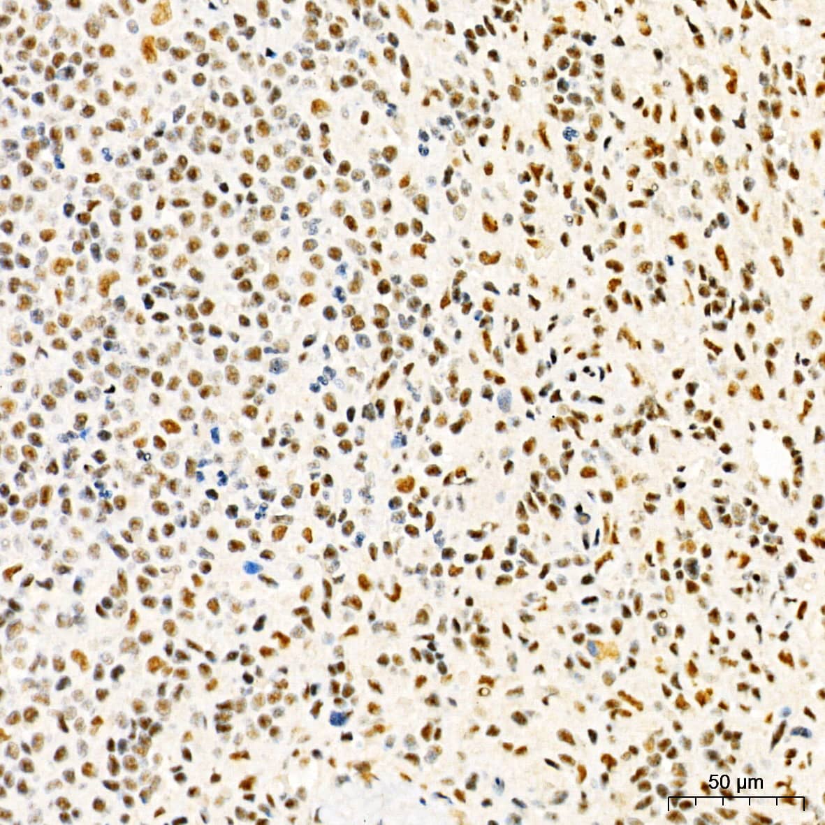
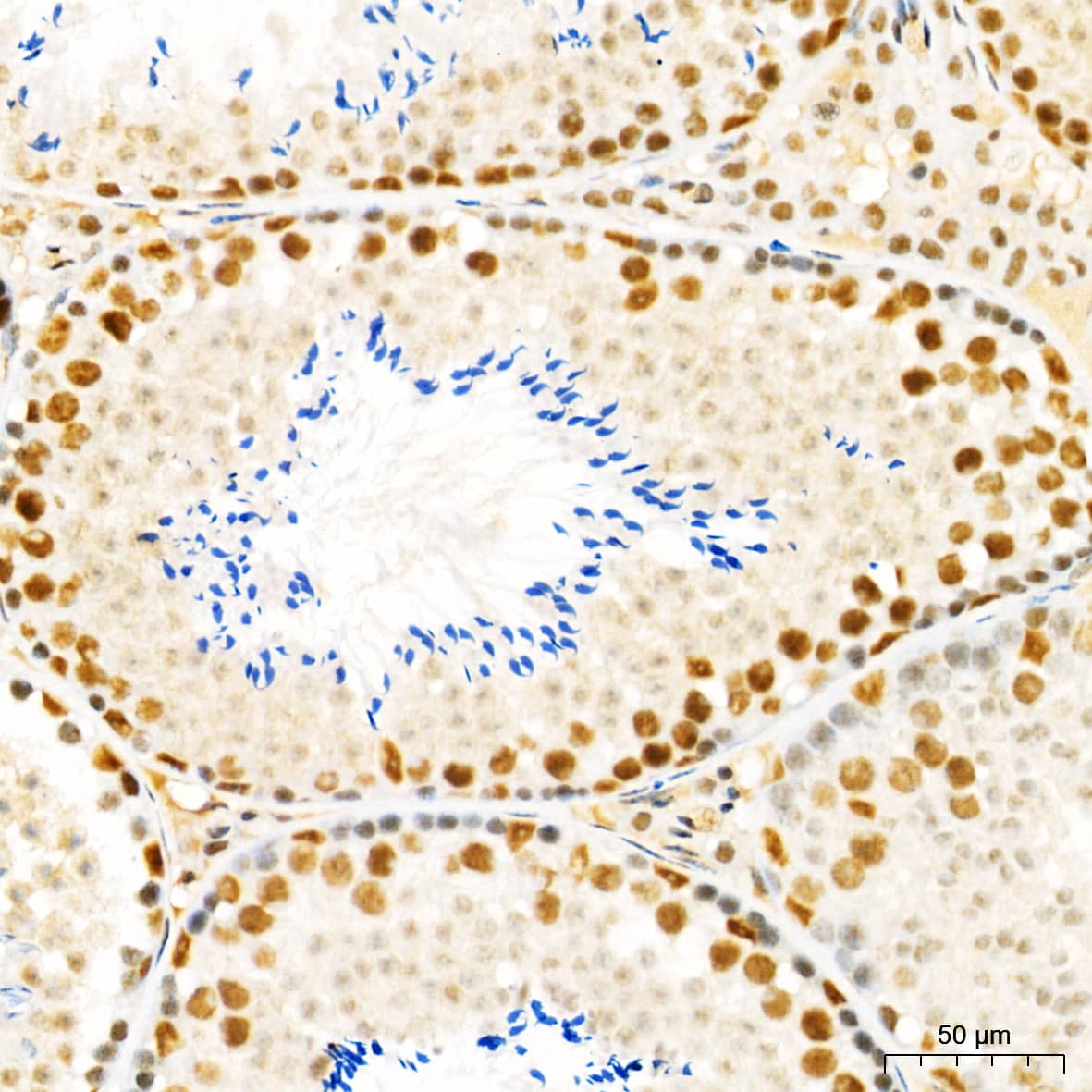
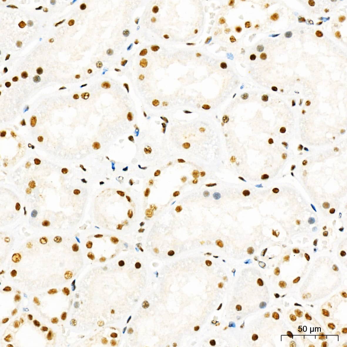


![Intracellular Staining by Flow Cytometry AKT [p Ser473] Antibody [Unconjugated] - Pan Specific](https://images.novusbio.com/images/antibody/Akt3_AF887_Flow_Cytometry_8283.jpg)
![Western Blot AKT [p Ser473] Antibody [Unconjugated] - Pan Specific](https://images.novusbio.com/images/af887_phospho-akt-s473-pan-specific-affinity-purified-pab-41202410485440.jpg)
![Western Blot AKT [p Ser473] Antibody [Unconjugated] - Pan Specific](https://images.novusbio.com/images/af887_phospho-akt-s473-pan-specific-affinity-purified-pab-8120255552843.jpg)


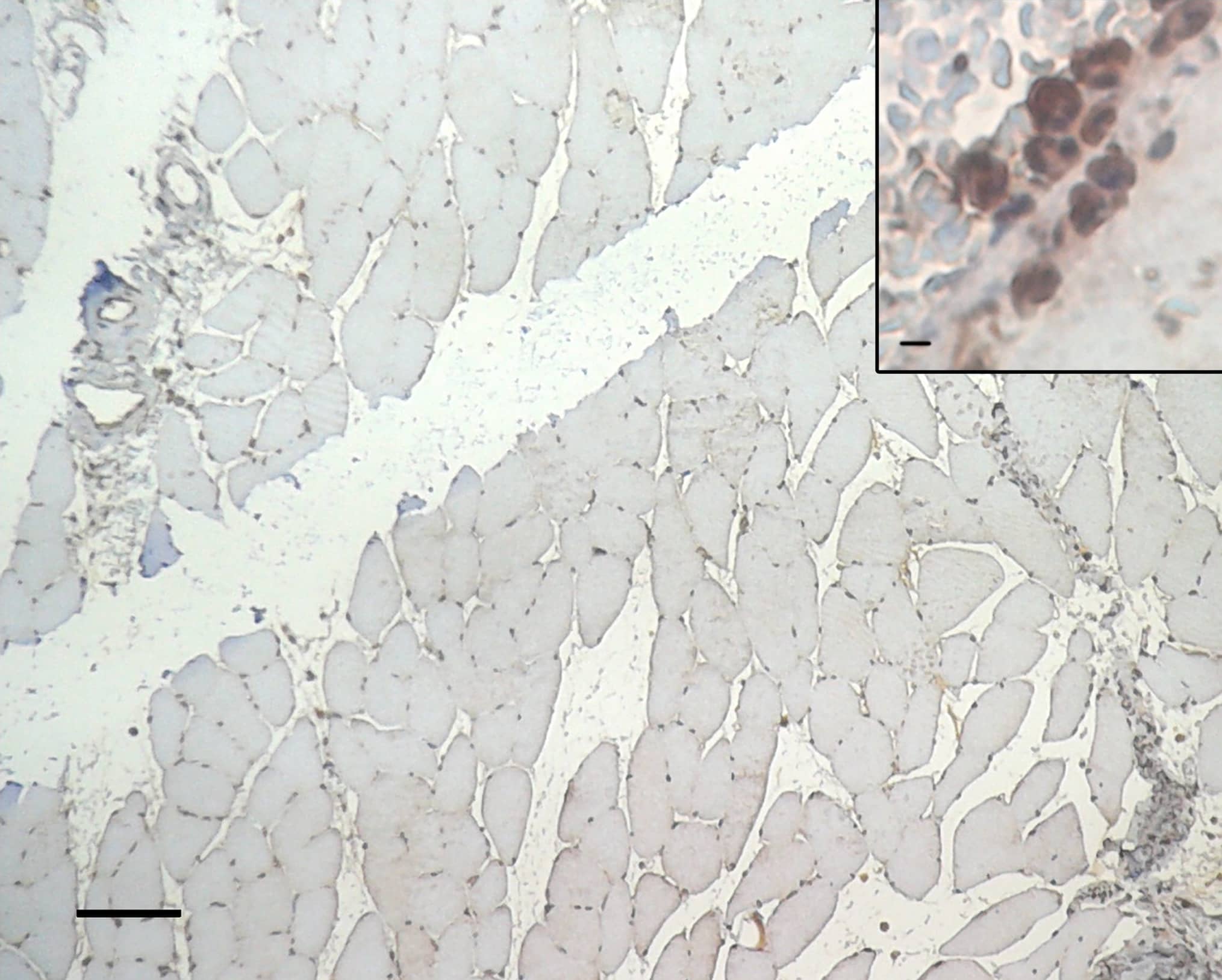
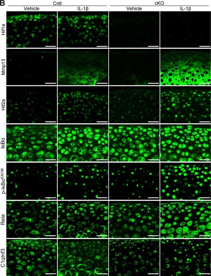
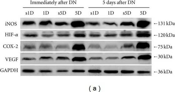


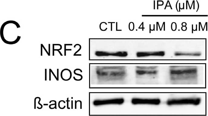


-(01-ml)_NB100-479_10081.gif)
-(01-ml)_NB100-479_9371.jpg)


 staining with HIF-1 alpha antibody and (B) blocking peptide used at 10-fold excess concentration. Images provided by Dr Yves Heremans. Please see the product review of NBC1-18422 for additional details.")
 using HIF-1 alpha antibody. Western blot image submitted by a verified customer review.")


 was performed with the indicated antibody (Ab). Primers flanking the HIF binding site in the S100A10 gene were used for qPCR. ChIP image submitted by a verified customer review.")
 cells, 20% oxygen and B. PC12 (rat) cells, 1% oxygen.")
. Lane 2: Untreated Cos-7 nuclear extract (50 ug, normoxic). 10 second ECL exposure.")
 incubation. Western blot image submitted by a verified customer review.")
 in RCC4 cells using NB100-479. Nuclei (Blue) were counterstained using Hoechst 33258.")


 using NB100-479 (1:100 dilution) followed by Anti-Rabbit Secondary Antibody (042-206, ProteinSimple). This experiment was conducted under standard assay conditions, and using the 12-230 kDa separation module (SW-W004). Non-specific interaction with the 230 kDa Simple Western standard seen with this antibody. Image from an internal validation.")
 or hypoxia (1% O2). Cell extracts were analysed by Western blots. Note that p-cofilin levels are reduced in c2 and c2 cells compared to wt and shC cells at 20% O2 and 1% O2.")
![Western Blot: HIF-1 alpha Antibody - BSA Free [NB100-479] - Hypoxic markers accumulate in miR-29-sponge Zebrafish hearts. (A) Representative western blot analysis of hypoxia inducible factor 1 alpha (HIF1 alpha ) expression in Wild Type & miR-29-sponge Zebrafish heart. In each condition, alpha -tubulin was used as loading control. Three independent experiments were performed. Full-length blot is presented in Supplementary Figure 6.(B) qRT-PCR mRNA analysis of hypoxia associated genes: erythropoietin alpha (epoa); hexokinase2 (hk2); heme oxygenase1a (hmox1a); lactate dehydrogenase A (ldha); cyclin-dependent kinase inhibitor 1B (p27) in Wild Type (black circles; n = 4) & miR-29-sponge (gray squares; n = 4) Zebrafish hearts expressed as fold-change versus Wild Type samples. *p N) localization in three microscopic viewing areas were counted as shown in the lower panel. The total counted cell numbers ranged from 35 to 64. The experiments were repeated more than twice, except for the cell localization count, & the results were reproduced. Image collected & cropped by CiteAb from the following publication (//pubmed.ncbi.nlm.nih.gov/28775317), licensed under a CC-BY license. Not internally tested by Novus Biologicals.](http://images.novusbio.com/fullsize/nb100-479_rabbit-polyclonal-hif-1-alpha-antibody-31020241554526.jpg "Western Blot: HIF-1 alpha Antibody - BSA Free [NB100-479] - Hypoxic markers accumulate in miR-29-sponge Zebrafish hearts. (A) Representative western blot analysis of hypoxia inducible factor 1 alpha (HIF1 alpha ) expression in Wild Type & miR-29-sponge Zebrafish heart. In each condition, alpha -tubulin was used as loading control. Three independent experiments were performed. Full-length blot is presented in Supplementary Figure 6.(B) qRT-PCR mRNA analysis of hypoxia associated genes: erythropoietin alpha (epoa); hexokinase2 (hk2); heme oxygenase1a (hmox1a); lactate dehydrogenase A (ldha); cyclin-dependent kinase inhibitor 1B (p27) in Wild Type (black circles; n = 4) & miR-29-sponge (gray squares; n = 4) Zebrafish hearts expressed as fold-change versus Wild Type samples. *p N) localization in three microscopic viewing areas were counted as shown in the lower panel. The total counted cell numbers ranged from 35 to 64. The experiments were repeated more than twice, except for the cell localization count, & the results were reproduced. Image collected & cropped by CiteAb from the following publication (//pubmed.ncbi.nlm.nih.gov/28775317), licensed under a CC-BY license. Not internally tested by Novus Biologicals.")
![Western Blot: HIF-1 alpha Antibody - BSA Free [NB100-479] - Hypoxia signaling & cytotoxicity to CdCl2. (a) Wild-type (WT) cells were left untreated (Ctrl) or exposed to 150 μM CoCl2 (Co2+), 1, 5 or 10 μM CdCl2 (Cd2+) or 150 μM CoCl2 & 10 μM CdCl2 for 24 hours. HIF1 alpha −/− cell extract treated with 150 μM CoCl2 was used as a negative control. Nuclear protein was extracted & separated by SDS-PAGE, transferred to nitrocellulose membrane & probed with a HIF1 alpha (upper panel) or beta -actin (lower panel) specific antibody. The bands observed in the cadmium only WT cells are nonspecific as they are also observed in the HIF1 alpha −/− cells. (b) BNip3 mRNA expression levels were analyzed by qRT-PCR in wild type (WT, white bars) & HIF1 alpha −/− cells (black bars). Cells were left untreated (0), or exposed to 5 μM CdCl2 (Cd2+) or 150 μM CoCl2 (Co2+) for 24 hours. Each value was normalized to the control level in the corresponding cell line. *P 0.05; *p 60 recipients/genotype). (C) When 500 WT & KO cells were implanted into the mammary fat pad, the median time until 50% of recipients developed tumors > 500 mm3 was 64 days for WT mice & 127 days for KO mice (n = 14 recipients/genotype, P < 0.001, logrank test). (D) Western blot for HIF-1 alpha in three independent tumors (500 to 750 mm3) per genotype. WT & KO cells that were cultured for 6 hours under hypoxic conditions served as positive & negative controls, respectively. CRM, cross-reactive material. MTEC, mammary tumor epithelial cell. (E) Mean fold change ± SEM in expression of HIF-1 targets in KO tumors as determined by qRT-PCR (n = 5 tumors/genotype). Decreased gene expression in KO tumors is presented as a negative fold-change relative to WT tumors. (F) An increase in Ki67+ cells in KO tumors is balanced by an increase in caspase 3-positive cells (n = 5 tumors/genotype, *P < 0.05, Student's t-test). Representative immunostaining images are shown in Additional file 2 Figure S4. Image collected & cropped by CiteAb from the following publication (//breast-cancer-research.biomedcentral.com/articles/10.1186/bcr3087), licensed under a CC-BY license. Not internally tested by Novus Biologicals.](http://images.novusbio.com/fullsize/nb100-479_rabbit-polyclonal-hif-1-alpha-antibody-310202415532013.jpg "Western Blot: HIF-1 alpha Antibody - BSA Free [NB100-479] - Hypoxia signaling & cytotoxicity to CdCl2. (a) Wild-type (WT) cells were left untreated (Ctrl) or exposed to 150 μM CoCl2 (Co2+), 1, 5 or 10 μM CdCl2 (Cd2+) or 150 μM CoCl2 & 10 μM CdCl2 for 24 hours. HIF1 alpha −/− cell extract treated with 150 μM CoCl2 was used as a negative control. Nuclear protein was extracted & separated by SDS-PAGE, transferred to nitrocellulose membrane & probed with a HIF1 alpha (upper panel) or beta -actin (lower panel) specific antibody. The bands observed in the cadmium only WT cells are nonspecific as they are also observed in the HIF1 alpha −/− cells. (b) BNip3 mRNA expression levels were analyzed by qRT-PCR in wild type (WT, white bars) & HIF1 alpha −/− cells (black bars). Cells were left untreated (0), or exposed to 5 μM CdCl2 (Cd2+) or 150 μM CoCl2 (Co2+) for 24 hours. Each value was normalized to the control level in the corresponding cell line. *P 0.05; *p 60 recipients/genotype). (C) When 500 WT & KO cells were implanted into the mammary fat pad, the median time until 50% of recipients developed tumors > 500 mm3 was 64 days for WT mice & 127 days for KO mice (n = 14 recipients/genotype, P < 0.001, logrank test). (D) Western blot for HIF-1 alpha in three independent tumors (500 to 750 mm3) per genotype. WT & KO cells that were cultured for 6 hours under hypoxic conditions served as positive & negative controls, respectively. CRM, cross-reactive material. MTEC, mammary tumor epithelial cell. (E) Mean fold change ± SEM in expression of HIF-1 targets in KO tumors as determined by qRT-PCR (n = 5 tumors/genotype). Decreased gene expression in KO tumors is presented as a negative fold-change relative to WT tumors. (F) An increase in Ki67+ cells in KO tumors is balanced by an increase in caspase 3-positive cells (n = 5 tumors/genotype, *P < 0.05, Student's t-test). Representative immunostaining images are shown in Additional file 2 Figure S4. Image collected & cropped by CiteAb from the following publication (//breast-cancer-research.biomedcentral.com/articles/10.1186/bcr3087), licensed under a CC-BY license. Not internally tested by Novus Biologicals.")
![Western Blot: HIF-1 alpha Antibody - BSA Free [NB100-479] - Effect of HIF-1 alpha silencing on alpha -MSH-induced apoptosis in B16-F10 melanoma cells during hypoxia.a Cells were transfected with shLacZ or shHIF-1 alpha plasmids for 48 h before harvest. HIF-1 alpha shRNA reduced the basal HIF-1 alpha mRNA & protein expression levels. b, c Relative mRNA expression levels were analyzed by real-time PCR. Data are expressed as fold change compared with control (means ± SD of triplicate experiments). d Cell lysates were analyzed by immunoblot using the indicated antibodies. beta -Actin was used as an internal control for loading & transfer. e The population of apoptotic cells was analyzed by flow cytometry & qualified as mean ± SD from triplicate experiments. *p < 0.05, **p < 0.01. Knockdown of HIF-1 alpha diminished alpha -MSH-induced apoptosis in B16-F10 melanoma cells during hypoxia Image collected & cropped by CiteAb from the following publication (//pubmed.ncbi.nlm.nih.gov/30062060), licensed under a CC-BY license. Not internally tested by Novus Biologicals.](http://images.novusbio.com/fullsize/nb100-479_rabbit-polyclonal-hif-1-alpha-antibody-310202415532077.jpg "Western Blot: HIF-1 alpha Antibody - BSA Free [NB100-479] - Effect of HIF-1 alpha silencing on alpha -MSH-induced apoptosis in B16-F10 melanoma cells during hypoxia.a Cells were transfected with shLacZ or shHIF-1 alpha plasmids for 48 h before harvest. HIF-1 alpha shRNA reduced the basal HIF-1 alpha mRNA & protein expression levels. b, c Relative mRNA expression levels were analyzed by real-time PCR. Data are expressed as fold change compared with control (means ± SD of triplicate experiments). d Cell lysates were analyzed by immunoblot using the indicated antibodies. beta -Actin was used as an internal control for loading & transfer. e The population of apoptotic cells was analyzed by flow cytometry & qualified as mean ± SD from triplicate experiments. *p < 0.05, **p < 0.01. Knockdown of HIF-1 alpha diminished alpha -MSH-induced apoptosis in B16-F10 melanoma cells during hypoxia Image collected & cropped by CiteAb from the following publication (//pubmed.ncbi.nlm.nih.gov/30062060), licensed under a CC-BY license. Not internally tested by Novus Biologicals.")
![Immunohistochemistry: HIF-1 alpha Antibody - BSA Free [NB100-479] - Hif1 alpha expression in neonatal & adult testis.(A) Section of testis from 5-day old (P5) male new born pups showing Hif1 alpha expression in MVH+ gonocytes within the seminiferous tubules (top) & negative control images without primary antibodies (bottom). Scale bar: 50μm. (B) Western blot analysis of Hif1 alpha expression in P5 testes (left) compared to extract of the adult brain sub-ventricular zone (SVZ) (right). Loading control ( beta -Actin) is shown below. (C) Section of adult (3 month old) testis showing Hif1 alpha expression in spermatogonia. Scale bar: 30μm. (D) Western blot analysis of whole adult testis. HEK293 cells treated with DFX were used as a positive control & intestinal tissue was used as a negative control. Loading control ( beta -Actin) is shown below. Image collected & cropped by CiteAb from the following publication (//pubmed.ncbi.nlm.nih.gov/27148974), licensed under a CC-BY license. Not internally tested by Novus Biologicals.](http://images.novusbio.com/fullsize/nb100-479_rabbit-polyclonal-hif-1-alpha-antibody-31020241682177.jpg "Immunohistochemistry: HIF-1 alpha Antibody - BSA Free [NB100-479] - Hif1 alpha expression in neonatal & adult testis.(A) Section of testis from 5-day old (P5) male new born pups showing Hif1 alpha expression in MVH+ gonocytes within the seminiferous tubules (top) & negative control images without primary antibodies (bottom). Scale bar: 50μm. (B) Western blot analysis of Hif1 alpha expression in P5 testes (left) compared to extract of the adult brain sub-ventricular zone (SVZ) (right). Loading control ( beta -Actin) is shown below. (C) Section of adult (3 month old) testis showing Hif1 alpha expression in spermatogonia. Scale bar: 30μm. (D) Western blot analysis of whole adult testis. HEK293 cells treated with DFX were used as a positive control & intestinal tissue was used as a negative control. Loading control ( beta -Actin) is shown below. Image collected & cropped by CiteAb from the following publication (//pubmed.ncbi.nlm.nih.gov/27148974), licensed under a CC-BY license. Not internally tested by Novus Biologicals.")
![Western Blot: HIF-1 alpha Antibody - BSA Free [NB100-479] - Expression of hypoxia-inducible factor (HIF) proteins after bilateral renal ischemia or sham ischemia. Immunoblots for HIF-1 alpha (A–C) & HIF-2 alpha (D–F) of tissue extracts from the cortex & outer & inner medulla of the left kidneys of rats 24 h & 5 days following recovery from either sham ischemia (○) or bilateral renal ischemia (●); n = 6 per group. G: typical image of the gel following electrophoresis. H: typical image of the nitrocellulose membrane following transfer. Values are expressed as medians (25th percentile, 75th percentile). Paired comparisons were performed using the Mann-Whitney U-test. Because paired comparisons were made at two time points, P values were conservatively adjusted using the Dunn-Sidak method with k = 2. PTr, PT, & PTr*T are the outcomes of two-way analysis of variance on ranking with the factors treatment (Tr) & time (T). AU, arbitrary unit; I1, 24 h after ischemia; I5, 5 days after ischemia; S1, 24 h after sham ischemia; S5, 5 days after sham ischemia. Image collected & cropped by CiteAb from the following publication (//pubmed.ncbi.nlm.nih.gov/30110566), licensed under a CC-BY license. Not internally tested by Novus Biologicals.](http://images.novusbio.com/fullsize/nb100-479_rabbit-polyclonal-hif-1-alpha-antibody-310202415535154.jpg "Western Blot: HIF-1 alpha Antibody - BSA Free [NB100-479] - Expression of hypoxia-inducible factor (HIF) proteins after bilateral renal ischemia or sham ischemia. Immunoblots for HIF-1 alpha (A–C) & HIF-2 alpha (D–F) of tissue extracts from the cortex & outer & inner medulla of the left kidneys of rats 24 h & 5 days following recovery from either sham ischemia (○) or bilateral renal ischemia (●); n = 6 per group. G: typical image of the gel following electrophoresis. H: typical image of the nitrocellulose membrane following transfer. Values are expressed as medians (25th percentile, 75th percentile). Paired comparisons were performed using the Mann-Whitney U-test. Because paired comparisons were made at two time points, P values were conservatively adjusted using the Dunn-Sidak method with k = 2. PTr, PT, & PTr*T are the outcomes of two-way analysis of variance on ranking with the factors treatment (Tr) & time (T). AU, arbitrary unit; I1, 24 h after ischemia; I5, 5 days after ischemia; S1, 24 h after sham ischemia; S5, 5 days after sham ischemia. Image collected & cropped by CiteAb from the following publication (//pubmed.ncbi.nlm.nih.gov/30110566), licensed under a CC-BY license. Not internally tested by Novus Biologicals.")
![Western Blot: HIF-1 alpha Antibody - BSA Free [NB100-479] - Hypoxia reduces inflammatory signaling pathways & NLRP3 expression & induces autophagy in IECs. a HT-29 cells were subjected to normoxia & hypoxia at the indicated times in the absence or presence of 10 µg/ml LPS. Autophagy was measured by variations in the ratio of LC3-II/LC3-I & the total amount of LC3 (LC3-I plus LC3-II) relative to beta -actin. Results are representative of two independent experiments. b, c & d HT-29 cells were subjected to normoxia & hypoxia for the indicated periods in the absence or presence of 10 µg/ml LPS, followed by transcript analysis. Statistical analysis was performed using one-way ANOVA followed by Tukey’s post-test. Results represent mean + s.e.m. of two independent experiments done in triplicate, *P < 0.05; **P < 0.01; ***P < 0.001; ns not significant. e HT-29 cells were subjected to normoxia or hypoxia in the presence & absence of 10 µg/ml LPS & 20 µM of MG132. Results are representative of two independent experiments. f & g Putative binding sites for HIF-1 alpha & NF-kappa B were found in the p62 f & NLRP3 g promoters using Genomatix software tools. Numbers under the boxes indicate the distance from the transcription start site. HT-29 cells were subjected to normoxia (21% O2) or hypoxia (0.2% O2) for 6 h & 24 h. ChIP analysis was performed using antibodies against HIF-1 alpha & NF-kappa B for immunoprecipitation. PCR was performed using the promoter-specific primers for the p62 f & NLRP3 g promoter binding sites of HIF-1 alpha & NF-kappa B. Aliquots taken prior to immunoprecipitation were used as input control. PCR products were run on 2% agarose gel. The results are representative of three independent experiments. Image collected & cropped by CiteAb from the following publication (//pubmed.ncbi.nlm.nih.gov/28740109), licensed under a CC-BY license. Not internally tested by Novus Biologicals.](http://images.novusbio.com/fullsize/nb100-479_rabbit-polyclonal-hif-1-alpha-antibody-310202415541975.jpg "Western Blot: HIF-1 alpha Antibody - BSA Free [NB100-479] - Hypoxia reduces inflammatory signaling pathways & NLRP3 expression & induces autophagy in IECs. a HT-29 cells were subjected to normoxia & hypoxia at the indicated times in the absence or presence of 10 µg/ml LPS. Autophagy was measured by variations in the ratio of LC3-II/LC3-I & the total amount of LC3 (LC3-I plus LC3-II) relative to beta -actin. Results are representative of two independent experiments. b, c & d HT-29 cells were subjected to normoxia & hypoxia for the indicated periods in the absence or presence of 10 µg/ml LPS, followed by transcript analysis. Statistical analysis was performed using one-way ANOVA followed by Tukey’s post-test. Results represent mean + s.e.m. of two independent experiments done in triplicate, *P < 0.05; **P < 0.01; ***P < 0.001; ns not significant. e HT-29 cells were subjected to normoxia or hypoxia in the presence & absence of 10 µg/ml LPS & 20 µM of MG132. Results are representative of two independent experiments. f & g Putative binding sites for HIF-1 alpha & NF-kappa B were found in the p62 f & NLRP3 g promoters using Genomatix software tools. Numbers under the boxes indicate the distance from the transcription start site. HT-29 cells were subjected to normoxia (21% O2) or hypoxia (0.2% O2) for 6 h & 24 h. ChIP analysis was performed using antibodies against HIF-1 alpha & NF-kappa B for immunoprecipitation. PCR was performed using the promoter-specific primers for the p62 f & NLRP3 g promoter binding sites of HIF-1 alpha & NF-kappa B. Aliquots taken prior to immunoprecipitation were used as input control. PCR products were run on 2% agarose gel. The results are representative of three independent experiments. Image collected & cropped by CiteAb from the following publication (//pubmed.ncbi.nlm.nih.gov/28740109), licensed under a CC-BY license. Not internally tested by Novus Biologicals.")
![Western Blot: HIF-1 alpha Antibody - BSA Free [NB100-479] - Transient hyperoxia does not sensitize normal human astrocytes to radiation.Results are shown for the colony forming assays for all previously assayed cell lines & a normal human astrocyte cell line (Astro). Cells were continuously maintained under normoxic conditions (NOx) or exposed to 25 min of hyperoxia (50% O2) & then returned to normoxic conditions for 25 min (HyperOx) before being treated with a 5 Gy dose of radiation. To allow for ease of comparisons among cell types, raw values are expressed as a percentage of the corresponding cells type’s negative (non-irradiated) control & the means & SEMs are plotted. Each result represents three independent samples, plated in triplicate (*p<0.05, Student’s t-test). Also shown are Western blots of nuclear HIF-1 alpha at the time of irradiation for each cell line. Corresponding Western blots of lamin A/C are shown as a loading control. Image collected & cropped by CiteAb from the following publication (//pubmed.ncbi.nlm.nih.gov/25350400), licensed under a CC-BY license. Not internally tested by Novus Biologicals.](http://images.novusbio.com/fullsize/nb100-479_rabbit-polyclonal-hif-1-alpha-antibody-310202415525255.jpg "Western Blot: HIF-1 alpha Antibody - BSA Free [NB100-479] - Transient hyperoxia does not sensitize normal human astrocytes to radiation.Results are shown for the colony forming assays for all previously assayed cell lines & a normal human astrocyte cell line (Astro). Cells were continuously maintained under normoxic conditions (NOx) or exposed to 25 min of hyperoxia (50% O2) & then returned to normoxic conditions for 25 min (HyperOx) before being treated with a 5 Gy dose of radiation. To allow for ease of comparisons among cell types, raw values are expressed as a percentage of the corresponding cells type’s negative (non-irradiated) control & the means & SEMs are plotted. Each result represents three independent samples, plated in triplicate (*p<0.05, Student’s t-test). Also shown are Western blots of nuclear HIF-1 alpha at the time of irradiation for each cell line. Corresponding Western blots of lamin A/C are shown as a loading control. Image collected & cropped by CiteAb from the following publication (//pubmed.ncbi.nlm.nih.gov/25350400), licensed under a CC-BY license. Not internally tested by Novus Biologicals.")
![Western Blot: HIF-1 alpha Antibody - BSA Free [NB100-479] - YC-1 attenuates pro-inflammatory differentiation of macrophages. (a,b) qRT-PCR analyses, revealing mRNA expression of the pro-inflammatory M1-differentiation markers NOS2 (a) & IL-6 (b) in murine J774A.1 macrophages under normoxia (left bars) & hypoxia (right bars). LPS exposure was carried out to induce M1-polarization in presence of YC-1 or vehicle control (DMSO; **P ≤ 0.01, n = 9). (c,d) Western blots of nuclear extracts, revealing protein levels of HIF-1 alpha (c) & HIF-2 alpha (d) in J774.A1 macrophages treated with vehicle (DMSO) or YC-1 under normoxic (left) or hypoxic (right) culture conditions. Simultaneous LPS-treatment was performed to induce pro-inflammatory activation. Note LPS-induced stabilization of HIF-1 alpha - (c), but not HIF-2 alpha -levels (d), & reversal of LPS-induced HIF-1 alpha -stabilization by YC-1 (c). Histone H3 was used as a loading control. (e,f) qRT-PCR analyses, revealing mRNA expression of the pro-inflammatory M1-differentiation markers NOS2 (e) & IL-6 (f) in BMDMs under normoxia (left bars) & hypoxia (right bars). LPS exposure was carried out to induce M1-polarization in presence of YC-1 or vehicle control (DMSO; ***P ≤ 0.001, **P ≤ 0.01, n = 6). Image collected & cropped by CiteAb from the following publication (//www.nature.com/articles/s41598-017-13638-z), licensed under a CC-BY license. Not internally tested by Novus Biologicals.](http://images.novusbio.com/fullsize/nb100-479_rabbit-polyclonal-hif-1-alpha-antibody-310202415532080.jpg "Western Blot: HIF-1 alpha Antibody - BSA Free [NB100-479] - YC-1 attenuates pro-inflammatory differentiation of macrophages. (a,b) qRT-PCR analyses, revealing mRNA expression of the pro-inflammatory M1-differentiation markers NOS2 (a) & IL-6 (b) in murine J774A.1 macrophages under normoxia (left bars) & hypoxia (right bars). LPS exposure was carried out to induce M1-polarization in presence of YC-1 or vehicle control (DMSO; **P ≤ 0.01, n = 9). (c,d) Western blots of nuclear extracts, revealing protein levels of HIF-1 alpha (c) & HIF-2 alpha (d) in J774.A1 macrophages treated with vehicle (DMSO) or YC-1 under normoxic (left) or hypoxic (right) culture conditions. Simultaneous LPS-treatment was performed to induce pro-inflammatory activation. Note LPS-induced stabilization of HIF-1 alpha - (c), but not HIF-2 alpha -levels (d), & reversal of LPS-induced HIF-1 alpha -stabilization by YC-1 (c). Histone H3 was used as a loading control. (e,f) qRT-PCR analyses, revealing mRNA expression of the pro-inflammatory M1-differentiation markers NOS2 (e) & IL-6 (f) in BMDMs under normoxia (left bars) & hypoxia (right bars). LPS exposure was carried out to induce M1-polarization in presence of YC-1 or vehicle control (DMSO; ***P ≤ 0.001, **P ≤ 0.01, n = 6). Image collected & cropped by CiteAb from the following publication (//www.nature.com/articles/s41598-017-13638-z), licensed under a CC-BY license. Not internally tested by Novus Biologicals.")
![N/A VEGF [HRP]](https://images.novusbio.com/images/elisa/VEGF_DVE00_ELISA_208.jpg)
![N/A VEGF [HRP]](https://images.novusbio.com/images/elisa/DATA_VEGF_DVE00_ELISA_871.jpg)
![N/A VEGF [HRP]](https://images.novusbio.com/images/elisa/DATA_VEGF_DVE00_ELISA_872.jpg)
![N/A Erythropoietin/EPO [HRP]](https://images.novusbio.com/images/elisa/Erythropoietin_MEP00_ELISA_421.jpg)
![N/A Erythropoietin/EPO [HRP]](https://images.novusbio.com/images/elisa/DATA_Erythropoietin_MEP00_ELISA_970.jpg)


![Western Blot ERK2 Antibody [Unconjugated]](https://images.novusbio.com/images/antibody/ERK2_AF1230_Western_Blot_5097.jpg)
![Knockout Validated ERK2 Antibody [Unconjugated]](https://images.novusbio.com/images/antibody/ERK2_AF1230_Knockout_Validated_22864.jpg)
![Immunohistochemistry ERK2 Antibody [Unconjugated]](https://images.novusbio.com/images/antibody/ERK2_AF1230_Immunohistochemistry_20696.jpg)
![Immunohistochemistry TOR/mTOR [p Ser2448] Antibody - BSA Free](https://images.novusbio.com/images/nb600-607_rabbit-polyclonal-tor-mtor-p-ser2448-antibody-235202318143142.jpg)
![Western Blot TOR/mTOR [p Ser2448] Antibody - BSA Free](https://images.novusbio.com/images/nb600-607_rabbit-polyclonal-tor-mtor-p-ser2448-antibody-31020241535647.jpg)
![Data TOR/mTOR [p Ser2448] Antibody - BSA Free](https://images.novusbio.com/images/TOR-mTOR-[p-Ser2448]-Antibody-N-A-NB600-607-img0008.jpg)
![N/A TIMP-1 [HRP]](https://images.novusbio.com/images/elisa/DATA_TIMP1_DTM100_ELISA_847.jpg)
![N/A TIMP-1 [HRP]](https://images.novusbio.com/images/elisa/TIMP-1_DTM100_ELISA_194.jpg)
![SDS-Page TNF-alpha [Unconjugated]](https://images.novusbio.com/images/protein/TNF-alpha_210-TA_256.jpg)
![Bioactivity TNF-alpha [Unconjugated]](https://images.novusbio.com/images/protein/TNFalpha_210TA_1658.jpg)
![SEC-MALS TNF-alpha [Unconjugated]](https://images.novusbio.com/images/210-ta_recombinant-human-tnf-alpha-protein-sec-mals-35202312244..jpg)
![Western Blot: Human Pancreas Whole Tissue Lysate (Adult Whole Normal) [NB820-59244] - man Pancreas Whole Tissue Lysate (Adult Normal) [NB820-59244] - Image from verified customer review.](https://images.novusbio.com/images/Human-Pancreas-Whole-Tissue-Lysate-Adult-Whole-Normal-Western-Blot-NB820-59244-img0002.jpg "Western Blot: Human Pancreas Whole Tissue Lysate (Adult Whole Normal) [NB820-59244] - man Pancreas Whole Tissue Lysate (Adult Normal) [NB820-59244] - Image from verified customer review.")
![Western Blot: Goat anti-Rabbit IgG (H+L) Secondary Antibody [HRP] [NB7160] - Western blot showing vemurafenib treatment in BRAFV600E CRC cells inhibits fission mediator DRP1 with no significant effect on fusion proteins (Mfn1 & 2) using MFN-1 antibody (NBP1-51841) and corresponding secondary antibody, goat anti-rabbit IgG-HRP (NB7160). Image collected and cropped by CiteAb from the following publication (https://pubmed.ncbi.nlm.nih.gov/33738242).](https://images.novusbio.com/images/Goat-anti-Rabbit-IgG-H+L-Secondary-Antibody-HRP-Western-Blot-NB7160-img0001.jpg "Western Blot: Goat anti-Rabbit IgG (H+L) Secondary Antibody [HRP] [NB7160] - Western blot showing vemurafenib treatment in BRAFV600E CRC cells inhibits fission mediator DRP1 with no significant effect on fusion proteins (Mfn1 & 2) using MFN-1 antibody (NBP1-51841) and corresponding secondary antibody, goat anti-rabbit IgG-HRP (NB7160). Image collected and cropped by CiteAb from the following publication (https://pubmed.ncbi.nlm.nih.gov/33738242).")
 followed by 30 min incubation with Goat anti Rabbit HRP conjugated secondary antibodies (Catalog # HAF008) at 1:20 dilution + DAB chromogen (brown). The tissue was counterstained with Hematoxylin (blue). Control was done by omitting primary antibody.")
![Flow Cytometry: Rabbit IgG Isotype Control [NBP2-24891] - An intracellular stain was performed on Raji cells with Adiponectin antibody NB100-65810 (blue) and a matched isotype control NBP2-24893 (orange). Cells were fixed with 4% PFA and then permeablized with 0.1% saponin. Cells were incubated in an antibody dilution of 1 ug/mL for 30 minutes at room temperature, followed by Dylight488-conjugated anti-rabbit secondary antibody. Image using the Azide Free form of this antibody.](https://images.novusbio.com/images/Rabbit--Mouse-IgG-Isotype-Control-Flow-Cytometry-NBP2-24891-img0006.jpg "Flow Cytometry: Rabbit IgG Isotype Control [NBP2-24891] - An intracellular stain was performed on Raji cells with Adiponectin antibody NB100-65810 (blue) and a matched isotype control NBP2-24893 (orange). Cells were fixed with 4% PFA and then permeablized with 0.1% saponin. Cells were incubated in an antibody dilution of 1 ug/mL for 30 minutes at room temperature, followed by Dylight488-conjugated anti-rabbit secondary antibody. Image using the Azide Free form of this antibody.")


