| Montes-Alvarado J, Garcia-Ibañez P, Moreno D et al. Brassica Extracts Prevent Benzo(a)pyrene-Induced Transformation by Modulating Reactive Oxygen Species and Autophagy. International Journal of Molecular Sciences 2025-10-16 [PMID: 41096784] |
|
|
| Vasukutty A, Bhattarai P, Choi H Ebselen Suppresses Breast Cancer Tumorigenesis by Inhibiting YTHDF1-Mediated c-Fos Expression. International Journal of Molecular Sciences 2025-10-16 [PMID: 41096691] |
|
|
| Augur Z, Fogo G, Arbery M et al. Genetic and proteomic analysis identifies BAG3 as an amyloid-responsive regulator of neuronal proteostasis. Acta neuropathologica 2025-10-14 [PMID: 41085772] |
|
|
| Singh J, Williams J, Elliott Q et al. Endosome transcriptomics reveal trafficking of Cajal bodies into multivesicular bodies. Proceedings of the National Academy of Sciences of the United States of America 2025-10-08 [PMID: 41060753] |
|
|
| Kosmac K, Wang R, Stewart J et al. Gastrocnemius myofiber type and mitochondrial alterations associated with peripheral artery disease severity. Function (Oxford, England) 2025-10-06 [PMID: 41051228] |
|
|
| Civiletto G, Brunetti D, Lizzo G et al. Herbal terpenoids activate autophagy and mitophagy through modulation of bioenergetics and protect from metabolic stress, sarcopenia and epigenetic aging Nature aging 2025-09-24 [PMID: 40993327] |
|
|
| Chen, H;Sun, YY;Li, QF;Du, YT;Hu, NN;Sui, AR;Luo, XQ;Huang, X;Zhu, C;Yang, G;Yao, LL;Tang, Y;Hu, H;Liu, CF;Tao, J;Feng, L;Kirchhoff, F;Huang, W;Li, S;Ma, QH; Impaired macroautophagy in oligodendrocyte precursor cells suppresses neuronal plasticity via a senescence-associated signaling Science advances 2025-09-26 [PMID: 40991686] |
|
|
| Augur, ZM;Fogo, GM;Arbery, MR;Stern, AM;Benoit, CR;Hsieh, YC;Young-Pearse, TL; Optineurin deficiency disrupts phosphorylated tau proteostasis and clusterin expression in human neurons Acta neuropathologica communications 2025-09-02 [PMID: 40898372] |
|
|
| Peng B, Lin H, Zhang M et al. Dnmt1 Alleviates S1PR1-Mediated Pyroptosis after Spinal Cord Injury through Regulating Pon3 Expression. Advanced science (Weinheim, Baden-Wurttemberg, Germany) 2025-08-30 [PMID: 40884250] |
|
|
| Duffy H, Byrnes C, Zhu H et al. A pathogenic alpha synuclein variant exacerbates disease progression in a neuron-specific Gba-KO mouse. Neurobiology of disease 2025-08-27 [PMID: 40882878] |
|
|
| Nakayama T, Suzuki K, Mitsutake N Calorie restriction in radiation-exposed mice affects the expression of autophagy-related protein p62. BMC Cancer 2025-08-28 [PMID: 40877886] |
|
|
| Lee Y, Kim Y, Park S et al. Fexuprazan mitigates NSAID-induced small intestinal injury by restoring intestinal barrier integrity in mice. Biomedicine & pharmacotherapy = Biomedecine & pharmacotherapie 2025-08-26 [PMID: 40753937] |
|
|
| Cinque L, Iavazzo M, Bonito G et al. FGF Signaling Promotes Lysosome Biogenesis in Chondrocytes via the Mannose Phosphate Receptor Pathway Traffic (Copenhagen, Denmark) 2025-08-01 [PMID: 40747612] |
|
|
| Seo, S;Park, MJ;Park, MG;Gwak, M;Kim, Y;Jang, J;Hong, N;Lee, BS;Kim, C;Jo, S;Shim, HB;Kim, HJ;Kim, MH;Yoo, SH;Yoon, S;Kim, S;Lee, JH;Choi, SH;Lee, SY;Yeon, GB;Park, SH;Kim, SH;Lee, H;Lee, JY;Kim, DS;Lee, BC;Park, JW;Kim, H; DHRS13 suppresses differentiation and mitophagy in glioma via retinoic acid and mitochondrial reactive oxygen species Nature communications 2025-07-30 [PMID: 40739132] |
|
|
| Mallick P, Maity S, Mondal R et al. The impact of ER UPR on mitochondrial integrity mediated by PDK4 Cell Death & Disease 2025-07-29 [PMID: 40730554] |
|
|
| Deshetty U, Oladapo A, Mohankumar Y et al. Protective Effects of N-Acetylcysteine in Alleviating Cocaine-Mediated Microglial Activation and Neuroinflammation. Biology 2025-07-20 [PMID: 40723450] |
|
|
| Deng K, Li W, Liang J et al. Peptide DFCPPGFNTK Mitigates Dry Eye Pathophysiology by Suppressing Oxidative Stress, Apoptosis, Inflammation, and Autophagy: Evidence from In Vitro and In Vivo Models. Current issues in molecular biology 2025-06-10 [PMID: 40699840] |
|
|
| Lee K, Lieu A, Lin C et al. Synthetic oleanolic acid derivative, RTA-408, overcome in TMZ-resistant glioblastoma cells by inducing apoptosis and G1 cell cycle arrest. Medical oncology (Northwood, London, England) 2025-07-20 [PMID: 40684395] |
|
|
| Collier C, Ruedi C, Thorne N et al. PINK1 Loss of Function Selectively Alters the Mitochondrial‐Derived Vesicle Pathway FASEB BioAdvances 2025-07-10 [PMID: 40641845] |
|
|
| Chen T, Ouyang A, Zou J et al. Rab27a regulates the transport of influenza virus membrane proteins to the plasma membrane. Nature Communications 2025-07-07 [PMID: 40623997] |
|
|
| Mitri F, Giansanti M, Melaiu O et al. Inhibition of autophagy enhances the antitumor efficacy of T/CAR T cell against neuroblastoma. Journal of Experimental & Clinical Cancer Research : CR 2025-07-04 [PMID: 40611330] |
|
|
| Lin Y, Chu C, Hsieh T et al. FGFR inhibitors promote the autophagic degradation of IFN-gamma -induced PD-L1 and alleviate the PD-L1-mediated transcriptional suppression of FGFR3-TACC3 in non-muscle-invasive bladder cancer. Cell death & disease 2025-07-03 [PMID: 40603902] |
|
|
| Madhu V, Hernandaz-Meadows M, Coleman A et al. The loss of OPA1 accelerates intervertebral disc degeneration and osteoarthritis in aged mice. Nature Communications 2025-07-02 [PMID: 40595596] |
|
|
| Zheng X, Luo Z, Zheng J et al. Electroacupuncture Attenuates High‐Fat Diet‐Exacerbated Alzheimer's Pathology by Enhancing TFEB / TFE3 ‐Mediated Autophagic Clearance of Tau and NLRP3 Inflammasome in 3xTg Mice CNS neuroscience & therapeutics 2025-07-01 [PMID: 40589337] |
|
|
| Maestri A, Ehrenborg E, Werngren O et al. Titration-WB: A methodology for accurate quantitative protein determination overcoming reproducibility errors. PLoS ONE 2025-06-12 [PMID: 40504814] |
|
|
| Carosi J, Martin A, Hein L et al. Autophagy across tissues of aging mice. PLoS ONE 2025-06-04 [PMID: 40465797] |
|
|
| Bonavita R, Prodomo A, Cortone G et al. Evidence of an unprecedented cytoplasmic function of DDX11, the Warsaw breakage syndrome DNA helicase, in regulating autophagy. Autophagy 2025-05-25 [PMID: 40413757] |
|
|
| Juliani J, Tran S, Harris T et al. BECLIN-1 is essential for the maintenance of gastrointestinal epithelial integrity by regulating endocytic trafficking, F-actin organization, and lysosomal function Autophagy Reports 2025-04-03 [PMID: 40395982] |
|
|
| Stavrides P, Goulbourne C, Peddy J et al. mTOR inhibition in Q175 Huntington’s disease model mice facilitates neuronal autophagy and mutant huntingtin clearance eLife 2025-05-20 [PMID: 40392702] |
|
|
| Williams J, Ngo J, Murugupandiyan A et al. Calpains orchestrate secretion of annexin-containing microvesicles during membrane repair. The Journal of Cell Biology 2025-05-16 [PMID: 40377476] |
|
|
| Marchese M, Bernardi S, Vivarelli R et al. CLN5 deficiency impairs glucose uptake and uncovers PHGDH as a potential biomarker in Batten disease. Molecular Psychiatry 2025-05-09 [PMID: 40346285] |
|
|
| Ramteke P, Watson B, Toci M et al. Sirt6 deficiency promotes senescence and age-associated intervertebral disc degeneration in mice Bone Research 2025-05-08 [PMID: 40335469] |
|
|
| Sayegh R, Wan J, Caër C et al. Defective autophagy in CD4 T cells drives liver fibrosis via type 3 inflammation Nature Communications 2025-04-24 [PMID: 40274816] |
|
|
| Bird, LE;Xu, B;Hobbs, AD;Ziegler, AR;Scott, NE;Newton, P;Thomas, DR;Edgington-Mitchell, LE;Newton, HJ; Coxiella burnetii manipulates the lysosomal protease cathepsin B to facilitate intracellular success Nature communications 2025-04-24 [PMID: 40274809] |
|
|
| Sansbury S, Serebrenik Y, Lapidot T et al. Pooled tagging and hydrophobic targeting of endogenous proteins for unbiased mapping of unfolded protein responses. Molecular Cell 2025-05-03 [PMID: 40273915] |
|
|
| García-Vázquez N, González-Robles T, Lane E et al. Stabilization of GTSE1 by cyclin D1–CDK4/6-mediated phosphorylation promotes cell proliferation with implications for cancer prognosis eLife 2025-04-24 [PMID: 40272409] |
|
|
| Son S, Siddiqi F, Lopez A et al. Alpha-synuclein mutations mislocalize cytoplasmic p300 compromising autophagy, which is rescued by ACLY inhibition. Neuron 2025-04-14 [PMID: 40262613] |
|
|
| Teng M, Guo J, Xu X et al. Circular RMST cooperates with lineage-driving transcription factors to govern neuroendocrine transdifferentiation. Cancer Cell 2025-04-15 [PMID: 40250444] |
|
|
| Fundora K, Zhuang Y, Hamamoto K et al. DBeQ derivative targets vacuolar protein sorting 4 functions in cancer cells and suppresses tumor growth in mice. The Journal of pharmacology and experimental therapeutics 2025-02-28 [PMID: 40147096] |
|
|
| Gao Y, Wang L, Doeswijk T et al. Intraneuronal A beta accumulation causes tau hyperphosphorylation via endolysosomal leakage Alzheimer's & dementia : the journal of the Alzheimer's Association 2025-03-01 [PMID: 40145397] |
|
|
| Liang W, Zhang C, Wang D et al. Inhibition of Salt‐Inducible Kinase 2 Protects Motor Neurons From Degeneration in ALS by Activating Autophagic Flux and Enhancing mTORC1 Activity CNS neuroscience & therapeutics 2025-03-26 [PMID: 40135564] |
|
|
| Liao K, Yu J, Akhmerov A et al. Long noncoding RNA BCYRN1 promotes cardioprotection by enhancing human and murine regulatory T cell dynamics The Journal of Clinical Investigation 2025-03-25 [PMID: 40131367] |
|
|
| Chaouki G, Parry L, Vituret C et al. Pre-cachectic changes in amino acid homeostasis precede activation of eIF2 alpha signaling in the liver at the onset of C26 cancer-induced cachexia iScience 2025-02-14 [PMID: 40124481] |
|
|
| Boonhok R, Senghoi W, Sangkanu S et al. Acanthamoeba castellanii –Mediated Reduction of Interleukin-1 beta Secretion and Its Association With Macrophage Autophagy Scientifica 2025-01-01 [PMID: 40109888] |
|
|
| Shao R, Liu W, Feng Y et al. LAMP2-FLOT2 interaction enhances autophagosome-lysosome fusion to protect the septic heart in response to ILC2. Autophagy 2025-03-11 [PMID: 40066518] |
|
|
| Feng Y, Cao S, Shi Y et al. Human herpesvirus-associated transposable element activation in human aging brains with Alzheimer's disease. Alzheimer's & dementia : the journal of the Alzheimer's Association 2025-02-22 [PMID: 39985481] |
|
|
| Jing J, Yang F, Wang K et al. UFMylation of NLRP3 Prevents Its Autophagic Degradation and Facilitates Inflammasome Activation. Advanced science (Weinheim, Baden-Wurttemberg, Germany) 2025-02-22 [PMID: 39985286] |
|
|
| Paul S, Dansithong W, Figueroa K et al. Staufen2 dysregulation in neurodegenerative disease. The Journal of Biological Chemistry 2025-04-08 [PMID: 39955058] |
|
|
| Li, X;Chaouhan, HS;Yu, SH;Wang, IK;Yu, TM;Chuang, YW;Chen, KB;Lin, FY;Chen, MY;Hsu, CH;Sun, KT;Li, CY; Hypoxia-Induced Metabolic and Functional Changes in Oral CSCs: Implications for Stemness and Viability Modulation Through BNIP3-Driven Mitophagy Journal of cellular and molecular medicine 2025-02-01 [PMID: 39945227] |
|
|
| Giamundo G, Intartaglia D, Prete E et al. Ezrin defines TSC complex activation at endosomal compartments through EGFR–AKT signaling eLife 2025-02-12 [PMID: 39937579] |
|
|
| Esawie M, Matboli M, Bushra M et al. ZBiotics ameliorates T2DM-induced histopathological damage in liver, kidney and adipose tissues by modulating the NOD-like receptor signaling in Wistar rats. Diabetology & Metabolic Syndrome 2025-02-04 [PMID: 39905472] |
|
|
| Shen S, Liang L, Shi T et al. Microglia-Derived Interleukin-6 Triggers Astrocyte Apoptosis in the Hippocampus and Mediates Depression-Like Behavior. Advanced science (Weinheim, Baden-Wurttemberg, Germany) 2025-01-30 [PMID: 39888279] |
|
|
| Park N, Jo D, Yang J et al. Activation of lysophagy by a TBK1-SCF FBXO3 -TMEM192-TAX1BP1 axis in response to lysosomal damage Nature Communications 2025-01-28 [PMID: 39875384] |
|
|
| Risi M, Cusimano L, Cundin X et al. D1 dopamine receptor antagonists as a new therapeutic strategy to treat autistic-like behaviours in lysosomal storage disorders. Molecular Psychiatry 2025-01-26 [PMID: 39865184] |
|
|
| Pepe S, Aprile D, Castroflorio E, Marte A et Al. TBC1D24 interacts with the v-ATPase and regulates intraorganellar pH in neurons iScience 2025-01-06 [PMID: 39758816] |
|
|
| Vecchia S, Imbrici P, Liantonio A et al. Dapagliflozin ameliorates Lafora disease phenotype in a zebrafish model Biomedicine & Pharmacotherapy 2025-02-01 [PMID: 39753095] |
|
|
| Hu X, Wang Y, Wang R et Al. The hybrid lipoplex induces cytoskeletal rearrangement via autophagy/RhoA signaling pathway for enhanced anticancer gene therapy Nat Commun 2025-01-02 [PMID: 39747218] |
|
|
| Zhu H, Lee YT, Byrnes C et Al. Reactivation of mTOR signaling slows neurodegeneration in a lysosomal sphingolipid storage disease Neurobiol Dis 2024-12-06 [PMID: 39647513] |
|
|
| Sul JH, Shin S, Kim HK et Al. Dopamine-conjugated extracellular vesicles induce autophagy in Parkinson's disease J Extracell Vesicles 2024-12-06 [PMID: 39641313] |
|
|
| Hamamoto K, Liang X, Ito A et Al. Unveiling the physiological impact of ESCRT-dependent autophagosome closure by targeting the VPS37A ubiquitin E2 variant-like domain Cell Rep 2024-11-27 [PMID: 39607828] |
|
|
| Wen J, Li Y, Qin Y et Al. Lycorine protects motor neurons against TDP-43 proteinopathy-induced degeneration in cross-species models with amyotrophic lateral sclerosis Pharmacol Res 2024-11-26 [PMID: 39603574] |
|
|
| Wei W, Gao X, Qian J et al. Beclin 1 prevents ISG15-mediated cytokine storms to secure fetal hematopoiesis and survival The Journal of clinical investigation 2025-02-03 [PMID: 39589832] |
|
|
| Ji Y, Jeon YG, Lee WT et Al. PKA regulates autophagy through lipolysis during fasting Mol Cells 2024-11-13 [PMID: 39547583] |
|
|
| Gallagher ER, Oloko PT, Fitch TC, Brown EM et Al. Lysosomal damage triggers a p38 MAPK-dependent phosphorylation cascade to promote lysophagy via the small heat shock protein HSP27 Curr Biol 2024-11-14 [PMID: 39541976] |
|
|
| Lopez A, Siddiqi FH, Villeneuve J et al. Carbonic anhydrase inhibition ameliorates tau toxicity via enhanced tau secretion. Nature chemical biology 2024-10-31 [PMID: 39482469] |
|
|
| Park, NY;Jo, DS;Park, HJ;Bae, JE;Kim, YH;Kim, JB;Lee, HJ;Kim, SH;Choi, H;Lee, HS;Yoshimori, T;Lee, DS;Lee, JA;Kim, P;Cho, DH; Deciphering melanophagy: role of the PTK2-ITCH-MLANA-OPTN cascade on melanophagy in melanocytes Autophagy 2024-10-30 [PMID: 39477686] |
|
|
| Pei CS, Hou XO, Ma ZY et al. alpha -Synuclein disrupts microglial autophagy through STAT1-dependent suppression of Ulk1 transcription Journal of Neuroinflammation 2024-10-26 [PMID: 39462396] |
|
|
| Li C, Li C, Wang Y et al. Polygoni Cuspidati Rhizoma et Radix extract activates TFEB and alleviates hepatic steatosis by promoting autophagy. Life sciences 2024-10-23 [PMID: 39454991] |
|
|
| Schapfl MA, LoMastro GM, Braun VZ et al. Centrioles are frequently amplified in early B cell development but dispensable for humoral immunity Nature Communications 2024-10-15 [PMID: 39406735] |
|
|
| Bonelli S, Lo Pinto M, Ye Y et Al. Proteomic Characterization of Ubiquitin Carboxyl-Terminal Hydrolase 19 Deficient Cells Reveals a Role for USP19 in the Secretion of Lysosomal Proteins Mol Cell Proteomics 2024-10-09 [PMID: 39389361] |
|
|
| Zareba J, Cattaneo EF, Villani A et al. NPC1 links cholesterol trafficking to microglial morphology via the gastrosome Nature Communications 2024-10-05 [PMID: 39366931] |
|
|
| Giong HK, Hyeon SJ, Lee JG et Al. Tau accumulation is cleared by the induced expression of VCP via autophagy Acta Neuropathol 2024-09-24 [PMID: 39316141] |
|
|
| Kang J, Li CM, Kim N et al. Non-autophagic Golgi-LC3 lipidation facilitates TFE3 stress response against Golgi dysfunction The EMBO Journal 2024-09-16 [PMID: 39284911] |
|
|
| Leytens A, Benítez-Fernández R, Jiménez-García C et Al. Targeted proteomics addresses selectivity and complexity of protein degradation by autophagy Autophagy 2024-09-08 [PMID: 39245437] |
|
|
| Zhang Z, Chen S, Jun S et Al. MLKL-USP7-UBA52 signaling is indispensable for autophagy in brain through maintaining ubiquitin homeostasis Autophagy 2024-08-28 [PMID: 39193909] |
|
|
| Zou Y, Zhang X, Chen XY et Al. Contactin -Associated protein1 Regulates Autophagy by Modulating the PI3K/AKT/mTOR Signaling Pathway and ATG4B Levels in Vitro and in Vivo Mol Neurobiol 2024-08-20 [PMID: 39164481] |
|
|
| Ye C, Yan C, Bian SJ et Al. Momordica charantia L.-derived exosome-like nanovesicles stabilize p62 expression to ameliorate doxorubicin cardiotoxicity J Nanobiotechnology 2024-08-02 [PMID: 39095755] |
|
|
| Kim S, Chun H, Kim Y et Al. Astrocytic autophagy plasticity modulates A? clearance and cognitive function in Alzheimer's disease Mol Neurodegener 2024-07-23 [PMID: 39044253] |
|
|
| Ivancevic A, Simpson DM, Joyner OM et Al. Endogenous retroviruses mediate transcriptional rewiring in response to oncogenic signaling in colorectal cancer Sci Adv 2024-07-17 [PMID: 39018396] |
|
|
| Long Z, Ge C, Zhao Y, Liu Y et Al. Enhanced autophagic clearance of amyloid-? via histone deacetylase 6-mediated V-ATPase assembly and lysosomal acidification protects against Alzheimer's disease in vitro and in vivo Neural Regen Res 2024-07-12 [PMID: 38993141] |
|
|
| Kopsidas CA, Lowe CC, McDaniel DP, Zhou X et Al. Sustained generation of neurons destined for neocortex with oxidative metabolic upregulation upon filamin abrogation iScience 2024-07-11 [PMID: 38989458] |
|
|
| Oishi Y, Asakawa K, Ishiwata Y et al. Autophagy in the corpus luteum correlates with tissue growth in pregnant rats The Journal of Reproduction and Development 2024-07-07 [PMID: 38972734] |
|
|
| Alejandro Soto-Avellaneda, Alexandra E. Oxford, Fabio Halla, Peyton Vasquez, Emily Oe, Anton D. Pugel, Alyssa M. Schoenfeld, Matthew C. Tillman, André Cuevas, Eric A. Ortlund, Brad E. Morrison, Yi Cao FABP5-binding lipids regulate autophagy in differentiated SH-SY5Y cells PLOS ONE 2024-06-20 [PMID: 38900831] |
|
|
| Anna Masato, Annapaola Andolfo, Giulia Favetta, Edoardo Niccolò Bellini, Susanna Cogo, Luisa Dalla Valle, Daniela Boassa, Elisa Greggio, Nicoletta Plotegher, Luigi Bubacco Sequestosome-1 (SQSTM1/p62) as a target in dopamine catabolite-mediated cellular dyshomeostasis Cell Death & Disease 2024-06-18 [PMID: 38890356] |
|
|
| Jahanian S, Pareja-Cajiao M, Gransee HM et Al. Autophagy markers LC3 and p62 in aging lumbar motor neurons Exp Gerontol 2024-06-18 [PMID: 38885913] |
|
|
| Ma X, Rawnsley D, Kovacs A et al. TRAF2, an Innate Immune Sensor, Reciprocally Regulates Mitophagy and Inflammation to Maintain Cardiac Myocyte Homeostasis JACC: Basic to Translational Science Dec 1 2021 (WB) |
WB |
|
| Wen L, Huang X, Cao Z et al. Nanoreceptors promote mutant p53 protein degradation by mimicking selective autophagy receptors Research Square 2021-12-01 (WB, Human) |
WB |
Human |
| Sampathkumar P, Jung H, Chen H et Al. Targeted protein degradation systems to enhance Wnt signaling Elife 2024-06-07 [PMID: 38847394] (Western Blot) |
Western Blot |
|
| Tetsushi Kataura, Lucia Sedlackova, Congxin Sun, Gamze Kocak, Niall Wilson, Peter Banks, Faisal Hayat, Sergey Trushin, Eugenia Trushina, Oliver D. K. Maddocks, John E. Oblong, Satomi Miwa, Masaya Imoto, Shinji Saiki, Daniel Erskine, Marie E. Migaud, Sovan Sarkar, Viktor I. Korolchuk Targeting the autophagy-NAD axis protects against cell death in Niemann-Pick type C1 disease models Cell Death & Disease 2024-05-31 [PMID: 38821960] |
|
|
| Ding SA, Liu H, Zheng R et Al. Downregulation of MYBL1 in endothelial cells contributes to atherosclerosis by repressing PLEKHM1-inducing autophagy Cell Biol Toxicol 2024-05-27 [PMID: 38797732] |
|
|
| Zhang S, Dong Y, Chen X et al. Toosendanin, A Novel Late-Stage Autophagy Inhibitor Sensitizes Triple-Negative Breast Cancer to Irinotecan cChemotherapy In Vitro and In Vivo Research Square Dec 14 2021 |
|
|
| Capucine de Talhouët, Noemi Esteras, Marc P. M. Soutar, Benjamin O’Callaghan, Helene Plun-Favreau KAT8 compound inhibition inhibits the initial steps of PINK1-dependant mitophagy Scientific Reports 2024-05-22 [PMID: 38777823] |
|
|
| Zhang W, Song Y, Yi L et Al. Tris(2-ethylhexyl) phosphate induces cytotoxicity in TM3 Leydig cells by modulating autophagy and endoplasmic reticulum stress Ecotoxicol Environ Saf 2024-06-06 [PMID: 38776784] |
|
|
| Carrasquillo Rodríguez JW, Uche O, Gao S, Lee S et Al. Differential reliance of CTD-nuclear envelope phosphatase 1 on its regulatory subunit in ER lipid synthesis and storage Mol Biol Cell 2024-05-22 [PMID: 38776127] |
|
|
| Maria Marchese, Sara Bernardi, Asahi Ogi, Rosario Licitra, Giada Silvi, Serena Mero, Daniele Galatolo, Nicola Gammaldi, Stefano Doccini, Gian Michele Ratto, Simona Rapposelli, Stephan C F Neuhauss, Jingjing Zang, Silvia Rocchiccioli, Elena Michelucci, Elisa Ceccherini, Filippo M Santorelli Targeting autophagy impairment improves the phenotype of a novel CLN8 zebrafish model. Neurobiology of disease 2024-05-17 [PMID: 38763444] |
|
|
| Zhao DY, Bäuerlein FJB, Saha I, Hartl FU et Al. Autophagy preferentially degrades non-fibrillar polyQ aggregates Mol Cell 2024-05-17 [PMID: 38759629] |
|
|
| Yujun Hou, Xixia Chu, Jae‐Hyeon Park, Qing Zhu, Mansoor Hussain, Zhiquan Li, Helena Borland Madsen, Beimeng Yang, Yong Wei, Yue Wang, Evandro F. Fang, Deborah L. Croteau, Vilhelm A. Bohr Urolithin A improves Alzheimer's disease cognition and restores mitophagy and lysosomal functions Alzheimer's & Dementia 2024-05-16 [PMID: 38753870] |
|
|
| Liu J, Huang Y, Qian T et Al. Exploring the neuroprotective role of artesunate in mouse models of anti-NMDAR encephalitis: insights from molecular mechanisms and transmission electron microscopy Cell Commun Signal 2024-05-14 [PMID: 38745240] |
|
|
| Wenting You, Kèvin Knoops, Tos T. J. M. Berendschot, Birke J. Benedikter, Carroll A. B. Webers, Chris P. M. Reutelingsperger, Theo G. M. F. Gorgels PGC-1a mediated mitochondrial biogenesis promotes recovery and survival of neuronal cells from cellular degeneration Cell Death Discovery 2024-04-17 [PMID: 38632223] |
|
|
| Jean-Louis A. Parmasad, Konrad M. Ricke, Benjamin Nguyen, Morgan G. Stykel, Brodie Buchner-Duby, Amanda Bruce, Haley M. Geertsma, Eric Lian, Nathalie A. Lengacher, Steve M. Callaghan, Alvin Joselin, Julianna J. Tomlinson, Michael G. Schlossmacher, William L. Stanford, Jiyan Ma, Patrik Brundin, Scott D. Ryan, Maxime W. C. Rousseaux Genetic and pharmacological reduction of CDK14 mitigates synucleinopathy Cell Death & Disease 2024-04-04 [PMID: 38575601] |
|
|
| Yongxia Zhao, Changbin Zhao, Yuelin Deng, Ming Pan, Guodong Mo, Zhiying Liao, Xiquan Zhang, Dexiang Zhang, Hongmei Li PMAIP1 promotes J subgroup avian leukosis virus replication by regulating mitochondrial function. Poultry science 2024-03-06 [PMID: 38547674] |
|
|
| Kyeong Eun Yang, Soo-Bin Nam, Ga-Eun Lee, Gabsik Yang, Mee-Hyun Lee, Geul Bang, Jung Hoon Choi, Yong-Yeon Cho, Cheol-Jung Lee, Jung-Rae Rho Induction of Autophagy by Extract from Corydalis heterocarpa for Skin Anti-Aging Marine Drugs 2024-03-08 [PMID: 38535468] |
|
|
| Li C, Zhang J, Dionigi G, Liang N et Al. Adiponectin Inhibits the Progression of Obesity-Associated Papillary Thyroid Carcinoma Through Autophagy Endocrinology 2024-03-16 [PMID: 38492235] |
|
|
| Ye Y, Liang X, Wang G et Al. Identification of membrane curvature sensing motifs essential for VPS37A phagophore recruitment and autophagosome closure Commun Biol 2024-03-15 [PMID: 38491121] (Western Blot) |
Western Blot |
|
| Ga-Eun Lee, Geul Bang, Jiin Byun, Cheol-Jung Lee, Weidong Chen, Dohyun Jeung, Hyun-Jung An, Han Chang Kang, Joo Young Lee, Hye Suk Lee, Young-Soo Hong, Dae Joon Kim, Megan Keniry, Jin Young Kim, Jin-Sung Choi, Manolis Fanto, Sung-Jun Cho, Kwang-Dong Kim, Yong-Yeon Cho Dysregulated CREB3 cleavage at the nuclear membrane induces karyoptosis-mediated cell death Experimental & Molecular Medicine 2024-03-13 [PMID: 38480902] |
|
|
| Prakash P. Praharaj, Srimanta Patra, Amruta Singh, Debasna P. Panigrahi, Hwa Y. Lee, Mohammad F. Kabir, Muhammad K. Hossain, Samir K. Patra, Birija S. Patro, Shankargouda Patil, Daniel J. Klionsky, Han J. Chae, Sujit K. Bhutia CLU (clusterin) and PPARGC1A/PGC1 alpha coordinately control mitophagy and mitochondrial biogenesis for oral cancer cell survival Autophagy 2024-03-06 [PMID: 38447939] |
|
|
| Ming Yan, DaWei Zhang, Min Yang Saikosaponin D alleviates inflammatory response of osteoarthritis and mediates autophagy via elevating microRNA-199-3p to target transcription Factor-4. Journal of orthopaedic surgery and research 2024-02-26 [PMID: 38389105] |
|
|
| Gabriele Zaffagnini, Shiya Cheng, Marion C Salzer, Barbara Pernaute, Juan Manuel Duran, Manuel Irimia, Melina Schuh, Elvan Böke Mouse oocytes sequester aggregated proteins in degradative super-organelles. Cell 2024-03-04 [PMID: 38382525] |
|
|
| Lee E, Tran S, Juliani J et al. BECLIN1 is essential for intestinal homeostasis Research Square 2023-11-08 [PMID: 38378743] (WB) |
WB |
|
| Seon Yong Lee, Sang-Hun Choi, Yoonji Kim, Hee-Sung Ahn, Young-Gyu Ko, Kyunggon Kim, Sung Wook Chi, Hyunggee Kim Migrasomal autophagosomes relieve endoplasmic reticulum stress in glioblastoma cells BMC Biology 2024-01-30 [PMID: 38287397] |
|
|
| Jenna Kerry, Erin J Specker, Morgan Mizzoni, Andrea Brumwell, Leslie Fell, Jenna Goodbrand, Michael N Rosen, James Uniacke Autophagy-dependent alternative splicing of ribosomal protein S24 produces a more stable isoform that aids in hypoxic cell survival. FEBS letters 2024-03-12 [PMID: 38281767] |
|
|
| Yong Hwan Kim, Na Yeon Park, Doo Sin Jo, Ji-Eun Bae, Joon Bum Kim, Kyuhee Park, Kwiwan Jeong, Pansoo Kim, Eunbyul Yeom, Dong-Hyung Cho Inhibition of VHL by VH298 Accelerates Pexophagy by Activation of HIF-1 alpha in HeLa Cells. Molecules (Basel, Switzerland) 2024-01-24 [PMID: 38257395] |
|
|
| Piao S, Kim S, Seo Y et Al. The relative isoform expression levels of isocitrate dehydrogenase in breast cancer: IDH2 is a potential target in MDA-MB-231 cells Korean J Clin Oncol 2023-12-01 [PMID: 38229490] |
|
|
| García-Miranda A, Montes-Alvarado JB, Sarmiento-Salinas FL et Al. Regulation of mitochondrial metabolism by autophagy supports leptin-induced cell migration Sci Rep 2024-01-18 [PMID: 38228661] |
|
|
| Kenneth A Wilson, Sudipta Bar, Eric B Dammer, Enrique M Carrera, Brian A Hodge, Tyler A U Hilsabeck, Joanna Bons, George W Brownridge, Jennifer N Beck, Jacob Rose, Melia Granath-Panelo, Christopher S Nelson, Grace Qi, Akos A Gerencser, Jianfeng Lan, Alexandra Afenjar, Geetanjali Chawla, Rachel B Brem, Philippe M Campeau, Hugo J Bellen, Birgit Schilling, Nicholas T Seyfried, Lisa M Ellerby, Pankaj Kapahi OXR1 maintains the retromer to delay brain aging under dietary restriction. Nature communications 2024-01-15 [PMID: 38212606] |
|
|
| Oh, SJ;Yu, JW;Ahn, JH;Choi, ST;Park, H;Yun, J;Shin, OS; Varicella zoster virus glycoprotein E facilitates PINK1/Parkin-mediated mitophagy to evade STING and MAVS-mediated antiviral innate immunity Cell death & disease 2024-01-06 [PMID: 38184594] |
|
|
| Abudu YP, Kournoutis A, Brenne HB, Lamark T et Al. MORG1 limits mTORC1 signaling by inhibiting Rag GTPases Mol Cell 2023-12-16 [PMID: 38103557] |
|
|
| Tina Rönn, Jones K. Ofori, Alexander Perfilyev, Alexander Hamilton, Karolina Pircs, Fabian Eichelmann, Sonia Garcia-Calzon, Alexandros Karagiannopoulos, Hans Stenlund, Anna Wendt, Petr Volkov, Matthias B. Schulze, Hindrik Mulder, Lena Eliasson, Sabrina Ruhrmann, Karl Bacos, Charlotte Ling Genes with epigenetic alterations in human pancreatic islets impact mitochondrial function, insulin secretion, and type 2 diabetes Nature Communications 2023-12-12 [PMID: 38086799] |
|
|
| Paparo L, Coppola S, Nocerino R et al. How advanced glycation end products could facilitate the occurrence of food allergy The Journal of allergy and clinical immunology 2023-11-30 [PMID: 38042501] (WB, Human) |
WB |
Human |
| Ellen R. Bowen, Phillip DiGiacomo, Hannah P. Fraser, Kevin Guttenplan, Benjamin A. H. Smith, Marlene L. Heberling, Laura Vidano, Nigam Shah, Mehrdad Shamloo, Jennifer L. Wilson, Kevin V. Grimes Beta-2 adrenergic receptor agonism alters astrocyte phagocytic activity and has potential applications to psychiatric disease Discover Mental Health 2023-11-30 [PMID: 38036718] |
|
|
| Wu MY, Ge YJ, Wang EJ et al. Enhancement of efferocytosis through biased FPR2 signaling attenuates intestinal inflammation EMBO molecular medicine 2023-11-22 [PMID: 37994307] |
|
|
| Li B, Ming H, Qin S et al. HSPA8 Activates Wnt/?-Catenin Signaling to Facilitate BRAF V600E Colorectal Cancer Progression by CMA-Mediated CAV1 Degradation Advanced science (Weinheim, Baden-Wurttemberg, Germany) 2023-11-16 [PMID: 37973552] |
|
|
| Wen-Jie Jiang, Song-Hee Lee, Geun Heo, Hak Jae Chung, Eun Seok Cho, Soo Jin Sa, Shinichi Hochi, Xiang-Shun Cui Knockdown of Y-box binding protein 1 induces autophagy in early porcine embryos Frontiers in Cell and Developmental Biology 2023-10-30 [PMID: 37965572] |
|
|
| Quintero-Espinosa D, Velez-Pardo C, Jimenez-Del-Rio M High Yield of Functional Dopamine-like Neurons Obtained in NeuroForsk 2.0 Medium to Study Acute and Chronic Rotenone Effects on Oxidative Stress, Autophagy, and Apoptosis International Journal of Molecular Sciences 2023-10-30 [PMID: 37958728] (Flow Cytometry, Human) |
Flow Cytometry |
Human |
| Kopiasz ?, Dziendzikowska K, Oczkowski M et al. Low-molar-mass oat beta-glucan impacts autophagy and apoptosis in early stages of induced colorectal carcinogenesis in rats International Journal of Biological Macromolecules 2023-11-01 [PMID: 37924897] (WB, Rat) |
WB |
Rat |
Ortigosa-Palomo A, Porras-Alcalá C, Quiñonero F et al. Antitumor activity of bengamide ii in a panel of human and murine tumor cell lines: In vitro and in vivo determination of effectiveness against lung cancer Biomedicine & Pharmacotherapy 2023-12-01 [PMID: 37924787] (WB)
Details:
Dilution 1:1000 |
WB |
|
Seo Y, Lim C, Lee J et al. Sakurasosaponin inhibits lung cancer cell proliferation by inducing autophagy via AMPK activation Oncology letters 2023-12-01 [PMID: 37920436] (ICC/IF, Human)
Details:
1:2000 ICC/IF diltuion |
ICC/IF |
Human |
| Xia Q, Zheng H, Li Y et al. SMURF1 controls the PPP3/calcineurin complex and TFEB at a regulatory node for lysosomal biogenesis Autophagy 2023-11-01 [PMID: 37909662] (ICC/IF, Human) |
ICC/IF |
Human |
| Naia L, Shimozawa M, Bereczki E et al. Mitochondrial hypermetabolism precedes impaired autophagy and synaptic disorganization in App knock-in Alzheimer mouse models Molecular psychiatry 2023-11-01 [PMID: 37907591] (Immunohistochemistry, Mouse) |
Immunohistochemistry |
Mouse |
| Hakim Bouamar, Larry Esteban Broome, Kate Ida Lathrop, Ismail Jatoi, Andrew Jacob Brenner, Alia Nazarullah, Karla Moncada Gorena, Michael Garcia, Yidong Chen, Virginia Kaklamani, Lu-Zhe Sun mTOR inhibition abrogates human mammary stem cells and early breast cancer progression markers. Breast cancer research : BCR 2023-11-01 [PMID: 37904250] |
|
|
| Kim J, Yang Y, Choi E et al. Effects of C-Peptide on Dexamethasone-Induced In Vitro and In Vivo Models as a Potential Therapeutic Agent for Muscle Atrophy International journal of molecular sciences 2023-10-21 [PMID: 37895113] (WB, Mouse) |
WB |
Mouse |
Sánchez-Torres S, Orozco-Barrios C, Salgado-Ceballos H et al. Tibolone Improves Locomotor Function in a Rat Model of Spinal Cord Injury by Modulating Apoptosis and Autophagy International journal of molecular sciences 2023-10-18 [PMID: 37894971] (WB, Rat)
Details:
Dilution 1:2000 |
WB |
Rat |
| Carrasquillo Rodríguez JW, Uche O, Gao S et al. Differential reliance of CTD-nuclear envelope phosphatase 1 on its regulatory subunit in ER lipid synthesis and storage bioRxiv : the preprint server for biology 2023-10-13 [PMID: 37873275] (WB, Human) |
WB |
Human |
| Fu Y, Sacco O, DeBitetto E et al. Mitochondrial DNA breaks activate an integrated stress response to reestablish homeostasis Molecular cell 2023-10-19 [PMID: 37832546] |
|
|
| Shuai Tang, Yichen Duan, Tao Yuan, Yuting Hu, Liang Yuan, Ning Shen, Yixian Fu, Congying Pu, Xiaomin Wang, Jun Xu, Xiaojing Lan, Ying Zheng, Yu Zhou, Hong Zhu, Jian Ding, Meiyu Geng, Min Huang Tetrandrine synergizes with MAPK inhibitors in treating KRAS-mutant pancreatic ductal adenocarcinoma via collaboratively modulating the TRAIL-death receptor axis. Pharmacological research 2023-11-21 [PMID: 37820855] |
|
|
| Santarelli R, Evangelista L, Pompili C et al. EBV infection of primary colonic epithelial cells causes inflammation, DDR and autophagy dysregulation, effects that may predispose to IBD and carcinogenesis Virus research 2023-10-12 [PMID: 37797746] (WB, Human) |
WB |
Human |
| Mubarak HA, Kamal MM, Mahmoud Y et al. The ameliorating effects of mesenchymal stem cells compared to ?-tocopherol on apoptosis and autophagy in streptozotocin-induced diabetic rats: Implication of PI3K/Akt signaling pathway and entero-insular axis Journal of cellular biochemistry 2023-10-05 [PMID: 37796145] (Immunohistochemistry) |
Immunohistochemistry |
|
| Lee H, Cho S, Kim MJ et al. ApoE4-dependent lysosomal cholesterol accumulation impairs mitochondrial homeostasis and oxidative phosphorylation in human astrocytes Cell reports 2023-10-31 [PMID: 37777962] |
|
|
| Kim JM, Dziobaka S, Yoon YE et al. OR2H2 Activates CAMKK?-AMPK-Autophagy Signaling Axis and Suppresses Senescence in VK2/E6E7 Cells Pharmaceuticals (Basel) 2023-08-29 [PMID: 37765029] (Immunocytochemistry/ Immunofluorescence, Mouse) |
Immunocytochemistry/ Immunofluorescence |
Mouse |
| Ko SH, Choi JH, Kim JM. Bacteroides fragilis Enterotoxin Induces Autophagy through an AMPK and FoxO3-Pathway, Leading to the Inhibition of Apoptosis in Intestinal Epithelial Cells Toxins (Basel) 2023-09-03 [PMID: 37755970] (Immunocytochemistry/ Immunofluorescence, Mouse) |
Immunocytochemistry/ Immunofluorescence |
Mouse |
| Li N, Rao L, Zhao X et al. Chlorpromazine affects autophagy in association with altered Rag GTPase-mTORC1-TFEB signaling Frontiers in cell and developmental biology 2023-09-08 [PMID: 37745295] (WB, Human) |
WB |
Human |
| Hattori T, Fundora KA, Hamamoto K et al. ER stress elicits non-canonical CASP8 (caspase 8) activation on autophagosomal membranes to induce apoptosis Autophagy 2023-09-21 [PMID: 37733908] (WB, Human) |
WB |
Human |
| Bullen CK, Singh AK, Krug S et al. MDA5 RNA sensing pathway activation by Mycobacterium tuberculosis promotes innate immune subversion and pathogen survival JCI insight 2023-09-19 [PMID: 37725440] (ICC/IF, Mouse) |
ICC/IF |
Mouse |
| Waisner H, Lasnier S, Suma SM, Kalamvoki M Effects on exocytosis by two HSV-1 mutants unable to block autophagy Journal of virology 2023-09-15 [PMID: 37712703] (WB, Human) |
WB |
Human |
| Chedrewih M, Thiery V, Gauthier A et Al. Incorporation of antimony-bearing mining wastes into clinker Portland raw feed: The difficulty of Sb analysis in calcium silicates J Microsc 2023-09-14 [PMID: 37706543] |
|
|
| Krishnamoorthi S, Iyaswamy A, Sreenivasmurthy SG et al. PPAR? Ligand Caudatin Improves Cognitive Functions and Mitigates Alzheimer's Disease Defects By Inducing Autophagy in Mice Models Journal of neuroimmune pharmacology : the official journal of the Society on NeuroImmune Pharmacology 2023-09-08 [PMID: 37682502] |
|
|
| Ravera S, Bertola N, Puddu A et al. Crosstalk between the Rod Outer Segments and Retinal Pigmented Epithelium in the Generation of Oxidative Stress in an In Vitro Model. Preprints 2023-06-30 [PMID: 37681906] (WB, Human) |
WB |
Human |
| Ye Y, Tyndall ER, Bui V et al. Multifaceted membrane interactions of human Atg3 promote LC3-phosphatidylethanolamine conjugation during autophagy Nat Commun 2023-09-07 [PMID: 37679347] (Immunocytochemistry/ Immunofluorescence, Mouse) |
Immunocytochemistry/ Immunofluorescence |
Mouse |
| Ershova ES, Savinova EA, Kameneva LV et al. Satellite III (1q12) Copy Number Variation in Cultured Human Skin Fibroblasts from Schizophrenic Patients and Healthy Controls Frontiers in bioscience (Landmark edition) 2023-08-31 [PMID: 37664948] (FLOW, Human) |
FLOW |
Human |
| Du Y, Shang Y, Qian Y et al. Plk1 promotes renal tubulointerstitial fibrosis by targeting autophagy/lysosome axis Cell Death & Disease 2023-08-29 [PMID: 37640723] (Immunocytochemistry/ Immunofluorescence, Mouse) |
Immunocytochemistry/ Immunofluorescence |
Mouse |
| Plaza-Zabala A, Sierra A Studying Autophagy in Microglia: Overcoming the Obstacles Methods in molecular biology (Clifton, N.J.) 2023-08-28 [PMID: 37639114] |
|
|
| Korsgen ME, Sun C, Seranova E et al. Analysis of autophagy deficiency and cytotoxicity in autophagy-deficient human embryonic stem cell-derived neurons STAR Protocols 2023-09-01 [PMID: 37624702] (Immunocytochemistry/ Immunofluorescence, Mouse) |
Immunocytochemistry/ Immunofluorescence |
Mouse |
| Pai YL, Lin YJ, Peng WH et al. The deubiquitinase Leon/USP5 interacts with Atg1/ULK1 and antagonizes autophagy Cell Death & Disease 2023-08-22 [PMID: 37607937] (Immunocytochemistry/ Immunofluorescence, Mouse) |
Immunocytochemistry/ Immunofluorescence |
Mouse |
| Zhang X, Shan G, Li N et al. An autophagy-inducing stapled peptide induces mitochondria dysfunction and triggers autotic cell death in triple-negative breast cancer Cell Death Discovery 2023-08-19 [PMID: 37598181] (Immunocytochemistry/ Immunofluorescence, Mouse) |
Immunocytochemistry/ Immunofluorescence |
Mouse |
| Sun, X;Zhou, L;Wang, Y;Deng, G;Cao, X;Ke, B;Wu, X;Gu, Y;Cheng, H;Xu, Q;Du, Q;Chen, H;Sun, Y; Single-cell analyses reveal cannabidiol rewires tumor microenvironment via inhibiting alternative activation of macrophage and synergizes with anti-PD-1 in colon cancer Journal of pharmaceutical analysis 2023-07-01 [PMID: 37577382] (Immunocytochemistry/ Immunofluorescence, Mouse) |
Immunocytochemistry/ Immunofluorescence |
Mouse |
| Sang Dang Huynh, Joanna Melonek, Catherine Colas des Francs-Small, Charles S Bond, Ian Small A unique C-terminal domain contributes to the molecular function of Restorer-of-fertility proteins in plant mitochondria. The New phytologist 2023-09-22 [PMID: 37551058] |
|
|
| Zhang W, Wei P, Liu L et al. AIE-enabled transfection-free identification and isolation of viable cell subpopulations differing in the level of autophagy Autophagy 2023-08-02 [PMID: 37533292] |
|
|
Keary KM, Gu QH, Chen J, Li Z Dendritic distribution of autophagosomes underlies pathway-selective induction of LTD Cell reports 2023-07-28 [PMID: 37516958] (WB, Mouse)
Details:
Dilutions: 1:500 |
WB |
Mouse |
| Ahmed M, Spicer C, Harley J et al. Amplifying the heat shock response ameliorates ALS and FTD pathology in mouse and human models. Research Square 2023-05-15 [PMID: 37516663] (Immunohistochemistry-Frozen, Mouse) |
Immunohistochemistry-Frozen |
Mouse |
| Ma Q, Xin J, Peng Q et al. UBQLN2 and HSP70 participate in Parkin-mediated mitophagy by facilitating outer mitochondrial membrane rupture EMBO reports 2023-07-28 [PMID: 37501540] |
|
|
| Anugula S, Li Z, Li Y et al. Rev1 deficiency induces a metabolic shift in MEFs that can be manipulated by the NAD+ precursor nicotinamide riboside Heliyon 2023-06-01 [PMID: 37484291] |
|
|
| Wang K, Fu S, Dong L et al. Periplocin suppresses the growth of colorectal cancer cells by triggering LGALS3 (galectin 3)-mediated lysophagy Autophagy 2023-07-23 [PMID: 37471054] |
|
|
| Vo MT, Choi CY, Choi YB The mitophagy receptor NIX induces vIRF-1 oligomerization and interaction with GABARAPL1 for the promotion of HHV-8 reactivation-induced mitophagy PLoS pathogens 2023-07-01 [PMID: 37459327] (WB, Human) |
WB |
Human |
| Perez-Abshana L, Mendivil-Perez M, Velez-Pardo C, Jimenez-Del-Rio M Rotenone Blocks the Glucocerebrosidase Enzyme and Induces the Accumulation of Lysosomes and Autophagolysosomes Independently of LRRK2 Kinase in HEK-293 Cells International Journal of Molecular Sciences 2023-06-24 [PMID: 37445771] (FLOW, Human) |
FLOW |
Human |
| Tamiya H, Urushihara N, Shizuma K et al. SHARPIN Enhances Ferroptosis in Synovial Sarcoma Cells via NF-?B- and PRMT5-Mediated PGC1? Reduction Cancers (Basel) 2023-07-04 [PMID: 37444594] (Immunocytochemistry/ Immunofluorescence, Mouse) |
Immunocytochemistry/ Immunofluorescence |
Mouse |
| McKinney JR, Seferovic MD, Major AM et al. Placental Autophagy and Viral Replication Co-localize in Human and Non-human Primate Placentae Following Zika Virus Infection: Implications for Therapeutic Interventions Frontiers in Virology 2021-09-17 [PMID: 37431450] (Immunocytochemistry/ Immunofluorescence, Mouse) |
Immunocytochemistry/ Immunofluorescence |
Mouse |
| Vámos A, Arianti R, Vinnai B et al. Human abdominal subcutaneous-derived active beige adipocytes carrying FTO rs1421085 obesity-risk alleles exert lower thermogenic capacity Frontiers in Cell and Developmental Biology 2023-06-14 [PMID: 37416800] (Immunocytochemistry/ Immunofluorescence, Mouse) |
Immunocytochemistry/ Immunofluorescence |
Mouse |
| Fernandes MGF, Mohammadnia A, Pernin F et al. Mechanisms of metabolic stress induced cell death of human oligodendrocytes: relevance for progressive multiple sclerosis Acta neuropathologica communications 2023-07-05 [PMID: 37408029] (IHC, Human) |
IHC |
Human |
| Calcagni' A, Staiano L, Zampelli N et al. Loss of the batten disease protein CLN3 leads to mis-trafficking of M6PR and defective autophagic-lysosomal reformation Nature communications 2023-07-03 [PMID: 37400440] (WB) |
WB |
|
| Nopper SL, Schmidt KW, Ligeon LA, Münz C Analysis of LC3-Associated Phagocytosis and Antigen Presentation in Macrophages and B Cells Methods in molecular biology (Clifton, N.J.) 2023-06-26 [PMID: 37365477] |
|
|
| Chae CW, Yoon JH, Lim JR et al. TRIM16-mediated lysophagy suppresses high-gluocse-accumulated neuronal A? Autophagy 2023-06-25 [PMID: 37357416] |
|
|
| Zhang X, Evans TD, Chen S et al. Loss of Macrophage mTORC2 Drives Atherosclerosis via FoxO1 and IL-1? Signaling Circulation research 2023-06-23 [PMID: 37350264] |
|
|
Korover N, Etzion S, Cherniak A et al. Functional defects in hiPSCs-derived cardiomyocytes from patients with a PLEKHM2-mutation associated with dilated cardiomyopathy and left ventricular non-compaction Biological research 2023-06-23 [PMID: 37349842] (ICC/IF, WB, Human)
Details:
1:200 ICC/IF |
ICC/IF, WB |
Human |
| Iemolo A, De Risi M, Giordano N et al. Synaptic mechanisms underlying onset and progression of memory deficits caused by hippocampal and midbrain synucleinopathy NPJ Parkinson's disease 2023-06-16 [PMID: 37328503] (WB, Mouse) |
WB |
Mouse |
| Kim M, Chen C, Yaari Z et al. Nanosensor-based monitoring of autophagy-associated lysosomal acidification in vivo Nature chemical biology 2023-06-15 [PMID: 37322156] |
|
|
| Le W, Wang P, Al-Nusaif M et al. Pathological characteristics of axons and proteome patterns in midbrain dopaminergic neurodegeneration induced by WDR45-deficiency Research Square 2023-06-21 [PMID: 37292937] (Immunocytochemistry/ Immunofluorescence, Mouse) |
Immunocytochemistry/ Immunofluorescence |
Mouse |
| Boutry M, DiGiovanni L, Demers N et al. Arf1-PI4KIII? positive vesicles regulate phosphatidylinositol-3-phosphate signalling to facilitate the fission of lysosomal tubules bioRxiv 2023-03-25 [PMID: 37289133] (WB, Human) |
WB |
Human |
| Rabinowitsch AI, Maretzky T, Weskamp G et al. Analysis of the function of ADAM17 in iRhom2-curly-bare (cub) and Tylosis with Oesophageal Cancer (TOC) mutant mice Journal of cell science 2023-06-07 [PMID: 37282854] |
|
|
| Bellese G, Tagliatti E, Gagliani M et al. Neratinib Is a TFEB and TFE3 Activator That Potentiates Autophagy and Unbalances Energy Metabolism in ERBB2 Breast Cancer Cells SSRN Electronic Journal 2023-04-18 [PMID: 37269887] |
|
|
| Zhang J, Jiang N, Du C et al. HSF4 Transcriptionally Activates Autophagy by Regulating ATG9a During Lens Terminal Differentiation Investigative ophthalmology & visual science 2023-06-01 [PMID: 37266953] (ICC/IF, WB, Mouse) |
ICC/IF, WB |
Mouse |
| Hou X, Chen TH, Koga S et al. Alpha-synuclein-associated changes in PINK1-PRKN-mediated mitophagy are disease context dependent Brain pathology (Zurich, Switzerland) 2023-05-31 [PMID: 37259617] (WB, Human) |
WB |
Human |
| Jeong G, Shin S, Kim S et al. Ginsenoside Re prevents 3-methyladenine-induced catagen phase acceleration by regulating Wnt/ beta -catenin signaling in human dermal papilla cells Journal of Ginseng Research 2022-11-01 [PMID: 37252273] (WB, Human) |
WB |
Human |
| Wang L, Liu L, Han C et al. Histone Deacetylase 4 Inhibition Reduces Rotenone-Induced Alpha-Synuclein Accumulation via Autophagy in SH-SY5Y Cells Brain Sciences 2023-04-16 [PMID: 37190635] (WB, Mouse) |
WB |
Mouse |
| Panes J, Nguyen T, Gao H et al. Partial Inhibition of Complex I Restores Mitochondrial Morphology and Mitochondria-ER Communication in Hippocampus of APP/PS1 Mice Preprint 2023-01-28 [PMID: 37190020] |
|
|
| Jeong SH, An HK, Ha S, Yu SW. Distinct Signaling Pathways for Autophagy-Driven Cell Death and Survival in Adult Hippocampal Neural Stem Cells International Journal of Molecular Sciences 2023-05-05 [PMID: 37175992] (Immunocytochemistry/ Immunofluorescence, Mouse) |
Immunocytochemistry/ Immunofluorescence |
Mouse |
| Kim BH, Koh HC The role of CK2 in the regulation of mitochondrial autophagy induced by rotenone Toxicology letters 2023-05-06 [PMID: 37156403] (WB, Human) |
WB |
Human |
| Harris, TJ;Liao, Y;Shi, W;Evangelista, M;Pal, B;Puthalakath, H;Aston, R;Mollard, R;Mariadason, JM;Lee, EF;Fairlie, WD; Induction of endoplasmic reticulum stress is associated with the anti-tumor activity of monepantel across cancer types Cancer medicine 2023-05-06 [PMID: 37148543] (Western Blot, Human) |
Western Blot |
Human |
| Kim YG, Seong C, Cho KA et al. Anti?tumor properties of FoxO1 in YD?9 oral squamous cell carcinoma cells Oncology reports 2023-06-01 [PMID: 37144504] (WB, Human) |
WB |
Human |
Zhou D, Sun MH, Jiang WJ et al. Knock-down of YME1L1 induces mitochondrial dysfunction during early porcine embryonic development Frontiers in cell and developmental biology 2023-04-13 [PMID: 37123411] (WB, Porcine)
Details:
Dilutions: 1:1000 |
WB |
Porcine |
| Lee E, Jung Y, Park Y et al. A distinct astrocyte subtype in the aging mouse brain characterized by impaired protein homeostasis Nature Aging 2022-08-01 [PMID: 37118130] (IHC-Fr, Mouse) |
IHC-Fr |
Mouse |
| Wang G, Yin W, Shin H Et al. Neuronal accumulation of peroxidated lipids promotes demyelination and neurodegeneration through the activation of the microglial NLRP3 inflammasome Nature Aging Nov 1 2021 |
|
|
| Aranda-Martínez P, Fernández-Martínez J, Ramírez-Casas Y et al. Chronodisruption and Loss of Melatonin Rhythm, Associated with Alterations in Daily Motor Activity and Mitochondrial Dynamics in Parkinsonian Zebrafish, Are Corrected by Melatonin Treatment Antioxidants (Basel, Switzerland) 2023-04-18 [PMID: 37107331] (Western Blot, Fish - Danio rerio (Zebrafish)) |
Western Blot |
Fish - Danio rerio (Zebrafish) |
| Sun C, Seranova E, Cohen MA et al. NAD depletion mediates cytotoxicity in human neurons with autophagy deficiency Cell reports 2023-04-20 [PMID: 37086404] (WB, Human) |
WB |
Human |
| Ben Salem I, Boussabbeh M, Pires Da Silva J et al. Effects of Dichlorvos on cardiac cells: Toxicity and molecular mechanism of action Chemosphere 2023-04-18 [PMID: 37080471] (WB, Rat) |
WB |
Rat |
| Conley MJ, Epifano I, Kirk A et al. Microwave hyperthermia represses human papillomavirus oncoprotein activity and induces cell death due to cell stress in 3D tissue models of anogenital precancers and cancers EBioMedicine 2023-04-15 [PMID: 37068348] (IHC-P, Mouse) |
IHC-P |
Mouse |
| Bertola N, Bruno S, Capanni C et al. Altered Mitochondrial Dynamic in Lymphoblasts and Fibroblasts Mutated for FANCA-A Gene: The Central Role of DRP1 International journal of molecular sciences 2023-03-31 [PMID: 37047537] (WB, Human) |
WB |
Human |
| Santelices J, Ou M, Maegawa GHB et al. USP8 inhibition regulates autophagy flux and controls Salmonella infection Frontiers in Cellular and Infection Microbiology 2023-03-21 [PMID: 37026055] (Immunocytochemistry/ Immunofluorescence, Mouse) |
Immunocytochemistry/ Immunofluorescence |
Mouse |
| Poetsch MS, Palus S, Van Linthout S et al. The small molecule ACM-001 improves cardiac function in a rat model of severe cancer cachexia European journal of heart failure 2023-03-31 [PMID: 36999379] (WB, Rat) |
WB |
Rat |
| Reyes-Perea AD, Guerrero-Netro HM, Meza-Serrano E et al. The Mycotoxin De-Epoxy-Deoxynivalenol (DOM-1) Increases Endoplasmic Reticulum Stress in Ovarian Theca Cells Toxins 2023-03-17 [PMID: 36977119] (WB, Bovine) |
WB |
Bovine |
| Bonavita R, Scerra G, Di Martino R et al. The HSPB1-p62/SQSTM1 functional complex regulates the unconventional secretion and transcellular spreading of the HD-associated mutant huntingtin protein Human molecular genetics 2023-03-27 [PMID: 36971475] |
|
|
| Shahin S, Lu B, Zhou Y et al. MFN1 augmentation prevents retinal degeneration in a Charcot-Marie-Tooth type 2A mouse model iScience 2023-02-01 [PMID: 36936780] (WB, IHC, Mouse) |
WB, IHC |
Mouse |
| Fan Z, Ardicoglu R, Batavia AA et al. The vascular gene Apold1 is dispensable for normal development but controls angiogenesis under pathological conditions Angiogenesis 2023-08-01 [PMID: 36933174] (Immunocytochemistry/ Immunofluorescence, Mouse) |
Immunocytochemistry/ Immunofluorescence |
Mouse |
| Bi Y, Yang G, Guo Z et al. Chronic high?salt intake induces cardiomyocyte autophagic vacuolization during left ventricular maladaptive remodeling in spontaneously hypertensive rats Experimental and Therapeutic Medicine 2023-02-16 [PMID: 36911373] (ICC/IF, Rat) |
ICC/IF |
Rat |
| Volpe AR, Carmignani M, Cesare P Hydroalcoholic extract of Buxus sempervirens shows antiproliferative effect on melanoma, colorectal carcinoma and prostate cancer cells by affecting the autophagic flow Frontiers in pharmacology 2023-02-20 [PMID: 36891266] (WB, Human) |
WB |
Human |
| Xie T, Feng M, Zhang X et al. Chicken CH25H inhibits ALV-J replication by promoting cellular autophagy Frontiers in Immunology 2023-02-15 [PMID: 36875122] (Immunocytochemistry/ Immunofluorescence, Mouse) |
Immunocytochemistry/ Immunofluorescence |
Mouse |
| Bensalem J, Hein LK, Hassiotis S et al. Modifying dietary protein impacts mTOR signaling and brain deposition of amyloid beta in a knock-in mouse model of Alzheimer's disease The Journal of nutrition 2023-03-02 [PMID: 36870538] (WB) |
WB |
|
| YW Jun, S Lee, BK Ban, JA Lee, FB Gao Non-muscle MYH10/myosin IIB recruits ESCRT-III to participate in autophagosome closure to maintain neuronal homeostasis Autophagy, 2023-02-27;0(0):1-17. 2023-02-27 [PMID: 36849436] (Immunocytochemistry/ Immunofluorescence, Mouse) |
Immunocytochemistry/ Immunofluorescence |
Mouse |
| Kim SH, Cho YS, Kim Y et al. Endolysosomal impairment by binding of amyloid beta or MAPT/Tau to V-ATPase and rescue via the HYAL-CD44 axis in Alzheimer disease Autophagy 2023-02-26 [PMID: 36843263] (WB, Mouse) |
WB |
Mouse |
| Seliger C, Rauer L, W�ster AL et al. Heterogeneity of Amino Acid Profiles of Proneural and Mesenchymal Brain-Tumor Initiating Cells International journal of molecular sciences 2023-02-06 [PMID: 36834608] (WB, Human) |
WB |
Human |
| Wang Y, Pan C, Zhang X et al. Mutant NPM1 maintains RASGRP3 protein stability via interaction with MID1 to promote acute myeloid leukemia cell proliferation and autophagy Journal of leukocyte biology 2023-02-24 [PMID: 36826998] (ICC/IF) |
ICC/IF |
|
| Charrin E, Dabaghie D, Sen I et al. Soluble Klotho protects against glomerular injury through regulation of ER stress response Communications biology 2023-02-22 [PMID: 36813870] (WB, Mouse) |
WB |
Mouse |
| Kuramoto K, Liang H, Hong JH, He C Exercise-activated hepatic autophagy via the FN1-?5?1 integrin pathway drives metabolic benefits of exercise Cell metabolism 2023-02-14 [PMID: 36812915] |
|
|
| Kim MJ, Choi GE, Chae CW et al. Melatonin-mediated FKBP4 downregulation protects against stress-induced neuronal mitochondria dysfunctions by blocking nuclear translocation of GR Cell death & disease 2023-02-21 [PMID: 36810730] (IHC, ICC/IF, Human) |
IHC, ICC/IF |
Human |
| Xia Q, Li Y, Xu W et al. Enhanced liquidity of p62 droplets mediated by Smurf1 links Nrf2 activation and autophagy Cell & bioscience 2023-02-21 [PMID: 36810259] (ICC/IF, Human) |
ICC/IF |
Human |
| Soto-Avellaneda A Autophagy Regulation by Lipid Factors with Implications for Parkinson's Disease Thesis 2021-11-19 (WB, Human) |
WB |
Human |
| Wen H, Zuo Y, Li L et al. Hypoxic postconditioning restores mitophagy against transient global cerebral ischemia via Parkin-induced posttranslational modification of TBK1 Neurobiology of disease 2023-02-15 [PMID: 36805078] (WB, Rat) |
WB |
Rat |
| Ritzel R, Li Y, Lei Z Et al. Age-related Dysregulation of Autophagy Contributes to Microglial Dysfunction and Chronic Neurobehavioral Deficits After Traumatic Brain Injury Research Square Nov 11 2021 (IHC, Mouse) |
IHC |
Mouse |
| Wang D, Wu F, Zhu Y et al. Stilbene Derivatives from the Leaves and Stems of Bletilla striata and their Cytotoxicity and Autophagy Activity Chemistry of Natural Compounds 2021-05-24 |
|
|
| |
|
|
| Qiao L, Qin S, Weng N et Al. Discovery of canine drug toceranib phosphate as a repurposed agent against human hepatocellular carcinoma Liver Int 2023-03-21 [PMID: 36776105] |
|
|
| |
|
|
| Yu J, Liu T, Guo Q et al. Disruption of the Intestinal Mucosal Barrier Induced by High Fructose and Restraint Stress Is Regulated by the Intestinal Microbiota and Microbiota Metabolites Microbiology spectrum 2023-01-31 [PMID: 36719201] (WB, Mouse) |
WB |
Mouse |
| Doganlar O, Doganlar ZB, Erdogan S, Delen E Antineoplastic multi-drug chemotherapy to sensitize tumors triggers multi-drug resistance and inhibits efficiency of maintenance treatment inglioblastoma cells EXCLI journal 2023-01-04 [PMID: 36660193] (WB, Human) |
WB |
Human |
| Evans S, Ma X, Wang X et al. Targeting the Autophagy-Lysosome Pathway in a Pathophysiologically Relevant Murine Model of Reversible Heart�Failure JACC: Basic to Translational Science 2022-12-01 [PMID: 36644282] (Immunocytochemistry/ Immunofluorescence, Mouse) |
Immunocytochemistry/ Immunofluorescence |
Mouse |
| S Wu, NC Hernandez, DW Sirkis, I Thomas-Wri, R Wade-Marti, R Schekman Unconventional secretion of alpha-synuclein mediated by palmitoylated DNAJC5 oligomers Elife, 2023-01-10;12(0):. 2023-01-10 [PMID: 36626307] (Immunocytochemistry/ Immunofluorescence, Mouse) |
Immunocytochemistry/ Immunofluorescence |
Mouse |
| Lee HJ, Kim SH, Kim YH et al. Nalfurafine Hydrochloride, a ?-Opioid Receptor Agonist, Induces Melanophagy via PKA Inhibition in B16F1 Cells Cells 2022-12-29 [PMID: 36611940] (Immunocytochemistry/ Immunofluorescence, Mouse) |
Immunocytochemistry/ Immunofluorescence |
Mouse |
| Hara K, Horikoshi Y, Morimoto M et al. TYRO3 promotes chemoresistance via increased LC3 expression in pancreatic cancer Translational oncology 2022-12-26 [PMID: 36577166] (IHC, Human) |
IHC |
Human |
Condello M, Vona R, Meschini S Prunus spinosa Extract Sensitized HCT116 Spheroids to 5-Fluorouracil Toxicity, Inhibiting Autophagy International journal of molecular sciences 2022-12-17 [PMID: 36555736] (WB, Human)
Details:
Dilution used in WB 1:1000 |
WB |
Human |
Ben-Zvi H, Rabinski T, Ofir R et al. PLEKHM2 Loss of Function Impairs the Activity of iPSC-Derived Neurons via Regulation of Autophagic Flux International journal of molecular sciences 2022-12-17 [PMID: 36555735] (WB, Human)
Details:
Dilution used in WB 1:500 |
WB |
Human |
| Wu Y, Kram H, Gempt J et al. ALDH1-Mediated Autophagy Sensitizes Glioblastoma Cells to Ferroptosis Cells 2022-12-12 [PMID: 36552781] (WB, Human) |
WB |
Human |
| Shi L, Song Z, Li C et al. HDAC6 Inhibition Alleviates Ischemia- and Cisplatin-Induced Acute Kidney Injury by Promoting Autophagy Cells 2022-12-07 [PMID: 36552715] (IHC-P, Mouse) |
IHC-P |
Mouse |
| Ying Z, Lv Q, Yao X et al. BAG3 promotes autophagy and suppresses NLRP3 inflammasome activation in Parkinson’s disease Annals of Translational Medicine 2022-11-01 [PMID: 36544667] (WB, Mouse) |
WB |
Mouse |
| Demers ND, Riccio V, Jo DS et al. PEX13 prevents pexophagy by regulating ubiquitinated PEX5 and peroxisomal ROS Autophagy 2023-06-01 [PMID: 36541703] |
|
|
| Qiu-Jiao Z, Jian W, Ying Li et al PRKCA Promotes Mitophagy through the miR-15a-5p/PDK4 Axis to Relieve Sepsis-Induced Acute Lung Injury Infection and immunity 2022-11-30 [PMID: 36448837] |
|
|
| Hung CH, Hsu HY, Chiou HC et al. Arsenic Induces M2 Macrophage Polarization and Shifts M1/M2 Cytokine Production via Mitophagy International journal of molecular sciences 2022-11-10 [PMID: 36430358] (ICC/IF, Human) |
ICC/IF |
Human |
Choi SW, Oh H, Park SY et al. Netrin-1 attenuates hepatic steatosis via UNC5b/PPAR gamma -mediated suppression of inflammation and ER stress Life sciences 2022-12-15 [PMID: 36400204] (WB, Mouse)
Details:
Dilution used in WB 1:1000 |
WB |
Mouse |
| Sun S, Hou H, Ma G et al. The interaction between E3 ubiquitin ligase Parkin and mitophagy receptor PHB2 links inner mitochondrial membrane ubiquitination to efficient mitophagy The Journal of biological chemistry 2022-11-12 [PMID: 36379251] (WB, Human) |
WB |
Human |
| Li A, Huang K, Guo J Et Al. SPAG1 Inhibits Cell Proliferation and Tumor Growth of Lung Adenocarcinoma via the AKT/mTORC1 Signaling Axis Research Square Jun 22 2021 (WB) |
WB |
|
| Liao YH, Wu JT, Hsieh IC et al. ARMS-NF-?B signaling regulates intracellular ROS to induce autophagy-associated cell death upon oxidative stress iScience 2023-02-17 [PMID: 36798436] (Immunocytochemistry/ Immunofluorescence, Mouse) |
Immunocytochemistry/ Immunofluorescence |
Mouse |
| Huang CF, Hsieh YH, Yang SF et al. Mitophagy Effects of Protodioscin on Human Osteosarcoma Cells by Inhibition of p38MAPK Targeting NIX/LC3 Axis Cells 2023-01-21 [PMID: 36766737] (Immunocytochemistry/ Immunofluorescence, Mouse) |
Immunocytochemistry/ Immunofluorescence |
Mouse |
| ER Gallagher, ELF Holzbaur The selective autophagy adaptor p62/SQSTM1 forms phase condensates regulated by HSP27 that facilitate the clearance of damaged lysosomes via lysophagy Cell Reports, 2023-01-25;42(2):112037. 2023-01-25 [PMID: 36701233] |
|
|
Shen WC, Yuh CH, Lu YT et al. Reduced Ribose-5-Phosphate Isomerase A-1 Expression in Specific Neurons and Time Points Promotes Longevity in Caenorhabditis elegans Antioxidants (Basel, Switzerland) 2023-01-04 [PMID: 36670987] (WB, C. elegans)
Details:
Dilution used in WB 1:1000 |
WB |
C. elegans |
| Hegdekar N, Sarkar C, Bustos S et al. Inhibition of autophagy in microglia and macrophages exacerbates innate immune responses and worsens brain injury outcomes Autophagy 2023-07-03 [PMID: 36652438] (Immunocytochemistry/ Immunofluorescence, Mouse) |
Immunocytochemistry/ Immunofluorescence |
Mouse |
| Meyers AK, Wang Z, Han W et al. Pyruvate dehydrogenase kinase supports macrophage NLRP3 inflammasome activation during acute inflammation Cell Reports 2023-01-31 [PMID: 36640341] (Immunocytochemistry/ Immunofluorescence, Mouse) |
Immunocytochemistry/ Immunofluorescence |
Mouse |
| Ma M, Cai B, Zhou Z et al. LncRNA-TBP mediates TATA-binding protein recruitment to regulate myogenesis and induce slow-twitch myofibers Research Square 2022-07-11 [PMID: 36635672] |
|
|
| Xu WN, Liu C, Zheng HL et al. Sesn2 Serves as a Regulator between Mitochondrial Unfolded Protein Response and Mitophagy in Intervertebral Disc Degeneration International Journal of Biological Sciences 2023-01-01 [PMID: 36632468] (Immunocytochemistry/ Immunofluorescence, Mouse) |
Immunocytochemistry/ Immunofluorescence |
Mouse |
| Beccari S, Sierra-Torre V, Valero J et al. Microglial phagocytosis dysfunction in stroke is driven by energy depletion and induction of autophagy Autophagy 2023-07-03 [PMID: 36622892] (Immunocytochemistry/ Immunofluorescence, Mouse) |
Immunocytochemistry/ Immunofluorescence |
Mouse |
| Lagos Orellana JA Regulation of autophagy by inflammatory signals and its impacts on antigen presentation in B Cells Thesis 2021-01-01 (WB, ICC/IF) |
WB, ICC/IF |
|
| Lee C, Tsai J, Lee H et al. Blockage of Autophagy Increases Timosaponin AIII-Induced Apoptosis of Glioma Cells In Vitro and In Vivo Cells 2022-12-30 [PMID: 36611961] (WB, Human) |
WB |
Human |
| Guo L, Reed K, Carter A et al. Sleep-Disturbance-Induced Microglial Activation Involves CRH-Mediated Galectin 3 and Autophagy Dysregulation Cells 2022-12-30 [PMID: 36611953] (WB, Mouse) |
WB |
Mouse |
| Higgins CB, Adams JA, Ward MH et al. The tetraspanin transmembrane protein CD53 mediates dyslipidemia and integrates inflammatory and metabolic signaling in hepatocytes The Journal of biological chemistry 2022-12-26 [PMID: 36581203] (WB, Mouse) |
WB |
Mouse |
| Belmonte-FernAndez A, Herrero-RuIz J, Galindo-Moreno M et al. Cisplatin-induced cell death increases the degradation of the MRE11-RAD50-NBS1 complex through the autophagy/lysosomal pathway Cell death and differentiation 2022-12-08 [PMID: 36477079] (ICC/IF, WB, Human) |
ICC/IF, WB |
Human |
| Yu XH, Xu XM, Zhang SX Low-dose dexamethasone promotes osteoblast viability by activating autophagy via the SGK1/FOXO3a signaling pathway Cell biology international 2022-12-01 [PMID: 36453461] (WB, Mouse) |
WB |
Mouse |
| Kataura T, Sedlackova L, Otten EG et al. Autophagy promotes cell survival by maintaining NAD levels Developmental cell 2022-11-21 [PMID: 36413951] |
|
|
| |
|
|
| Lu Y, Yuan R, Huang H et al. A Six Autophagy-Related LncRNA Signature Associated with Clinical Prognosis in Prostate Cancer Genetic Testing and Molecular Biomarkers Sep 1 2021 (WB, Human) |
WB |
Human |
| Lagos J, Sagadiev S, Diaz J et al. Autophagy Induced by Toll-like Receptor Ligands Regulates Antigen Extraction and Presentation by B Cells Cells 2022-12-01 [PMID: 36497137] (Immunocytochemistry/ Immunofluorescence, Mouse) |
Immunocytochemistry/ Immunofluorescence |
Mouse |
| Lu Y, Yuan R, Huang H et al. A Six Autophagy-Related LncRNA Signature Associated with Clinical Prognosis in Prostate Cancer Genetic Testing and Molecular Biomarkers Sep 1 2021 (WB, Human) |
WB |
Human |
Lyu P, Jiang H RNA-Sequencing Reveals Upregulation and a Beneficial Role of Autophagy in Myoblast Differentiation and Fusion Cells 2022-11-10 [PMID: 36428978] (WB, Mouse)
Details:
Dilution used for WB 1:1000 |
WB |
Mouse |
| Choi HMC, Li Y, Suraj D et Al. Autophagy protein ULK1 interacts with and regulates SARM1 during axonal injury Proc Natl Acad Sci U S A 2022-11-14 [PMID: 36375051] |
|
|
| Imam KMSU, Tian Y, Xin F et al. Lactucin, a Bitter Sesquiterpene from Cichorium intybus, Inhibits Cancer Cell Proliferation by Downregulating the MAPK and Central Carbon Metabolism Pathway Molecules (Basel, Switzerland) 2022-10-29 [PMID: 36364182] (WB, Human) |
WB |
Human |
| Carotenuto P, Romano A, Barbato A et al. Targeting the MITF/APAF-1 axis as salvage therapy for MAPK inhibitors in resistant melanoma Cell reports 2022-11-08 [PMID: 36351409] (WB, ICC/IF, Human) |
WB, ICC/IF |
Human |
| Moore TM, Cheng L, Wolf DM et al. Parkin regulates adiposity by coordinating mitophagy with mitochondrial biogenesis in white adipocytes Nature communications 2022-11-04 [PMID: 36333379] (WB, Mouse) |
WB |
Mouse |
| Hu J, Deng Y, Ding T et al. Lpcat3 deficiency promotes palmitic acid-induced 3T3-L1 mature adipocyte inflammation through enhanced ROS generation Acta Biochimica et Biophysica Sinica 2022-11-25 [PMID: 36331295] (Immunocytochemistry/ Immunofluorescence, Mouse) |
Immunocytochemistry/ Immunofluorescence |
Mouse |
| Wang YC, Tsai MC, Chen YS et al. NPRL2 down-regulation facilitates the growth of hepatocellular carcinoma via the mTOR pathway and autophagy suppression Hepatology communications 2022-12-01 [PMID: 36321403] (WB, IHC-P, Human) |
WB, IHC-P |
Human |
| Schlotawa L, Lopez A, Sanchez-Elexpuru G et al. An inducible expression system for the manipulation of autophagic flux in vivo Autophagy 2023-05-04 [PMID: 36310368] (Immunocytochemistry/ Immunofluorescence, Mouse) |
Immunocytochemistry/ Immunofluorescence |
Mouse |
| Calis S, Dogan B, Durdagi S et al. A novel BH3 mimetic Bcl-2 inhibitor promotes autophagic cell death and reduces in vivo Glioblastoma tumor growth Cell death discovery 2022-10-29 [PMID: 36309485] (ICC/IF, Human) |
ICC/IF |
Human |
| Dong J, Guo C, Yang Z et al. Follicle-Stimulating Hormone Alleviates Ovarian Aging by Modulating Mitophagy- and Glycophagy-Based Energy Metabolism in Hens Cells 2022-10-18 [PMID: 36291137] (IHC-P, Chicken) |
IHC-P |
Chicken |
| Rampogu S, Balasubramaniyam T, Lee JH. et Al. Phytotherapeutic applications of alkaloids in treating breast cancer Biomed Pharmacother 2022-10-25 [PMID: 36271547] |
|
|
| Kim YS, Ko B, Kim DJ et al. Induction of the hepatic aryl hydrocarbon receptor by alcohol dysregulates autophagy and phospholipid metabolism via PPP2R2D Nature communications 2022-10-14 [PMID: 36241614] |
|
|
| Schmitt D, Bozkurt S, Henning-Domres P et al. Lipid and protein content profiling of isolated native autophagic vesicles EMBO reports 2022-10-10 [PMID: 36215690] (ICC/IF, FLOW) |
ICC/IF, FLOW |
|
| Hekman KE, Koss KM, Ivancic DZ et al. Autophagy Enhances Longevity of Induced Pluripotent Stem Cell-Derived Endothelium via mTOR-Independent ULK1 Kinase Stem cells translational medicine 2022-09-29 [PMID: 36173887] (WB, Human) |
WB |
Human |
| Dowdell AS, Cartwright IM, Kitzenberg DA et al. Essential role for epithelial HIF-mediated xenophagy in control of Salmonella infection and dissemination Cell Reports 2022-09-27 [PMID: 36170839] (Immunocytochemistry/ Immunofluorescence, Mouse) |
Immunocytochemistry/ Immunofluorescence |
Mouse |
| Dengler F, Sternberg F, Grages M et al. Adaptive mechanisms in no flow vs. low flow ischemia in equine jejunum epithelium: Different paths to the same destination Frontiers in Veterinary Science 2022-09-08 [PMID: 36157182] (Immunocytochemistry/ Immunofluorescence, Mouse) |
Immunocytochemistry/ Immunofluorescence |
Mouse |
| Glover ZK, Wecksler A, Aryal B et al. Physicochemical and biological impact of metal-catalyzed oxidation of IgG1 monoclonal antibodies and antibody-drug conjugates via reactive oxygen species mAbs 2022-12-31 [PMID: 36151884] (Immunocytochemistry/ Immunofluorescence, Mouse) |
Immunocytochemistry/ Immunofluorescence |
Mouse |
| Paul S, Dansithong W, Gandelman M et al. Staufen Impairs Autophagy in Neurodegeneration Annals of Neurology 2023-02-01 [PMID: 36151701] (Immunocytochemistry/ Immunofluorescence, Mouse) |
Immunocytochemistry/ Immunofluorescence |
Mouse |
| Fan YM, Zhang YL, Bahreyni A et al. Coxsackievirus Protease 2A Targets Host Protease ATG4A to Impair Autophagy Viruses 2022-09-13 [PMID: 36146840] (Immunocytochemistry/ Immunofluorescence, Mouse) |
Immunocytochemistry/ Immunofluorescence |
Mouse |
| Kim YH, Jo DS, Park NY et al. Inhibition of BRD4 Promotes Pexophagy by Increasing ROS and ATM Activation Cells 2022-09-12 [PMID: 36139416] (Immunocytochemistry/ Immunofluorescence, Mouse) |
Immunocytochemistry/ Immunofluorescence |
Mouse |
| Lo WT, Belabed H, K�c�kdisli M et al. Development of selective inhibitors of phosphatidylinositol 3-kinase C2? Nature Chemical Biology 2023-01-01 [PMID: 36109648] (Immunocytochemistry/ Immunofluorescence, Mouse) |
Immunocytochemistry/ Immunofluorescence |
Mouse |
| Wu HY, Trevino JG, Fang BL et al. Patient-Derived Pancreatic Cancer Cells Induce C2C12 Myotube Atrophy by Releasing Hsp70 and Hsp90 Cells 2022-09-03 [PMID: 36078164] (Immunocytochemistry/ Immunofluorescence, Mouse) |
Immunocytochemistry/ Immunofluorescence |
Mouse |
| Kshirsagar S, Alvir RV, Pradeepkiran JA et al. A Combination Therapy of Urolithin A+EGCG Has Stronger Protective Effects than Single Drug Urolithin A in a Humanized Amyloid Beta Knockin Mice for Late-Onset Alzheimer's Disease Cells 2022-08-27 [PMID: 36078067] (Immunocytochemistry/ Immunofluorescence, Mouse) |
Immunocytochemistry/ Immunofluorescence |
Mouse |
| Jang J, Koh JH, Kim Y et al. Balanced Free Essential Amino Acids and Resistance Exercise Training Synergistically Improve Dexamethasone-Induced Impairments in Muscle Strength, Endurance, and Insulin Sensitivity in Mice International Journal of Molecular Sciences 2022-08-27 [PMID: 36077132] (Immunocytochemistry/ Immunofluorescence, Mouse) |
Immunocytochemistry/ Immunofluorescence |
Mouse |
| Fevga C, Tesson C, Carreras Mascaro A et al. PTPA variants and impaired PP2A activity in early-onset parkinsonism with intellectual disability Brain 2023-04-19 [PMID: 36073231] (Immunocytochemistry/ Immunofluorescence, Mouse) |
Immunocytochemistry/ Immunofluorescence |
Mouse |
| Ainembabazi D, Geng X, Gavande NS et al. Synthesis and Biological Evaluation of Cardiac Glycosides for Cancer Therapy by Targeting the DNA Damage Response ChemMedChem 2022-11-04 [PMID: 36054918] (Immunocytochemistry/ Immunofluorescence, Mouse) |
Immunocytochemistry/ Immunofluorescence |
Mouse |
Zhang F, Chen Q, Chen P et al. The lncRNA CRNDE is regulated by E2F6 and sensitizes gastric cancer cells to chemotherapy by inhibiting autophagy Journal of Cancer 2022-08-08 [PMID: 36046639] (WB, Human)
Details:
1:1000 WB dilution |
WB |
Human |
| Imberechts D, Kinnart I, Wauters F et al. DJ-1 is an essential downstream mediator in PINK1/parkin-dependent mitophagy Brain 2022-12-19 [PMID: 36039535] (Immunocytochemistry/ Immunofluorescence, Mouse) |
Immunocytochemistry/ Immunofluorescence |
Mouse |
| Kim HJ, Lee PCW, Hong JH Lamin-A/C Is Modulated by the Involvement of Histamine-Mediated Calcium/Calmodulin-Dependent Kinase II in Lung Cancer Cells International journal of molecular sciences 2022-08-13 [PMID: 36012358] (WB, Human) |
WB |
Human |
| Wang S, Wang G, Wu W et al. Autophagy activation by dietary piceatannol enhances the efficacy of immunogenic chemotherapy Frontiers in Immunology 2022-08-01 [PMID: 35979349] (Immunocytochemistry/ Immunofluorescence, Mouse) |
Immunocytochemistry/ Immunofluorescence |
Mouse |
| Sun Y, Guan X, Zhang T et al. Regulation of the sensitivity of hepatocarcinoma cells by ORMDL3, to sorafenib by autophagy Medical oncology (Northwood, London, England) 2022-08-16 [PMID: 35972600] (IHC-P, Mouse) |
IHC-P |
Mouse |
Su M, Chen C, Li S Et al. Gasdermin D-dependent platelet pyroptosis exacerbates NET formation and inflammation in severe sepsis Nat Cardiovasc Res 2022-08-15 [PMID: 35967457] (FLOW, Human)
Details:
Citation using the Alexa Fluor 405 version of this antibody. |
FLOW |
Human |
Yu J, Liu T, Gao Z et al. Akkermansia muciniphila Colonization Alleviating High Fructose and Restraint Stress-Induced Jejunal Mucosal Barrier Disruption Nutrients 2022-07-30 [PMID: 35956340] (WB, Mouse)
Details:
Dilution used 1:1000, jejunum samples from female C57BL/6J mice |
WB |
Mouse |
Bertola N, Degan P, Cappelli E, Ravera S Mutated FANCA Gene Role in the Modulation of Energy Metabolism and Mitochondrial Dynamics in Head and Neck Squamous Cell Carcinoma Cells 2022-07-30 [PMID: 35954197] (WB, Human)
Details:
Dilution used 1:1000 |
WB |
Human |
| Oh H, Cho W, Abd El-Aty A et al. Resolvin D3 Improves the Impairment of Insulin Signaling in Skeletal Muscle and Nonalcoholic Fatty Liver Disease Through AMPK/Autophagy-Associated Attenuation of ER Stress SSRN Electronic Journal 2022-07-10 [PMID: 35948170] |
|
|
Bock J, KUhnle N, Knopf JD et al. Rhomboid protease RHBDL4 promotes retrotranslocation of aggregation-prone proteins for degradation Cell reports 2022-08-09 [PMID: 35947953] (WB)
Details:
Supplementary Figure S5. |
WB |
|
Li Y, Lei Z, Ritzel RM Et al. Impairment of autophagy after spinal cord injury potentiates neuroinflammation and motor function deficit in mice Theranostics 2022-08-01 [PMID: 35910787] (IHC-P, WB, Mouse)
Details:
Citation using the Alexa Fluor 647 version of this antibody. |
IHC-P, WB |
Mouse |
| Baytas O, Kauer J, Morrow E Loss of mitochondrial enzyme GPT2 causes early neurodegeneration in locus coeruleus Neurobiology of Disease 2022-07-01 [PMID: 35908744] (IHC-Fr, Mouse) |
IHC-Fr |
Mouse |
| Dong AQ, Yang YP, Jiang SM et al. Pramipexole inhibits astrocytic NLRP3 inflammasome activation via Drd3-dependent autophagy in a mouse model of Parkinson's disease Acta pharmacologica Sinica 2022-07-27 [PMID: 35896696] (WB, Mouse) |
WB |
Mouse |
RamIrez-Pardo I, Villarejo-Zori B, JimEnez-Loygorri JI et al. Ambra1 haploinsufficiency in CD1 mice results in metabolic alterations and exacerbates age-associated retinal degeneration Autophagy 2022-07-24 [PMID: 35875981] (ICC/IF, Mouse)
Details:
Dilutions: 1:200 |
ICC/IF |
Mouse |
Peischard S, Moller M, Disse P Et al. Virus-induced inhibition of cardiac pacemaker channel HCN4 triggers bradycardia in human-induced stem cell system Cell Mol Life Sci 2022-07-21 [PMID: 35864219] (ICC, Human)
Details:
Citation using the Alexa Fluor 350 version of this antibody. |
ICC |
Human |
| Danese A, Patergnani S, Maresca A et al. Pathological mitophagy disrupts mitochondrial homeostasis in Leber's hereditary optic neuropathy Cell reports 2022-07-19 [PMID: 35858578] |
|
|
| Das P, Brahmachari G, Chatterjee K, Choudhuri T. Synthetic antioxidants from a natural source can overtake the oncogenic stress management system and activate the�stress?sensitized death of KSHV?infected cancer cells International Journal of Molecular Medicine 2022-07-13 [PMID: 35856421] (Immunocytochemistry/ Immunofluorescence, Mouse) |
Immunocytochemistry/ Immunofluorescence |
Mouse |
| yang C, Su C, Iyaswamy A et al. Celastrol enhances transcription factor EB (TFEB)-mediated autophagy and mitigates Tau pathology: Implications for Alzheimer’s disease therapy Acta Pharm Sin B 2022-07-18 [PMID: 35847498] |
|
|
Walter PB, Hohman LS, Rokeby A et al. The effects of glutamine supplementation on markers of apoptosis and autophagy in sickle cell disease peripheral blood mononuclear cells Complementary therapies in medicine 2022-07-14 [PMID: 35843474] (WB, Human)
Details:
1:1000 5% milk in TBST, Fig. 1 |
WB |
Human |
Wrobel L, Hill SM, Djajadikerta A et al. Compounds activating VCP D1 ATPase enhance both autophagic and proteasomal neurotoxic protein clearance Nature communications 2022-07-16 [PMID: 35842429] (WB, Human)
Details:
Supplementary fig. 3, Dilution 1:1000 |
WB |
Human |
| Silva-Rojas R, P�rez-Gu�rdia L, Lafabrie E et al. Silencing of the Ca(2+) Channel ORAI1 Improves the Multi-Systemic Phenotype of Tubular Aggregate Myopathy (TAM) and Stormorken Syndrome (STRMK) in Mice International Journal of Molecular Sciences 2022-06-23 [PMID: 35805973] (Immunocytochemistry/ Immunofluorescence, Mouse) |
Immunocytochemistry/ Immunofluorescence |
Mouse |
| Ruz C, Alcantud J, Vives F et al. Seventy-Two-Hour LRRK2 Kinase Activity Inhibition Increases Lysosomal GBA Expression in H4, a Human Neuroglioma Cell Line International Journal of Molecular Sciences 2022-06-22 [PMID: 35805938] |
|
|
| Scoles DR, Gandelman M, Paul S et al. A quantitative high throughput screen identifies compounds that lower expression of the SCA2- and ALS-associated gene ATXN2 The Journal of biological chemistry 2022-07-01 [PMID: 35787375] (Immunocytochemistry/ Immunofluorescence, Mouse) |
Immunocytochemistry/ Immunofluorescence |
Mouse |
| Borgo GM, Burke TP, Tran CJ et al. A patatin-like phospholipase mediates Rickettsia parkeri escape from host membranes Nature communications 2022-06-27 [PMID: 35760786] |
|
|
| Singh VK, Khan A, Xu Y et al. Antibody-Mediated LILRB2-Receptor Antagonism Induces Human Myeloid-Derived Suppressor Cells to Kill Mycobacterium tuberculosis Frontiers in Immunology 2022-06-10 [PMID: 35757769] (Immunocytochemistry/ Immunofluorescence, Mouse) |
Immunocytochemistry/ Immunofluorescence |
Mouse |
| Wu M, An R, Zhou N et al. Toxoplasma gondii CDPK3 Controls the Intracellular Proliferation of Parasites in Macrophages Frontiers in Immunology 2022-06-10 [PMID: 35757711] (Immunocytochemistry/ Immunofluorescence, Mouse) |
Immunocytochemistry/ Immunofluorescence |
Mouse |
| Zhang H, Li X, Li Y et al. CREB Ameliorates Osteoarthritis Progression Through Regulating Chondrocytes Autophagy via the miR-373/METTL3/TFEB Axis Frontiers in Cell and Developmental Biology 2022-06-09 [PMID: 35756079] (Immunocytochemistry/ Immunofluorescence, Mouse) |
Immunocytochemistry/ Immunofluorescence |
Mouse |
| Guo Y, Chomiak AA, Hong Y et al. Histone H2A ubiquitination resulting from Brap loss of function connects multiple aging hallmarks and accelerates neurodegeneration iScience 2022-07-15 [PMID: 35754718] (WB, Mouse) |
WB |
Mouse |
| Bei R, Benvenuto M, Focaccetti C et al. Combined treatment with inhibitors of ErbB Receptors and Hh signaling pathways is more effective than single treatment in reducing the growth of malignant mesothelioma both in vitro and in vivo Journal of translational medicine 2022-06-25 [PMID: 35752861] (WB, Mouse, Human) |
WB |
Mouse, Human |
| Carracedo M, Pawelzik SC, Artiach G et al. The tyrosine kinase inhibitor nilotinib targets discoidin domain receptor 2 in calcific aortic valve stenosis British journal of pharmacology 2022-06-25 [PMID: 35751904] (WB, Human) |
WB |
Human |
| Duan P, Tan J, Miao Y, Zhang Q PINK1/Parkin-Mediated Mitophagy Plays a Protective Role in Albumin Overload-Induced Renal Tubular Cell Injury Frontiers in bioscience (Landmark edition) 2022-06-07 [PMID: 35748260] (Human) |
|
Human |
| Yang S, Liu T, Hu C et al. Ginsenoside Compound K Protects against Obesity through Pharmacological Targeting of Glucocorticoid Receptor to Activate Lipophagy and Lipid Metabolism Pharmaceutics 2022-06-02 [PMID: 35745765] (WB, Mouse) |
WB |
Mouse |
| Della Vecchia S, Ogi A, Licitra R et al. Trehalose Treatment in Zebrafish Model of Lafora Disease International journal of molecular sciences 2022-06-20 [PMID: 35743315] (WB, Zebrafish) |
WB |
Zebrafish |
| Gonnella R, Zarrella R, Santarelli R et al. Mechanisms of Sensitivity and Resistance of Primary Effusion Lymphoma to Dimethyl Fumarate (DMF) International journal of molecular sciences 2022-06-17 [PMID: 35743211] (WB, Human) |
WB |
Human |
| Zhang Y, Yuan F, Li P et al. Resveratrol inhibits HeLa cell proliferation by regulating mitochondrial function Ecotoxicology and environmental safety 2022-06-20 [PMID: 35738103] (WB, ICC/IF) |
WB, ICC/IF |
|
| Fu Y, Xiang Y, Wu W et al. Persistent Activation of Autophagy After Cisplatin Nephrotoxicity Promotes Renal Fibrosis and Chronic Kidney Disease Frontiers in pharmacology 2022-05-30 [PMID: 35707397] (IHC, Mouse) |
IHC |
Mouse |
| Di Carlo S, H�cker G, Gentle IE. GM-CSF suppresses antioxidant signaling and drives IL-1? secretion through NRF2 downregulation EMBO reports 2022-08-03 [PMID: 35695080] (Immunocytochemistry/ Immunofluorescence, Mouse) |
Immunocytochemistry/ Immunofluorescence |
Mouse |
| Lin WY, Li LH, Hsiao YY et al. Repositioning of the Angiotensin II Receptor Antagonist Candesartan as an Anti-Inflammatory Agent With NLRP3 Inflammasome Inhibitory Activity Frontiers in Immunology 2022-05-20 [PMID: 35669789] (Immunocytochemistry/ Immunofluorescence, Mouse) |
Immunocytochemistry/ Immunofluorescence |
Mouse |
| Jiang R, Shimozawa M, Mayer J et al. Autophagy Impairment in App Knock-in Alzheimer's Model Mice Frontiers in aging neuroscience 2022-05-19 [PMID: 35663567] (IHC-P, Human) |
IHC-P |
Human |
| Chaudhary, R;Liu, B;Bensalem, J;Sargeant, T;Page, A;Wittert, G;Hutchison, A;Heilbronn, L; Intermittent fasting activates markers of autophagy in mouse liver, but not muscle from mouse or humans Nutrition [PMID: 35660501] |
|
|
| Liu K, Huang J, Liu J et al. Induction of autophagy-dependent ferroptosis to eliminate drug-tolerant human retinoblastoma cells Cell death & disease 2022-06-02 [PMID: 35654783] (WB, Human) |
WB |
Human |
| Gund R, Christiano AM Impaired autophagy promotes hair loss in the C3H/HeJ mouse model of alopecia areata Autophagy 2022-06-02 [PMID: 35652954] |
|
|
| Teyssou E, Chartier L, Roussel D et al. The Amyotrophic Lateral Sclerosis M114T PFN1 Mutation Deregulates Alternative Autophagy Pathways and Mitochondrial Homeostasis International Journal of Molecular Sciences 2022-05-19 [PMID: 35628504] (Immunocytochemistry/ Immunofluorescence, Mouse) |
Immunocytochemistry/ Immunofluorescence |
Mouse |
| Mecchia A, Palumbo C, De Luca A et al. High glucose induces an early and transient cytoprotective autophagy in retinal M�ller cells Endocrine 2022-05-25 [PMID: 35612691] (Immunocytochemistry/ Immunofluorescence, Mouse) |
Immunocytochemistry/ Immunofluorescence |
Mouse |
| Kumpanat Pomlok, Supansa Pata, Mattapong Kulaphisit, Rachan Pangnuchar, Jiraprapa Wipasa, Duncan R Smith, Watchara Kasinrerk, Pathrapol Lithanatudom An IgM monoclonal antibody against domain 1 of CD147 induces non-canonical RIPK-independent necroptosis in a cell type specific manner in hepatocellular carcinoma cells. Biochimica et biophysica acta. Molecular cell research 2022-09-01 [PMID: 35598753] |
|
|
| Buel GR, Dang HQ, Asara JM et al. Prolonged deprivation of arginine or leucine induces PI3K/Akt-dependent reactivation of mTORC1 Journal of Biological Chemistry 2022-06-01 [PMID: 35577075] (Immunocytochemistry/ Immunofluorescence, Mouse) |
Immunocytochemistry/ Immunofluorescence |
Mouse |
| Ma X, Wang Q. Short-Chain Fatty Acids Attenuate Renal Fibrosis and Enhance Autophagy of Renal Tubular Cells in Diabetic Mice Through the HDAC2/ULK1 Axis Endocrinology and Metabolism 2022-06-30 [PMID: 35574586] (Immunocytochemistry/ Immunofluorescence, Mouse) |
Immunocytochemistry/ Immunofluorescence |
Mouse |
| Iwasaki M, Kawahara Y, Okubo C et al. Multi-omics approach reveals post-transcriptionally regulated genes are essential for human pluripotent stem cells iScience 2022-04-01 [PMID: 35573189] (Simple Western) |
Simple Western |
|
| Lu X, Ding F, Chen Y, Ke S et Al. Deficiency of C1QL1 Reduced Murine Ovarian Follicle Reserve Through Intraovarian and Endocrine Control Endocrinology 2022-05-13 [PMID: 35560215] |
|
|
| Ricciardi S, Guarino AM, Giaquinto L et al. The role of NSP6 in the biogenesis of the SARS-CoV-2 replication organelle Nature 2022-06-23 [PMID: 35551511] (Immunocytochemistry/ Immunofluorescence, Mouse) |
Immunocytochemistry/ Immunofluorescence |
Mouse |
Yan J, Shen M, Sui B Et al. Autophagic LC3(+) calcified extracellular vesicles initiate cartilage calcification in osteoarthritis Sci Adv 2022-05-11 [PMID: 35544558]
Details:
Citation using the FITC version of this antibody. |
|
|
| Dong Y, Zhu G, Wang S et al. Toosendanin, a novel potent vacuolar-type H+-translocating ATPase inhibitor, sensitizes cancer cells to chemotherapy by blocking protective autophagy International Journal of Biological Sciences 2022-04-08 [PMID: 35541921] (WB, Human) |
WB |
Human |
| Li J, Ding R, Gao H et al. New spirobisnaphthalenes from an endolichenic fungus strain CGMCC 3.15192 and their anticancer effects through the P53-P21 pathway RSC Adv 2022-05-11 [PMID: 35540656] |
|
|
| Wang F, Jin S, Mayca Pozo F et al. Chemical screen identifies shikonin as a broad DNA damage response inhibitor that enhances chemotherapy through inhibiting ATM and ATR Acta Pharmaceutica Sinica B 2022-03-01 [PMID: 35530159] (Immunocytochemistry/ Immunofluorescence, Mouse) |
Immunocytochemistry/ Immunofluorescence |
Mouse |
| Chen J, Zhu Q, Cai C et al. alpha-mangostin derivative 4e as a PDE4 inhibitor promote proteasomal degradation of alpha-synuclein in Parkinson's disease models through PKA activation Phytomedicine 2022-07-01 [PMID: 35525236] |
|
|
| Zhang S, Dong Y, Chen X et al. Toosendanin, a late-stage autophagy inhibitor, sensitizes triple-negative breast cancer to irinotecan chemotherapy Chinese Medicine 2022-05-06 [PMID: 35524271] (Immunocytochemistry/ Immunofluorescence, Mouse) |
Immunocytochemistry/ Immunofluorescence |
Mouse |
| Livingston MJ, Shu S, Fan Y et al. Tubular cells produce FGF2 via autophagy after acute kidney injury leading to fibroblast activation and renal fibrosis Autophagy 2022-05-18 [PMID: 35491858] (IHC-P, WB, Human, Mouse) |
IHC-P, WB |
Human, Mouse |
| Pearl P. Y. Lie, Lang Yoo, Chris N. Goulbourne, Martin J. Berg, Philip Stavrides, Chunfeng Huo, Ju-Hyun Lee, Ralph A. Nixon Axonal transport of late endosomes and amphisomes is selectively modulated by local Ca 2+ efflux and disrupted by PSEN1 loss of function Science Advances 2022-04-01 [PMID: 35486730] |
|
|
| Shi L, Xiao C, Zhang Y et al. Vitamin D/vitamin D receptor/Atg16L1 axis maintains podocyte autophagy and survival in diabetic kidney disease Renal failure 2022-12-01 [PMID: 35469547] (WB, Mouse, Rat) |
WB |
Mouse, Rat |
| Kim KR, Cho EJ, Eom JW et al. S-Nitrosylation of cathepsin B affects autophagic flux and accumulation of protein aggregates in neurodegenerative disorders Cell death and differentiation 2022-04-24 [PMID: 35462559] (WB, Human, Mouse) |
WB |
Human, Mouse |
| Cunha A, Gaubert A, Verget J et al. Trehalose-Based Nucleolipids as Nanocarriers for Autophagy Modulation: An In Vitro Study Pharmaceutics [PMID: 35456691] (WB, Human) |
WB |
Human |
| Shahin S, Xu H, Lu B et al. AAV-CRISPR/Cas9 Gene Editing Preserves Long-Term Vision in the P23H Rat Model of Autosomal Dominant Retinitis Pigmentosa Pharmaceutics 2022-04-09 [PMID: 35456659] (IF/IHC) |
IF/IHC |
|
| Tayyeb A, Dihazi GH, Tampe B et al. Calreticulin Shortage Results in Disturbance of Calcium Storage, Mitochondrial Disease, and Kidney Injury Cells 2022-04-13 [PMID: 35456008] (IF/IHC, WB, Mouse) |
IF/IHC, WB |
Mouse |
| Schwarz L, Fitzgerald JC Steady-State Levels of Miro1 Linked to Phosphorylation at Serine 156 and Mitochondrial Respiration in Dopaminergic Neurons Cells [PMID: 35455950] (WB, Human) |
WB |
Human |
| Ritzel RM, Li Y, Lei Z et al. Functional and transcriptional profiling of microglial activation during the chronic phase of TBI identifies an age-related driver of poor outcome in old mice GeroScience 2022-04-22 [PMID: 35451674] |
|
|
| Cai B, Ma M, Zhang J et al. Long noncoding RNA ZFP36L2-AS functions as a metabolic modulator to regulate muscle development Cell death & disease 2022-04-21 [PMID: 35449125] (WB, Chicken) |
WB |
Chicken |
| Stolla MC, Reilly A, Bergantinos R et Al. ATG4A regulates human erythroid maturation and mitochondrial clearance Blood Adv 2022-06-28 [PMID: 35443024] |
|
|
| Liang S, Zhu C, Suo C et al. Mitochondrion-Localized SND1 Promotes Mitophagy and Liver Cancer Progression Through PGAM5 Frontiers in Oncology 2022-03-31 [PMID: 35433434] (Immunocytochemistry/ Immunofluorescence, Mouse) |
Immunocytochemistry/ Immunofluorescence |
Mouse |
| Lu X, Ding F, Chen Y et al. Deficiency of C1QL1 reduced murine ovarian follicle reserve through intraovarian and endocrine control Endocrinology 2022-04-16 [PMID: 35429158] |
|
|
| Bowers RR, Andrade MF, Jones CM et al. Autophagy modulating therapeutics inhibit ovarian cancer colony generation by polyploid giant cancer cells (PGCCs) BMC cancer 2022-04-14 [PMID: 35421971] (WB, Human) |
WB |
Human |
| Wang KX, Yan C, Yang X et al. Enhanced autophagy promotes radiosensitivity by mediating Sirt1 downregulation in RM-1 prostate cancer cells Biochemical and biophysical research communications 2022-04-06 [PMID: 35421633] (WB, Mouse) |
WB |
Mouse |
| Ma X, Rawnsley D, Kovacs A et al. TRAF2, an Innate Immune Sensor, Reciprocally Regulates Mitophagy and Inflammation to Maintain Cardiac Myocyte Homeostasis JACC Basic Transl Sci 2022-04-12 [PMID: 35411325] (Immunocytochemistry/ Immunofluorescence, Mouse) |
Immunocytochemistry/ Immunofluorescence |
Mouse |
| Heged�s L, Sz�cs KD, Kudla M et al. Nintedanib and Dasatinib Treatments Induce Protective Autophagy as a Potential Resistance Mechanism in MPM Cells Frontiers in Cell and Developmental Biology 2022-03-22 [PMID: 35392170] (Immunocytochemistry/ Immunofluorescence, Mouse) |
Immunocytochemistry/ Immunofluorescence |
Mouse |
| Ishikawa M, Nakazawa T, Kunikata H et al. The Enantiomer of Allopregnanolone Prevents Pressure-Mediated Retinal Degeneration Via Autophagy Frontiers in Pharmacology 2022-03-16 [PMID: 35370641] (WB, Rat) |
WB |
Rat |
| Park SY, Cho W, Abd El-Aty AM et al. Valdecoxib attenuates lipid-induced hepatic steatosis through autophagy-mediated suppression of endoplasmic reticulum stress Biochemical pharmacology 2022-03-28 [PMID: 35358477] |
|
|
| Desler C, Durhuus JA, Hansen TL et al. Partial inhibition of mitochondrial-linked pyrimidine synthesis increases tumorigenic potential and lysosome accumulation Mitochondrion 2022-03-25 [PMID: 35346867] (WB, Human) |
WB |
Human |
| Lars Tebbe, Haarthi Sakthivel, Mustafa S. Makia, Mashal Kakakhel, Shannon M. Conley, Muayyad R. Al-Ubaidi, Muna I. Naash Prph2 disease mutations lead to structural and functional defects in the RPE FASEB journal : official publication of the Federation of American Societies for Experimental Biology 2023-09-20 [PMID: 35344225] |
|
|
| Zhang, Y;Zhang, T;Li, Y;Guo, Y;Liu, B;Tian, Y;Wu, P;Shi, H; Metformin attenuates early brain injury after subarachnoid hemorrhage in rats via AMPK-dependent mitophagy Experimental neurology [PMID: 35341746] (WB) |
WB |
|
| VAmos, A;Shaw, A;Varga, K;CsomOs, I;MocsAr, G;Balajthy, Z;LAnyi, C;Bacso, Z;SzatmAri-TOth, M;KristOf, E; Mitophagy Mediates the Beige to White Transition of Human Primary Subcutaneous Adipocytes Ex Vivo Pharmaceuticals (Basel, Switzerland) [PMID: 35337160] (Immunocytochemistry/ Immunofluorescence, Mouse) |
Immunocytochemistry/ Immunofluorescence |
Mouse |
| Santarelli R, Pompili C, Gilardini Montani MS et al. 3,4-Dihydroxyphenylethanol (DPE or Hydroxytyrosol) Counteracts ERK1/2 and mTOR Activation, Pro-Inflammatory Cytokine Release, Autophagy and Mitophagy Reduction Mediated by Benzo[a]pyrene in Primary Human Colonic Epithelial Cells Pharmaceutics 2022-03-17 [PMID: 35336037] |
|
|
| Tsang T, Gu X, Davis CI et al. BRAFV600E-Driven Lung Adenocarcinoma Requires Copper to Sustain Autophagic Signaling and Processing Molecular cancer research : MCR 2022-07-06 [PMID: 35320362] (WB, IHC-P, Mouse) |
WB, IHC-P |
Mouse |
| Arotcarena ML, Soria FN, Cunha A et al. Acidic nanoparticles protect against alpha-synuclein-induced neurodegeneration through the restoration of lysosomal function Aging cell 2022-03-23 [PMID: 35318803] (WB, Human) |
WB |
Human |
| Lelieveld, LT;Gerhardt, S;Maas, S;Zwiers, KC;de Wit, C;Beijk, EH;Ferraz, MJ;Artola, M;Meijer, AH;Tudorache, C;Salvatori, D;Boot, RG;Aerts, JMFG; Consequences of excessive glucosylsphingosine in glucocerebrosidase-deficient zebrafish Journal of lipid research [PMID: 35315333] (WB, Zebrafish) |
WB |
Zebrafish |
| Wang T, Long K, Zhou y et al. Optochemical Control of mTOR Signaling and mTOR-Dependent Autophagy ACS Pharmacology & Translational Science 2022-02-08 [PMID: 35311017] |
|
|
| Corral-Juan M, Casquero P, Giraldo Restrepo N et al. New spinocerebellar ataxia subtype caused by SAMD9L mutation triggering mitochondrial dysregulation (SCA49) Brain Communications 2022-02-10 [PMID: 35310830] (WB, Human) |
WB |
Human |
| Ng L, Wang X, Yang C et al. Celastrol Downmodulates Alpha-Synuclein-Specific T Cell Responses by Mediating Antigen Trafficking in Dendritic Cells Frontiers in immunology 2022-03-02 [PMID: 35309340] (ICC/IF, Human) |
ICC/IF |
Human |
| Hwang HJ, Ha H, Lee BS et al. LC3B is an RNA-binding protein to trigger rapid mRNA degradation during autophagy Nature communications 2022-03-17 [PMID: 35302060] |
|
|
| Retnakumar SV, Geesala R, Bretin A et al. Targeting the endo-lysosomal autophagy pathway to treat inflammatory bowel diseases Journal of autoimmunity 2022-04-01 [PMID: 35298976] (WB, Mouse) |
WB |
Mouse |
| Palumbo C, Mecchia A, Bocedi A et al. Revisited role of TRAF2 and TRAF2 C-terminal domain in endoplasmic reticulum stress-induced autophagy in HAP1 leukemia cells The international journal of biochemistry & cell biology 2022-04-01 [PMID: 35257890] (WB, Human) |
WB |
Human |
| Chen y, Chen S, Li T et al. 4-Acetylantroquinonol B enhances cell death and inhibits autophagy by downregulating the PI3K/Akt/MDR1 pathway in gemcitabine-resistant pancreatic cancer cells Oncology Letters 2022-02-18 [PMID: 35251348] |
|
|
| Zhao M, Yao P, Mao Y et al. Malic enzyme 2 maintains protein stability of mutant p53 through 2-hydroxyglutarate Nature metabolism 2022-02-01 [PMID: 35228743] (WB, Human) |
WB |
Human |
| Yang HL, Lai ZZ, Shi JW et al. A defective lysophosphatidic acid-autophagy axis increases miscarriage risk by restricting decidual macrophage residence Autophagy 2022-02-27 [PMID: 35220880] (ICC/IF) |
ICC/IF |
|
| Zhu Z, Liu LF, Su CF et al. Corynoxine B derivative CB6 prevents Parkinsonian toxicity in mice by inducing PIK3C3 complex-dependent autophagy Acta pharmacologica Sinica 2022-02-25 [PMID: 35217810] |
|
|
| Cai CZ, Zhuang XX, Zhu Q et al. Enhancing autophagy maturation with CCZ1-MON1A complex alleviates neuropathology and memory defects in Alzheimer disease models Theranostics 2022-01-24 [PMID: 35198070] (WB) |
WB |
|
| Wang yT, Liu Ty, Shen CH et al. K48/K63-linked polyubiquitination of ATG9A by TRAF6 E3 ligase regulates oxidative stress-induced autophagy Cell reports 2022-02-22 [PMID: 35196483] (WB, Mouse, Human) |
WB |
Mouse, Human |
| Vessey KA, Jobling AI, Tran MX et al. Treatments targeting autophagy ameliorate the age-related macular degeneration phenotype in mice lacking APOE (apolipoprotein E) Autophagy 2022-02-23 [PMID: 35196199] (IF/IHC, Mouse) |
IF/IHC |
Mouse |
| Zhou Z, Wang H, Zhang X et al. Defective autophagy contributes to endometrial epithelial-mesenchymal transition in intrauterine adhesions Autophagy 2022-02-23 [PMID: 35196191] |
|
|
| Singh AK, Praharaj M, Lombardo KA et al. Re-engineered BCG overexpressing cyclic di-AMP augments trained immunity and exhibits improved efficacy against bladder cancer Nature communications 2022-02-15 [PMID: 35169141] |
|
|
| Eltayeb S, Ciarimboli G, Beul K et al. Role of Organic Cation Transporter 2 in Autophagy Induced by Platinum Derivatives International Journal of Molecular Sciences 2022-01-19 [PMID: 35163014] (WB, Human) |
WB |
Human |
| Viveiros A, Kadam V, Monyror J et al. In-Cell Labeling Coupled to Direct Analysis of Extracellular Vesicles in the Conditioned Medium to Study Extracellular Vesicles Secretion with Minimum Sample Processing and Particle Loss Cells 2022-01-20 [PMID: 35159161] (Immunocytochemistry/ Immunofluorescence, Mouse) |
Immunocytochemistry/ Immunofluorescence |
Mouse |
| Xie D, Zhao T, Zhang X et al. Autophagy Contributes to the Rapamycin-Induced Improvement of Otitis Media Frontiers in Cellular Neuroscience 2022-01-28 [PMID: 35153674] (Immunocytochemistry/ Immunofluorescence, Mouse) |
Immunocytochemistry/ Immunofluorescence |
Mouse |
| Mondal A, Appu AP, Sadhukhan T et al. Ppt1-deficiency dysregulates lysosomal Ca++ -homeostasis contributing to pathogenesis in a mouse model of CLN1 disease Journal of inherited metabolic disease 2022-02-12 [PMID: 35150145] |
|
|
| Shi D, Ding J, Xie S et al. Myocardin/microRNA-30a/Beclin1 signaling controls the phenotypic modulation of vascular smooth muscle cells by regulating autophagy Cell death & disease 2022-02-08 [PMID: 35136037] (WB, Human) |
WB |
Human |
| Duan W, Sun Q, Wu X et al. Cervical Vagus Nerve Stimulation Improves Neurologic Outcome After Cardiac Arrest in Mice by Attenuating Oxidative Stress and Excessive Autophagy Neuromodulation : journal of the International Neuromodulation Society 2022-02-04 [PMID: 35131154] (WB, Mouse) |
WB |
Mouse |
| Capuozzo A, Montefusco S, Cacace V et al. Fluoxetine ameliorates mucopolysaccharidosis type IIIA Molecular therapy : the journal of the American Society of Gene Therapy 2022-02-02 [PMID: 35121108] |
|
|
| Dong K, Zhu y, Deng Q et al. Salmonella pSLT-encoded effector SpvB promotes RIPK3-dependent necroptosis in intestinal epithelial cells Cell death discovery 2022-02-02 [PMID: 35110556] (WB, Human) |
WB |
Human |
| Hu X, Xiang J, Li Y et al. Inhibition of stearoyl-CoA desaturase 1 potentiates anti-tumor activity of amodiaquine in non-small cell lung cancer Biological & pharmaceutical bulletin 2022-02-03 [PMID: 35110426] (WB, Human) |
WB |
Human |
| Zhang Y, Higgins C, Van Tine B et al. Pegylated arginine deiminase drives arginine turnover and systemic autophagy to dictate energy metabolism Cell reports. Medicine 2022-01-01 [PMID: 35106510] |
|
|
| Xu F, Xi H, Liao M et al. Repurposed antipsychotic chlorpromazine inhibits colorectal cancer and pulmonary metastasis by inducing G2/M cell cycle arrest, apoptosis, and autophagy Cancer chemotherapy and pharmacology 2022-01-24 [PMID: 35067737] (WB) |
WB |
|
| Guti E, Regdon Z, Sturniolo I et al. The multitargeted receptor tyrosine kinase inhibitor sunitinib induces resistance of HER2 positive breast cancer cells to trastuzumab-mediated ADCC Cancer Immunology, Immunotherapy 2022-09-01 [PMID: 35066605] (Immunocytochemistry/ Immunofluorescence, Mouse) |
Immunocytochemistry/ Immunofluorescence |
Mouse |
| Krstic J, Reinisch I, Schindlmaier K et al. Fasting improves therapeutic response in hepatocellular carcinoma through p53-dependent metabolic synergism Science advances 2022-01-21 [PMID: 35061544] (WB, Human) |
WB |
Human |
| Djudjaj S, Kavvadas P, Prakoura N et al. Activation of Notch3 in Renal Tubular Cells Leads to Progressive Cystic Kidney Disease International journal of molecular sciences 2022-01-14 [PMID: 35055068] (IHC-P, Mouse) |
IHC-P |
Mouse |
| Graziani S, Scorrano L, Pontarin G. Transient Exposure of Endothelial Cells to Doxorubicin Leads to Long-Lasting Vascular Endothelial Growth Factor Receptor 2 Downregulation Cells 2022-01-08 [PMID: 35053325] (Immunocytochemistry/ Immunofluorescence, Mouse) |
Immunocytochemistry/ Immunofluorescence |
Mouse |
| Goldsmith J, Ordureau A, Harper JW, Holzbaur ELF Brain-derived autophagosome profiling reveals the engulfment of nucleoid-enriched mitochondrial fragments by basal autophagy in neurons Neuron 2022-01-13 [PMID: 35051374] |
|
|
| Kim Y, Park S, Lee J et al. Essential Amino Acid-Enriched Diet Alleviates Dexamethasone-Induced Loss of Muscle Mass and Function through Stimulation of Myofibrillar Protein Synthesis and Improves Glucose Metabolism in Mice Metabolites 2022-01-16 [PMID: 35050206] (WB, Mouse) |
WB |
Mouse |
| Xu Y, Cheng S, Zeng H et al. ARF GTPases activate Salmonella effector SopF to ADP-ribosylate host V-ATPase and inhibit endomembrane damage-induced autophagy Nature structural & molecular biology 2022-01-01 [PMID: 35046574] (ICC/IF, Human) |
ICC/IF |
Human |
| Nishida K, Sekida S, Anada T, Tanaka M Modulation of Biological Responses of Tumor Cells Adhered to Poly(2-methoxyethyl acrylate) with Increasing Cell Viability under Serum-Free Conditions ACS biomaterials science & engineering 2022-01-17 [PMID: 35037460] |
|
|
| Spix B, Butz ES, Chen CC et al. Lung emphysema and impaired macrophage elastase clearance in mucolipin 3 deficient mice Nature communications 2022-01-14 [PMID: 35031603] (WB, Mouse) |
WB |
Mouse |
| Buchacher T, Honkimaa A, Valikangas T et al. Persistent coxsackievirus B1 infection triggers extensive changes in the transcriptome of human pancreatic ductal cells iScience 2022-01-01 [PMID: 35024587] (WB) |
WB |
|
| Cai B, Ma M, Zhang J et al. LncEDCH1 improves mitochondrial function to reduce muscle atrophy by interacting with SERCA2 Molecular Therapy - Nucleic Acids 2022-03-01 [PMID: 35024244] (WB, Chicken) |
WB |
Chicken |
| Nah J, Shirakabe A, Mukai R et al. Ulk1-dependent alternative mitophagy plays a protective role during pressure overload in the heart Cardiovascular research 2022-01-09 [PMID: 35018428] |
|
|
| Park CH, Lee B, Han M et al. Canagliflozin protects against cisplatin-induced acute kidney injury by AMPK-mediated autophagy in renal proximal tubular cells Cell death discovery 2022-01-10 [PMID: 35013111] (IHC-P, Mouse) |
IHC-P |
Mouse |
| Mavroeidi P, Arvanitaki F, Vetsi M et Al. Autophagy mediates the clearance of oligodendroglial SNCA/alpha-synuclein and TPPP/p25A in multiple system atrophy models Autophagy 2022-01-09 [PMID: 35000546] |
|
|
| Liu YT, Tzang BS, Yow J et al. Traditional Chinese medicine formula T33 inhibits the proliferation of human colorectal cancer cells by inducing autophagy Environmental toxicology 2022-01-07 [PMID: 34995006] (WB, Human) |
WB |
Human |
| Xie C, Zhuang XX, Niu Z et al. Amelioration of Alzheimer's disease pathology by mitophagy inducers identified via machine learning and a cross-species workflow Nature biomedical engineering 2022-01-01 [PMID: 34992270] (WB, Human, Mouse) |
WB |
Human, Mouse |
| Dong J, Guo C, Zhou S et al. Leukemia inhibitory factor prevents chicken follicular atresia through PI3K/AKT and Stat3 signaling pathways Molecular and cellular endocrinology 2022-01-04 [PMID: 34990741] (ICC/IF, Chicken) |
ICC/IF |
Chicken |
| Chan H, Li Q, Wang X et al. Vitamin D3 and carbamazepine protect against Clostridioides difficile infection in mice by restoring macrophage lysosome acidification Autophagy 2022-01-06 [PMID: 34989311] |
|
|
| Khandagale A, Lindahl B, Lind SB et al. Plasma-derived extracellular vesicles from myocardial infarction patients inhibits tumor necrosis factor-alpha induced cardiac cell death Current research in translational medicine 2021-12-31 [PMID: 34979484] (WB, Human) |
WB |
Human |
| Zhang W, Zhou L, Qin S et al. Sertaconazole provokes proapoptotic autophagy via stabilizing TRADD in nonsmall cell lung cancer cells MedComm 2021-12-01 [PMID: 34977879] (ICC/IF, WB, Human) |
ICC/IF, WB |
Human |
| Li Z, Yao Q, Tian Y et al. Trehalose protects against cisplatin-induced cochlear hair cell damage by activating TFEB-mediated autophagy Biochemical pharmacology 2021-12-28 [PMID: 34971589] |
|
|
| Liu J, Zhu S, Zeng L et al. DCN released from ferroptotic cells ignites AGER-dependent immune responses Autophagy 2021-12-29 [PMID: 34964698] |
|
|
| Klein M, Kaleem A, Oetjen S et al. Converging roles of PSENEN/PEN2 and CLN3 in the autophagy-lysosome system Autophagy 2021-12-29 [PMID: 34964690] (WB) |
WB |
|
| Manganelli V, Salvatori I, Costanzo M et al. Overexpression of Neuroglobin Promotes Energy Metabolism and Autophagy Induction in Human Neuroblastoma SH-SY5Y Cells Cells 2021-12-02 [PMID: 34943907] (WB, Human) |
WB |
Human |
| Deng Q, Yang S, Sun L et al. A detrimental role of NLRP6 in host iron metabolism during Salmonella infection Redox biology 2021-12-18 [PMID: 34942528] (WB, Mouse) |
WB |
Mouse |
| Virenque A Characterizing Autophagy in the Cold Preservation of Small Bowel Grafts Thesis 2023-01-01 [PMID: 34876200] (IHC, Rat) |
IHC |
Rat |
| Huang PJ, Chiu CC, Hsiao MH et al. Potential of antiviral drug oseltamivir for the treatment of liver cancer International journal of oncology 2021-12-01 [PMID: 34859259] (WB, Human) |
WB |
Human |
| Park SJ, Frake RA, Karabiyik C Et al. Vinexin contributes to autophagic decline in brain ageing across species Cell death and differentiation 2021-11-30 [PMID: 34848853] (WB, Mouse, Human) |
WB |
Mouse, Human |
| Marcelo A, Afonso IT, Afonso-Reis R Et al. Autophagy in Spinocerebellar ataxia type 2, a dysregulated pathway, and a target for therapy Cell death & disease 2021-11-29 [PMID: 34845184] (IHC-P, WB, Mouse) |
IHC-P, WB |
Mouse |
| Streubel-Gallasch L, Zysk M, Beretta C, Erlandsson A Traumatic brain injury in the presence of A beta pathology affects neuronal survival, glial activation and autophagy Scientific reports 2021-11-26 [PMID: 34837024] (ICC/IF) |
ICC/IF |
|
| Tsai MF, Chen SM, Ong AZ Et al. Shikonin Induced Program Cell Death through Generation of Reactive Oxygen Species in Renal Cancer Cells Antioxidants (Basel, Switzerland) 2021-11-18 [PMID: 34829701] (WB, Human) |
WB |
Human |
| Rojas-Sanchez G, GarcIa-Miranda A, Montes-Alvarado JB Et al. Chloroquine Induces ROS-mediated Macrophage Migration Inhibitory Factor Secretion and Epithelial to Mesenchymal Transition in ER-positive Breast Cancer Cell Lines Journal of mammary gland biology and neoplasia 2021-11-23 [PMID: 34813005] |
|
|
| Tu HY, Yuan BS, Hou XO Et al. alpha-synuclein suppresses microglial autophagy and promotes neurodegeneration in a mouse model of Parkinson's disease Aging cell 2021-11-22 [PMID: 34811872] |
|
|
| Yang K, Luan L, Li X Et al. ERK inhibition in glioblastoma is associated with autophagy activation and tumorigenesis suppression Journal of neuro-oncology 2021-11-19 [PMID: 34797524] |
|
|
| Saha A, Saleem S, Kumar PR, Biswas SC BH3-only proteins Puma and Beclin1 regulate autophagic death in neurons in response to Amyloid-beta Cell death discovery 2021-11-15 [PMID: 34782612] (Immunocytochemistry/ Immunofluorescence, Mouse) |
Immunocytochemistry/ Immunofluorescence |
Mouse |
| Wang CC, Lin SY, Huang YH Et al. Paired-like homeodomain 2B contributes to tumour progression and anti-autophagy in human lung cancer American journal of cancer research 2021-10-15 [PMID: 34765299] (WB, Human) |
WB |
Human |
| Patra T, Meyer K, Ray RB et al. Akt inhibitor augments anti-proliferative efficacy of a dual mTORC1/2 inhibitor by FOXO3a activation in p53 mutated hepatocarcinoma cells Cell Death & Disease 2021-11-10 [PMID: 34759291] (Immunocytochemistry/ Immunofluorescence, Mouse) |
Immunocytochemistry/ Immunofluorescence |
Mouse |
| Willemsen J, Neuhoff MT, Hoyler T Et al. TNF leads to mtDNA release and cGAS/STING-dependent interferon responses that support inflammatory arthritis Cell reports 2021-11-09 [PMID: 34758308] |
|
|
| Shao R, Wang X, Xu T Et al. The balance between AIM2-associated inflammation and autophagy: the role of CHMP2A in brain injury after cardiac arrest Journal of neuroinflammation 2021-11-05 [PMID: 34740380] (WB, ICC/IF, Rat) |
WB, ICC/IF |
Rat |
| Kim NY, Hwang SH, Yang Y, Kim Y Temozolomide abrogates the aggressiveness of urothelial carcinoma cells by enhancing senescence and depleting the side population Oncology letters 2021-12-01 [PMID: 34733363] (WB, Human) |
WB |
Human |
| Liu Z, Xiong J, Gao S et al. Ameliorating cancer cachexia by inhibiting cancer cell release of Hsp70 and Hsp90 with omeprazole Journal of Cachexia, Sarcopenia and Muscle 2022-02-01 [PMID: 34729960] (Immunocytochemistry/ Immunofluorescence, Mouse) |
Immunocytochemistry/ Immunofluorescence |
Mouse |
| Gong Y, Tang N, Liu P Et al. Newcastle disease virus degrades SIRT3 via PINK1-PRKN-dependent mitophagy to reprogram energy metabolism in infected cells Autophagy 2021-10-31 [PMID: 34720029] (WB, Human) |
WB |
Human |
| Shu S, Wang H, Zhu J Et al. Reciprocal regulation between ER stress and autophagy in renal tubular fibrosis and apoptosis Cell death & disease 2021-10-29 [PMID: 34716302] (WB, IF/IHC, Human, Mouse) |
WB, IF/IHC |
Human, Mouse |
| Ulferts R, Marcassa E, Timimi L Et al. Subtractive CRISPR screen identifies the ATG16L1/vacuolar ATPase axis as required for non-canonical LC3 lipidation Cell reports 2021-10-26 [PMID: 34706226] |
|
|
| Markus Krane, Martina Dre beta en, Gianluca Santamaria, Ilaria My, Christine M. Schneider, Tatjana Dorn, Svenja Laue, Elisa Mastantuono, Riccardo Berutti, Hilansi Rawat, Ralf Gilsbach, Pedro Schneider, Harald Lahm, Sascha Schwarz, Stefanie A. Doppler, Sharon Paige, Nazan Puluca, Sophia Doll, Irina Neb, Thomas Brade, Zhong Zhang, Claudia Abou-Ajram, Bernd Northoff, Lesca M. Holdt, Stefanie Sudhop, Makoto Sahara, Alexander Goedel, Andreas Dendorfer, Fleur V.Y. Tjong, Maria E. Rijlaarsdam, Julie Cleuziou, Nora Lang, Christian Kupatt, Connie Bezzina, Rüdiger Lange, Neil E. Bowles, Matthias Mann, Bruce D. Gelb, Lia Crotti, Lutz Hein, Thomas Meitinger, Sean Wu, Daniel Sinnecker, Peter J. Gruber, Karl-Ludwig Laugwitz, Alessandra Moretti Sequential Defects in Cardiac Lineage Commitment and Maturation Cause Hypoplastic Left Heart Syndrome Circulation 2021-10-26 [PMID: 34694888] |
|
|
| Guo W, Zhong W, Hao L Et al. Activation of mTORC1 by Free Fatty Acids Suppresses LAMP2 and Autophagy Function via ER Stress in Alcohol-Related Liver Disease Cells 2021-10-13 [PMID: 34685712] (WB, Human, Mouse) |
WB |
Human, Mouse |
| Korolenko TA, Dubrovina NI, Ovsyukova MV Et al. Treatment with Autophagy Inducer Trehalose Alleviates Memory and Behavioral Impairments and Neuroinflammatory Brain Processes in db/db Mice Cells 2021-09-27 [PMID: 34685538] (IF/IHC, Mouse) |
IF/IHC |
Mouse |
| Cheng Y, Kim WK, Wellman LL Et al. Short-Term Sleep Fragmentation Dysregulates Autophagy in a Brain Region-Specific Manner Life (Basel, Switzerland) 2021-10-16 [PMID: 34685469] (Mouse) |
|
Mouse |
| Guo ML, Chivero ET, Callen SE, Buch S NLRP3 Inflammasome Is Involved in Cocaine-Mediated Potentiation on Behavioral Changes in CX3CR1-Deficient Mice Journal of personalized medicine 2021-09-27 [PMID: 34683104] (WB, Mouse) |
WB |
Mouse |
| Li M, Shen Y, Xiong Y Et al. Loss of SMARCB1 promotes autophagy and facilitates tumour progression in chordoma by transcriptionally activating ATG5 Cell proliferation 2021-10-20 [PMID: 34668612] (ICC/IF, IF/IHC, WB, Human) |
ICC/IF, IF/IHC, WB |
Human |
| Anderson R, Agarwal A, Ghosh A Et al. eIF2A-knockout mice reveal decreased life span and metabolic syndrome FASEB journal : official publication of the Federation of American Societies for Experimental Biology 2021-11-01 [PMID: 34665898] (WB, Mouse) |
WB |
Mouse |
| Carolina Ortiz-Cordero, Claudia Bincoletto, Neha R. Dhoke, Sridhar Selvaraj, Alessandro Magli, Haowen Zhou, Do-Hyung Kim, Anne G. Bang, Rita C.R. Perlingeiro Defective autophagy and increased apoptosis contribute toward the pathogenesis of FKRP-associated muscular dystrophies Stem Cell Reports 2021-10-14 [PMID: 34653404] |
|
|
| Zhou X, Zhao X, Zhou W Et al. Impaired placental mitophagy and oxidative stress are associated with dysregulated BNIP3 in preeclampsia Scientific reports 2021-10-14 [PMID: 34650122] (WB, Human) |
WB |
Human |
| Bagherian A, Roudi B, Masoudian N, Mirzaei H Anti-glioblastoma effects of nanomicelle-curcumin plus erlotinib Food & function 2021-11-01 [PMID: 34647945] |
|
|
| Peng ZL, Yin BX, Ren RM et al. Altered metabolic state impedes limb regeneration in salamanders Zoological Research 2021-11-18 [PMID: 34643071] (Immunocytochemistry/ Immunofluorescence, Mouse) |
Immunocytochemistry/ Immunofluorescence |
Mouse |
| Lohanadan K, Molt S, Dierck F Et al. Isoform-specific functions of synaptopodin-2 variants in cytoskeleton stabilization and autophagy regulation in muscle under mechanical stress Experimental cell research 2021-10-11 [PMID: 34637763] |
|
|
| Wang H, Li B, Asha K Et al. The ion channel TRPM7 regulates zinc-depletion-induced MDMX degradation The Journal of biological chemistry 2021-10-08 [PMID: 34627839] (WB, Human) |
WB |
Human |
| Parry H, Randall R, Hyatt H et al. Short and long-term effect of reproduction on mitochondrial dynamics and autophagy in rats Heliyon 2021-09-01 [PMID: 34622072] (WB, Rat) |
WB |
Rat |
| Jing Y, Jiang X, Lei L Et al. Mutant NPM1-regulated lncRNA HOTAIRM1 promotes leukemia cell autophagy and proliferation by targeting EGR1 and ULK3 Journal of experimental & clinical cancer research : CR 2021-10-06 [PMID: 34615546] (ICC/IF, Human) |
ICC/IF |
Human |
| Han D, Li S, Xia Q et al. Overexpressed Smurf1 is degraded in glioblastoma cells through autophagy in a p62-dependent manner FEBS Open Bio 2022-01-01 [PMID: 34614303] (Immunocytochemistry/ Immunofluorescence, Mouse) |
Immunocytochemistry/ Immunofluorescence |
Mouse |
| Trapika IGMGSC, Liu XT, Chung LH et al. Ceramide Regulates Anti-Tumor Mechanisms of Erianin in Androgen-Sensitive and Castration-Resistant Prostate Cancers Frontiers in Oncology 2021-09-17 [PMID: 34604081] (Immunocytochemistry/ Immunofluorescence, Mouse) |
Immunocytochemistry/ Immunofluorescence |
Mouse |
| Wang H, Newton G, Wu L Et al. CD47 antibody blockade suppresses microglia-dependent phagocytosis and monocyte transition to macrophages impairing recovery in EAE JCI insight 2021-09-30 [PMID: 34591795] (FLOW, Mouse) |
FLOW |
Mouse |
| Lan CN, Cai WJ, Shi J, Yi ZJ MAPK inhibitors protect against early stage osteoarthritis by activating autophagy Molecular medicine reports 2021-12-01 [PMID: 34590154] (WB) |
WB |
|
| Peng Y, Liou B, Lin Y et al. Substrate Reduction Therapy Reverses Mitochondrial, mTOR, and Autophagy Alterations in a Cell Model of Gaucher Disease Cells 2021-09-02 [PMID: 34571934] (WB, Mouse) |
WB |
Mouse |
| Perez-Iturralde A, Carte B, Aldabe R CONSEQUENCES OF mTOR INHIBITION ON AAV HEPATIC TRANSDUCTION EFFICACY Human gene therapy 2021-09-24 [PMID: 34555962] |
|
|
| Hsu CC, Zhang X, Wang G et al. Inositol serves as a natural inhibitor of mitochondrial fission by directly targeting AMPK Molecular cell 2021-09-16 [PMID: 34547240] (Immunocytochemistry/ Immunofluorescence, Mouse) |
Immunocytochemistry/ Immunofluorescence |
Mouse |
| King WT, Axelrod CL, Zunica ERM et al. Dynamin-related protein 1 regulates substrate oxidation in skeletal muscle by stabilizing cellular and mitochondrial calcium dynamics The Journal of biological chemistry 2021-09-13 [PMID: 34529976] (WB, Mouse) |
WB |
Mouse |
| Campbell GR, Zhuang J, Zhang G et al. CD4+ T cell-mimicking nanoparticles encapsulating DIABLO/SMAC mimetics broadly neutralize HIV-1 and selectively kill HIV-1-infected cells Theranostics 2021-08-25 [PMID: 34522224] (WB, Human) |
WB |
Human |
| Gandelman M, Dansithong W, Kales SC et al. The AKT modulator A-443654 reduces ?-synuclein expression and normalizes ER stress and autophagy Journal of Biological Chemistry 2021-10-01 [PMID: 34520759] (Immunocytochemistry/ Immunofluorescence, Mouse) |
Immunocytochemistry/ Immunofluorescence |
Mouse |
| Sun T, Peng H, Mao W et al. Autophagy-mediated negative feedback attenuates the oncogenic activity of YAP in pancreatic cancer International Journal of Biological Sciences 2021-08-21 [PMID: 34512171] (Immunocytochemistry/ Immunofluorescence, Mouse) |
Immunocytochemistry/ Immunofluorescence |
Mouse |
| Shi Y, Hu Y, Wang Y et al. Blockade of Autophagy Prevents the Development and Progression of Peritoneal Fibrosis Frontiers in Pharmacology 2021-08-23 [PMID: 34497522] (Immunocytochemistry/ Immunofluorescence, Mouse) |
Immunocytochemistry/ Immunofluorescence |
Mouse |
| Tian Y, Wu J, Zeng L et al. Huaier polysaccharides suppress triple-negative breast cancer metastasis and epithelial-mesenchymal transition by inducing autophagic degradation of Snail Cell & bioscience 2021-09-04 [PMID: 34481526] (WB, Human, Mouse) |
WB |
Human, Mouse |
| Braatz EM, Andr� EA, Liu JP, Zeitlin SO. Characterization of a Knock-In Mouse Model with a Huntingtin Exon 1 Deletion Journal of Huntington's Disease 2021-11-09 [PMID: 34459410] (Immunocytochemistry/ Immunofluorescence, Mouse) |
Immunocytochemistry/ Immunofluorescence |
Mouse |
| Hu F, Wu Y, Liu C Et al. Penicillin disrupts mitochondrial function and induces autophagy in colorectal cancer cell lines Oncology letters 2021-10-01 [PMID: 34457046] (WB, Human) |
WB |
Human |
| Casteels T, Zhang Y, Frogne T Et al. An inhibitor-mediated beta cell dedifferentiation model reveals distinct roles for FoxO1 in glucagon repression and insulin maturation Molecular metabolism 2021-08-25 [PMID: 34454092] |
|
|
| Abokyi S, Shan SW, Lam CH et al. Targeting Lysosomes to Reverse Hydroquinone-Induced Autophagy Defects and Oxidative Damage in Human Retinal Pigment Epithelial Cells International Journal of Molecular Sciences 2021-08-22 [PMID: 34445748] (Immunocytochemistry/ Immunofluorescence, Mouse) |
Immunocytochemistry/ Immunofluorescence |
Mouse |
| Xiang P, Mohamud Y, Luo H SNAP47 Interacts with ATG14 to Promote VP1 Conjugation and CVB3 Propagation Cells 2021-08-20 [PMID: 34440910] (WB, Human) |
WB |
Human |
| Yang CB, Liu J, Tong BC Et al. TFEB, a master regulator of autophagy and biogenesis, unexpectedly promotes apoptosis in response to the cyclopentenone prostaglandin 15d-PGJ2 Acta pharmacologica Sinica 2021-08-20 [PMID: 34417577] |
|
|
| He Z, Chen H, Zhong Y, Yang Q et Al. MicroRNA 223 Targeting ATG16L1 Affects Microglial Autophagy in the Kainic Acid Model of Temporal Lobe Epilepsy Front Neurol 2021-08-12 [PMID: 34381417] |
|
|
| Trebucq L, Cardama G, Lorenzano Menna P Et Al. Timing of Novel Drug 1A-116 to Circadian Rhythms Improves Therapeutic Effects against Glioblastoma Pharmaceutics 2021-07-16 [PMID: 34371781] (WB) |
WB |
|
| Silva-Rojas R, Charles A, Djeddi S Et Al. Pathophysiological Effects of Overactive STIM1 on Murine Muscle Function and Structure Cells 2021-07-08 [PMID: 34359900] (WB) |
WB |
|
| Yoon Y, Go G, Yoon S Et Al. Melatonin Treatment Improves Renal Fibrosis via miR-4516/SIAH3/PINK1 Axis Cells 2021-07-03 [PMID: 34359852] (Immunocytochemistry/ Immunofluorescence, Mouse) |
Immunocytochemistry/ Immunofluorescence |
Mouse |
| Condello M, D'Avack G, Vona R et al. Electrochemotherapy with Mitomycin C Potentiates Apoptosis Death by Inhibiting Autophagy in Squamous Carcinoma Cells Cancers (Basel) 2021-07-31 [PMID: 34359775] (Immunocytochemistry/ Immunofluorescence, Mouse) |
Immunocytochemistry/ Immunofluorescence |
Mouse |
| Prins MMC, van Roest M, Vermeulen JLM et al. Applicability of different cell line-derived dendritic cell-like cells in autophagy research Journal of immunological methods 2021-07-26 [PMID: 34324891] (ICC/IF) |
ICC/IF |
|
| Cabezudo S, Sanz-Flores M, Caballero A Et Al. G alpha q activation modulates autophagy by promoting mTORC1 signaling Nature communications 2021-07-27 [PMID: 34315875] (SDS-Page) |
SDS-Page |
|
| Tong Bc, Wu Aj, Huang As Et Al. Lysosomal TPCN (two pore segment channel) inhibition ameliorates beta-amyloid pathology and mitigates memory impairment in Alzheimer disease Autophagy 2021-07-27 [PMID: 34313551] (WB) |
WB |
|
| Anagnostopoulos G, Montegut L, Chen H et al. Immunoblot-based assays for assessing autophagy in the turquoise killifish Nothobranchius furzeri Methods Cell Biol 2021-07-27 [PMID: 34311861] |
|
|
| Hattersley Kj, Carosi Jm, Hein Lk Et Al. PICALM regulates cathepsin D processing and lysosomal function Biochemical and biophysical research communications 2021-07-23 [PMID: 34311200] |
|
|
| Nazio F, Po A, Abballe L Et Al. Targeting cancer stem cells in medulloblastoma by inhibiting AMBRA1 dual function in autophagy and STAT3 signalling Acta neuropathologica 2021-07-24 [PMID: 34302498] |
|
|
| Zhong C, Wu K, Wang S Et Al. Autophagy-related circRNA evaluation reveals hsa_circ_0001747 as a potential favorable prognostic factor for biochemical recurrence in patients with prostate cancer Cell death & disease 2021-07-22 [PMID: 34294687] (WB) |
WB |
|
| Ariella Zehender, Yi-Nan Li, Neng-Yu Lin, Adrian Stefanica, Julian Nüchel, Chih-Wei Chen, Hsiao-Han Hsu, Honglin Zhu, Xiao Ding, Jingang Huang, Lichong Shen, Andrea-Hermina Györfi, Alina Soare, Simon Rauber, Christina Bergmann, Andreas Ramming, Markus Plomann, Beate Eckes, Georg Schett, Jörg H. W. Distler TGF beta promotes fibrosis by MYST1-dependent epigenetic regulation of autophagy Nature Communications 2021-07-20 [PMID: 34285225] |
|
|
| Scerra G, De Pasquale V, Pavone L et al. Early onset effects of single substrate accumulation recapitulate major features of LSD in patient-derived lysosomes iScience 2021-07-01 [PMID: 34258549] (Human) |
|
Human |
| Weng SW, Liu TT, Eng HL et al. Autophagy Plays a Role in the CUL4A-Related Poor Prognosis of Intrahepatic Cholangiocarcinoma Pathology and Oncology Research 2021-02-23 [PMID: 34257560] (Immunocytochemistry/ Immunofluorescence, Mouse) |
Immunocytochemistry/ Immunofluorescence |
Mouse |
| Shixuan Wang, Shougang Zhuang, Zheng Dong IFT88 deficiency in proximal tubular cells exaggerates cisplatin-induced injury by suppressing autophagy American Journal of Physiology - Renal Physiology 2021-09-01 [PMID: 34251272] |
|
|
| Fontaine A, Bellanger D, Guibon R Et Al. Lipophagy and prostate cancer: association with disease aggressiveness and proximity to periprostatic adipose tissue The Journal of pathology 2021-07-05 [PMID: 34219239] (IF/IHC) |
IF/IHC |
|
| Akoumianaki T, Vaporidi K, Diamantaki E et al. Uncoupling of IL-6 signaling and LC3-associated phagocytosis drives immunoparalysis during sepsis Cell host & microbe 2021-06-25 [PMID: 34214493] |
|
|
| Bell S, McCarty V, Peng H et Al. Lesch-Nyhan disease causes impaired energy metabolism and reduced developmental potential in midbrain dopaminergic cells Stem Cell Reports 2021-07-01 [PMID: 34214487] |
|
|
| Wu Z, Wang F, Hu L et al. Inhibition of endoplasmic reticulum stress-related autophagy attenuates MCLR-induced apoptosis in zebrafish testis and mouse TM4 cells Ecotoxicology and environmental safety 2021-06-25 [PMID: 34175825] (IF/IHC, ICC/IF, Mouse) |
IF/IHC, ICC/IF |
Mouse |
| Cachon-Gonzalez Mb, Wang S, Cox Tm Expression of Ripk1 and DAM genes correlates with severity and progression of Krabbe disease Human molecular genetics 2021-06-25 [PMID: 34172992] (WB) |
WB |
|
| Towers CG, Wodetzki DK, Thorburn J et al. Mitochondrial-derived vesicles compensate for loss of LC3-mediated mitophagy Developmental Cell 2021-07-01 [PMID: 34171288] (Immunocytochemistry/ Immunofluorescence, Mouse) |
Immunocytochemistry/ Immunofluorescence |
Mouse |
| Jiang X, MacArthur MR, TreviNo-Villarreal JH et al. Intracellular H2S production is an autophagy-dependent adaptive response to DNA damage Cell chemical biology 2021-06-12 [PMID: 34166610] |
|
|
| Saumil Sethna, Patrick A. Scott, Arnaud P. J. Giese, Todd Duncan, Xiaoying Jian, Sheikh Riazuddin, Paul A. Randazzo, T. Michael Redmond, Steven L. Bernstein, Saima Riazuddin, Zubair M. Ahmed CIB2 regulates mTORC1 signaling and is essential for autophagy and visual function Nature Communications 2021-06-23 [PMID: 34162842] |
|
|
| Deng J, Thennavan A, Dolgalev I et al. ULK1 inhibition overcomes compromised antigen presentation and restores antitumor immunity in LKB1-mutant lung cancer Nature Cancer 2021-05-01 [PMID: 34142094] |
|
|
| Amegandjin CA, Choudhury M, Jadhav V et al. Sensitive period for rescuing parvalbumin interneurons connectivity and social behavior deficits caused by TSC1 loss Nature communications 2021-06-16 [PMID: 34135323] (WB, Mouse) |
WB |
Mouse |
| De Risi M, Tufano M, Alvino FG et al. Altered heparan sulfate metabolism during development triggers dopamine-dependent autistic-behaviours in models of lysosomal storage disorders Nature communications 2021-06-09 [PMID: 34108486] (WB, Mouse) |
WB |
Mouse |
| Wang Y, Zhang S, Ni H et Al. Autophagy is involved in the neuroprotective effect of nicotiflorin J Ethnopharmacol 2021-12-07 [PMID: 34087402] |
|
|
| Chen J, Wang D, Zong Y, Yang X. DHA Protects Hepatocytes from Oxidative Injury through GPR120/ERK-Mediated Mitophagy International Journal of Molecular Sciences 2021-05-26 [PMID: 34073582] (Immunocytochemistry/ Immunofluorescence, Mouse) |
Immunocytochemistry/ Immunofluorescence |
Mouse |
| Liu EA, Mori E, Hamasaki F, Lieberman AP. TDP-43 proteinopathy occurs independently of autophagic substrate accumulation and underlies nuclear defects in Niemann-Pick C disease Neuropathology and Applied Neurobiology 2021-12-01 [PMID: 34048071] (Immunocytochemistry/ Immunofluorescence, Mouse) |
Immunocytochemistry/ Immunofluorescence |
Mouse |
| Davis LJ, Bright NA, Edgar JR et al. Organelle tethering, pore formation and SNARE compensation in the late endocytic pathway Journal of cell science 2021-05-15 [PMID: 34042162] |
|
|
| Kuramoto K, Kim YJ, Hong JH, He C The autophagy protein Becn1 improves insulin sensitivity by promoting adiponectin secretion via exocyst binding Cell reports 2021-05-25 [PMID: 34038729] |
|
|
| Abudu YP, Shrestha BK, Zhang W et al. SAMM50 acts with p62 in piecemeal basal- and OXPHOS-induced mitophagy of SAM and MICOS components The Journal of cell biology 2021-08-02 [PMID: 34037656] (Immunocytochemistry/ Immunofluorescence, Mouse) |
Immunocytochemistry/ Immunofluorescence |
Mouse |
| W El Manaa, E Duplan, T Goiran, I Lauritzen, L Vaillant B, S Lacas-Gerv, VA Morais, H You, L Qi, M Salazar, U Ozcan, M Chami, F Checler, C Alves da C Transcription- and phosphorylation-dependent control of a functional interplay between XBP1s and PINK1 governs mitophagy and potentially impacts Parkinson disease pathophysiology Autophagy, 2021-05-24;0(0):1-23. 2021-05-24 [PMID: 34030589] (Immunocytochemistry/ Immunofluorescence, Mouse) |
Immunocytochemistry/ Immunofluorescence |
Mouse |
| Tripathi R, Aggarwal T, Fredriksson R SLC38A10 Transporter Plays a Role in Cell Survival Under Oxidative Stress and Glutamate Toxicity Frontiers in molecular biosciences 2021-05-05 [PMID: 34026845] (WB, Mouse) |
WB |
Mouse |
| Lytridou, A A, Demetriadou, A Et al. Stbd1 promotes glycogen clustering during endoplasmic reticulum stress and supports survival of mouse myoblasts. J Cell Sci 2020-01-01 [PMID: 34005315] |
|
|
| Santarelli R, Pompili C, Gilardini Montani MS et al. Lovastatin reduces PEL cell survival by phosphorylating ERK1/2 that blocks the autophagic flux and engages a cross-talk with p53 to activate p21 IUBMB life 2021-05-14 [PMID: 33987937] |
|
|
| Li X, Yang KB, Chen W et al. CUL3 (cullin 3)-mediated ubiquitination and degradation of BECN1 (beclin 1) inhibit autophagy and promote tumor progression Autophagy 2021-05-12 [PMID: 33977871] |
|
|
| Lora J, Weskamp G, Li T et al. Targeted truncation of the ADAM17 cytoplasmic domain in mice results in protein destabilization and a hypomorphic phenotype Journal of Biological Chemistry 2021-05-01 [PMID: 33957124] |
|
|
| Lin F, Liu Y, Tang L et al. Rapamycin protects against aristolochic acid nephropathy in mice by potentiating mammalian target of rapamycin mediated autophagy Molecular medicine reports Jul 1 2021 [PMID: 33955513] (ICC/IF, Human) |
ICC/IF |
Human |
| Lin F, Liu Y, Tang L et al. Rapamycin protects against aristolochic acid nephropathy in mice by potentiating mammalian target of rapamycin?mediated autophagy Molecular Medicine Reports 2021-05-05 [PMID: 33955513] (Immunocytochemistry/ Immunofluorescence, Mouse) |
Immunocytochemistry/ Immunofluorescence |
Mouse |
| Ye Q, Zhou L, Jin P et al. Guaiazulene Triggers ROS-Induced Apoptosis and Protective Autophagy in Non-small Cell Lung Cancer Frontiers in Pharmacology 2021-04-15 [PMID: 33935713] |
|
|
| Hegdekar N, Lipinski M, Sarkar C N-Acetyl-l-leucine improves functional recovery and attenuates cortical cell death and neuroinflammation after traumatic brain injury in mice Scientific Reports 2021-12-01 [PMID: 33927281] (IF/IHC, WB, Mouse) |
IF/IHC, WB |
Mouse |
| ArgUello G, Balboa E, Tapia P et al. Genistein Activates Transcription Factor EB and Corrects NiemannPick C Phenotype International Journal of Molecular Sciences 2021-04-19 [PMID: 33921734] |
|
|
| Guerra-Librero A, Fernandez-Gil BI, Florido J et al. Melatonin Targets Metabolism in Head and Neck Cancer Cells by Regulating Mitochondrial Structure and Function Antioxidants (Basel) 2021-04-30 [PMID: 33919790] |
|
|
| Shi M, Shepard S, Zhou Z et al. High Dietary Phosphate Exacerbates and Acts Independently of Low Autophagy Activity in Pathological Cardiac Remodeling and Dysfunction Cells 2021-04-01 [PMID: 33915953] |
|
|
| Lie PPY, Yang DS, Stavrides P et al. Post-Golgi carriers, not lysosomes, confer lysosomal properties to pre-degradative organelles in normal and dystrophic axons Cell reports 2021-04-27 [PMID: 33910020] |
|
|
| Poon A, Saini H, Sethi S et al. The role of SQSTM1 (p62) in mitochondrial function and clearance in human cortical neurons Stem cell reports 2021-04-13 [PMID: 33891871] (WB, Human) |
WB |
Human |
Guo J, Peng L, Zeng J et al. Paeoniflorin suppresses allergic and inflammatory responses by promoting autophagy in rats with urticaria Exp Ther Med 2021-04-22 [PMID: 33884028] (IF/IHC, Rat)
Details:
Citation using the Biotin format of this antibody. |
IF/IHC |
Rat |
| Nthiga TM, Shrestha BK, Bruun JA et al. Regulation of Golgi turnover by CALCOCO1-mediated selective autophagy The Journal of cell biology 2021-06-07 [PMID: 33871553] |
|
|
| Yoon JY, Brezden-Masley C, Streutker CJ. Autophagic Heterogeneity in Gastric Adenocarcinoma Frontiers in Oncology 2021-03-30 [PMID: 33859932] (Immunocytochemistry/ Immunofluorescence, Mouse) |
Immunocytochemistry/ Immunofluorescence |
Mouse |
| RUhl P, Rosato AS, Urban N et al. Estradiol analogs attenuate autophagy, cell migration and invasion by direct and selective inhibition of TRPML1, independent of estrogen receptors Scientific reports 2021-04-15 [PMID: 33859333] (WB, Human) |
WB |
Human |
| Simoneschi D, Rona G, Zhou N et al. CRL4AMBRA1 is a master regulator of D-type cyclins Nature 2021-04-01 [PMID: 33854235] |
|
|
| Li XL, Zhou J, Xia CJ et al. PRIMA 1met induces autophagy in colorectal cancer cells through upregulation of the mTOR/AMPK ULK1 Vps34 signaling cascade Oncology reports 2021-05-01 [PMID: 33846805] (WB, Human) |
WB |
Human |
| Tyutyunyk-Massey L, Sun Y, Dao N et al. Autophagy-Dependent Sensitization of Triple-Negative Breast Cancer Models to Topoisomerase II Poisons by Inhibition of the Nucleosome Remodeling Factor Molecular Cancer Research 2021-08-01 [PMID: 33811160] (Immunocytochemistry/ Immunofluorescence, Mouse) |
Immunocytochemistry/ Immunofluorescence |
Mouse |
| Napolitano F, Bruno G, Terracciano C et al. Rare Variants in Autophagy and Non-Autophagy Genes in Late-Onset Pompe Disease: Suggestions of Their Disease-Modifying Role in Two Italian Families International Journal of Molecular Sciences 2021-03-31 [PMID: 33807278] (Immunocytochemistry/ Immunofluorescence, Mouse) |
Immunocytochemistry/ Immunofluorescence |
Mouse |
| Arcones A, MartInez-Cignoni M, Vila-Bedmar R et al. Cardiac GRK2 Protein Levels Show Sexual Dimorphism during Aging and Are Regulated by Ovarian Hormones Cells 2021-03-17 [PMID: 33803070] (WB, Mouse, Rat) |
WB |
Mouse, Rat |
| Xu S, Zhou Z, Peng X et al. EBV-LMP1 promotes radioresistance by inducing protective autophagy through BNIP3 in nasopharyngeal carcinoma Cell death & disease 2021-04-01 [PMID: 33795637] (WB, Human) |
WB |
Human |
| Li D, Vogel P, Li-Harms X et al. ATG14 and RB1CC1 play essential roles in maintaining muscle homeostasis Autophagy 2021-04-14 [PMID: 33794726] |
|
|
| Bensalem J, Fourrier C, Hein LK et al. Inhibiting mTOR activity using AZD2014 increases autophagy in the mouse cerebral cortex Neuropharmacology 2021-03-29 [PMID: 33794244] (WB, Mouse) |
WB |
Mouse |
| GarcIa-Miranda A, Solano-AlcalA KA, Montes-Alvarado JB et al. Autophagy Mediates Leptin-Induced Migration and ERK Activation in Breast Cancer Cells Frontiers in cell and developmental biology 2021-03-08 [PMID: 33763424] (WB, Human) |
WB |
Human |
| Zhou X, Sun T, Meng Y et al. BET inhibitors combined with chemotherapy synergistically inhibit the growth of NSCLC cells Oncology reports 2021-05-01 [PMID: 33760217] |
|
|
| Dai H, Hu W, Zhang L et al. FGF21 facilitates autophagy in prostate cancer cells by inhibiting the PI3K-Akt-mTOR signaling pathway Cell death & disease 2021-03-22 [PMID: 33753729] (ICC/IF, Human) |
ICC/IF |
Human |
| Su H, Yang F, Fu R et al. Cancer cells escape autophagy inhibition via NRF2-induced macropinocytosis Cancer cell 2021-03-10 [PMID: 33740421] |
|
|
| Tavassoly O, Del Cid Pellitero E, Larroquette F et al. Pharmacological Inhibition of Brain EGFR Activation By a BBB-penetrating Inhibitor, AZD3759, Attenuates alpha-synuclein Pathology in a Mouse Model of alpha-Synuclein Propagation Neurotherapeutics : the journal of the American Society for Experimental NeuroTherapeutics 2021-03-12 [PMID: 33713002] |
|
|
| Mao JY, Su LX, Li DK et al. The effects of UCP2 on autophagy through the AMPK signaling pathway in septic cardiomyopathy and the underlying mechanism Annals of Translational Medicine 2021-03-12 [PMID: 33708886] (Immunocytochemistry/ Immunofluorescence, Mouse) |
Immunocytochemistry/ Immunofluorescence |
Mouse |
| Lee, S H, Shim, K S Et al. Characterization of the role of autophagy in retinal ganglion cell survival over time using a rat model of chronic ocular hypertension. Sci Rep 2021-03-11 [PMID: 33707562] (WB, Mouse, Human) |
WB |
Mouse, Human |
| Wang C, Wang W, Han X et al. Methyltransferase like 1 regulates lung adenocarcinoma A549 cell proliferation and autophagy via the AKT/mTORC1 signaling pathway Oncology Letters 2021-02-25 [PMID: 33692862] (WB, Human) |
WB |
Human |
| Laure-Anne Ligeon, Maria Pena-Francesch, Liliana Danusia Vanoaica, Nicolás Gonzalo Núñez, Deepti Talwar, Tobias P. Dick, Christian Münz Oxidation inhibits autophagy protein deconjugation from phagosomes to sustain MHC class II restricted antigen presentation Nature Communications 2021-03-08 [PMID: 33686057] |
|
|
| De Leo MG, Berger P, Mayer A. WIPI1 promotes fission of endosomal transport carriers and formation of autophagosomes through distinct mechanisms Autophagy 2021-11-02 [PMID: 33685363] (Immunocytochemistry/ Immunofluorescence, Mouse) |
Immunocytochemistry/ Immunofluorescence |
Mouse |
| Yu Y, Men S, Zhang Y miR-20a-5p ameliorates ovalbumin (OVA)-induced mouse model of allergic asthma through targeting ATG7-regulated cell death, fibrosis and inflammation International immunopharmacology 2021-03-06 [PMID: 33684878] (ICC/IF, WB, Mouse) |
ICC/IF, WB |
Mouse |
| Guo W, Wang Q, Pan S et al. The ERK1/2-ATG13-FIP200 signaling cascade is required for autophagy induction to protect renal cells from hypoglycemia-induced cell death Journal of cellular physiology 2021-03-08 [PMID: 33682133] |
|
|
| Nakkas H, Ocal B, Kipel S et al. Ubiquitin Proteasome System and Autophagy associated proteins in human testicular tumors Tissue and Cell 2021-02-01 [PMID: 33677201] (IF/IHC, Human) |
IF/IHC |
Human |
| Ryu HY, Kim LE, Jeong H et al. GSK3B induces autophagy by phosphorylating ULK1 Experimental & molecular medicine 2021-03-02 [PMID: 33654220] |
|
|
| Liu N, Lin MM, Huang SS et al. NADPH and Mito-Apocynin Treatment Protects Against KA-Induced Excitotoxic Injury Through Autophagy Pathway Frontiers in Cell and Developmental Biology 2021-02-11 [PMID: 33644049] (Immunocytochemistry/ Immunofluorescence, Mouse) |
Immunocytochemistry/ Immunofluorescence |
Mouse |
| Zheng X, Lin W, Jiang Y et al. Electroacupuncture ameliorates beta-amyloid pathology and cognitive impairment in Alzheimer disease via a novel mechanism involving activation of TFEB (transcription factor EB) Autophagy 2021-02-23 [PMID: 33622188] (WB) |
WB |
|
| Murdocca M, Spitalieri P, De Masi C et al. Functional analysis of POLD1 p.ser605del variant: the aging phenotype of MDPL syndrome is associated with an impaired DNA repair capacity Aging 2021-02-22 [PMID: 33618333] |
|
|
Kim YJ, Kong Q, Yamamoto S et al. An autophagy-related protein Becn2 regulates cocaine reward behaviors in the dopaminergic system Science advances 2021-02-01 [PMID: 33608268] (Immunocytochemistry/ Immunofluorescence, Mouse)
Details:
Co-immunoprecipitaiton performed to analyze Becn2-GASP1 interactions in mouse Neuro2A cells. |
Immunocytochemistry/ Immunofluorescence |
Mouse |
| Zhang Z, Zhu D, Zhang X et al. Tanshinone IIA regulates fibroblast proliferation and migration and post-surgery arthrofibrosis through the autophagy-mediated PI3K and AMPK-mTOR signaling pathway American journal of translational research 2021-02-15 [PMID: 33594310] (IF/IHC) |
IF/IHC |
|
Plaza-Zabala, A, Sierra-Torre, V Et al. Assessing Autophagy in Microglia: A Two-Step Model to Determine Autophagosome Formation, Degradation, and Net Turnover. Front Immunol 2021-02-16 [PMID: 33584716] (ICC/IF, Mouse)
Details:
Citation using the Alexa Fluor 488 format of this antibody. |
ICC/IF |
Mouse |
| Shao Z, Dou S, Zhu J et al. Apelin-36 Protects HT22 Cells Against Oxygen-Glucose Deprivation/Reperfusion-Induced Oxidative Stress and Mitochondrial Dysfunction by Promoting SIRT1-Mediated PINK1/Parkin-Dependent Mitophagy Neurotox Res 2021-02-13 [PMID: 33580874] (WB, Mouse) |
WB |
Mouse |
| Uchida K, Inoue K, Hasegawa Y et al. Endogenous testosterone reduces hepatic lipid accumulation in protein-restricted male rats Nutrition 2020-12-01 [PMID: 33545537] (WB, Rat) |
WB |
Rat |
| Gonnella R, Gilardini Montani MS, Guttieri L et al. IRE1 Alpha/XBP1 Axis Sustains Primary Effusion Lymphoma Cell Survival by Promoting Cytokine Release and STAT3 Activation Biomedicines 2021-01-27 [PMID: 33513694] (WB, Human) |
WB |
Human |
| Hill SM, Wrobel L, Ashkenazi A et al. VCP/p97 regulates Beclin-1-dependent autophagy initiation Nature chemical biology 2021-01-28 [PMID: 33510452] |
|
|
| Yu SH, Palanisamy K, Sun KT, et al. Human antigen R regulates hypoxia-induced mitophagy in renal tubular cells through PARKIN/BNIP3L expressions Journal of cellular and molecular medicine 2021-01-26 [PMID: 33496385] (ICC/IF, WB, Human) |
ICC/IF, WB |
Human |
| Li XJ, Zhang YY, Fu YH et al. Gossypol, a novel modulator of VCP, induces autophagic degradation of mutant huntingtin by promoting the formation of VCP/p97-LC3-mHTT complex Acta pharmacologica Sinica 2021-01-25 [PMID: 33495516] |
|
|
| Huang L, Fu Q, Dai JM, et al. High-content screening of diterpenoids from Isodon species as autophagy modulators and the functional study of their antiviral activities Cell biology and toxicology 2021-01-23 [PMID: 33486680] |
|
|
| Wang P, Chen X, Wang Y, et al. Essential role for autophagy protein VMP1 in maintaining neuronal homeostasis and preventing axonal degeneration Cell death & disease 2021-01-22 [PMID: 33483473] (WB, ICC/IF) |
WB, ICC/IF |
|
| Choi GE, Lee HJ, Chae CW, et al. BNIP3L/NIX-mediated mitophagy protects against glucocorticoid-induced synapse defects Nature communications 2021-01-20 [PMID: 33473105] (WB, Mouse) |
WB |
Mouse |
| Chambraud B, Daguinot C, Guillemeau K, et al. Decrease of neuronal FKBP4/FKBP52 modulates perinuclear lysosomal positioning and MAPT/Tau behavior during MAPT/Tau-induced proteotoxic stress Autophagy 2021-01-25 [PMID: 33459145] (Human) |
|
Human |
| Wilhelm R, Eckes T, Imre G, et al. C6 Ceramide (d18:1/6:0) as a Novel Treatment of Cutaneous T Cell Lymphoma Cancers 2021-01-13 [PMID: 33450826] (WB, Human) |
WB |
Human |
| Ye Y, Tyndall ER, Bui V, et al. An N-terminal conserved region in human Atg3 couples membrane curvature sensitivity to conjugase activity during autophagy Nature communications 2021-01-14 [PMID: 33446636] (WB) |
WB |
|
| Fei M, Zhang L, Wang H et al. Inhibition of Cathepsin S Induces Mitochondrial Apoptosis in Glioblastoma Cell Lines Through Mitochondrial Stress and Autophagosome Accumulation Frontiers in Oncology 2020-12-23 [PMID: 33425712] (Immunocytochemistry/ Immunofluorescence, Mouse) |
Immunocytochemistry/ Immunofluorescence |
Mouse |
| Wu CH, Gan CH, Li LH et al. A Synthetic Small Molecule F240B Decreases NLRP3 Inflammasome Activation by Autophagy Induction Frontiers in Immunology 2020-12-18 [PMID: 33424855] (Immunocytochemistry/ Immunofluorescence, Mouse) |
Immunocytochemistry/ Immunofluorescence |
Mouse |
| Stojakovic A, Trushin S, Sheu A et al. Partial inhibition of mitochondrial complex I ameliorates Alzheimer's disease pathology and cognition in APP/PS1 female mice Communications Biology 2021-01-08 [PMID: 33420340] (Immunocytochemistry/ Immunofluorescence, Mouse) |
Immunocytochemistry/ Immunofluorescence |
Mouse |
| Zhao T, Zheng T, Yu H, et al. Autophagy impairment as a key feature for acetaminophen-induced ototoxicity Cell death & disease 2021-01-04 [PMID: 33414397] (WB, Mouse) |
WB |
Mouse |
| Meng F, Zhou M, Xiao Y, et al. A deafness-associated tRNA mutation caused pleiotropic effects on the m1G37 modification, processing, stability and aminoacylation of tRNAIle and mitochondrial translation Nucleic acids research 2021-01-25 [PMID: 33398350] (WB, Human) |
WB |
Human |
| Ling J, Cai H, Lin M, et al. RTN1-C mediates cerebral ischemia/reperfusion injury via modulating autophagy Acta biochimica et biophysica Sinica 2020-12-29 [PMID: 33372676] (WB, Rat) |
WB |
Rat |
| Chen X, Wang Y, Qu N, et al. PLC gamma 1 inhibition-driven autophagy of IL-1 beta -treated chondrocyte confers cartilage protection against osteoarthritis, involving AMPK, Erk and Akt Journal of cellular and molecular medicine 2020-12-28 [PMID: 33372388] (IF/IHC, WB, Human) |
IF/IHC, WB |
Human |
| Sin, T K, Zhang, G Et al. Cancer-induced muscle wasting requires p38beta MAPK activation of p300. Cancer Res 2020-12-22 [PMID: 33355181] (ICC/IF, Human) |
ICC/IF |
Human |
| Dong X, Yang Y, Zou Z et al. Sorting nexin 5 mediates virus-induced autophagy and immunity Nature 2020-12-16 [PMID: 33328639] |
|
|
| Yang J, Li ZD, Hou CY et al. EM-2 inhibited autophagy and promoted G2/M phase arrest and apoptosis by activating the JNK pathway in hepatocellular carcinoma cells Acta pharmacologica Sinica 2020-12-14 [PMID: 33318625] |
|
|
| Fourrier C, Bryksin V, Hattersley K et al. Comparison of chloroquine-like molecules for lysosomal inhibition and measurement of autophagic flux in the brain Biochemical and biophysical research communications 2020-12-11 [PMID: 33316543] (WB, Mouse) |
WB |
Mouse |
| Lee SE, Shin N, Kook MG et al. Human iNSC-derived brain organoid model of lysosomal storage disorder in Niemann-Pick disease type C Cell death & disease 2020-12-12 [PMID: 33311479] (WB, Human) |
WB |
Human |
Cassandrini D, Merlini L, Pilla F et al. Protein aggregates and autophagy involvement in a family with a mutation in Z-band alternatively spliced PDZ-motif protein Neuromuscular Disorders 2020-11-01 [PMID: 33308939] (WB, Human)
Details:
Muscle biopsy underwent Western blot analysis |
WB |
Human |
| Gu Q, Jiao S, Duan K et al. The BAD-BAX-caspase-3 cascade modulates synaptic vesicle pools via autophagy The Journal of neuroscience : the official journal of the Society for Neuroscience 2020-12-10 [PMID: 33303681] |
|
|
| Pareja-Cajiao M, Gransee HM, Stowe JM et al. Age-related impairment of autophagy in cervical motor neurons Experimental gerontology 2020-12-05 [PMID: 33290859] (WB, ICC/IF, Mouse) |
WB, ICC/IF |
Mouse |
| Pyun DH, Kim TJ, Kim MJ et al. Endogenous metabolite, kynurenic acid, attenuates nonalcoholic fatty liver disease via AMPK/autophagy- and AMPK/ORP150-mediated signaling Journal of cellular physiology 2020-12-07 [PMID: 33283879] |
|
|
| Hwang JR, Kim WY, Cho YJ et al. Chloroquine reverses chemoresistance via upregulation of p21WAF1/CIP1 and autophagy inhibition in ovarian cancer Cell death & disease 2020-12-04 [PMID: 33277461] (Immunocytochemistry/ Immunofluorescence, Mouse) |
Immunocytochemistry/ Immunofluorescence |
Mouse |
| Shao ZQ, Dou SS, Zhu JG et al. Apelin-13 inhibits apoptosis and excessive autophagy in cerebral ischemia/reperfusion injury Neural Regen Res 2020-12-03 [PMID: 33269749] (WB, IF/IHC, Rat) |
WB, IF/IHC |
Rat |
| Leal NS, Dentoni G, Schreiner B et al. Amyloid beta-Peptide Increases Mitochondria-Endoplasmic Reticulum Contact Altering Mitochondrial Function and Autophagosome Formation in Alzheimer's Disease-Related Models Cells 2020-11-28 [PMID: 33260715] (WB, Mouse) |
WB |
Mouse |
| Zuliani I, Lanzillotta C, Tramutola A et al. The Dysregulation of OGT/OGA Cycle Mediates Tau and APP Neuropathology in Down Syndrome Neurotherapeutics : the journal of the American Society for Experimental NeuroTherapeutics 2020-11-30 [PMID: 33258073] (WB, Mouse) |
WB |
Mouse |
Yang T, Tran KC, Zeng AY et al. Small molecule modulation of the p75 neurotrophin receptor inhibits multiple amyloid beta-induced tau pathologies Sci Rep 2020-11-23 [PMID: 33230162] (WB, Mouse)
Details:
Western blot analysis performed on Thy-1 hAPP 41B C57BL/6 transgenic mice hippocampi. |
WB |
Mouse |
| Zhao Y, Zou Z, Sun D et al. GLIPR2 is a negative regulator of autophagy and the BECN1-ATG14-containing phosphatidylinositol 3-kinase complex Autophagy 2020-11-23 [PMID: 33222586] (WB) |
WB |
|
| Raj P, Song R, Zhu H et al. Deep sequencing reveals a DAP1 regulatory haplotype that potentiates autoimmunity in systemic lupus erythematosus Genome Biol 2020-11-19 [PMID: 33213505] (WB, Human) |
WB |
Human |
Deng R, Zhang HL, Huang JH et al. MAPK1/3 kinase-dependent ULK1 degradation attenuates mitophagy and promotes breast cancer bone metastasis Autophagy 2020-11-19 [PMID: 33213267] (WB, Human)
Details:
Western blot analysis performed on knockdown and knockout ULK1 HEK293T cell. |
WB |
Human |
| Santos-Galdiano M, González-Rodríguez P, Font-Belmonte E, Ugidos IF et Al. Celecoxib-Dependent Neuroprotection in a Rat Model of Transient Middle Cerebral Artery Occlusion (tMCAO) Involves Modifications in Unfolded Protein Response (UPR) and Proteasome Mol Neurobiol 2020-11-13 [PMID: 33184783] |
|
|
| Goyala A, Baruah A, Mukhopadhyay A The genetic paradigms of dietary restriction fail to extend life span in cep-1(gk138) mutant of C. elegans p53 due to possible background mutations PLoS One 2020-11-12 [PMID: 33180887] |
|
|
| Bensalem J, Hattersley KJ, Hein LK et al. Measurement of autophagic flux in humans: an optimized method for blood samples PLoS One 2020-11-05 [PMID: 33164641] (IHC-P, Human) |
IHC-P |
Human |
Chang CF, Islam A, Liu PF et al. Capsaicin acts through tNOX (ENOX2) to induce autophagic apoptosis in p53-mutated HSC-3 cells but autophagy in p53-functional SAS oral cancer cells Am J Cancer Res 2020-10-01 [PMID: 33163267] (WB, Human)
Details:
Western blot analysis of human SAS and HSC-3 cells treated with capsaicin. |
WB |
Human |
| Mohamud Y, Shi J, Tang H et al. Coxsackievirus infection induces a non-canonical autophagy independent of the ULK and PI3K complexes Sci Rep 2020-11-04 [PMID: 33149253] (WB) |
WB |
|
| AragonEs G, Dasuri K, Olukorede O et al. Autophagic receptor p62 protects against glycation-derived toxicity and enhances viability Aging Cell 2020-11-01 [PMID: 33146912] |
|
|
| Meagan J. McGrath, Matthew J. Eramo, Rajendra Gurung, Absorn Sriratana, Stefan M. Gehrig, Gordon S. Lynch, Sonia Raveena Lourdes, Frank Koentgen, Sandra J. Feeney, Michael Lazarou, Catriona A. McLean, Christina A. Mitchell Defective lysosome reformation during autophagy causes skeletal muscle disease The Journal of Clinical Investigation 2021-01-04 [PMID: 33119550] |
|
|
| Bondy-Chorney E, Abramchuk I, Nasser R, Holinier C A broad response to intracellular long-chain polyphosphate in human cells Cell Rep 2020-10-28 [PMID: 33113373] |
|
|
| HY Chou, YT Lee, YJ Lin, JK Wen, WH Peng, PL Hsieh, SY Lin, CC Hung, GC Chen PTPN9-mediated dephosphorylation of VTI1B promotes ATG16L1 precursor fusion and autophagosome formation Autophagy, 2020-10-28;0(0):1-16. 2020-10-28 [PMID: 33112705] |
|
|
Zhang Y, Xu X, Hu M et al SPATA33 is an autophagy mediator for cargo selectivity in germline mitophagy Cell Death Differ 2020-10-22 [PMID: 33087875] (Mouse)
Details:
Citation using the Alexa Fluor 488 version of this antibody. |
|
Mouse |
| Noh JJ, Kim MS, Cho YJ et al. Anti-Cancer Activity of As4O6 and its Efficacy in a Series of Patient-Derived Xenografts for Human Cervical Cancer Pharmaceutics 2020-10-19 [PMID: 33086573] (WB, Human) |
WB |
Human |
| Chen BH, Chang YJ, Lin S, Yang WY Hsc70/Stub1 promotes the removal of individual oxidatively stressed peroxisomes Nat Commun 2020-10-19 [PMID: 33077711] (WB, Mouse) |
WB |
Mouse |
| Tiwari SK, Dang JW, Lin N et al. Zika virus depletes neural stem cells and evades selective autophagy by suppressing the Fanconi anemia protein FANCC EMBO Rep 2020-10-19 [PMID: 33073500] (WB, Mouse, Human) |
WB |
Mouse, Human |
| Bai C, Zhang Z, Zhou L et al. Repurposing Ziyuglycoside II Against Colorectal Cancer via Orchestrating Apoptosis and Autophagy Frontiers in Pharmacology 2020-09-18 [PMID: 33071789] (Immunocytochemistry/ Immunofluorescence, Mouse) |
Immunocytochemistry/ Immunofluorescence |
Mouse |
| Yu W, Wang B, Zhou L, Xu G. Endoplasmic Reticulum Stress-Mediated p62 Downregulation Inhibits Apoptosis via c-Jun Upregulation Biomolecules & Therapeutics 2021-03-01 [PMID: 33046662] (Immunocytochemistry/ Immunofluorescence, Mouse) |
Immunocytochemistry/ Immunofluorescence |
Mouse |
| Colella P, Sellier P, Gomez MJ et al. Gene therapy with secreted acid alpha-glucosidase rescues Pompe disease in a novel mouse model with early-onset spinal cord and respiratory defects EBioMedicine 2020-10-09 [PMID: 33039711] |
|
|
| Strobbe D, Pecorari R, Conte O et al. NH-sulfoximine: A novel pharmacological inhibitor of the mitochondrial F(1) F(o) -ATPase, which suppresses viability of cancerous cells British Journal of Pharmacology 2021-01-01 [PMID: 33037618] (Immunocytochemistry/ Immunofluorescence, Mouse) |
Immunocytochemistry/ Immunofluorescence |
Mouse |
| Strobbe, D, Pecorari, R Et al. NH-sulfoximine: A novel pharmacological inhibitor of the mitochondrial F1 Fo -ATPase, which suppresses viability of cancerous cells. Br J Pharmacol Oct 10 2020 [PMID: 33037618] (SW, Common marmoset) |
SW |
Common marmoset |
| Lu H, Yang HL, Zhou WJ et al. Rapamycin prevents spontaneous abortion by triggering decidual stromal cell autophagy-mediated NK cell residence Autophagy 2020-11-01 [PMID: 33030400] (WB, IF/IHC, Human, Mouse) |
WB, IF/IHC |
Human, Mouse |
| Hoover AA, Hufnagel DH, Harris W et al. Lupeol Counteracts the Proinflammatory Signalling Triggered in Macrophages by 7-Keto-Cholesterol: New Perspectives in the Therapy of Atherosclerosis BMC Cancer 2020-09-27 [PMID: 33028251] (WB, Human) |
WB |
Human |
| Bhushan J, Radke JB, Perng YC et al. ISG15 Connects Autophagy and IFN-gamma-Dependent Control of Toxoplasma gondii Infection in Human Cells mBio 2020-10-06 [PMID: 33024031] (ICC/IF, Human) |
ICC/IF |
Human |
| Tripathi A, Thangaraj A, Chivero ET et al. N-Acetylcysteine Reverses Antiretroviral-Mediated Microglial Activation by Attenuating Autophagy-Lysosomal Dysfunction Frontiers in Neurology 2020-09-04 [PMID: 33013619] (Immunocytochemistry/ Immunofluorescence, Mouse) |
Immunocytochemistry/ Immunofluorescence |
Mouse |
| Verusingam ND, Chen YC, Lin HF et al. Generation of osimertinib-resistant cells from epidermal growth factor receptor L858R/T790M mutant non-small cell lung carcinoma cell line Journal of the Chinese Medical Association 2021-03-01 [PMID: 33009209] (Immunocytochemistry/ Immunofluorescence, Mouse) |
Immunocytochemistry/ Immunofluorescence |
Mouse |
Bressan, C, Pecora, A Et al. The dynamic interplay between ATP/ADP levels and autophagy sustain neuronal migration in vivo. Elife 2020-09-28 [PMID: 32985978] (IF/IHC, Mouse)
Details:
Citation using the Alexa Fluor 405 format of this antibody. |
IF/IHC |
Mouse |
| Gambardella G, Staiano L, Moretti MN et al. GADD34 is a modulator of autophagy during starvation Sci Adv 2020-09-01 [PMID: 32978159] (WB, KO, ICC/IF, Human) |
WB, KO, ICC/IF |
Human |
| Geng X, Wang F, Tian D et al. Cardiac glycosides inhibit cancer through Na/K-ATPase-dependent cell death induction Biochem. Pharmacol. 2020-09-22 [PMID: 32976831] (WB, Human) |
WB |
Human |
| Huang R, Li A SPAG5 is associated with unfavorable prognosis in patients with lung adenocarcinoma and promotes proliferation, motility and autophagy in A549 cells Experimental and therapeutic medicine 2020-11-01 [PMID: 32968434] (WB, Human) |
WB |
Human |
| Lytridou AA, Demetriadou A, Christou M et al. Stbd1 promotes glycogen clustering during endoplasmic reticulum stress and supports survival of mouse myoblasts Journal of Cell Science 2020-10-15 [PMID: 32958708] (Immunocytochemistry/ Immunofluorescence, Mouse) |
Immunocytochemistry/ Immunofluorescence |
Mouse |
| Fang, Y, An, N Et al. Autophagy-Sirt3 axis decelerates hematopoietic aging. Aging Cell 2020-10-01 [PMID: 32951306] (WB, Rat) |
WB |
Rat |
| Park HJ, Jo DS, Choi H et al. Melasolv induces melanosome autophagy to inhibit pigmentation in B16F1 cells PloS one 2020-09-17 [PMID: 32941497] (WB, Mouse) |
WB |
Mouse |
| Rahim I, Sayed RK, Fernandez-Ortiz M et al. Melatonin alleviates sepsis-induced heart injury through activating the Nrf2 pathway and inhibiting the NLRP3 inflammasome Naunyn Schmiedebergs Arch. Pharmacol. 2020-09-16 [PMID: 32936353] |
|
|
| Zhang G, Campbell G. R, et al. CD4+ T Cell-Mimicking Nanoparticles Broadly Neutralize HIV-1 and Suppress Viral Replication through Autophagy. MBio 2020-09-15 [PMID: 32934078] (WB, Human) |
WB |
Human |
| Szatm�ri-T�th M, Shaw A, Csom�s I et al. Thermogenic Activation Downregulates High Mitophagy Rate in Human Masked and Mature Beige Adipocytes International Journal of Molecular Sciences 2020-09-10 [PMID: 32927882] (Immunocytochemistry/ Immunofluorescence, Mouse) |
Immunocytochemistry/ Immunofluorescence |
Mouse |
| Ogasawara Y, Cheng J, Tatematsu T et al. Long-term autophagy is sustained by activation of CCT beta 3 on lipid droplets Nature communications 2020-09-08 [PMID: 32900992] (ICC/IF, WB, Mouse) |
ICC/IF, WB |
Mouse |
| Li X, Hu Z, Shi H et al. Inhibition of VEGFA Increases the Sensitivity of Ovarian Cancer Cells to Chemotherapy by Suppressing VEGFA-Mediated Autophagy Onco Targets Ther 2020-09-05 [PMID: 32884298] |
|
|
| de Carvalho NZM, Chiarotto GB, Bernardes D et al. Neuroprotection by dimethyl fumarate following ventral root crush in C57BL/6 J mice Brain Res. Bull. 2020-08-28 [PMID: 32866558] (WB) |
WB |
|
| Gao Y, Zhang W, Zeng L et al. Exercise and dietary intervention ameliorate high-fat diet-induced NAFLD and liver aging by inducing lipophagy Redox Biology 2020-01-01 [PMID: 32863214] |
|
|
| Assar EA, Tumbarello DA Loss of the Essential Autophagy Regulators FIP200 or Atg5 Leads to Distinct Effects on Focal Adhesion Composition and Organization Front Cell Dev Biol 2020-08-04 [PMID: 32850845] (WB, ICC/IF) |
WB, ICC/IF |
|
| Xu M, Hu Y, Ding W et al. Rationally designed rapamycin-encapsulated ZIF-8 nanosystem for overcoming chemotherapy resistance Biomaterials 2020-08-08 [PMID: 32841911] (WB, Human) |
WB |
Human |
| Mukhopadhyay S, Praharaj PP, Naik PP et al. Identification of Annexin A2 as a key mTOR target to induce autophagy to autophagy dependent cell death in stress Biochim Biophys Acta Mol Basis Dis 2020-08-22 [PMID: 32841734] (WB) |
WB |
|
| Lauterbach MA, Saavedra V, Mangan MSJ et al. 1-Deoxysphingolipids cause autophagosome and lysosome accumulation and trigger NLRP3 inflammasome activation Autophagy 2020-08-24 [PMID: 32835606] (WB, Mouse) |
WB |
Mouse |
| Thomas A, Samykutty A, Gomez-Gutierrez JG et al. Actively Targeted Nanodelivery of Echinomycin Induces Autophagy-Mediated Death in Chemoresistant Pancreatic Cancer In Vivo Cancers (Basel) 2020-08-14 [PMID: 32823919] (WB, Human) |
WB |
Human |
| Li C, Zhao W, Pan X et al. LncRNA KTN1-AS1 promotes the progression of non-small cell lung cancer via sponging of miR-130a-5p and activation of PDPK1 Oncogene 2020-08-20 [PMID: 32820252] (WB, Human) |
WB |
Human |
| McCauley ME, O'Rourke JG, Yanez A et al. C9orf72 in myeloid cells suppresses STING-induced inflammation Nature 2020-09-01 [PMID: 32814898] (WB, Mouse) |
WB |
Mouse |
| Hattori T, Takahashi Y, Chen L Et Al. Targeting the ESCRT-III component CHMP2A for noncanonical Caspase-8 activation on autophagosomal membranes Cell Death Differ. 2020-08-17 [PMID: 32807832] |
|
|
| Park HJ, Jo DS, Choi DS et al. Ursolic acid inhibits pigmentation by increasing melanosomal autophagy in B16F1 cells Biochem. Biophys. Res. Commun. 2020-08-10 [PMID: 32792197] (WB, Mouse) |
WB |
Mouse |
| Su Z, Lin M, Su Y et al. Oxidized low-density lipoprotein inhibits the degradation of cyclophilin A via the lysosome in vascular smooth muscle cells Am J Transl Res 2020-07-15 [PMID: 32774749] |
|
|
| Abokyi S, Shan S, To C et al. Autophagy Upregulation by the TFEB Inducer Trehalose Protects against Oxidative Damage and Cell Death Associated with NRF2 Inhibition in Human RPE Cells Oxidative Medicine and Cellular Longevity 2020-07-22 [PMID: 32774677] (WB, Human) |
WB |
Human |
| Fiorito F, Nocera F, Cantiello A et al. Bovine herpesvirus-1 infection in mouse neuroblastoma (Neuro-2A) cells Veterinary Microbiology 2020-08-01 [PMID: 32768214] (ICC/IF, Mouse) |
ICC/IF |
Mouse |
| Yang K, Wei M, Yang Z Et Al. Activation of dopamine receptor D1 inhibits glioblastoma tumorigenicity by regulating autophagic activity Cell Oncol (Dordr) 2020-08-06 [PMID: 32761562] (WB, IHC-P, Human) |
WB, IHC-P |
Human |
| Patra S, Bhol CS, Panigrahi DP et al. Gamma irradiation promotes chemo-sensitization potential of gallic acid through attenuation of autophagic flux to trigger apoptosis in an NRF2 inactivation signalling pathway Free Radic. Biol. Med. 2020-08-02 [PMID: 32755671] (Human) |
|
Human |
| ZL Li, HL Zhang, Y Huang, JH Huang, P Sun, NN Zhou, YH Chen, J Mai, Y Wang, Y Yu, LH Zhou, X Li, D Yang, XD Peng, GK Feng, J Tang, XF Zhu, R Deng Autophagy deficiency promotes triple-negative breast cancer resistance to T cell-mediated cytotoxicity by blocking tenascin-C degradation Nat Commun, 2020-07-30;11(1):3806. 2020-07-30 [PMID: 32732922] |
|
|
| Lanzillotta C, Zuliani I, Vasavda C et al. BVR-A Deficiency Leads to Autophagy Impairment through the Dysregulation of AMPK/mTOR Axis in the Brain-Implications for Neurodegeneration Antioxidants (Basel) 2020-07-27 [PMID: 32727065] (KO, WB, Mouse) |
KO, WB |
Mouse |
| Campbell GR, To RK, Zhang G, Spector SA SMAC mimetics induce autophagy-dependent apoptosis of HIV-1-infected macrophages Cell Death Dis 2020-07-27 [PMID: 32719312] (WB) |
WB |
|
| Cinque L, De Leonibus C, Iavazzo M et al. MiT/TFE factors control ER-phagy via transcriptional regulation of FAM134B EMBO J. 2020-09-01 [PMID: 32716134] (WB, Mouse) |
WB |
Mouse |
| SH Lee, HJ Moon, YS Lee, CD Kang, SH Kim Potentiation of TRAIL?induced cell death by nonsteroidal anti?inflammatory drug in human hepatocellular carcinoma cells through the ER stress?dependent autophagy pathway Oncol. Rep., 2020-06-25;44(3):1136-1148. 2020-06-25 [PMID: 32705218] (Immunocytochemistry/ Immunofluorescence, Mouse) |
Immunocytochemistry/ Immunofluorescence |
Mouse |
| Wandrer F, Frangez z, Liebig S et al. Autophagy alleviates amiodarone-induced hepatotoxicity Arch. Toxicol. 2020-07-10 [PMID: 32651653] (WB, Human) |
WB |
Human |
| Reed CB, Frick LR, Weaver A et al. Deletion of Calcineurin in Schwann Cells Does Not Affect Developmental Myelination, But Reduces Autophagy and Delays Myelin Clearance after Peripheral Nerve Injury J. Neurosci. 2020-07-08 [PMID: 32641402] (Immunocytochemistry/ Immunofluorescence, Mouse) |
Immunocytochemistry/ Immunofluorescence |
Mouse |
| Casalena GA, Yu L, Gil R et al. The diabetic microenvironment causes mitochondrial oxidative stress in glomerular endothelial cells and pathological crosstalk with podocytes Cell Commun. Signal 2020-07-08 [PMID: 32641054] (ICC/IF, Mouse) |
ICC/IF |
Mouse |
| Wu J, Dobbs N, Yang K, Yan N Interferon-Independent Activities of Mammalian STING Mediate Antiviral Response and Tumor Immune Evasion Immunity 2020-07-14 [PMID: 32640258] |
|
|
| Saegusa C, Hosoya M, Nishiyama T et al. Low dose rapamycin induced autophagy in cochlear outer sulcus cells Laryngoscope Investigative Otolaryngology 2020-05-29 [PMID: 32596496] (IF/IHC, Mouse) |
IF/IHC |
Mouse |
| Wang F, Mayca Pozo F, Tian D et al. Shikonin Inhibits Cancer Through P21 Upregulation and Apoptosis Induction Front Pharmacol 2020-06-09 [PMID: 32581812] (WB, Human) |
WB |
Human |
| Zhou N, Wei Z, Qi Z, Chen L Abscisic Acid-Induced Autophagy Selectively via MAPK/JNK Signalling Pathway in Glioblastoma Cell. Mol. Neurobiol. 2020-06-23 [PMID: 32577848] (ICC/IF, WB, Human) |
ICC/IF, WB |
Human |
| Kim Y, Stahl MC, Huang X, Connor JR H63D variant of the homeostatic iron regulator (HFE) gene alters alpha-synuclein expression, aggregation, and toxicity J. Neurochem. 2020-06-23 [PMID: 32574378] |
|
|
| Xi X, He T Inhibition of JK184-Induced Cytoprotective Autophagy Potentiates JK184 Antitumor Effects in Breast Cancer J Oncol 2020-06-02 [PMID: 32565796] (ICC/IF, WB, Human) |
ICC/IF, WB |
Human |
| Santarelli R, Arteni Amb, Gilardini Montani Ms Et Al. KSHV dysregulates bulk macroautophagy, mitophagy and UPR to promote endothelial to mesenchymal transition and CCL2 release, key events in viral-driven sarcomagenesis Int. J. Cancer 2020-06-19 [PMID: 32559816] (Human) |
|
Human |
| Mookherjee D, Das S, Mukherjee R et al. RETREG1/FAM134B mediated autophagosomal degradation of AMFR/GP78 and OPA1 -a dual organellar turnover mechanism Autophagy 2020-07-01 [PMID: 32559118] (WB, KD, ICC/IF, Monkey) |
WB, KD, ICC/IF |
Monkey |
| Neharika Jain, Haroon Kalam, Lakshyaveer Singh, Vartika Sharma, Saurabh Kedia, Prasenjit Das, Vineet Ahuja, Dhiraj Kumar Mesenchymal stem cells offer a drug-tolerant and immune-privileged niche to Mycobacterium tuberculosis Nature Communications 2020-06-16 [PMID: 32546788] |
|
|
| Cai CZ, Yang C, Zhuang XX et al. NRBF2 is a RAB7 effector required for autophagosome maturation and mediates the association of APP-CTFs with active form of RAB7 for degradation Autophagy 2020-06-16 [PMID: 32543313] (IP, WB, Mouse) |
IP, WB |
Mouse |
| Rahman AA, Soto-Avellaneda A, Yong Jin H et al. Enhanced Hyaluronan Signaling and Autophagy Dysfunction by VPS35 D620N Neuroscience 2020-06-13 [PMID: 32540366] (WB, Human) |
WB |
Human |
| Shen H, Zhu H, Panja D et al. Autophagy controls the induction and developmental decline of NMDAR-LTD through endocytic recycling Nat Commun 2020-06-12 [PMID: 32532981] (ICC/IF) |
ICC/IF |
|
| Yefimova, M G, LefEvre, C Et al. Granulosa cells provide elimination of apoptotic oocytes through unconventional autophagy-assisted phagocytosis. Hum Reprod 2020-06-01 [PMID: 32531067] (WB, Rat) |
WB |
Rat |
| Sacitharan P, Bou-Gharios G, Edwards J SIRT1 directly activates autophagy in human chondrocytes Cell Death Discov. 2020-12-01 [PMID: 32528730] (WB, Human) |
WB |
Human |
| Nthiga TM, Kumar Shrestha B, Sjottem E et al. CALCOCO1 acts with VAMP-associated proteins to mediate ER-phagy EMBO J. 2020-06-11 [PMID: 32525583] (IF/IHC, Human) |
IF/IHC |
Human |
| De Leo E, Elmonem MA, Berlingerio SP et al. Cell-Based Phenotypic Drug Screening Identifies Luteolin as Candidate Therapeutic for Nephropathic Cystinosis Journal of the American Society of Nephrology 2020-07-01 [PMID: 32503896] (Immunocytochemistry/ Immunofluorescence, Mouse) |
Immunocytochemistry/ Immunofluorescence |
Mouse |
| Pandey K, Yu XW, Steinmetz A, Alberini CM Autophagy coupled to translation is required for long-term memory Autophagy 2020-06-05 [PMID: 32501746] (ICC/IF, Rat) |
ICC/IF |
Rat |
| Dan X, Babbar M, Moore A et al. DNA damage invokes mitophagy through a pathway involving Spata18 Nucleic Acids Res. 2020-05-26 [PMID: 32453416] (WB, Human) |
WB |
Human |
| Fakih W, Mroueh A, Salah H et al. Dysfunctional cerebrovascular tone contributes to cognitive impairment in a non-obese rat model of prediabetic challenge: Role of suppression of autophagy and modulation by anti-diabetic drugs Biochem Pharmacol 2020-05-23 [PMID: 32439335] |
|
|
| Chae CW, Lee HJ, Choi GE et al. High glucose-mediated PICALM and mTORC1 modulate processing of amyloid precursor protein via endosomal abnormalities Br. J. Pharmacol. 2020-05-20 [PMID: 32436237] |
|
|
| Flower, T G, Takahashi, Y Et al. A helical assembly of human ESCRT-I scaffolds reverse-topology membrane scission. Nat Struct Mol Biol 2020-06-01 [PMID: 32424346] (IF/IHC, Mouse) |
IF/IHC |
Mouse |
| Shiozaki y, Miyazaki-Anzai S, Okamura K et al. GPAT4-Generated Saturated LPAs Induce Lipotoxicity through Inhibition of Autophagy by Abnormal Formation of Omegasomes iScience. 2020-05-22 [PMID: 32408172] (WB, Mouse) |
WB |
Mouse |
| Choudhary E, Bullen CK, Goel R et al. Relative and Quantitative Phosphoproteome Analysis of Macrophages in Response to Infection by Virulent and Avirulent Mycobacteria Reveals a Distinct Role of the Cytosolic RNA Sensor RIG-I in Mycobacterium tuberculosis Pathogenesis J. Proteome Res. 2020-05-14 [PMID: 32407090] (WB) |
WB |
|
| Macchiaiolo M, Buonuomo PS, Mastrogiorgio G et al. Very mild isolated intellectual disability caused by adenylosuccinate lyase deficiency: a new phenotype Mol Genet Metab Rep 2020-06-01 [PMID: 32405461] (WB, Human) |
WB |
Human |
| Correnti J, Lin C, Brettschneider J et al. Liver-specific ceramide reduction alleviates steatosis and insulin resistance in alcohol-fed mice J. Lipid Res. 2020-05-12 [PMID: 32398264] (WB, Mouse) |
WB |
Mouse |
| Ruan C, Wang C, Gong X et al. An integrative multi-omics approach uncovers the regulatory role of CDK7 and CDK4 in autophagy activation induced by silica nanoparticles Autophagy 2020-05-23 [PMID: 32397800] |
|
|
| Zhang R, Li SW, Liu L et al. TRIM11 facilitates chemoresistance in nasopharyngeal carcinoma by activating the beta-catenin/ABCC9 axis via p62-selective autophagic degradation of Daple Oncogenesis 2020-05-07 [PMID: 32382014] (Human) |
|
Human |
| Du H, Ye C, Wu D et al. The Cation Channel TMEM63B Is an Osmosensor Required for Hearing Cell Rep 2020-05-05 [PMID: 32375046] (IF/IHC, Mouse) |
IF/IHC |
Mouse |
| Bratt�s PL, Hersbach BA, Madsen S et al. Impact of differential and time-dependent autophagy activation on therapeutic efficacy in a model of Huntington disease Autophagy 2021-06-03 [PMID: 32374203] (Immunocytochemistry/ Immunofluorescence, Mouse) |
Immunocytochemistry/ Immunofluorescence |
Mouse |
| Nah J, Zhai P, Huang CY et al. Upregulation of Rubicon promotes autosis during myocardial ischemia/reperfusion injury J. Clin. Invest. 2020-06-01 [PMID: 32364533] (WB) |
WB |
|
| Guo M, Wang J, Zhao Y et al. Microglial exosomes facilitate alpha-synuclein transmission in Parkinson's disease Brain 2020-05-01 [PMID: 32355963] (WB, Mouse) |
WB |
Mouse |
| Parker, S J, Amendola, C R Et al. Selective Alanine Transporter Utilization Creates a Targetable Metabolic Niche in Pancreatic Cancer. Cancer Discov 2020-07-01 [PMID: 32341021] (IF/IHC, Mouse) |
IF/IHC |
Mouse |
| Besemer AS, Maus J, Ax MDA et al. Receptor-mediated endocytosis 8 (RME-8)/DNAJC13 is a novel positive modulator of autophagy and stabilizes cellular protein homeostasis Cell. Mol. Life Sci. 2020-04-22 [PMID: 32322926] (WB, Human) |
WB |
Human |
| Tadepalle N, Robers L et al. Microtubule-dependent and independent roles of spastin in lipid droplet dispersion and biogenesis. Life Sci Alliance 2020-01-06 [PMID: 32321733] (WB, Mouse) |
WB |
Mouse |
| Zhang J, Zhu S et al. Combination therapy with ropivacaine-loaded liposomes and nutrient deprivation for simultaneous cancer therapy and cancer pain relief. Theranostics 2020-04-21 [PMID: 32308756] (WB, ICC/IF, Human) |
WB, ICC/IF |
Human |
| Scoles DR, Dansithong W, Pflieger LT et al. ALS-associated genes in SCA2 mouse spinal cord transcriptomes Hum. Mol. Genet. 2020-04-20 [PMID: 32307524] (WB, Mouse) |
WB |
Mouse |
| D'Adamo S, Cetrullo S, Guidotti S et al. Spermidine rescues the deregulated autophagic response to oxidative stress of osteoarthritic chondrocytes Free Radic. Biol. Med. 2020-04-16 [PMID: 32305648] |
|
|
| Cui, D, Drake, J C Et al. A novel voluntary weightlifting model in mice promotes muscle adaptation and insulin sensitivity with simultaneous enhancement of autophagy and mTOR pathway. FASEB J 2020-06-01 [PMID: 32304342] (WB, Human) |
WB |
Human |
| Barnhill LM, Khuansuwan S, Juarez D et al. Diesel Exhaust Extract Exposure Induces Neuronal Toxicity by Disrupting Autophagy Toxicological Sciences 2020-07-01 [PMID: 32298450] (Immunocytochemistry/ Immunofluorescence, Mouse) |
Immunocytochemistry/ Immunofluorescence |
Mouse |
| Jang J E, Eom J I et al. PERK/NRF2 and autophagy form a resistance mechanism against G9a inhibition in leukemia stem cells. J Exp Clin Cancer Res 2020-04-15 [PMID: 32293500] (WB, Human) |
WB |
Human |
| Khobrekar NV, Quintremil S, Dantas TJ, Vallee RB The Dynein Adaptor RILP Controls Neuronal Autophagosome Biogenesis, Transport, and Clearance Dev. Cell 2020-04-20 [PMID: 32275887] |
|
|
| Tsang YH, Wang Y, Kong K et al. Differential expression of MAGEA6 toggles autophagy to promote pancreatic cancer progression Elife 2020-04-09 [PMID: 32270762] (IP, ICC/IF, Human) |
IP, ICC/IF |
Human |
| Madhu V, Boneski PK, Silagi E et al. Hypoxic Regulation of Mitochondrial Metabolism and Mitophagy in Nucleus Pulposus Cells Is Dependent on HIF-1?-BNIP3 Axis Journal of Bone and Mineral Research 2020-08-01 [PMID: 32251541] (Immunocytochemistry/ Immunofluorescence, Mouse) |
Immunocytochemistry/ Immunofluorescence |
Mouse |
| Du Y, Qian Y, Tang X et al. Chloroquine attenuates lithium-induced NDI and proliferation of renal collecting duct cells Am. J. Physiol. Renal Physiol. 2020-05-01 [PMID: 32249612] (WB, Mouse) |
WB |
Mouse |
| Lv XF, Zhang YJ, Liu X et al. TMEM16A ameliorates vascular remodeling by suppressing autophagy via inhibiting Bcl-2-p62 complex formation Theranostics 2020-03-04 [PMID: 32226533] (IHC-Fr, Mouse) |
IHC-Fr |
Mouse |
| Tsang T, Posimo JM, Gudiel AA et al. Copper is an essential regulator of the autophagic kinases ULK1/2 to drive lung adenocarcinoma Nat Cell Biol 2020-03-24 [PMID: 32203415] |
|
|
| Kobayashi M, Yoshino O, Nakashima A et al. Inhibition of autophagy in theca cells induces CYP17A1 and PAI-1 expression via ROS/p38 and JNK signalling during the development of polycystic ovary syndrome. Mol Cell Endocrinol. 2020-03-19 [PMID: 32199904] |
|
|
| Intartaglia D, Giamundo G, Marrocco E et Al. Retinal Degeneration in MPS-IIIA Mouse Model Front Cell Dev Biol 2020-03-04 [PMID: 32195255] (WB, Mouse) |
WB |
Mouse |
| Mutvei Ap, Nagiec Mj, Hamann Jc et Al. Rap1-GTPases control mTORC1 activity by coordinating lysosome organization with amino acid availability Nat Commun 2020-03-17 [PMID: 32184389] (WB, Mouse, Human) |
WB |
Mouse, Human |
| Jin L, Yuan F, Dai G et Al. Blockage of O-linked GlcNAcylation induces AMPK-dependent autophagy in bladder cancer cells Cell. Mol. Biol. Lett. 2020-03-10 [PMID: 32174982] (WB) |
WB |
|
| Luningschror P, Werner G, Stroobants S et Al. The FTLD Risk Factor TMEM106B Regulates the Transport of Lysosomes at the Axon Initial Segment of Motoneurons Cell Rep 2020-03-10 [PMID: 32160553] (EM, WB, Mouse) |
EM, WB |
Mouse |
| Liang JR, Lingeman E, Luong T et al. A Genome-wide ER-phagy Screen Highlights Key Roles of Mitochondrial Metabolism and ER-Resident UFMylation Cell 2020-03-19 [PMID: 32160526] (WB, Human) |
WB |
Human |
| Towers Cg, Wodetzki D, Thorburn A Autophagy-dependent cancer cells circumvent loss of the upstream regulator RB1CC1/FIP200 and loss of LC3 conjugation by similar mechanisms Autophagy 2020-03-18 [PMID: 32160093] (WB, Human) |
WB |
Human |
| Xie B, Zhao M, Song D et Al. Induction of autophagy and suppression of type I IFN secretion by CSFV Autophagy 2020-03-21 [PMID: 32160078] |
|
|
| Naso F, Intartaglia D, Falanga D et Al. Light-responsive microRNA miR-211 targets Ezrin to modulate lysosomal biogenesis and retinal cell clearance EMBO J. 2020-03-10 [PMID: 32154600] (WB, Mouse) |
WB |
Mouse |
| Pla-Martin D, Schatton D, Wiederstein Jl et Al. CLUH granules coordinate translation of mitochondrial proteins with mTORC1 signaling and mitophagy EMBO J. 2020-03-09 [PMID: 32149416] (WB, Mouse) |
WB |
Mouse |
| Zhang X, Sergin I, Evans TD et al. High-protein diets increase cardiovascular risk by activating macrophage mTOR to suppress mitophagy Nature Metabolism 2020-03-05 [PMID: 32128508] (Immunocytochemistry/ Immunofluorescence, Mouse) |
Immunocytochemistry/ Immunofluorescence |
Mouse |
| Domercq M Functional and Metabolic Characterization of Microglia Culture in a Defined Medium Front Cell Neurosci 2020-02-07 [PMID: 32116565] (ICC/IF, Rat) |
ICC/IF |
Rat |
| Micale L, Morlino S, Biagini T Et Al. Insights into the molecular pathogenesis of cardiospondylocarpofacial syndrome: MAP3K7 c.737-7A > G variant alters the TGF beta-mediated alpha-SMA cytoskeleton assembly and autophagy Biochim Biophys Acta Mol Basis Dis 2020-02-24 [PMID: 32105826] (ICC/IF, Human) |
ICC/IF |
Human |
| Lee JH, Wolfe DM, Darji S et al. Beta 2-adrenergic agonists rescue lysosome acidification and function in PSEN1 deficiency by reversing defective ER to lysosome delivery of ClC-8 J. Mol. Biol. 2020-02-24 [PMID: 32105735] |
|
|
| Wang Z, Yang C, Liu J et al. A Curcumin Derivative Activates TFEB and Protects Against Parkinsonian Neurotoxicity inVitro Int J Mol Sci 2020-02-22 [PMID: 32098449] (ICC/IF, Rat, Human, Mouse) |
ICC/IF |
Rat, Human, Mouse |
| Franco-Iborra S, Plaza-Zabala A, Montpeyo M et al. Mutant HTT (huntingtin) impairs mitophagy in a cellular model of Huntington disease Autophagy 2020-02-24 [PMID: 32093570] |
|
|
| Wu W, Wu Y, Mayer K et al. Lipid Peroxidation Plays an Important Role in Chemotherapeutic Effects of Temozolomide and the Development of Therapy Resistance in Human Glioblastoma Transl Oncol 2020-02-19 [PMID: 32087559] (WB, Human) |
WB |
Human |
| Georgila K, Gounis M, Havaki S et al. mTORC1-dependent protein synthesis and autophagy uncouple in the regulation of Apolipoprotein A-I expression Metab. Clin. Exp. 2020-02-18 [PMID: 32084429] |
|
|
| Iaconis D, Crina C, Brillante S et al. The HOPS Complex Subunit VPS39 controls ciliogenesis through autophagy Hum. Mol. Genet. 2020-02-20 [PMID: 32077937] (ICC/IF, WB, Human) |
ICC/IF, WB |
Human |
| Ishikawa M, Takaseki S, Yoshitomi T et al. The neurosteroid allopregnanolone protects retinal neurons by effects on autophagy and GABRs/GABAA receptors in rat glaucoma models Autophagy 2020-02-27 [PMID: 32070183] (WB, Rat) |
WB |
Rat |
| POtsch MS, Ishida J, Palus S et al. MT-102 prevents tissue wasting and improves survival in a rat model of severe cancer cachexia J Cachexia Sarcopenia Muscle 2020-02-17 [PMID: 32067370] (WB, Rat) |
WB |
Rat |
| Hale A, Merchant M, White M Detection and analysis of autophagy in the American alligator (Alligator mississippiensis) J. Exp. Zool. B Mol. Dev. Evol. 2020-02-15 [PMID: 32061056] (WB, Alligator) |
WB |
Alligator |
| Weskamp, G, TUshaus, J Et al. ADAM17 stabilizes its interacting partner inactive Rhomboid 2 (iRhom2) but not inactive Rhomboid 1 (iRhom1). J Biol Chem 2020-03-27 [PMID: 32060096] (WB) |
WB |
|
| Liu ZQ, Liu N, Huang SS et al. NADPH protects against kainic acid-induced excitotoxicity via autophagy-lysosome pathway in rat striatum and primary cortical neurons Toxicology 2020-02-11 [PMID: 32057834] (WB, Rat) |
WB |
Rat |
| Dumas K, Ayachi C, Gilleron J et al. REDD1 deficiency protects against nonalcoholic hepatic steatosis induced by high-fat diet FASEB J. 2020-02-11 [PMID: 32043636] (WB, Mouse) |
WB |
Mouse |
| Yang C, Zhu Z, Tong B et al. A stress response p38 MAP kinase inhibitor SB202190 promoted TFEB/TFE3-dependent autophagy and lysosomal biogenesis independent of p38 Redox Biology 2020-01-01 [PMID: 32037305] (WB, Human) |
WB |
Human |
| Jiang X SILAC-based proteomic profiling of the suppression of TGF-beta 1-induced lung fibroblast-to-myofibroblast differentiation by trehalose Toxicol. Appl. Pharmacol. 2020-01-01 [PMID: 32035996] |
|
|
| Wang R, Zhu Y, Ren C et al. Influenza A virus protein PB1-F2 impairs innate immunity by inducing mitophagy Autophagy 2020-02-11 [PMID: 32013669] |
|
|
| Josifovska N, Albert R, Nagymihaly R et al. Resveratrol as Inducer of Autophagy, Pro-Survival, and Anti-Inflammatory Stimuli in Cultured Human RPE Cells Int J Mol Sci 2020-01-27 [PMID: 32012692] (WB) |
WB |
|
| Fischer A, Luersen K, Schultheib G et al. Supplementation with nitrate only modestly affects lipid and glucose metabolism in genetic and dietary-induced murine models of obesity J Clin Biochem Nutr 2020-01-01 [PMID: 32001953] (WB, Mouse) |
WB |
Mouse |
| Canivet CM, Bonnafous S, Rousseau D et al. Hepatic FNDC5 is a potential local protective factor against Non-Alcoholic Fatty Liver Biochim Biophys Acta Mol Basis Dis 2020-01-27 [PMID: 32001301] (WB, Mouse) |
WB |
Mouse |
| Simsek F, Eren E, Bahceci S, Aladag I High PI3K/mTOR and low MAPK/JNK activity results in decreased apoptosis and autophagy in nasal polyposis Braz J Otorhinolaryngol 2020-01-15 [PMID: 32001208] (IHC-P, Human) |
IHC-P |
Human |
| Joe Y, Chen Y, Park J et al. Cross-talk between CD38 and TTP Is Essential for Resolution of Inflammation during Microbial Sepsis Cell Rep 2020-01-28 [PMID: 31995750] (WB, Mouse) |
WB |
Mouse |
| Elswood, J, Pearson, S J Et al. Autophagy regulates functional differentiation of mammary epithelial cells. Autophagy 2021-02-01 [PMID: 31983267] (IF/IHC, Drosophila melanogaster) |
IF/IHC |
Drosophila melanogaster |
| Stefely JA, Zhang Y, Freiberger EC et al. Mass spectrometry proteomics reveals a function for mammalian CALCOCO1 in MTOR-regulated selective autophagy Autophagy 2020-01-23 [PMID: 31971854] (Human) |
|
Human |
| Zhang Y, Wang J, Wang X et al. The autophagic-lysosomal and ubiquitin proteasome systems are simultaneously activated in the skeletal muscle of gastric cancer patients with cachexia Am. J. Clin. Nutr. 2020-01-22 [PMID: 31968072] |
|
|
| Jo DS, Park SJ, Kim AK et al. Loss of HSPA9 induces peroxisomal degradation by increasing pexophagy Autophagy 2020-01-22 [PMID: 31964216] (WB, KD, Human) |
WB, KD |
Human |
| Cotzomi-Ortega I, Rosas-Cruz A, Ramirez-Ramirez D et al. Autophagy Inhibition Induces the Secretion of Macrophage Migration Inhibitory Factor (MIF) with Autocrine and Paracrine Effects on the Promotion of Malignancy in Breast Cancer Biology (Basel) 2020-01-18 [PMID: 31963754] (WB, Mouse) |
WB |
Mouse |
| Enyart DS, Crocker CL, Stansell JR et al. Low-dose caffeine administration increases fatty acid utilization and mitochondrial turnover in C2C12 skeletal myotubes Physiol Rep 2020-01-01 [PMID: 31960608] (WB, ICC/IF, Mouse) |
WB, ICC/IF |
Mouse |
| Chang KT, Lin YL, Lin CT et al. Neuroprotection in the Acute Stage Enables Functional Recovery Following Repair of Chronic Cervical Root Transection After a 3-Week Delay Neurosurgery 2020-01-20 [PMID: 31960049] (Rat) |
|
Rat |
| Ren YZ, Zhang BZ, Zhao XJ, Zhang ZY Resolvin D1 ameliorates cognitive impairment following traumatic brain injury via protecting astrocytic mitochondria J. Neurochem. 2020-01-17 [PMID: 31951012] (WB, Mouse) |
WB |
Mouse |
| Delaney JR, Patel CB, Bapat J et al. Autophagy gene haploinsufficiency drives chromosome instability, increases migration, and promotes early ovarian tumors PLoS Genet. 2020-01-01 [PMID: 31923184] (WB, Human) |
WB |
Human |
| Bragato C, Carra S, Blasevich F et al. Glycogen storage in a zebrafish Pompe disease model is reduced by 3-BrPA treatment Biochim Biophys Acta Mol Basis Dis 2020-01-07 [PMID: 31917327] (WB, Zebrafish) |
WB |
Zebrafish |
| Hong CJ, Yeon J, Yeo BK et al. Fas-apoptotic inhibitory molecule 2 localizes to the lysosome and facilitates autophagosome-lysosome fusion through the LC3 interaction region motif-dependent interaction with LC3 FASEB J. 2020-01-01 [PMID: 31914609] (WB, ICC/IF, Human) |
WB, ICC/IF |
Human |
| Lee DH, Park JS, Lee YS et al. SQSTM1/p62 activates NFE2L2/NRF2 via ULK1-mediated autophagic KEAP1 degradation and protects mouse liver from lipotoxicity Autophagy 2020-01-10 [PMID: 31913745] (WB, Human, Mouse) |
WB |
Human, Mouse |
| Schaub T, GUrgen D, Maus D mTORC1 and mTORC2 Differentially Regulate Cell Fate Programs to Coordinate Osteoblastic Differentiation in Mesenchymal Stromal Cells Sci Rep 2019-12-27 [PMID: 31882658] (ICC/IF, KD, WB, Mouse, Human) |
ICC/IF, KD, WB |
Mouse, Human |
| Oestreich AK, Chadchan SB, Popli P, et al. The autophagy gene Atg16L1 is necessary for endometrial decidualization Endocrinology 2019-12-26 [PMID: 31875883] (WB) |
WB |
|
| Zhou N, Wei Z X et al. Inhibition of autophagy triggers melatonin-induced apoptosis in glioblastoma cells. BMC Neurosci 2019-12-23 [PMID: 31870319] (ICC/IF, WB, Human) |
ICC/IF, WB |
Human |
| Samanta D, Clemente TM, Schuler BE, Gilk SD Coxiella burnetii Type 4B Secretion System-dependent manipulation of endolysosomal maturation is required for bacterial growth PLoS Pathog. 2019-12-01 [PMID: 31869379] (WB, Human) |
WB |
Human |
| Tomczyk S, Suknovic N, Schenkelaars Q Deficient autophagy in epithelial stem cells drives aging in the freshwater cnidarian Hydra Development 2019-12-20 [PMID: 31862842] (WB, Invertebrate) |
WB |
Invertebrate |
| Shrestha BK, Skytte Rasmussen M, Abudu YP, et al. NIMA-related kinase 9-mediated phosphorylation of the microtubule-associated LC3B protein at Thr-50 suppresses selective autophagy of p62/sequestosome 1 J. Biol. Chem. 2019-12-19 [PMID: 31857374] (Human) |
|
Human |
| Chung LY, Tang SJ, Wu YC, et al. Platinum-based combination chemotherapy triggers cancer cell death through induction of BNIP3 and ROS, but not autophagy J. Cell. Mol. Med. 2019-12-19 [PMID: 31856355] (WB, Human) |
WB |
Human |
| Rossi F, Noren H, Sarria L, et al. Combination therapies enhance immunoregulatory properties of MIAMI cells Stem Cell Res Ther 2019-12-18 [PMID: 31852519] (IB, Rat) |
IB |
Rat |
| Nguyen APT, Tsika E, Kelly K, et al. Antibody Therapy Targeting RAN Proteins Rescues C9 ALS/FTD Phenotypes in C9orf72 Mouse Model Neuron 2019-12-04 [PMID: 31831332] |
|
|
| Yang S, Qu Y, Zhang H et al. Hypoglycemic effects of polysaccharides from Gomphidiaceae rutilus fruiting bodies and their mechanisms Food Funct. 2019-11-29 [PMID: 31828269] |
|
|
| Wang H, Wang N, Xu D Oxidation of multiple MiT/TFE transcription factors links oxidative stress to transcriptional control of autophagy and lysosome biogenesis Autophagy 2019-12-18 [PMID: 31826695] (IB, Human) |
IB |
Human |
| Scotto Rosato A, Montefusco S, Soldati C et al. TRPML1 links lysosomal calcium to autophagosome biogenesis through the activation of the CaMKKB/VPS34 pathway Nat Commun 2019-12-10 [PMID: 31822666] (WB) |
WB |
|
| Slade L, Biswas D, Ihionu F A Lysosome independent role for TFEB in activating DNA repair and inhibiting apoptosis in breast cancer cells Biochem. J. 2019-12-10 [PMID: 31820786] |
|
|
| An HK, Chung KM, Park H, et al. CASP9 (caspase 9) is essential for autophagosome maturation through regulation of mitochondrial homeostasis Autophagy 2019-12-10 [PMID: 31818185] (WB, Human, Rat) |
WB |
Human, Rat |
| Xiaojuan Zhang, Kien Pham, Danmeng Li, Ryan J. Schutte, David Hernandez Gonzalo, Penghui Zhang, Regina Oshins, Weihong Tan, Mark Brantly, Chen Liu, David A. Ostrov A Novel Small Molecule Inhibits Intrahepatocellular Accumulation of Z-Variant Alpha 1-Antitrypsin In Vitro and In Vivo Cells 2019-12-06 [PMID: 31817705] |
|
|
| Piao S, Nagar H, Kim S, et al. CRIF1 deficiency induced mitophagy via p66shc-regulated ROS in endothelial cells Biochem. Biophys. Res. Commun. 2019-12-02 [PMID: 31806365] (WB, Human) |
WB |
Human |
| Jiang H, Wang H, Zou W et al. Sufentanil impairs autophagic degradation and inhibits cell migration in NCI H460 in-vitro Oncol Lett 2019 Oct 17 [PMID: 31788126] (ICC/IF, Human) |
ICC/IF |
Human |
| Jiang H, Wang H, Zou W et al. Sufentanil impairs autophagic degradation and inhibits cell migration in NCI-H460 in vitro Oncology Letters 2019-10-17 [PMID: 31788126] (Immunocytochemistry/ Immunofluorescence, Mouse) |
Immunocytochemistry/ Immunofluorescence |
Mouse |
| El-Mayet FS, Sawant L, Thunuguntla P et al. Two Pioneer Transcription Factors, KrUppel-Like Transcription Factor 4 and Glucocorticoid Receptor, Cooperatively Transactivate the Bovine Herpesvirus 1 ICP0 Early Promoter and Stimulate Productive Infection J. Virol. 2020-01-31 [PMID: 31776270] |
|
|
| Skoupilova H, Bartosik M, Sommerova L et al. Ferrocenes as new anticancer drug candidates: Determination of the mechanism of action Eur. J. Pharmacol. 2019-11-23 [PMID: 31770527] (WB, Human) |
WB |
Human |
| Tian W, Alsaadi R, Guo Z et al. An antibody for analysis of autophagy induction Nat. Methods 2019-11-25 [PMID: 31768061] (ICC/IF, WB, Human) |
ICC/IF, WB |
Human |
| Fang EF, Hou Y, Lautrup S et al. NAD+ augmentation restores mitophagy and limits accelerated aging in Werner syndrome Nat Commun 2019-11-21 [PMID: 31754102] (WB, KD, Mouse, Human) |
WB, KD |
Mouse, Human |
| VAzquez-VElez GE, Gonzales KA, Revelli JP et al. Doublecortin like kinase 1 regulates alpha-Synuclein levels and toxicity J. Neurosci. 2019-11-20 [PMID: 31748376] (WB, Human) |
WB |
Human |
| Vidal R, Wagner JUG, Braeuning C et al. Transcriptional heterogeneity of fibroblasts is a hallmark of the aging heart JCI Insight 2019-11-14 [PMID: 31723062] (ICC/IF, Human) |
ICC/IF |
Human |
| Aivazidis S, Jain A, Rauniyar AK et al. SNARE proteins rescue impaired autophagic flux in Down syndrome PLoS ONE 2019-11-12 [PMID: 31714914] (WB, ICC/IF, Human) |
WB, ICC/IF |
Human |
| Yu Z, Chen Y, Liang C Eriocalyxin B Induces Apoptosis and Autophagy Involving Akt/Mammalian Target of Rapamycin (mTOR) Pathway in Prostate Cancer Cells Med. Sci. Monit. 2019-11-12 [PMID: 31714902] (WB, Human) |
WB |
Human |
| Evans, T D, Zhang, X Et al. TFEB drives PGC-1 alpha expression in adipocytes to protect against diet-induced metabolic dysfunction. Sci Signal 2019-11-05 [PMID: 31690633] (WB, Porcine) |
WB |
Porcine |
| Zhou R, Sun X, Li Y et al. Low-dose Dexamethasone Increases Autophagy in Cerebral Cortical Neurons of Juvenile Rats with Sepsis Associated Encephalopathy Neuroscience 2019-11-01 [PMID: 31682824] (IF/IHC, WB, Rat) |
IF/IHC, WB |
Rat |
| Laforet P, Inoue M, Goillot E et al. Deep morphological analysis of muscle biopsies from type III glycogenesis (GSDIII), debranching enzyme deficiency, revealed stereotyped vacuolar myopathy and autophagy impairment Acta Neuropathol Commun 2019-10-28 [PMID: 31661040] (WB, Human) |
WB |
Human |
| Mietto BS, de Souza BJ, Pessolani MCV et al. Myelin breakdown favors Mycobacterium leprae survival in Schwann cells Cell. Microbiol. 2019-10-25 [PMID: 31652371] |
|
|
| Hu B, Song W, Tang Y et al. Induction of Chemerin on Autophagy and Apoptosis in Dairy Cow Mammary Epithelial Cells Animals (Basel) 2019-10-21 [PMID: 31640289] (WB, Bovine) |
WB |
Bovine |
| Sciarretta F, Fulci C, Palumbo C et al. Effects of Glutathione Transferase-Targeting Nitrobenzoxadiazole Compounds in Relation to PD-L1 Status in Human Melanoma Cells Chemotherapy 2019-10-22 [PMID: 31639786] (WB, Human) |
WB |
Human |
| Cabrita MA, Renart LI, Lau R, Pratt MAC Intrinsically Disordered SRC-3/AIB1 Protein Undergoes Homeostatic Nuclear Extrusion by Nuclear Budding While Ectopic Expression Induces Nucleophagy Cells 2019-10-19 [PMID: 31635050] (ICC/IF, IB, Human) |
ICC/IF, IB |
Human |
| Wang J, Wu MY, Su H et al. iNOS Interacts with Autophagy Receptor p62 and is Degraded by Autophagy in Macrophages Cells 2019-10-15 [PMID: 31618870] (WB, Mouse) |
WB |
Mouse |
| Jiang X, Chen LL, Lan Z et al. Icariin Ameliorates Amyloid Pathologies by Maintaining Homeostasis of Autophagic Systems in A beta 1-42-Injected Rats Neurochem. Res. 2019-10-15 [PMID: 31612304] |
|
|
| Martisova A, Sommerova L, Kuricova K et al. AGR2 silencing contributes to metformin-dependent sensitization of colorectal cancer cells to chemotherapy Oncol Lett 2019-11-01 [PMID: 31612008] (WB, KO, Human) |
WB, KO |
Human |
| EngstrOm P, Burke TP, Mitchell G et al. Evasion of autophagy mediated by Rickettsia surface protein OmpB is critical for virulence Nat Microbiol 2019-10-14 [PMID: 31611642] (ICC/IF, Mouse, Human) |
ICC/IF |
Mouse, Human |
| Olivas-Aguirre M, Torres-Lopez L, Valle-Reyes JS et al. Cannabidiol directly targets mitochondria and disturbs calcium homeostasis in acute lymphoblastic leukemia Cell Death Dis 2019-10-16 [PMID: 31611561] (WB, Human) |
WB |
Human |
| Minguzzi M, Guidotti S, Platano D et al. Polyamine supplementation reduces DNA damage in adipose stem cells cultured in 3-D Sci Rep 2019-10-03 [PMID: 31582764] (WB, Human) |
WB |
Human |
| Bandyopadhaya A, Tzika AA, Rahme LG Pseudomonas aeruginosa Quorum Sensing Molecule Alters Skeletal Muscle Protein Homeostasis by Perturbing the Antioxidant Defense System MBio 2019-10-01 [PMID: 31575771] (WB, Mouse) |
WB |
Mouse |
| Irungbam K, Churin Y, Matono T et al. Cannabinoid receptor 1 knockout alleviates hepatic steatosis by downregulating perilipin 2 Lab. Invest. 2019-09-30 [PMID: 31570772] (WB, Mouse) |
WB |
Mouse |
| Tripathi A, Thangaraj A, Chivero ET et al. Antiretroviral-Mediated Microglial Activation Involves Dysregulated Autophagy and Lysosomal Dysfunction Cells 2019-09-28 [PMID: 31569373] (ICC/IF, Rat) |
ICC/IF |
Rat |
| Granato M, Gilardini Montani MS, Zompetta C et al. Quercetin Interrupts the Positive Feedback Loop Between STAT3 and IL-6, Promotes Autophagy, and Reduces ROS, Preventing EBV-Driven B Cell Immortalization Biomolecules 2019-09-12 [PMID: 31547402] (WB, Human) |
WB |
Human |
| Hwang S, Shin DM, Hong JH Drug Repurposing as an Antitumor Agent: Disulfiram-Mediated Carbonic Anhydrase 12 and Anion Exchanger 2 Modulation to Inhibit Cancer Cell Migration Molecules 2019-09-19 [PMID: 31546841] (WB, Human) |
WB |
Human |
| Barroso-Chinea P, Luis-Ravelo D, Fumagallo-Reading F et al. DRD3 (dopamine receptor D3) but not DRD2 activates autophagy through MTORC1 inhibition preserving protein synthesis Autophagy 2019-09-20 [PMID: 31538542] (WB, Mouse) |
WB |
Mouse |
| Cai L, Jeong YW, Hyun SH et al. Trehalose supplementation during porcine oocytes in vitro maturation improves the developmental capacity of parthenotes Theriogenology [PMID: 31521883] (ICC/IF, Human) |
ICC/IF |
Human |
| Hadadi-Bechor S, Haim Y, Pecht T et al. Autophagy differentially regulates macrophage lipid handling depending on the lipid substrate (oleic acid vs. acetylated-LDL) and inflammatory activation state Biochim Biophys Acta Mol Cell Biol Lipids 2019-09-11 [PMID: 31520777] (WB, Mouse) |
WB |
Mouse |
| Takahashi Y, Liang X, Hattori T et al. VPS37A directs ESCRT recruitment for phagophore closure J. Cell Biol. 2019-09-13 [PMID: 31519728] (IB) |
IB |
|
| Zahedi S, Fitzwalter BE, Morin A et al. Effect of early-stage autophagy inhibition in BRAFV600E autophagy-dependent brain tumor cells Cell Death Dis 2019-09-12 [PMID: 31515514] (WB, Human) |
WB |
Human |
| Tang C, Han H, Liu Z et al. Activation of BNIP3-mediated mitophagy protects against renal ischemia-reperfusion injury Cell Death Dis 2019-09-12 [PMID: 31515472] (ICC/IF, KO, Mouse) |
ICC/IF, KO |
Mouse |
| Li C, Wang X, Li X et al. Proteasome Inhibition Activates Autophagy-Lysosome Pathway Associated With TFEB Dephosphorylation and Nuclear Translocation Front Cell Dev Biol 2019-08-22 [PMID: 31508418] (WB, Human) |
WB |
Human |
| Lian C, Lou H, Zhang J et al. MicroRNA-24 protects retina from degeneration in rats by down-regulating chitinase-3-like protein 1 Exp. Eye Res. 2019-09-03 [PMID: 31491426] (WB, Human, Rat) |
WB |
Human, Rat |
| Han NK, Jung MG, Jeong YJ et al. Plasma Fibrinogen-Like 1 as a Potential Biomarker for Radiation-Induced Liver Injury Cells 2019-09-06 [PMID: 31489941] (WB, Mouse) |
WB |
Mouse |
| Shin JH, Cho DH TMP21 regulates autophagy by modulating ROS production and mTOR activation Biochem. Biophys. Res. Commun. 2019-08-28 [PMID: 31472964] (WB, Human) |
WB |
Human |
| Juanes MA, Isnardon D, Badache A et al. The role of APC-mediated actin assembly in microtubule capture and focal adhesion turnover Journal of Cell Biology 2019-10-07 [PMID: 31471457] (Immunocytochemistry/ Immunofluorescence, Mouse) |
Immunocytochemistry/ Immunofluorescence |
Mouse |
| Arotcarena ML, Bourdenx M, Dutheil N et al. Transcription factor EB overexpression prevents neurodegeneration in experimental synucleinopathies JCI Insight 2019-08-22 [PMID: 31434803] (IF/IHC, ICC/IF, Mouse) |
IF/IHC, ICC/IF |
Mouse |
| Li YH, Zhang WL, Zhou HY et al. Halofuginone protects against advanced glycation end products induced injury of H9C2 cells via alleviating endoplasmic reticulum stress Mol Med Rep 2019-08-06 [PMID: 31432112] (ICC/IF, Rat) |
ICC/IF |
Rat |
| La Belle Flynn A, Calhoun BC, Sharma A et Al. Autophagy inhibition elicits emergence from metastatic dormancy by inducing and stabilizing Pfkfb3 expression Nat Commun 2019-08-14 [PMID: 31413316] (Western Blot) |
Western Blot |
|
| Tang Z, Takahashi Y, He H et al. TOM40 Targets Atg2 to Mitochondria-Associated ER Membranes for Phagophore Expansion Cell Rep 2019-08-13 [PMID: 31412244] (ICC/IF, Human) |
ICC/IF |
Human |
| Zhang Z, Gao W, Zhou L et al. Repurposing Brigatinib for the Treatment of Colorectal Cancer Based on Inhibition of ER-phagy Theranostics 2019-07-09 [PMID: 31410188] (WB, Human) |
WB |
Human |
| Vanzo R, Bartkova J, Merchut-Maya JM et al. Autophagy role(s) in response to oncogenes and DNA replication stress Cell Death Differ. 2019-08-14 [PMID: 31409894] (Immunocytochemistry/ Immunofluorescence, Mouse) |
Immunocytochemistry/ Immunofluorescence |
Mouse |
| Jin P, Jiang J et al. MCT1 relieves osimertinib-induced CRC suppression by promoting autophagy through the LKB1/AMPK signaling. Cell Death Dis 2019-08-13 [PMID: 31409796] (WB, IF/IHC, Human) |
WB, IF/IHC |
Human |
| Liu YT, Hsiao CH, Tzang BS, Hsu TC In vitro and in vivo effects of traditional Chinese medicine formula T33 in human breast cancer cells BMC Complement Altern Med 2019-08-13 [PMID: 31409331] (WB, Human) |
WB |
Human |
| Duan J, Du J, Jin R et al. Iron oxide nanoparticles promote vascular endothelial cells survival from oxidative stress by enhancement of autophagy Regenerative Biomaterials 2019-06-30 [PMID: 31404327] |
|
|
| Towers CG, Fitzwalter BE, Regan D et al. Cancer Cells Upregulate NRF2 Signaling to Adapt to Autophagy Inhibition Developmental Cell 2019-09-01 [PMID: 31378590] (Immunocytochemistry/ Immunofluorescence, Mouse) |
Immunocytochemistry/ Immunofluorescence |
Mouse |
| Pellegrini C, Columbaro M, Schena E et al. Altered adipocyte differentiation and unbalanced autophagy in type 2 Familial Partial Lipodystrophy: an in vitro and in vivo study of adipose tissue browning Exp. Mol. Med. 2019-08-02 [PMID: 31375660] (WB, ICC/IF, Human) |
WB, ICC/IF |
Human |
| Hao C, Liu G, Tian G Autophagy inhibition of cancer stem cells promotes the efficacy of cisplatin against non-small cell lung carcinoma Ther Adv Respir Dis 2019-08-02 [PMID: 31368411] (WB, Human) |
WB |
Human |
| Perez-Gonzalez R, Sahoo S, Gauthier SA et al. Neuroprotection mediated by cystatin C-loaded extracellular vesicles Sci Rep 2019-07-31 [PMID: 31367000] (ICC/IF, Mouse) |
ICC/IF |
Mouse |
| Niranjan N, Mareedu S, Tian Y et al. Sarcolipin overexpression impairs myogenic differentiation in Duchenne muscular dystrophy Am. J. Physiol., Cell Physiol. 2019-07-31 [PMID: 31365291] |
|
|
| Cui X, Zhang Y, Wang Z et al. High-intensity interval training changes the expression of muscle RING-finger protein-1 and muscle atrophy F-box proteins and proteins involved in the mechanistic target of rapamycin pathway and autophagy in rat skeletal muscle Exp. Physiol. 2019-07-29 [PMID: 31357248] |
|
|
| Pasquier A, Vivot K, Erbs E et al. Lysosomal degradation of newly formed insulin granules contributes to beta cell failure in diabetes Nat Commun 2019-07-25 [PMID: 31346174] (WB, Human, Rat) |
WB |
Human, Rat |
| Tardif N, Polia F, Tjader I et al. Autophagy flux in critical illness, a translational approach Sci Rep 2019-07-24 [PMID: 31341174] (ICC/IF, Human) |
ICC/IF |
Human |
| Liao X, Fan Y, Hou J et al. Identification of Chaetocin as a Potent non-ROS-mediated Anticancer Drug Candidate for Gastric Cancer J Cancer. 2019-06-08 [PMID: 31333785] (WB, Human) |
WB |
Human |
| Bordi M, Darji S, Sato Y et al. mTOR hyperactivation in Down Syndrome underlies deficits in autophagy induction, autophagosome formation, and mitophagy Cell Death Dis 2019-07-22 [PMID: 31332166] (WB, Human) |
WB |
Human |
| Fang Y, Zhu L, An N et al. Blood autophagy defect causes accelerated non-hematopoietic organ aging Aging (Albany NY) 2019-07-21 [PMID: 31327762] (WB, Mouse) |
WB |
Mouse |
| Kim D, Cho J, Kang H Protective Effect of Exercise Training against the Progression of Alzheimer's Disease in 3xTg-AD Mice Behav. Brain Res. 2019-07-17 [PMID: 31325514] |
|
|
| He X, Zhu Y et al. RNF34 functions in immunity and selective mitophagy by targeting MAVS for autophagic degradation. EMBO J 2019-07-15 [PMID: 31304625] (WB, Human) |
WB |
Human |
| Runwal G, Stamatakou E, Siddiqi FH et al. LC3-positive structures are prominent in autophagy-deficient cells Sci Rep 2019-07-12 [PMID: 31300716] (WB, Human) |
WB |
Human |
| Li Y, Jones JW, M C Choi H et al. cPLA2 activation contributes to lysosomal defects leading to impairment of autophagy after spinal cord injury Cell Death Dis 2019-07-11 [PMID: 31296844] (WB, Mouse) |
WB |
Mouse |
| Amano M, Bulut H, Tamiya S et al. Amino-acid inserts of HIV-1 capsid (CA) induce CA degradation and abrogate viral infectivity: Insights for the dynamics and mechanisms of HIV-1 CA decomposition Sci Rep 2019-07-08 [PMID: 31285456] (WB, Monkey, Human) |
WB |
Monkey, Human |
| Sarkar C, Jones JW, Hegdekar N, et al. PLA2G4A/cPLA2-mediated lysosomal membrane damage leads to inhibition of autophagy and neurodegeneration after brain trauma Autophagy 2019-06-25 [PMID: 31238788] (WB, Mouse) |
WB |
Mouse |
| Jung S, Choe S, Woo H et al. Autophagic death of neural stem cells mediates chronic stress-induced decline of adult hippocampal neurogenesis and cognitive deficits Autophagy 2019-06-24 [PMID: 31234698] (WB, Mouse) |
WB |
Mouse |
| Zhang L, Wei PF, Song YH et al. MnFe2O4 nanoparticles accelerate the clearance of mutant huntingtin selectively through ubiquitin-proteasome system Biomaterials 2019-09-01 [PMID: 31226569] |
|
|
| Gilardini Montani MS, Falcinelli L, Santarelli R et al. Kaposi Sarcoma Herpes Virus (KSHV) infection inhibits macrophage formation and survival by counteracting Macrophage Colony-Stimulating Factor (M-CSF)-induced increase of Reactive Oxygen Species (ROS), c-Jun N-terminal kinase (JNK) phosphorylation and auto Int. J. Biochem. Cell Biol. 2019-06-17 [PMID: 31220583] (WB, Human) |
WB |
Human |
| Chai M, Jiang M, Vergnes L et al. Stimulation of Hair Growth by Small Molecules that Activate Autophagy Cell Rep 2019-06-18 [PMID: 31216464] (WB, Mouse) |
WB |
Mouse |
| He H, Zhu Y, Zhang Y, et al. RNF34 functions in immunity and selective mitophagy by targeting MAVS for autophagic degradation EMBO J. 2019-06-17 [PMID: 31209065] (WB, IP, Human) |
WB, IP |
Human |
| Yang JY, Madrakhimov SB, Ahn DH et al. mTORC1 and mTORC2 are differentially engaged in the development of laser-induced CNV Cell Commun Signal. 2019-06-14 [PMID: 31200728] (WB, Mouse) |
WB |
Mouse |
| David A. Bader, Sean M. Hartig, Vasanta Putluri, Christopher Foley, Mark P. Hamilton, Eric A. Smith, Pradip K. Saha, Anil Panigrahi, Christopher Walker, Lin Zong, Heidi Martini-Stoica, Rui Chen, Kimal Rajapakshe, Cristian Coarfa, Arun Sreekumar, Nicholas Mitsiades, James A. Bankson, Michael M. Ittmann, Bert W. O’Malley, Nagireddy Putluri, Sean E. McGuire Mitochondrial pyruvate import is a metabolic vulnerability in androgen receptor-driven prostate cancer Nature metabolism 2019-04-15 [PMID: 31198906] |
|
|
| Hu Y, Zhang H, Dong L et al. Enhancing tumor chemotherapy and overcoming drug resistance through autophagy mediated intracellular dissolution of zinc oxide nanoparticles Nanoscale. [PMID: 31184642] (Mouse) |
|
Mouse |
| Bankston AN, Forston MD, Howard RM, Andres KR et Al. Autophagy is essential for oligodendrocyte differentiation, survival, and proper myelination Glia 2019-06-05 [PMID: 31162728] |
|
|
| Saint-Martin A, Martinez-Rios J, Castaneda-Patlan MC et al. Functional Interaction of Hypoxia-Inducible Factor 2-Alpha and Autophagy Mediates Drug Resistance in Colon Cancer Cells Cancers (Basel) 2019-05-30 [PMID: 31151160] (ICC/IF, Human) |
ICC/IF |
Human |
| Majumder S, Ren L, Pushpakumar S, Sen U Autophagy Mediates Astrogenesis in Adult Hippocampal Neural Stem Cells Exp Neurobiol 2019-04-01 [PMID: 31138991] (WB, Human) |
WB |
Human |
Song J, Zhao X, Feng Y et al. Involvement of proapoptotic genes in autophagic cell death induced by irradiation Cell Death Discov. 2017-12-04 [PMID: 31098300] (WB, Human)
Details:
Citation used the HRP format of this antibody. |
WB |
Human |
| Zhu L, Zhu Y, Han S et al. Impaired autophagic degradation of lncRNA ARHGAP5-AS1 promotes chemoresistance in gastric cancer Cell Death Dis 2019-05-16 [PMID: 31097692] (WB, ICC/IF, Human) |
WB, ICC/IF |
Human |
| Liao FX, Huang F, Ma WG et al. The New Role of Sirtuin1 in Human Osteoarthritis Chondrocytes by Regulating Autophagy Cartilage 2019-05-09 [PMID: 31072129] (IP, Human) |
IP |
Human |
| Meyer AR, Engevik AC, Willet SG et al. Cystine/Glutamate Antiporter (xCT) Is Required for Chief Cell Plasticity After Gastric Injury Cell Mol Gastroenterol Hepatol 2019-05-06 [PMID: 31071489] (IHC-P, Mouse) |
IHC-P |
Mouse |
| Livingston MJ, Wang J, Zhou J et al. Clearance of damaged mitochondria via mitophagy is important to the protective effect of ischemic preconditioning in kidneys Autophagy 2019-05-22 [PMID: 31066324] (WB, IHC-P, KO, Mouse) |
WB, IHC-P, KO |
Mouse |
| Piccolo M, Misso G, Ferraro MG, et al. Exploring cellular uptake, accumulation and mechanism of action of a cationic Ru-based nanosystem in human preclinical models of breast cancer Sci Rep 2019-05-07 [PMID: 31065032] (ICC/IF, WB, Human) |
ICC/IF, WB |
Human |
| Zheng W, Xie W, Yin D, et al. ATG5 and ATG7 induced autophagy interplays with UPR via PERK signaling Cell Commun. Signal 2019-05-06 [PMID: 31060556] (WB, Human) |
WB |
Human |
| Soutar MPM, Kempthorne L, Annuario E et al. FBS/BSA media concentration determines CCCP's ability to depolarize mitochondria and activate PINK1-PRKN mitophagy Autophagy 2019-05-07 [PMID: 31060423] (WB, Human) |
WB |
Human |
| Ying J, Sato Y, Im E et al. Lysosomal dysfunction in Down syndrome is APP-dependent and mediated by APP-beta CTF (C99) J. Neurosci. 2019-05-01 [PMID: 31043483] (ICC/IF, WB, Human) |
ICC/IF, WB |
Human |
| Roy, BC;Ahmed, I;Ramalingam, S;Jala, V;Haribabu, B;Ramamoorthy, P;Ashcraft, J;Valentino, J;Anant, S;Sampath, V;Umar, S; Co-localization of autophagy-related protein p62 with cancer stem cell marker dclk1 may hamper dclk1's elimination during colon cancer development and progression Oncotarget 2019-03-22 [PMID: 31040926] (ICC/IF, Mouse) |
ICC/IF |
Mouse |
| Park JS, Lee YS, Lee DH, Bae SH et al. Repositioning of niclosamide ethanolamine (NEN), an anthelmintic drug, for the treatment of lipotoxicity. Free Radic Biol Med. 2019-04-26 [PMID: 31035006] (WB, Human) |
WB |
Human |
| Yefimova M G, Buschiazzo A et al. Autophagy is increased in cryptorchid testis resulting in abnormal spermatozoa. Asian J Androl 2019-04-24 [PMID: 31031333] (ICC/IF, Human) |
ICC/IF |
Human |
| Xu J, Wang H et al. Inhibition of CaMKII alpha Activity Enhances Antitumor Effect of Fullerene C60 Nanocrystals by Suppression of Autophagic Degradation. Adv Sci (Weinh) 2019-04-17 [PMID: 31016106] (WB, Human) |
WB |
Human |
| Valcz, G;Buzas, EI;Kittel, a;Krenacs, T;Visnovitz, T;Spisak, S;Torok, G;Homolya, L;Zsigrai, S;Kiszler, G;Antalffy, G;Paloczi, K;Szallasi, Z;Szabo, V;Sebestyen, A;Solymosi, N;Kalmar, A;Dede, K;Lorincz, P;Tulassay, Z;Igaz, P;Molnar, B; En bloc release of MVB-like small extracellular vesicle clusters by colorectal carcinoma cells J Extracell Vesicles 2019-04-08 [PMID: 31007874] (IF/IHC, Human) |
IF/IHC |
Human |
| Thangaraj, A;Periyasamy, P;Guo, ML;Chivero, ET;Callen, S;Buch, S; Mitigation of cocaine-mediated mitochondrial damage, defective mitophagy and microglial activation by superoxide dismutase mimetics Autophagy 2019-04-16 [PMID: 30990365] (WB, Mouse) |
WB |
Mouse |
| Hoem, G;Bowitz Larsen, K;Overvatn, A;Brech, A;Lamark, T;Sjottem, E;Johansen, T; The FMRpolyGlycine Protein Mediates Aggregate Formation and Toxicity Independent of the CGG mRNA Hairpin in a Cellular Model for FXTAS Front Genet 2019-03-28 [PMID: 30984240] (ICC/IF, Human) |
ICC/IF |
Human |
| Neubert, P;Weichselbaum, A;Reitinger, C;Schatz, V;Schroder, A;Ferdinand, JR;Simon, M;Bar, AL;Brochhausen, C;Gerlach, RG;Tomiuk, S;Hammer, K;Wagner, S;van Zandbergen, G;Binger, KJ;Muller, DN;Kitada, K;Clatworthy, MR;Kurts, C;Titze, J;Abdullah, Z;Jantsch, J HIF1A and NFAT5 coordinate Na+-boosted antibacterial defense via enhanced autophagy and autolysosomal targeting Autophagy 2019-04-14 [PMID: 30982460] (WB, Mouse) |
WB |
Mouse |
| DeVorkin, L;Pavey, N;Carleton, G;Comber, A;Ho, C;Lim, J;McNamara, E;Huang, H;Kim, P;Zacharias, LG;Mizushima, N;Saitoh, T;Akira, S;Beckham, W;Lorzadeh, A;Moksa, M;Cao, Q;Murthy, A;Hirst, M;DeBerardinis, RJ;Lum, JJ; Autophagy Regulation of Metabolism Is Required for CD8+ T Cell Anti-tumor Immunity Cell Rep 2019-04-09 [PMID: 30970253] (WB, Mouse) |
WB |
Mouse |
| Carolina D'Alesio, Grazia Bellese, Maria Cristina Gagliani, Anastasia Lechiara, Martina Dameri, Elena Grasselli, Luisa Lanfrancone, Katia Cortese, Patrizio Castagnola The chromodomain helicase CHD4 regulates ERBB2 signaling pathway and autophagy in ERBB2 + breast cancer cells Biology Open 2019-04-15 [PMID: 30967373] |
|
|
| Thayer, JA;Awad, O;Hegdekar, N;Sarkar, C;Tesfay, H;Burt, C;Zeng, X;Feldman, RA;Lipinski, MM; The PARK10 gene USP24 is a negative regulator of autophagy and ULK1 protein stability Autophagy 2019-04-07 [PMID: 30957634] (WB, Human) |
WB |
Human |
| Jeppesen, DK;Fenix, AM;Franklin, JL;Higginbotham, JN;Zhang, Q;Zimmerman, LJ;Liebler, DC;Ping, J;Liu, Q;Evans, R;Fissell, WH;Patton, JG;Rome, LH;Burnette, DT;Coffey, RJ; Reassessment of Exosome Composition Cell 2019-04-04 [PMID: 30951670] (ICC/IF, Human) |
ICC/IF |
Human |
| Wauters, F;Cornelissen, T;Imberechts, D;Martin, S;Koentjoro, B;Sue, C;Vangheluwe, P;Vandenberghe, W; LRRK2 mutations impair depolarization-induced mitophagy through inhibition of mitochondrial accumulation of RAB10 Autophagy 2019-04-04 [PMID: 30945962] (ICC/IF, Human) |
ICC/IF |
Human |
| Milewska, M;Domoradzki, T;Majewska, A;Btaszczyk, M;Gajewska, M;Hulanicka, M;Ciecierska, A;Grzelkowska-Kowalczyk, K; Interleukin-8 enhances myocilin expression, Akt-FoxO3 signaling and myogenic differentiation in rat skeletal muscle cells J. Cell. Physiol. 2019-04-03 [PMID: 30945300] (WB, Human) |
WB |
Human |
| Carracedo M, Persson O, Saliba-Gustafsson P et al. Upregulated Autophagy in Calcific Aortic Valve Stenosis Confers Protection of Valvular Interstitial Cells Int J Mol Sci 2019-03-25 [PMID: 30934548] (WB, Human) |
WB |
Human |
| Shoemaker, CJ;Huang, TQ;Weir, NR;Polyakov, NJ;Schultz, SW;Denic, V; CRISPR screening using an expanded toolkit of autophagy reporters identifies TMEM41B as a novel autophagy factor PLoS Biol. 2019-04-01 [PMID: 30933966] (Human) |
|
Human |
| Qian Q, Zhang Z, Mark Li et al. Hepatic Lysosomal iNOS Activity Impairs Autophagy in Obesity Cell Mol Gastroenterol Hepatol 2019-03-27 [PMID: 30926581] (WB, Mouse) |
WB |
Mouse |
| Wang, X;Wu, WKK;Gao, J;Li, Z;Dong, B;Lin, X;Li, Y;Li, Y;Gong, J;Qi, C;Peng, Z;Yu, J;Shen, L; Autophagy inhibition enhances PD-L1 expression in gastric cancer J. Exp. Clin. Cancer Res. 2019-03-29 [PMID: 30925913] (IHC-P, Human) |
IHC-P |
Human |
| Park JS, Lee DH, Lee YS et al. Dual roles of ULK1 (unc-51 like autophagy activating kinase 1) in cytoprotection against lipotoxicity Autophagy 2019-03-25 [PMID: 30907226] (ICC/IF, Human) |
ICC/IF |
Human |
| Lin YJ, Liang WM, Chen CJ et al. Network analysis and mechanisms of action of Chinese herb-related natural compounds in lung cancer cells Phytomedicine 2019-03-13 [PMID: 30901663] (WB, Human) |
WB |
Human |
| Li L, Liu Y, Li S et al. Signal regulatory protein alpha protects podocytes through promoting autophagic activity JCI Insight 2019-03-19 [PMID: 30888336] (WB, IHC-Fr, Human) |
WB, IHC-Fr |
Human |
| Wang Y, Shi P, Chen Q et al. Mitochondrial ROS promote macrophage pyroptosis by inducing GSDMD oxidation J Mol Cell Biol 2019-03-12 [PMID: 30860577] (WB, Human) |
WB |
Human |
| Wu H, Ploeger JM, Kamarajugadda S, et al. Evidence for a Novel Regulatory Interaction Involving Cyclin D1, Lipid Droplets, Lipolysis, and Cell Cycle Progression in Hepatocytes. Hepatol Commun. 2019-01-30 [PMID: 30859152] (WB, Mouse) |
WB |
Mouse |
| Li Z, Tian Y, Qu L et al. AAV-Mig-6 Increase the Efficacy of TAE in VX2 Rabbit Model, Is Associated With JNK Mediated Autophagy J Cancer. 2019-01-28 [PMID: 30854112] (WB, IHC-P, Human) |
WB, IHC-P |
Human |
| Park H, Chung KM, An HK et al. Parkin Promotes Mitophagic Cell Death in Adult Hippocampal Neural Stem Cells Following Insulin Withdrawal Front Mol Neurosci. 2019-02-21 [PMID: 30853892] (ICC/IF, WB, Human) |
ICC/IF, WB |
Human |
| Jin R, Liu L, Zhu W et al. Iron oxide nanoparticles promote macrophage autophagy and inflammatory response through activation of toll-like Receptor-4 signaling Biomaterials 2019-02-01 [PMID: 30851490] (IF/IHC, Mouse) |
IF/IHC |
Mouse |
| Carballo-Carbajal I, Laguna A, et al. Brain tyrosinase overexpression implicates age-dependent neuromelanin production in Parkinson's disease pathogenesis. Nat Commun 2019-03-07 [PMID: 30846695] (WB, Human, Rat) |
WB |
Human, Rat |
| Shin JH, Park SJ, Jo DS et al. Down-regulated TMED10 in Alzheimer disease induces autophagy via ATG4B activation Autophagy 2019-03-01 [PMID: 30821607] (WB, Human) |
WB |
Human |
| Li C, Huang L, Sun W et al. Saikosaponin D suppresses enterovirus A71 infection by inhibiting autophagy Signal Transduct Target Ther 2019-02-22 [PMID: 30820356] (ICC/IF, Human) |
ICC/IF |
Human |
| Novais EM, Diekman BO, Shapiro IM, Risbud MV. p16Ink4a deletion in cells of the intervertebral disc affects their matrix homeostasis and senescence associated secretory phenotype without altering onset of senescence Matrix Biol. 2019-02-24 [PMID: 30811968] (IHC-P, Mouse) |
IHC-P |
Mouse |
| Szatmari-Toth M, Ilmarinen T, Mikhailova A et al. Human Embryonic Stem Cell-Derived Retinal Pigment Epithelium-Role in Dead Cell Clearance and Inflammation Int J Mol Sci 2019-02-20 [PMID: 30791639] (WB, Human) |
WB |
Human |
| Luo Z, Xu X, Sho T et al. Effects of n-acetyl-cysteine supplementation in late gestational diet on maternal-placental redox status, placental NLRP3 inflammasome and fecal microbiota in sows J. Anim. Sci. 2019-02-21 [PMID: 30789643] (Porcine) |
|
Porcine |
| Huang X, Gan G, Wang X et al. The HGF-MET axis coordinates liver cancer metabolism and autophagy for chemotherapeutic resistance Autophagy 2019-02-20 [PMID: 30786811] (WB, Human) |
WB |
Human |
| Losier TT, Akuma M, McKee-Muir OC et al. AMPK Promotes Xenophagy through Priming of Autophagic Kinases upon Detection of Bacterial Outer Membrane Vesicles Cell Rep 2019-02-19 [PMID: 30784596] (ICC/IF, Human) |
ICC/IF |
Human |
| Llaguno SA, Sun D, Pedraza AM et al. Cell-of-origin susceptibility to glioblastoma formation declines with neural lineage restriction Nat. Neurosci. 2019-02-18 [PMID: 30778149] (IHC-P, Mouse) |
IHC-P |
Mouse |
| Chang CH, Bijian K, Wernic D et al. A novel orally available seleno-purine molecule suppresses triple-negative breast cancer cell proliferation and progression to metastasis by inducing cytostatic autophagy Autophagy 2019-03-01 [PMID: 30773992] (WB, Human) |
WB |
Human |
| Ma X, Mani K, Liu H et al. Transcription Factor EB Activation Rescues Advanced aB-Crystallin Mutation-Induced Cardiomyopathy by Normalizing Desmin Localization J Am Heart Assoc 2019-02-19 [PMID: 30773991] (WB, Mouse) |
WB |
Mouse |
| Zhang Y, Jiang Q, Xie S et al. Lead Induced Ototoxicity and Neurotoxicity in Adult Guinea Pig BioMed Research International 2019-01-15 [PMID: 30766882] (WB, Guinea Pig) |
WB |
Guinea Pig |
| Ryzhikov M, Ehlers A, Steinberg D et al. Diurnal Rhythms Spatially and Temporally Organize Autophagy Cell Rep 2019-02-12 [PMID: 30759397] (WB, Mouse) |
WB |
Mouse |
| Khuansuwan S, Barnhill LM, Cheng S, Bronstein JM. A novel transgenic zebrafish line allows for in vivo quantification of autophagic activity in neurons Autophagy 2019-03-01 [PMID: 30755067] (WB, Human) |
WB |
Human |
| Foggetti G, Ottaggio L, Russo D et al. Autophagy induced by SAHA affects mutant P53 degradation and cancer cell survival Biosci. Rep. 2019-02-28 [PMID: 30745455] (WB, Human) |
WB |
Human |
| Ji C, Tang M, Zeidler C et al. BAG3 and SYNPO (synaptopodin) facilitate phospho-MAPT/Tau degradation via autophagy in neuronal processes. bioRxiv 1905-07-11 [PMID: 30744518] (WB, Rat) |
WB |
Rat |
| Fang EF, Hou Y, Palikaras K et al. Mitophagy inhibits amyloid-b and tau pathology and reverses cognitive deficits in models of Alzheimer's disease Nat. Neurosci. 2019-03-01 [PMID: 30742114] (Immunocytochemistry/ Immunofluorescence, Mouse) |
Immunocytochemistry/ Immunofluorescence |
Mouse |
| Fang EF, Hou Y, Palikaras K, et al Mitophagy inhibits amyloid-beta and tau pathology and reverses cognitive deficits in models of Alzheimer's disease. Nat Neurosci. 2019 Feb 11 [PMID: 30742114] (WB) |
WB |
|
| Fang EF, Hou Y, Palikaras K, et al Mitophagy inhibits amyloid-b and tau pathology and reverses cognitive deficits in models of Alzheimer's disease Nat. Neurosci. 2019 Feb 11 [PMID: 30742114] (WB) |
WB |
|
| Wang R, Zhu Y, Lin X, et al. Influenza M2 protein regulates MAVS-mediated signaling pathway through interacting with MAVS and increasing ROS production. Autophagy. 2019-02-20 [PMID: 30741586] (WB, Human) |
WB |
Human |
| Schoonover KE, Farmer CB, Cash AE, Roberts RC. Pathology of white matter integrity in three major white matter fasciculi: A postmortem study of schizophrenia and treatment status Br. J. Pharmacol. 2019-02-08 [PMID: 30735241] (WB, Human) |
WB |
Human |
| Angelioudaki I, Koniaris E, Kataki A, Loukas Stoupis A Autophagy-related LC3B protein is highly expressed in panNENs Conference Jan 1 2022 (IHC-P, Human) |
IHC-P |
Human |
| Araki K, Kawauchi K, Sugimoto W et al. Mitochondrial protein E2F3d, a distinctive E2F3 product, mediates hypoxia-induced mitophagy in cancer cells. Commun Biol 2018-12-01 [PMID: 30740539] (ICC/IF, Human) |
ICC/IF |
Human |
| Li YP, Sin TK, Zhu JZ, Zhang G. p300 mediates muscle wasting in Lewis lung carcinoma Cancer Res. 2019-01-31 [PMID: 30705122] (WB, Mouse) |
WB |
Mouse |
| Kapahi P, Wilson K, Bar S et al. OXR1 maintains the retromer to delay brain aging under dietary restriction Research Square 2022-04-07 |
|
|
| Bouamar H, Broome L, Lathrop K et al. mTOR Inhibition Abrogates Human Mammary Stem Cells and Early Breast Cancer Progression Markers SSRN Electronic Journal Jul 10 2022 (Flow, Human) |
Flow |
Human |
| Allen SP, Hall B, Castelli LM et al. Astrocyte adenosine deaminase loss increases motor neuron toxicity in amyotrophic lateral sclerosis Brain 2019-03-01 [PMID: 30698736] (WB, Mouse) |
WB |
Mouse |
| Cudre-Cung HP, Remacle N, do Vale-Pereira S et al. Ammonium accumulation and chemokine decrease in culture media of Gcdh-/- 3D reaggregated brain cell cultures. Mol. Genet. Metab. 2019-01-18 [PMID: 30686684] (WB, Mouse) |
WB |
Mouse |
| Zhou L, Gao W, Wang K et al. Brefeldin A inhibits colorectal cancer growth by triggering Bip/Akt-regulated autophagy. FASEB J. 2019-01-22 [PMID: 30668917] (WB, Human) |
WB |
Human |
| Li D, Wang H L et al. Identification and Functional Characterization of a New Splicing Variant of EZH2 in the Central Nervous System. Int J Biol Sci 2019-01-22 [PMID: 30662348] (WB, Rat) |
WB |
Rat |
Das LM, Binko AM, Traylor ZP et al. Vitamin D improves sunburns by increasing autophagy in M2 macrophages Autophagy. 2019-05-15 [PMID: 30661440] (WB, ICC/IF, Mouse)
Details:
Citation using the Alexa Fluor 488 form of this antibody. |
WB, ICC/IF |
Mouse |
| Sayin M, Simsek F. Pregabalin administration induces alterations in neural tube development during early embryonic stages. Turkish Neurosurgery 2018-10-17 [PMID: 30649814] (IF/IHC, Chicken) |
IF/IHC |
Chicken |
| Torres-Lopez L, Maycotte P, Linan-Rico A et al. Tamoxifen induces toxicity, causes autophagy, and partially reverses dexamethasone resistance in Jurkat T cells J Leukoc Biol 2019-01-16 [PMID: 30645008] (WB, Human) |
WB |
Human |
| Anthony Jalin AMA, Jin R, Wang M, Li G. EPPS treatment attenuates traumatic brain injury in mice by reducing Ab burden and ameliorating neuronal autophagic flux. Exp. Neurol. 2019-01-09 [PMID: 30639321] (WB, Mouse) |
WB |
Mouse |
| Colussi DJ, Jacobson MA. Patient-Derived Phenotypic High-Throughput Assay to Identify Small Molecules Restoring Lysosomal Function in Tay-Sachs Disease. SLAS Discov 2019-01-07 [PMID: 30616450] (WB, Human) |
WB |
Human |
| Zhuang J, Lu J, Wang X et al. Purple sweet potato color protects against high-fat diet-induced cognitive deficits through AMPK-mediated autophagy in mouse hippocampus J. Nutr. Biochem. 2019-03-01 [PMID: 30616064] (WB, Mouse) |
WB |
Mouse |
| Hu W, Zhang L, Li MX et al. Vitamin D3 activates the autolysosomal degradation function against Helicobacter pylori through the PDIA3 receptor in gastric epithelial cells. Autophagy 2019-01-06 [PMID: 30612517] (WB, Human) |
WB |
Human |
| Seranova E, Ward C, Chipara M et al. In Vitro Screening Platforms for Identifying Autophagy Modulators in Mammalian Cells. Methods Mol. Biol. 2019-01-06 [PMID: 30610712] (Human) |
|
Human |
| Pagano M, Jeong Y, Simoneschi D et al. The ULK1-FBXW5-SEC23B nexus controls autophagy bioRxiv 2018-10-12 [PMID: 30596474] (ICC/IF, WB, Human) |
ICC/IF, WB |
Human |
| Huber K, Hofer DC, Trefely S et al. N-acetylaspartate pathway is nutrient responsive and coordinates lipid and energy metabolism in brown adipocytes Biochim Biophys Acta Mol Cell Res. 2018-08-30 [PMID: 30595160] (WB, Mouse) |
WB |
Mouse |
| Granato M, Gilardini Montani MS, Angiolillo C et al. Cytotoxic Drugs Activate KSHV Lytic Cycle in Latently Infected PEL Cells by Inducing a Moderate ROS Increase Controlled by HSF1, NRF2 and p62/SQSTM1 Viruses 2018-12-24 [PMID: 30586869] (WB, Human) |
WB |
Human |
| Pupyshev AB, Tikhonova MA, Akopyan AA et al. Therapeutic activation of autophagy by combined treatment with rapamycin and trehalose in a mouse MPTP-induced model of Parkinson's disease Pharmacol. Biochem. Behav. 2018-12-21 [PMID: 30582934] (IF/IHC, Mouse) |
IF/IHC |
Mouse |
| Rawat P, Teodorof-Diedrich C, Spector SA. Human immunodeficiency virus Type-1 single-stranded RNA activates the NLRP3 inflammasome and impairs autophagic clearance of damaged mitochondria in human microglia Glia 2018-12-24 [PMID: 30582668] (Immunocytochemistry/ Immunofluorescence, Mouse) |
Immunocytochemistry/ Immunofluorescence |
Mouse |
| Xu J, Zhang G, Tong Y et al. Corilagin induces apoptosis, autophagy and ROS generation in gastric cancer cells in vitro Int. J. Mol. Med. 2018-12-18 [PMID: 30569134] (ICC/IF, WB, Human) |
ICC/IF, WB |
Human |
| Forrester A, De Leonibus C, Grumati P et al. A selective ER-phagy exerts procollagen quality control via a Calnexin-FAM134B complex. EMBO J. 2019-01-03 [PMID: 30559329] (ICC/IF, Human) |
ICC/IF |
Human |
| Zhao SJ, Kong FQ, Cai W et al. GIT1 contributes to autophagy in osteoclast through disruption of the binding of Beclin1 and Bcl2 under starvation condition Cell Death Dis 2018-12-13 [PMID: 30546041] (WB, Human) |
WB |
Human |
| Wang R, Zhu Y, Zhao J et al. Autophagy benefits the replication of influenza A virus in vitro J. Virol. 2018-12-12 [PMID: 30541828] (WB, Human) |
WB |
Human |
| Wu R, Dang F, Li P et al. The Circadian Protein Period2 Suppresses mTORC1 Activity via Recruiting Tsc1 to mTORC1 Complex Cell Metab. 2018-12-06 [PMID: 30527742] (WB, Human) |
WB |
Human |
| Lee JW, Nam H, Kim LE et al. TLR4 (toll-like receptor 4) activation suppresses autophagy through inhibition of FOXO3 and impairs phagocytic capacity of microglia Autophagy 2018-12-07 [PMID: 30523761] (WB, Mouse) |
WB |
Mouse |
| Pyo KE, Kim CR, Lee M et al. ULK1 O-GlcNAcylation Is Crucial for Activating VPS34 via ATG14L during Autophagy Initiation Cell Rep 2018-12-04 [PMID: 30517873] (WB, Human) |
WB |
Human |
| Luo Z, Xu X, Sho T et al. ROS-induced autophagy regulates porcine trophectoderm cell apoptosis, proliferation and differentiation Am. J. Physiol., Cell Physiol. 2018-11-28 [PMID: 30485137] (WB, Porcine) |
WB |
Porcine |
| Benvenuto M, Mattera R, Sticca JI et al. Effect of the BH3 Mimetic Polyphenol (-)-Gossypol (AT-101) on the in vitro and in vivo Growth of Malignant Mesothelioma. Front Pharmacol. 2018-11-06 [PMID: 30459622] (WB, Mouse) |
WB |
Mouse |
| Lu Y, Li R, Zhu J et al. Fibroblast growth factor 21 facilitates peripheral nerve regeneration through suppressing oxidative damage and autophagic cell death. J. Cell. Mol. Med. 2018-11-18 [PMID: 30450828] (WB, Rat) |
WB |
Rat |
| Zheng ZY, Li J, Li F et al. Induction of N-Ras degradation by flunarizine-mediated autophagy. Sci Rep. 2018-11-16 [PMID: 30446677] (ICC/IF, Human) |
ICC/IF |
Human |
| Choi GE, Oh JY, Lee HJ et al. Glucocorticoid-mediated ER-mitochondria contacts reduce AMPA receptor and mitochondria trafficking into cell terminus via microtubule destabilization Cell Death Dis. 2018-11-13 [PMID: 30429451] (ICC/IF, WB, Human) |
ICC/IF, WB |
Human |
| Yang Y, Ma F, Liu Z et al. The ER-localized Ca2+-binding protein calreticulin couples ER stress to autophagy by associating with microtubule-associated protein 1A/1B light chain 3. J. Biol. Chem. 2018-11-14 [PMID: 30429217] (WB, Human) |
WB |
Human |
| Maria Anele Romeo, Laura Masuelli, Aurelia Gaeta, Cristina Nazzari, Marisa Granato, Maria Saveria Gilardini Montani, Alberto Faggioni, Mara Cirone Impact of HHV-6A and HHV-6B lytic infection on autophagy and endoplasmic reticulum stress. The Journal of general virology 2019-10-23 [PMID: 30427305] |
|
|
| Oh JY, Choi GE, Lee HJ et al. 17b-Estradiol protects mesenchymal stem cells against high glucose-induced mitochondrial oxidants production via Nrf2/Sirt3/MnSOD signaling. Free Radic. Biol. Med. 2018-11-06 [PMID: 30412732] (ICC/IF, Human) |
ICC/IF |
Human |
| Gong Y, Zhang Z, Chang Z et al. Inactivation of glycogen synthase kinase-3a is required for mitochondria-mediated apoptotic germ cell phagocytosis in Sertoli cells. aging. 2018-11-06 [PMID: 30398976] (WB, Mouse) |
WB |
Mouse |
| Rivera-Reyes A, Ye S, E Marino G et al. YAP1 enhances NF-kb-dependent and independent effects on clock-mediated unfolded protein responses and autophagy in sarcoma. Cell Death Dis. 2018-10-31 [PMID: 30382078] (IHC-P, Human) |
IHC-P |
Human |
| von Eisenhart-Rothe P, Grubman A et al. Failure of Autophagy-Lysosomal Pathways in Rod Photoreceptors Causes the Early Retinal Degeneration Phenotype Observed in Cln6nclf Mice. Invest Ophthalmol Vis Sci 2018-01-10 [PMID: 30372735] (IF/IHC, Mouse) |
IF/IHC |
Mouse |
| Lopez A, Fleming A, Rubinsztein DC. Seeing is believing: methods to monitor vertebrate autophagy in vivo Drug Des Devel Ther 2018-10-02 [PMID: 30355753] (ICC/IF) |
ICC/IF |
|
| Gkirtzimanaki K, Kabrani E, Nikoleri D et al. IFNa Impairs Autophagic Degradation of mtDNA Promoting Autoreactivity of SLE Monocytes in a STING-Dependent Fashion Cell Rep 2018-10-23 [PMID: 30355498] (WB, Human) |
WB |
Human |
| Zhang Y, Mun SR, Linares JF et al. ZZ-dependent regulation of p62/SQSTM1 in autophagy. Nat Commun. 2018-10-22 [PMID: 30349045] (WB, Human) |
WB |
Human |
| Campbell GR, Bruckman RS, Chu YL et al. SMAC Mimetics Induce Autophagy-Dependent Apoptosis of HIV-1-Infected Resting Memory CD4+ T Cells. Cell Host Microbe. 2018-10-16 [PMID: 30344003] (WB, Human) |
WB |
Human |
| Cui Z, Zhang Y, Xia k et al. Nanodiamond autophagy inhibitor allosterically improves the arsenical-based therapy of solid tumors. Nat Commun. 2018-10-19 [PMID: 30341298] (WB, Mouse) |
WB |
Mouse |
| Kim HJ, Joe Y, Rah SY et al. Carbon monoxide-induced TFEB nuclear translocation enhances mitophagy/mitochondrial biogenesis in hepatocytes and ameliorates inflammatory liver injury. Cell Death Dis. 2018-10-17 [PMID: 30333475] (WB, Mouse) |
WB |
Mouse |
| Gilardini Montani MS, Santarelli R, Granato M et al. EBV reduces autophagy, intracellular ROS and mitochondria to impair monocyte survival and differentiation. Autophagy. 2018-10-16 [PMID: 30324853] (WB, Monkey) |
WB |
Monkey |
| Nawas AF, Mistry R, Narayanan S et al. IL-1 induces p62/SQSTM1 and autophagy in ERa+ /PR+ BCa cell lines concomitant with ERa and PR repression, conferring an ERa- /PR- BCa-like phenotype. J. Cell. Biochem. 2018-10-15 [PMID: 30324661] (IP, Human) |
IP |
Human |
| Zhang Y, Sha R, Zhang L et al. Harnessing copper-palladium alloy tetrapod nanoparticle-induced pro-survival autophagy for optimized photothermal therapy of drug-resistant cancer. Nat Commun. 2018-10-12 [PMID: 30315154] (WB, Human) |
WB |
Human |
| Shu S, Zhu J, Liu Z et al. Endoplasmic reticulum stress is activated in post-ischemic kidneys to promote chronic kidney disease. EBioMedicine. 2018-10-09 [PMID: 30314894] (IHC-P, Mouse) |
IHC-P |
Mouse |
| Civiletto G, Dogan SA, Cerutti R et al. Rapamycin rescues mitochondrial myopathy via coordinated activation of autophagy and lysosomal biogenesis. EMBO Mol Med 2018-11-15 [PMID: 30309855] (WB, Mouse) |
WB |
Mouse |
| McCann HD, Johnson CE, Errington RJ et al. Energy Stress-Mediated Cytotoxicity in Tuberous Sclerosis Complex 2-Deficient Cells with Nelfinavir and Mefloquine Treatment Cancers (Basel) 2018-10-10 [PMID: 30308940] (WB, Human) |
WB |
Human |
| Liu M, Zeng T, Zhang X et al. ATR/Chk1 signaling induces autophagy through sumoylated RhoB-mediated lysosomal translocation of TSC2 after DNA damage. Nat Commun. 2018-10-08 [PMID: 30297842] (WB, Human) |
WB |
Human |
| Lee JH, Rao MV, Yang DS et al. Transgenic expression of a ratiometric autophagy probe specifically in neurons enables the interrogation of brain autophagy in vivo Autophagy 2019-03-01 [PMID: 30269645] (WB, Human) |
WB |
Human |
| Ligeon LA, Loi M, Munz C. LC3-Associated Phagocytosis and Antigen Presentation. Curr Protoc Immunol 2018-09-25 [PMID: 30253073] (ICC/IF, Human) |
ICC/IF |
Human |
| MMK Wong, SD Hoekstra, J Vowles, LM Watson, G Fuller, AH Németh, SA Cowley, O Ansorge, K Talbot, EBE Becker Neurodegeneration in SCA14 is associated with increased PKC? kinase activity, mislocalization and aggregation Acta Neuropathol Commun, 2018-09-24;6(1):99. 2018-09-24 [PMID: 30249303] |
|
|
| Brose RD, Lehrmann E, Zhang Y et al. Hydroxyurea attenuates oxidative, metabolic, and excitotoxic stress in rat hippocampal neurons and improves spatial memory in a mouse model of Alzheimer's disease. Neurobiol. Aging 2018-08-29 [PMID: 30245242] (WB, Rat) |
WB |
Rat |
| Yan J, Porch MW, Court-Vazquez B et al. Activation of autophagy rescues synaptic and cognitive deficits in fragile X mice. Proc. Natl. Acad. Sci. U.S.A. 2018-09-21 [PMID: 30242133] (WB, Mouse) |
WB |
Mouse |
| Searfoss GH, Paisley BM, Goldstein KM et al. The integrated stress response regulates cell health of cardiac progenitors. Toxicol. Sci. 2018-09-12 [PMID: 30215789] (WB, Human) |
WB |
Human |
| Auge E, Duran J, Guinovart JJ et al. Exploring the elusive composition of corpora amylacea of human brain. Sci Rep 2018-09-10 [PMID: 30202002] (IF/IHC, Human) |
IF/IHC |
Human |
| Arbogast F, Arnold J, Hammann P et al. ATG5 is required for B cell polarization and presentation of particulate antigens Autophagy 2019-02-01 [PMID: 30196744] |
|
|
| Paul S, Dansithong W, Figueroa KP et al. Staufen1 links RNA stress granules and autophagy in a model of neurodegeneration. Nat Commun 2018-09-07 [PMID: 30194296] (WB, Human) |
WB |
Human |
| Wong WT, Li LH, Rao YK et al. Repositioning of the B-Blocker Carvedilol as a Novel Autophagy Inducer That Inhibits the NLRP3 Inflammasome. Front Immunol 2018-08-22 [PMID: 30186288] (WB, Mouse) |
WB |
Mouse |
| Gotzl JK, Colombo AV, Fellerer K et al. Early lysosomal maturation deficits in microglia triggers enhanced lysosomal activity in other brain cells of progranulin knockout mice. Mol Neurodegener 2018-09-04 [PMID: 30180904] (WB, Mouse) |
WB |
Mouse |
| Lin CW, Chen YS, Lin CC et al. Significant predictors of overall survival in patients with hepatocellular carcinoma after surgical resection. PLoS One 2018-09-04 [PMID: 30180193] (IF/IHC, Human) |
IF/IHC |
Human |
| Kaur G, Gauthier SA, Perez-Gonzalez R et al. Cystatin C prevents neuronal loss and behavioral deficits via the endosomal pathway in a mouse model of down syndrome. Neurobiol. Dis. 2018-09-01 [PMID: 30176349] (WB, Mouse) |
WB |
Mouse |
| Negoita F, Blomdahl J, Wasserstrom S et al. PNPLA3 variant M148 causes resistance to starvation-mediated lipid droplet autophagy in human hepatocytes. J Cell Biochem. [PMID: 30171718] (WB, ICC/IF, Human) |
WB, ICC/IF |
Human |
| Seibler P, Burbulla LF, Dulovic M et al. Iron overload is accompanied by mitochondrial and lysosomal dysfunction in WDR45 mutant cells. Brain 2018-10-01 [PMID: 30169597] (WB, Human) |
WB |
Human |
| Dower CM, Bhat N, Gebru MT et al. Targeted inhibition of ULK1 promotes apoptosis and suppresses tumor growth and metastasis in neuroblastoma. Mol. Cancer Ther. 2018-08-30 [PMID: 30166400] (WB, Human) |
WB |
Human |
| Teodorof-Diedrich C, Spector SA. Human Immunodeficiency Virus Type-1 gp120 and Tat Induce Mitochondrial Fragmentation and Incomplete Mitophagy in Human Neurons. J. Virol. 2018-08-29 [PMID: 30158296] (WB, Human) |
WB |
Human |
| Thomas HE, Zhang Y, Stefely JA et al. Mitochondrial Complex I Activity Is Required for Maximal Autophagy Cell Rep 2018-08-28 [PMID: 30157433] (WB, Human) |
WB |
Human |
| Mun Juinn Chow, Cynthia Licona, Giorgia Pastorin, Georg Mellitzer, Wee Han Ang, Christian Gaiddon Structural tuning of organoruthenium compounds allows oxidative switch to control ER stress pathways and bypass multidrug resistance †Electronic supplementary information (ESI) available: Preparation, characterization protocol and data for RAS-1H and RAS-1T, and the protocol and data for in vitro studies. See DOI: 10.1039/c6sc00268d Chemical Science 2016-07-01 [PMID: 30155055] |
|
|
| Bai Y D, Yang Y R et al. Hydrogen Sulfide Alleviates Acute Myocardial Ischemia Injury by Modulating Autophagy and Inflammation Response under Oxidative Stress. Oxid Med Cell Longev 2018-08-30 [PMID: 30154948] (WB, Rat) |
WB |
Rat |
| Wang C, Wang H, Zhang D et al. Phosphorylation of ULK1 affects autophagosome fusion and links chaperone-mediated autophagy to macroautophagy Nat Commun 2018-08-28 [PMID: 30154410] (ICC/IF, Human) |
ICC/IF |
Human |
| Sil S, Niu F, Tom E et al. Cocaine Mediated Neuroinflammation: Role of Dysregulated Autophagy in Pericytes Mol. Neurobiol. 2018-08-27 [PMID: 30151726] (ICC/IF, WB, Mouse) |
ICC/IF, WB |
Mouse |
| Wu D, Hao Z, Ren H et al. Loss of VAPB Regulates Autophagy in a Beclin 1-Dependent Manner Neurosci Bull 2018-08-24 [PMID: 30143980] (WB) |
WB |
|
| Higgins CB, Zhang Y, Mayer AL et al. Hepatocyte ALOXE3 is induced during adaptive fasting and enhances insulin sensitivity by activating hepatic PPARg JCI Insight 2018-08-23 [PMID: 30135298] (WB, Mouse) |
WB |
Mouse |
| Popp L, Tran V, Patel R et al. Autophagic response to cellular exposure to titanium dioxide nanoparticles Acta Biomater 2018-08-19 [PMID: 30134208] (WB, Human) |
WB |
Human |
| Thomas MB, Gnanadhas DP, Dash PK et al. Modulating cellular autophagy for controlled antiretroviral drug release Nanomedicine (Lond) 2018-08-21 [PMID: 30129397] (WB, Human) |
WB |
Human |
| Moretti F, Bergman P, Dodgson S et al. TMEM41B is a novel regulator of autophagy and lipid mobilization EMBO Rep. 2018-08-20 [PMID: 30126924] (WB, Human) |
WB |
Human |
| Dogan SA, Cerutti R, Beninca C et al. Perturbed Redox Signaling Exacerbates a Mitochondrial Myopathy. Cell Metab 2018-08-09 [PMID: 30122554] (WB, Mouse) |
WB |
Mouse |
| Wei X, Zhang H, Wei G et al. Honokiol Protects against Anti-B1-Adrenergic Receptor Autoantibody-Induced Myocardial Dysfunction via Activation of Autophagy. Oxidative Medicine and Cellular Longevity 2018-07-18 [PMID: 30116474] (WB, Rat) |
WB |
Rat |
| Li C, Zhang Y, Cheng X et al. PINK1 and PARK2 Suppress Pancreatic Tumorigenesis through Control of Mitochondrial Iron-Mediated Immunometabolism Dev. Cell 2018-08-20 [PMID: 30100261] (WB, Mouse) |
WB |
Mouse |
| Li , Wang C, Li Z et al. Nano-sized Al2O3 particle-induced autophagy reduces osteolysis in aseptic loosening of total hip arthroplasty by negative feedback regulation of RANKL expression in fibroblasts Cell Death Dis 2018-08-12 [PMID: 30082761] (WB, Human) |
WB |
Human |
| Tang Y, Blomenkamp KS, Fickert P et al. NorUDCA promotes degradation of a1-antitrypsin mutant Z protein by inducing autophagy through AMPK/ULK1 pathway PLoS ONE 2018-08-01 [PMID: 30067827] (WB, Human) |
WB |
Human |
| Wechman SL, Rao XM, Gomez-Gutierrez JG et al. The role of JNK phosphorylation as a molecular target to enhance adenovirus replication, oncolysis and cancer therapeutic efficacy Cancer Biol. Ther. 2018-08-01 [PMID: 30067431] (WB, Human) |
WB |
Human |
| Li CX, Cui LH, Zhuo YZ et al. Inhibiting autophagy promotes collagen degradation by regulating matrix metalloproteinases in pancreatic stellate cells. Life Sci. 2018-07-26 [PMID: 30056017] (ICC/IF, Rat) |
ICC/IF |
Rat |
| Mamais A, Manzoni C, Nazish I et al. Analysis of macroautophagy related proteins in G2019S LRRk2 Parkinson's disease brains with Lewy Body pathology. Brain Res. 2018-07-25 [PMID: 30055128] (WB, Human) |
WB |
Human |
| Pavel M, Renna M, Park SJ et al. Contact inhibition controls cell survival and proliferation via YAP/TAZ-autophagy axis. Nat Commun. 2018-07-27 [PMID: 30054475] (WB, Mouse, Human) |
WB |
Mouse, Human |
| Dethlefsen MM, Halling JF, Moller HD et al. Regulation of apoptosis and autophagy in mouse and human skeletal muscle with aging and lifelong exercise training. Exp. Gerontol. 2018-07-17 [PMID: 30030137] (WB, Human, Mouse) |
WB |
Human, Mouse |
| Tufan AB Identification of Essential Components and Novel Regulators of Non-canonical and DNA damage-induced NF-kappaB Pathways Thesis Jan 1 2021 |
|
|
| Zhang Y RGD-RE-1 Bifunctional Short Peptide Enhances the Interaction Between Rare Earth Nanomaterials and Cancer Cells and the Effect of Cell Autophagy Springer Theses 2022-01-01 |
|
|
| Takahashi Y, He H, Tang Z et al. An autophagy assay reveals the ESCRT-III component CHMP2A as a regulator of phagophore closure. Nat Commun. 2018-07-20 [PMID: 30030437] (WB, Human) |
WB |
Human |
| Zhang J, Dai W, Geng P et al. Midazolam Enhances Mutant Huntingtin Protein Accumulation via Impairment of Autophagic Degradation In Vitro. Cell. Physiol. Biochem. 2018-07-19 [PMID: 30025395] (WB, ICC/IF, Rat) |
WB, ICC/IF |
Rat |
| Schiavon CR, Griffin ME, Pirozzi M et al. Compositional Complexity of Rods and Rings. Mol. Biol. Cell. 2018-07-19 [PMID: 30024290] (ICC/IF, Human) |
ICC/IF |
Human |
| Ibanez-Cabellos JS, Aguado C, Perez-Cremades D et al. Extracellular histones activate autophagy and apoptosis via mTOR signaling in human endothelial cells. Biochim. Biophys. Acta. 2018-07-11 [PMID: 30006152] (WB, Human) |
WB |
Human |
| Sun W, Yue J. TPC2 mediates autophagy progression and extracellular vesicle secretion in cancer cells. Exp. Cell Res. 2018-07-07 [PMID: 29990474] (WB, Human) |
WB |
Human |
| Chen H, Qian Y, Yu S et al. Early onset developmental delay and epilepsy in pediatric patients with WDR45 variants. Eur J Med Genet. 2018-07-06 [PMID: 29981852] (ICC/IF, Human) |
ICC/IF |
Human |
| Roperto S, Russo V, Rosati A et al. Chaperone-assisted selective autophagy in healthy and papillomavirus-associated neoplastic urothelium of cattle Veterinary Microbiology 2018-07-01 [PMID: 29981700] (WB, IF/IHC, Bovine) |
WB, IF/IHC |
Bovine |
| Latomanski EA, Newton HJ. Interaction between autophagic vesicles and the Coxiella-containing vacuole requires CLTC (clathrin heavy chain). Autophagy 2018-07-29 [PMID: 29973118] (WB, Human) |
WB |
Human |
| Woo H, Hong CJ, Jung S et al. Chronic restraint stress induces hippocampal memory deficits by impairing insulin signaling. Mol Brain. 2018-07-03 [PMID: 29970188] (WB, Mouse) |
WB |
Mouse |
| Thangaraj A, Periyasamy P, Liao K et al. HIV-1 TAT-mediated microglial activation: role of mitochondrial dysfunction and defective mitophagy Autophagy. 2018-09-15 [PMID: 29966509] (WB, Rat) |
WB |
Rat |
| Zhang G, Luk BT, Hamidy M et al. Induction of a Na+/K+-ATPase-dependent form of autophagy triggers preferential cell death of human immunodeficiency virus type-1-infected macrophages Autophagy. 2018-07-22 [PMID: 29962265] (WB, Human) |
WB |
Human |
| Dethlefsen MM, kristensen CM, Tondering AS et al. Impact of liver PGC-1a on exercise and exercise training-induced regulation of hepatic autophagy and mitophagy in mice on HFF. Physiol Rep. 2018-07-01 [PMID: 29962089] (WB, Mouse) |
WB |
Mouse |
| Lin HH, Chung Y, Cheng CT et al. Autophagic reliance promotes metabolic reprogramming in oncogenic KRAS-driven tumorigenesis Autophagy. 2018-10-31 [PMID: 29956571] (WB, IHC-P, Mouse) |
WB, IHC-P |
Mouse |
| Shen T, Zhu W et al. Lactosylated N-Alkyl polyethylenimine coated iron oxide nanoparticles induced autophagy in mouse dendritic cells. Regen Biomater 2018-01-06 [PMID: 29942646] (WB, Mouse) |
WB |
Mouse |
| Wang W, Xia Z, FarrE JC et al. TRIM37 deficiency induces autophagy through deregulating the MTORC1-TFEB axis Autophagy. 2018-09-20 [PMID: 29940807] (WB, Human) |
WB |
Human |
| Park SM, Lim JS, Ramakrishina S et al. Brain Somatic Mutations in MTOR Disrupt Neuronal Ciliogenesis, Leading to Focal Cortical Dyslamination Neuron 2018-06-09 [PMID: 29937275] (ICC/IF, Human) |
ICC/IF |
Human |
| Al-Ramahi I, Lu B, Di Paola S et al. High-Throughput Functional Analysis Distinguishes Pathogenic, Nonpathogenic, and Compensatory Transcriptional Changes in Neurodegeneration Cell Syst 2018-06-06 [PMID: 29936182] (Human) |
|
Human |
| Tong Y, Zhang G, Li Y et al. Corilagin inhibits breast cancer growth via reactive oxygen species-dependent apoptosis and autophagy J. Cell. Mol. Med. 2018-06-19 [PMID: 29923307] (Human) |
|
Human |
| Lotfi P, Tse DY, Di Ronza A et al. Trehalose reduces retinal degeneration, neuroinflammation and storage burden caused by a lysosomal hydrolase deficiency. Autophagy. 2018-07-23 [PMID: 29916295] (WB, Mouse) |
WB |
Mouse |
| de Mattos Barbosa MG, de Andrade Silva BJ, Assis TQ et al. Autophagy Impairment Is Associated With Increased Inflammasome Activation and Reversal Reaction Development in Multibacillary Leprosy Front Immunol 2018-06-04 [PMID: 29915584] (ICC/IF, Human) |
ICC/IF |
Human |
| Li M, Pan B, Shi Y et al. Increased expression of CHOP and LC3B in newborn rats with bronchopulmonary dysplasia Int. J. Mol. Med. 2018-06-08 [PMID: 29901175] (Rat) |
|
Rat |
| Chiang W C, Wei Y et al. High-Throughput Screens To Identify Autophagy Inducers That Function by Disrupting Beclin 1/Bcl-2 Binding. ACS Chem Biol 2018-08-17 [PMID: 29878747] (WB, Human) |
WB |
Human |
| Carr D, Lau R, Hnatykiw AD et al. cIAP2 Is an Independent Signaling and Survival Factor during Mammary Lactational Involution and Tumorigenesis J Mammary Gland Biol Neoplasia 2018-06-06 [PMID: 29876871] (Mouse) |
|
Mouse |
| Li XR, Fu Q, Zhou M et al. Isoscoparins R and S, two new ent-clerodane diterpenoids from Isodon scoparius J Asian Nat Prod Res 2018-06-06 [PMID: 29873248] (WB, Human) |
WB |
Human |
| Stamatakou E, Zhu Y, Rubinsztein DC. Assessing Autophagic Activity and Aggregate Formation of Mutant Huntingtin in Mammalian Cells Methods Mol. Biol. 2018-06-02 [PMID: 29856012] (WB) |
WB |
|
| Bhutia SK, Praharaj PP, Bhol CS et al. Monitoring and Measuring Mammalian Autophagy Methods Mol. Biol. 2018-06-01 [PMID: 29855817] (ICC/IF, Human) |
ICC/IF |
Human |
| Petersen Dennis R, Saba Laura M, Sayin Volkan I et al. Elevated Nrf-2 responses are insufficient to mitigate protein carbonylation in hepatospecific PTEN deletion mice. PloS One 2018-05-25 [PMID: 29799837] (WB, Mouse) |
WB |
Mouse |
| Maurin A, Parry L, B’chir W et al. GCN2 upregulates autophagy in response to short-term deprivation of a single essential amino acid Autophagy Reports Dec 31 2022 (WB) |
WB |
|
| Choi M, Byun N, Hwang J et al. Effect of hydroxychloroquine and chloroquine on syncytial differentiation and autophagy in primary human trophoblasts Biomedicine & Pharmacotherapy May 1 2022 (WB, Human) |
WB |
Human |
| Li BX, Wang HB, Qiu MZ et al. Correction to: Novel smac mimetic APG-1387 elicits ovarian cancer cell killing through TNF-alpha, Ripoptosome and autophagy mediated cell death pathway J. Exp. Clin. Cancer Res. 2018-05-24 [PMID: 29793514] (WB) |
WB |
|
| Hamlin AN, Chinnarasu S, Ding Y et al. Low density lipoprotein receptor-related protein-1 dysfunction synergizes with dietary cholesterol to accelerate steatohepatitis progression J. Biol. Chem. 2018-05-11 [PMID: 29752404] (Mouse) |
|
Mouse |
| Ryabaya O, Prokofieva A, Khochenkov D et al. Inhibition of endoplasmic reticulum stress-induced autophagy sensitizes melanoma cells to temozolomide treatment Oncol. Rep. 2018-07-01 [PMID: 29749510] (WB, Human) |
WB |
Human |
| Gradzka S, Thomas O. S, et al. Inhibitor of apoptosis proteins are required for effective fusion of autophagosomes with lysosomes. Cell Death Dis 2018-05-01 [PMID: 29743550] (ICC/IF, Mouse) |
ICC/IF |
Mouse |
| Nobis S, Goichon A, Achamrah N et al. Alterations of proteome, mitochondrial dynamic and autophagy in the hypothalamus during activity-based anorexia Sci Rep 2018-05-08 [PMID: 29740148] (WB, Mouse) |
WB |
Mouse |
| Deshpande S, Abdollahi M, Wang M et al. Reduced Autophagy by a microRNA-mediated Signaling Cascade in Diabetes-induced Renal Glomerular Hypertrophy Sci Rep 2018-05-03 [PMID: 29725042] (WB) |
WB |
|
| Wang Y, Zhao Z, Wei F et al. Combining autophagy-inducing peptides and brefeldin A delivered by perinuclear-localized mesoporous silica nanoparticles: a manipulation strategy for ER-phagy. Nanoscale 2018-04-11 [PMID: 29713715] (Human) |
|
Human |
| Zuliani I, Di Domenico F, Gianni S Aberrant protein O-GlcNAcylation promotes Alzheimer-like neuropathology Thesis 2022-01-01 (WB, Mouse) |
WB |
Mouse |
| Li N, Wang H, Jiang C, Zhang M. Renal ischemia/reperfusion-induced mitophagy protects against renal dysfunction via Drp1-dependent-pathway Exp. Cell Res. 2018-04-26 [PMID: 29704468] (WB) |
WB |
|
| Su Q, Zheng B, Wang CY et al. Oxidative Stress Induces Neuronal Apoptosis Through Suppressing Transcription Factor EB Phosphorylation at Ser467. Cell. Physiol. Biochem. 2018-04-19 [PMID: 29689560] (Human) |
|
Human |
| Liao Y, Zhang P, Yuan B et al. Pravastatin Protects Against Avascular Necrosis of Femoral Head via Autophagy. Front Physiol 2018-04-09 [PMID: 29686621] (WB, Rat) |
WB |
Rat |
| Liu S, Li Y, Choi HMC et al. Lysosomal damage after spinal cord injury causes accumulation of RIPK1 and RIPK3 proteins and potentiation of necroptosis. Cell Death Dis 2018-04-23 [PMID: 29686269] (WB, Mouse) |
WB |
Mouse |
| FitzPatrick LM, Hawkins KE, Delhove JMKM et al. NF-kB Activity Initiates Human ESC-Derived Neural Progenitor Cell Differentiation by Inducing a Metabolic Maturation Program. Stem Cell Reports 2018-04-17 [PMID: 29681545] (WB, Human) |
WB |
Human |
| Wu Z, Zhao J, Qiu M et al. CRISPR/Cas9 mediated GFP knock-in at the MAP1LC3B locus in 293FT cells is better for bona fide monitoring cellular autophagy. Biotechnol J 2018-04-19 [PMID: 29673078] (Human) |
|
Human |
| Coant N, Garcia-Barros M, Zhang Q et al. AKT as a key target for growth promoting functions of neutral ceramidase in colon cancer cells. Oncogene 2018-04-17 [PMID: 29662189] (Human) |
|
Human |
| Walker A, Singh A, Tully E et al. Nrf2 signaling and autophagy are complementary in protecting breast cancer cells during glucose deprivation. Free Radic. Biol. Med. 2018-04-09 [PMID: 29649567] (WB, Human) |
WB |
Human |
| Lahuerta M, Aguado C, Sanchez-Martin P et al. Degradation of altered mitochondria by autophagy is impaired in Lafora disease. FEBS J. 2018-04-12 [PMID: 29645350] (WB, Human) |
WB |
Human |
| Vega-Rubin-de-Celis S, Zou Z, Fernandez AF et al. Increased autophagy blocks HER2-mediated breast tumorigenesis Proc Natl Acad Sci U S A 2019-04-17 [PMID: 29610308] |
|
|
| Kim S, Lee AJ, Yeo MK et al. Clinicopathological Profiling of LC3B, an Autophagy Marker, and ESRRA (Estrogen-related Receptor-alpha) in Muscle-invasive Bladder Cancer. Anticancer Res. 2018-04-01 [PMID: 29599373] (IF/IHC, Human) |
IF/IHC |
Human |
| Sanjurjo L, Aran G, Tellez E et al. CD5L Promotes M2 Macrophage Polarization through Autophagy-Mediated Upregulation of ID3 Front Immunol 2018-03-12 [PMID: 29593730] (WB, ICC/IF, Human) |
WB, ICC/IF |
Human |
| Li W, Li S, Li Y et al. Immunofluorescence Staining Protocols for Major Autophagy Proteins Including LC3, P62, and ULK1 in Mammalian Cells in Response to Normoxia and Hypoxia. Methods Mol. Biol. 2018-03-27 [PMID: 29582370] (ICC/IF) |
ICC/IF |
|
| Manzoni C, Mamais A, Dihanich S et al. mTOR independent alteration in ULK1 Ser758 phosphorylation following chronic LRRK2 kinase inhibition. Biosci Rep 2018-04-26 [PMID: 29563162] |
|
|
| Peker N, Donipadi V et al. Loss of Parkin impairs mitochondrial function and leads to muscle atrophy. Am J Physiol Cell Physiol 2018-01-08 [PMID: 29561660] (WB, Mouse) |
WB |
Mouse |
| Ji J, Yu Y, Li Z, Chen M. XIAP Limits Autophagic Degradation of Sox2 and Is A Therapeutic Target in Nasopharyngeal Carcinoma Stem Cells. Theranostics. 2018-02-19 [PMID: 29556337] (ICC/IF, Human) |
ICC/IF |
Human |
| Sachdev R, Kappes-Horn K, Paulsen L et al. Endoplasmic Reticulum Stress Induces Myostatin High Molecular Weight Aggregates and Impairs Mature Myostatin Secretion. Mol. Neurobiol. 2018-03-15 [PMID: 29546591] (Human) |
|
Human |
| Sha Z, Schnell HM, Ruoff K, Goldberg A. Rapid induction of p62 and GABARAPL1 upon proteasome inhibition promotes survival before autophagy activation. J. Cell Biol. 2018-03-13 [PMID: 29535191] (WB) |
WB |
|
| Fitzwalter BE, Towers CG, Sullivan KD et al. Autophagy Inhibition Mediates Apoptosis Sensitization in Cancer Therapy by Relieving FOXO3a Turnover. Dev. Cell. 2018-03-12 [PMID: 29533771] (WB) |
WB |
|
| Li BX, Wang HB, Qiu MZ et al. Novel smac mimetic APG-1387 elicits ovarian cancer cell killing through TNF-alpha, Ripoptosome and autophagy mediated cell death pathway. J. Exp. Clin. Cancer Res. 2018-03-12 [PMID: 29530056] (WB, Human) |
WB |
Human |
| Marrone L, Bus C, Schondorf D et al. Generation of iPSCs carrying a common LRRK2 risk allele for in vitro modeling of idiopathic Parkinson's disease. PLoS ONE. 2018-03-07 [PMID: 29513666] (WB, Human) |
WB |
Human |
| Chen J, Zhang L, Zhou H et al. Inhibition of autophagy promotes cisplatin-induced apoptotic cell death through Atg5 and Beclin 1 in A549 human lung cancer cells. Mol Med Rep. 2018-03-06 [PMID: 29512762] (Human) |
|
Human |
| Hsu CL, Lee EX, Gordon KL et al. MAP4K3 mediates amino acid-dependent regulation of autophagy via phosphorylation of TFEB. Nat Commun. 2018-03-05 [PMID: 29507340] (WB, Human) |
WB |
Human |
| Geng X, Ren Y, Wang F et al. Harmines inhibit cancer cell growth through coordinated activation of apoptosis and inhibition of autophagy. Biochem. Biophys. Res. Commun. 2018-03-25 [PMID: 29501493] (WB, Human) |
WB |
Human |
| Campbell GR, Bruckman RS, Herns SD et al. Induction of autophagy by PI3K/MTOR and PI3K/MTOR/BRD4 inhibitors suppresses HIV-1 replication. J. Biol. Chem. 2018-02-23 [PMID: 29475942] (WB) |
WB |
|
| Zhu, Y, Shan, X Et al. Pharmacological Inhibition of O-GlcNAcase Enhances Autophagy in Brain through an mTOR-Independent Pathway. ACS Chem Neurosci 2018-06-20 [PMID: 29460617] (ICC/IF, Mouse) |
ICC/IF |
Mouse |
| Lei Z, Deng M, Yi Z, Sun Q. cGAS-mediated autophagy protects the liver from ischemia/reperfusion injury independent of STING. Am. J. Physiol. Gastrointest. Liver Physiol. 2018-02-15 [PMID: 29446653] (Mouse) |
|
Mouse |
| Puddighinu Giovanni, D'amario Domenico, Foglio Eleonora et al. Molecular mechanisms of cardioprotective effects mediated by transplanted cardiac ckit+ cells through the activation of an inflammatory hypoxia-dependent reparative response. Oncotarget 2018-01-01 [PMID: 29416668] (ICC/IF, Mouse) |
ICC/IF |
Mouse |
| Minamoto T, Nakayama K, Nakamura K et al. Loss of beclin 1 expression in ovarian cancer: A potential biomarker for predicting unfavorable outcomes Oncology Letters 2017-11-09 [PMID: 29399172] (IF/IHC, Human) |
IF/IHC |
Human |
| Goiran T, Duplan E, Rouland L et al. Nuclear p53-mediated repression of autophagy involves PINK1 transcriptional down-regulation Cell Death Differ. 2018-05-01 [PMID: 29352272] (WB, Mouse) |
WB |
Mouse |
| Sil S, Periyasamy P, Guo ML et al. Morphine-Mediated Brain Region-Specific Astrocytosis Involves the ER Stress-Autophagy Axis Molecular Neurobiology 2018-08-01 [PMID: 29344928] (Immunocytochemistry/ Immunofluorescence, Mouse) |
Immunocytochemistry/ Immunofluorescence |
Mouse |
| Mukhopadhyay R, Venkatadri R, Katsnelson J et al. Digitoxin Suppresses Human Cytomegalovirus Replication via Na+, K+/ATPase a1 Subunit-Dependent AMP-Activated Protein Kinase and Autophagy Activation J. Virol. 2018-03-15 [PMID: 29321306] (WB, Human) |
WB |
Human |
| Hui kY, Fernandez-Hernandez H, Hu J et al. Functional variants in the LRRk2 gene confer shared effects on risk for Crohn's disease and Parkinson's disease. Sci Transl Med. 2018-01-10 [PMID: 29321258] (WB, Human) |
WB |
Human |
| Panda PK, Naik PP, Meher BR, Das DN. PUMA dependent mitophagy by Abrus agglutinin contributes to apoptosis through ceramide generation. Biochim Biophys Acta 1905-07-09 [PMID: 29229477] (Human) |
|
Human |
| Zhang L, Gong H, Sun Q et al. Spermidine-activated satellite cells are associated with hypoacetylation in ACVR2B and Smad3 binding to myogenic genes in mice. J. Agric. Food Chem. 2017-12-10 [PMID: 29224337] (Mouse) |
|
Mouse |
| Yen JB, Wei LH, Chen LW et al. Subcellular localization and functional characterization of GII.4 norovirus-encoded NTPase. J. Virol. 2017-12-06 [PMID: 29212938] (WB) |
WB |
|
Guo XL, Hu F, Wang H et al. Inhibition of autophagy in hepatocarcinoma cells promotes chemotherapeutic agent-induced apoptosis during nutrient deprivation Oncol Rep. 2018-02-01 [PMID: 29207161] (Human)
Details:
Citation using the HRP form of this antibody. |
|
Human |
| Hartupee J, Szalai GD, Wang W et al. Impaired Protein Quality Control During Left Ventricular Remodeling in Mice With Cardiac Restricted Overexpression of Tumor Necrosis Factor. Circ Heart Fail 2017-12-01 [PMID: 29203562] (WB, Mouse) |
WB |
Mouse |
| Lin CW, Lin CC, Lee PH et al. The autophagy marker LC3 strongly predicts immediate mortality after surgical resection for hepatocellular carcinoma Oncotarget. 2017-11-02 [PMID: 29190884] (IHC-P, Human) |
IHC-P |
Human |
| Diaz-Casado ME, Rusanova I, Aranda P et al. In Vivo Determination of Mitochondrial Respiration in 1-Methyl-4-Phenyl-1,2,3,6-Tetrahydropyridine-Treated Zebrafish Reveals the Efficacy of Melatonin in Restoring Mitochondrial Normalcy. Zebrafish 2017-11-29 [PMID: 29185873] (Zebrafish) |
|
Zebrafish |
| Sun H, Krauss RM, Chang JT, Teng BB. PCSK9 deficiency reduces atherosclerosis, apolipoprotein B secretion and endothelial dysfunction. J. Lipid Res. 2017-11-27 [PMID: 29180444] (Mouse) |
|
Mouse |
| Granato M, Gilardini Montani MS, Santarelli R et al. Apigenin, by activating p53 and inhibiting STAT3, modulates the balance between pro-apoptotic and pro-survival pathways to induce PEL cell death J. Exp. Clin. Cancer Res. 2017-11-28 [PMID: 29179721] (WB, Human) |
WB |
Human |
| Naik PP, Mukhopadhyay S, Panda PK et al. Autophagy regulates cisplatin-induced stemness and chemoresistance via the upregulation of CD44, ABCB1 and ADAM17 in oral squamous cell carcinoma Cell Prolif. 2017-11-23 [PMID: 29171106] (ICC/IF, Human) |
ICC/IF |
Human |
| Masuelli L, Granato M, Benvenuto M et al. Chloroquine supplementation increases the cytotoxic effect of curcumin against Her2/neu overexpressing breast cancer cells in vitro and in vivo in nude mice while counteracts it in immune competent mice Oncoimmunology. 2017-07-30 [PMID: 29147611] (WB, Mouse) |
WB |
Mouse |
| Lora JN Regulation of ADAM17, a Key Molecule in Inflammation and EGF-R Signaling, by Its Cytoplasmic Domain Thesis Jan 1 2022 |
|
|
| Stoughton WB, Li J, Balog-Alvarez C, Kornegay JN. Impaired Autophagy Correlates with Golden Retriever Muscular Dystrophy Phenotype. Muscle Nerve. 2018-03-07 [PMID: 29513363] (WB, Canine) |
WB |
Canine |
| qiao l, Qin S, Weng N et al. Discovery of Canine Drug Toceranib Phosphate as a Repurposed Agent against Human Hepatocellular Carcinoma Research Square Jan 31 2022 (WB, ICC/IF, Human) |
WB, ICC/IF |
Human |
| Khan MW, Layden BT, Chakrabarti P. Inhibition of mTOR complexes protects cancer cells from glutamine starvation induced cell death by restoring Akt stability. Biochim Biophys Acta Mol Basis Dis 2018-06-01 [PMID: 29555211] |
|
|
| Hu Y, Sun Q, Hu Y et al. Corticosterone-Induced Lipogenesis Activation and Lipophagy Inhibition in Chicken Liver Are Alleviated by Maternal Betaine Supplementation. J. Nutr. 2018-03-01 [PMID: 29546310] (Avian) |
|
Avian |
| Hong T, Ge Z, Meng R et al. Erythropoietin alleviates hepatic steatosis by activating SIRT1-mediated autophagy. Biochim Biophys Acta Mol Cell Biol Lipids 2018-06-01 [PMID: 29522896] (Mouse) |
|
Mouse |
| Danyukova T, Ariunbat K, Thelen M et al. Loss of CLN7 results in depletion of soluble lysosomal proteins and impaired mTOR reactivation. Hum Mol Genet 2018-05-15 [PMID: 29514215] |
|
|
| Wang P, Jiang L, Zhou N et al. Resveratrol ameliorates autophagic flux to promote functional recovery in rats after spinal cord injury. Oncotarget 2018-02-02 [PMID: 29492205] (ICC/IF, Rat) |
ICC/IF |
Rat |
| Delorme-Axford E, Abernathy E, Lennemann NJ et al. The exoribonuclease Xrn1 is a post-transcriptional negative regulator of autophagy. Autophagy 2018-03-21 [PMID: 29465287] |
|
|
| Li YL, Cai WF, Wang L et al. Identification of the Functional Autophagy-Regulatory Domain in HCLS1-Associated Protein X-1 That Resists Against Oxidative Stress. DNA Cell Biol 2018-05-01 [PMID: 29461873] (Human) |
|
Human |
| Panda PK, Naik PP, Praharaj PP, Meher BR. Abrus agglutinin stimulates BMP-2-dependent differentiation through autophagic degradation of b-catenin in colon cancer stem cells. Mol. Carcinog. 2018-02-19 [PMID: 29457276] (Human) |
|
Human |
| Zheng Z, Zhang L, Qu Y, Xiao G. Mesenchymal stem cells protect against hypoxia-ischemia brain damage by enhancing autophagy through BDNF/mTOR signaling pathway. Stem Cells. 2018-02-16 [PMID: 29451335] (Rat) |
|
Rat |
| Agod Z, Pazmandi K, Bencze D, Vereb G. Signaling Lymphocyte Activation Molecule Family 5 Enhances Autophagy and Fine-Tunes Cytokine Response in Monocyte-Derived Dendritic CellsviaStabilization of Interferon Regulatory Factor 8. Front Immunol. 2018-01-26 [PMID: 29434592] (WB, Human) |
WB |
Human |
| Liu J, Liu W, Lu Y, Tian H. Piperlongumine restores the balance of autophagy and apoptosis by increasing BCL2 phosphorylation in rotenone-induced Parkinson disease models. Autophagy. 2018-02-13 [PMID: 29433359] (Mouse) |
|
Mouse |
| Liuzzi JP, Narayanan V, Doan H, Yoo C. Effect of zinc intake on hepatic autophagy during acute alcohol intoxication. Biometals. 2018-02-01 [PMID: 29392448] (WB, Mouse) |
WB |
Mouse |
| Zhou Z, Ribas V, Rajbhandari P, Drew BG. Estrogen receptor alpha protects pancreatic beta cells from apoptosis by preserving mitochondrial function and suppressing endoplasmic reticulum stress. J. Biol. Chem. 2018-01-29 [PMID: 29378845] (WB, Mouse) |
WB |
Mouse |
| Sun T, Jiao L, Wang Y et al. SIRT1 induces epithelial-mesenchymal transition by promoting autophagic degradation of E-cadherin in melanoma cells Cell Death Dis 2018-01-26 [PMID: 29374154] (WB, Human) |
WB |
Human |
| Inoue N, Ogura S, Kasai A et al. Knockdown of the mitochondria-localized protein p13 protects against experimental parkinsonism EMBO Rep. 2018-01-25 [PMID: 29371327] (WB, Human) |
WB |
Human |
| Katreddy RR, Bollu LR, Su F et al. Targeted reduction of the EGFR protein, but not inhibition of its kinase activity, induces mitophagy and death of cancer cells through activation of mTORC2 and Akt Oncogenesis 2018-01-23 [PMID: 29358623] (WB, Human) |
WB |
Human |
| Miranda AM, Lasiecka ZM, Xu Y et al. Neuronal lysosomal dysfunction releases exosomes harboring APP C-terminal fragments and unique lipid signatures. Nat Commun 2018-01-18 [PMID: 29348617] (WB, Mouse) |
WB |
Mouse |
| Sil S, Periyasamy P, Guo ML et al. Morphine-Mediated Brain Region-Specific Astrocytosis Involves the ER Stress-Autophagy Axis Mol. Neurobiol. 2018 Jan 17 [PMID: 29344928] (Primate) |
|
Primate |
| Wang JP, Chi RF, Wang K, Ma T. Oxidative stress impairs myocyte autophagy, resulting in myocyte hypertrophy. Exp. Physiol. 2018-01-12 [PMID: 29327381] (WB, Rat) |
WB |
Rat |
| Sebastiani A, Golz C, Sebastiani PG et al. Sequestosome 1 Deficiency Delays, but Does Not Prevent Brain Damage Formation Following Acute Brain Injury in Adult Mice Front Neurosci 2017-12-19 [PMID: 29311767] (WB, Mouse) |
WB |
Mouse |
| Toubai T, Rossi C et al. IAPs protect host target tissues from graft-versus-host disease in mice. Blood Adv 2017-08-22 [PMID: 29296793] (FLOW, Mouse) |
FLOW |
Mouse |
| Greiner GF, Conraux C, Collard M, Picart P. [Value of pendular rotational methods in the study of vestibular reequilibration]. Rev Otoneuroophtalmol 2018-01-01 [PMID: 29290944] (WB, Human) |
WB |
Human |
| Falcon B, Noad J, McMahon H et al. Galectin-8-mediated selective autophagy protects against seeded tau aggregation. J. Biol. Chem. 2017-12-27 [PMID: 29282296] (WB) |
WB |
|
| Evans T, Kok WL, Cowan K et al. Accumulation of beta-synuclein in cortical neurons is associated with autophagy attenuation in the brains of dementia with Lewy body patients. Brain Res. 2017-12-23 [PMID: 29278715] (Human) |
|
Human |
| Shen YQ, Guerra-Librero A, Fernandez-Gil BI et al. Combination of melatonin and rapamycin for head and neck cancer therapy: Suppression of AKT/mTOR pathway activation, and activation of mitophagy and apoptosis via mitochondrial function regulation. J. Pineal Res. 2017-12-16 [PMID: 29247557] (Mouse) |
|
Mouse |
| Pupyshev AB, Korolenko TA, Akopyan AA et al. Suppression of autophagy in the brain of transgenic mice with overexpression of capital A, Cyrillic53capital TE, Cyrillic-mutant alpha-synuclein as an early event at synucleinopathy progression. Neurosci Lett 2018-04-13 [PMID: 29203207] (Mouse) |
|
Mouse |
| Wang LY, Fan YC, Zhao J et al. Increased BATF expression is associated with the severity of liver damage in patients with chronic hepatitis B. Clin. Exp. Med. [PMID: 29164410] (WB, ICC/IF, Human) |
WB, ICC/IF |
Human |
| Matos L, Gouveia AM, Almeida H. Erratum to Resveratrol Attenuates Copper-Induced Senescence by Improving Cellular Proteostasis Oxid Med Cell Longev. 2017-09-24 [PMID: 29147466] (WB, Human) |
WB |
Human |
| Sun L, Meng Z, Zhu Y et al. TM9SF4 is a novel factor promoting autophagic flux under amino acid starvation Cell Death Differ. 2017-11-10 [PMID: 29125601] (Mouse) |
|
Mouse |
| She T, Feng J, Lian S et al. Sarsaparilla (Smilax Glabra Rhizome) Extract Activates Redox-Dependent ATM/ATR Pathway to Inhibit Cancer Cell Growth by S Phase Arrest, Apoptosis, and Autophagy Nutr Cancer. 2017-11-07 [PMID: 29111814] (Human) |
|
Human |
| Necchi V, Sommi P, Vanoli A et al. Natural history of Helicobacter pylori VacA toxin in human gastric epithelium in vivo: vacuoles and beyond Sci Rep. 2017-11-06 [PMID: 29109534] (Human) |
|
Human |
| Jabir MS, Sulaiman GM, Taqi ZJ, Li D. Iraqi propolis increases degradation of IL-1beta and NLRC4 by autophagy following Pseudomonas aeruginosa infection. Microbes Infect 2018-02-01 [PMID: 29104144] (WB) |
WB |
|
| Wang B, Iyengar R, Li-Harms X et al. The Autophagy-Inducing Kinases, ULK1 and ULK2, Regulate Axon Guidance in the Developing Mouse Forebrain via a Noncanonical Pathway Autophagy. 2017-11-03 [PMID: 29099309] (WB, Mouse) |
WB |
Mouse |
| Rostami J, Holmqvist S, Lindstrom V et al. Human astrocytes transfer aggregated alpha-synuclein via tunneling nanotubes J. Neurosci. 2017-10-31 [PMID: 29089438] (Human) |
|
Human |
| Zowalaty AEE, Ye X Seipin deficiency leads to defective parturition in mice Biol Reprod. 2017-08-13 [PMID: 29088395] (IF/IHC, Mouse) |
IF/IHC |
Mouse |
| Luningschror P, Binotti B, Dombert B et al. Plekhg5-regulated autophagy of synaptic vesicles reveals a pathogenic mechanism in motoneuron disease Nat Commun. 2017-10-30 [PMID: 29084947] (WB, Mouse) |
WB |
Mouse |
| Gauster M, Maninger S, Siwetz M et al. Downregulation of p53 drives autophagy during human trophoblast differentiation Cellular and Molecular Life Sciences 2018-05-01 [PMID: 29080089] (Immunocytochemistry/ Immunofluorescence, Mouse) |
Immunocytochemistry/ Immunofluorescence |
Mouse |
| Li X, Wu XQ, Deng R et al. CaMKII-mediated Beclin 1 phosphorylation regulates autophagy that promotes degradation of Id and neuroblastoma cell differentiation. Nat Commun. 2017-10-27 [PMID: 29079782] (WB, Human) |
WB |
Human |
| Qian Q, Zhang Z, Orwig A et al. S-Nitrosoglutathione Reductase Dysfunction Contributes to Obesity-Associated Hepatic Insulin Resistance via Regulating Autophagy Diabetes 2017-10-26 [PMID: 29074597] (WB, Mouse) |
WB |
Mouse |
| Fiorito F, Iovane V, Cantiello A et al. MG-132 reduces virus release in Bovine herpesvirus-1 infection. Sci Rep. 2017-10-17 [PMID: 29042667] (WB, ICC/IF, Bovine) |
WB, ICC/IF |
Bovine |
| Shabaninia M, Tourchi A, Di Carlo H, Gearhart J. Autophagy, Apoptosis and Cell Proliferation in Exstrophy-Epispadias Complex. Urology. 2017-10-03 [PMID: 28986224] (Human) |
|
Human |
| Yang C, Cai CZ, Song JX et al. NRBF2 is involved in the autophagic degradation process of APP-CTFs in Alzheimer disease models. Autophagy. 2017-10-05 [PMID: 28980867] (Mouse) |
|
Mouse |
| Jiao L, Zhang HL, Li DD et al. Regulation of Glycolytic Metabolism by Autophagy in Liver Cancer Involves Selective Autophagic Degradation of HK2 (hexokinase 2). Autophagy. 2017-10-05 [PMID: 28980855] (Human) |
|
Human |
| Bricard G, Cadassou O, Cassagnes LE et al. The cytosolic 5'-nucleotidase cN-II lowers the adaptability to glucose deprivation in human breast cancer cells Oncotarget. 2017-09-14 [PMID: 28978040] (WB, Human) |
WB |
Human |
| Boselli M, Lee BH, Robert J et al. An inhibitor of the proteasomal deubiquitinating enzyme USP14 induces tau elimination in cultured neurons. J. Biol. Chem. 2017-09-26 [PMID: 28972160] (WB) |
WB |
|
| Fakouri NB, Durhuus JA, Regnell CE et al. Rev1 contributes to proper mitochondrial function via the PARP-NAD(+)-SIRT1-PGC1a axis. Sci Rep. 2017-10-02 [PMID: 28970491] (WB, Mouse) |
WB |
Mouse |
| An Z, Qiao F, Lu Q et al. Interleukin-6 downregulated vascular smooth muscle cell contractile proteins via ATG4B-mediated autophagy in thoracic aortic dissection. Heart Vessels. 2017-09-30 [PMID: 28965202] (Human) |
|
Human |
| Granato M, Gilardini Montani MS, Romeo MA et al. Metformin triggers apoptosis in PEL cells and alters bortezomib-induced Unfolded Protein Response increasing its cytotoxicity and inhibiting KSHV lytic cycle activation. Cell. Signal. 2017-12-01 [PMID: 28964970] (WB) |
WB |
|
| SK Kuss-Duerk, J Wang, I Mena, K White, G Metreveli, R Sakthivel, MA Mata, R Muñoz-More, X Chen, F Krammer, MS Diamond, ZJ Chen, A García-Sas, BMA Fontoura Influenza virus differentially activates mTORC1 and mTORC2 signaling to maximize late stage replication PLoS Pathog., 2017-09-27;13(9):e1006635. 2017-09-27 [PMID: 28953980] (Immunocytochemistry/ Immunofluorescence, Mouse) |
Immunocytochemistry/ Immunofluorescence |
Mouse |
| Xue R, Jiang J, Dong B et al. DJ-1 activates autophagy in the repression of cardiac hypertrophy Arch. Biochem. Biophys. 2017-09-21 [PMID: 28941803] (WB, Rat) |
WB |
Rat |
| Wei H, Cui R, Bahr J et al. miR-130a deregulates PTEN and stimulates tumor growth Cancer Res. 2017-09-21 [PMID: 28935812] (WB, Mouse) |
WB |
Mouse |
| Zhang G, Liu Z, Ding H et al. Tumor induces muscle wasting in mice through releasing extracellular Hsp70 and Hsp90 Nat Commun 2017-09-19 [PMID: 28928431] (WB, Mouse) |
WB |
Mouse |
| Park Tae Kwann, Lee Si Hyung, Choi Jun Sub et al. Adeno-Associated Viral Vector-Mediated mTOR Inhibition by Short Hairpin RNA Suppresses Laser-Induced Choroidal Neovascularization. Molecular Therapy. Nucleic Acids 2017-09-15 [PMID: 28918027] (IF/IHC, Mouse) |
IF/IHC |
Mouse |
| Lin TW, Chen MT, Lin LT et al. TDP-43/HDAC6 axis promoted tumor progression and regulated nutrient deprivation-induced autophagy in glioblastoma Oncotarget 2017-08-22 [PMID: 28915616] (WB, Human) |
WB |
Human |
| Feng Y, Madungwe NB, da Cruz Junho CV, Bopassa JC. Activation of G protein-coupled oestrogen receptor 1 at the onset of reperfusion protects the myocardium against ischemia/reperfusion injury by reducing mitochondrial dysfunction and mitophagy. Br. J. Pharmacol. 2017-12-01 [PMID: 28906548] (WB, Rat) |
WB |
Rat |
| Rudnick ND, Griffey CJ, Guarnieri P et al. Distinct roles for motor neuron autophagy early and late in the SOD1(G93A) mouse model of ALS Proc. Natl. Acad. Sci. U.S.A. 2017-09-26 [PMID: 28904095] (Mouse) |
|
Mouse |
| Rogers RS, Tungtur S, Tanaka T et al. Impaired Mitophagy Plays a Role in Denervation of Neuromuscular Junctions in ALS Mice Front Neurosci 2017-09-11 [PMID: 28890682] (IF/IHC, Mouse) |
IF/IHC |
Mouse |
| Ndoye A, Budina-Kolomets A, Kugel CH et al. ATG5 mediates a positive feedback loop between Wnt signaling and autophagy in melanoma Cancer Res. 2017-09-08 [PMID: 28887323] (Mouse) |
|
Mouse |
| Bartolomeo R, Cinque L, De Leonibus C mTORC1 hyperactivation arrests bone growth in lysosomal storage disorders by suppressing autophagy. J Clin Invest. 2017-10-02 [PMID: 28872463] (ICC/IF, Mouse) |
ICC/IF |
Mouse |
| Szabo DJ, Nagymihaly R, Vereb Z et al. Ex vivo 3D human corneal stroma model for Schnyder corneal dystrophy - role of autophagy in its pathogenesis and resolution. Histol Histopathol 2018-05-01 [PMID: 28872183] (Human) |
|
Human |
| Sparrer KMJ, Gableske S, Zurenski MA et al. TRIM23 mediates virus-induced autophagy via activation of TBK1 Nat Microbiol 2017-09-04 [PMID: 28871090] (Immunocytochemistry/ Immunofluorescence, Mouse) |
Immunocytochemistry/ Immunofluorescence |
Mouse |
| Kim SW, Ehrman J, Ahn MR et al. Shear stress induces noncanonical autophagy in intestinal epithelial monolayers Mol. Biol. Cell 2017-11-01 [PMID: 28855375] (ICC/IF, Human) |
ICC/IF |
Human |
| Liu H, Javaheri A, Godar RJ et al. Intermittent Fasting Preserves Beta-Cell Mass in Obesity-induced Diabetes via the Autophagy-Lysosome Pathway Autophagy 2017-08-30 [PMID: 28853981] (Mouse) |
|
Mouse |
| Lee A, Rayner SL, Gwee SSL et al. Pathogenic mutation in the ALS/FTD gene, CCNF, causes elevated Lys48-linked ubiquitylation and defective autophagy. Cell Mol Life Sci 2018-01-01 [PMID: 28852778] (Human) |
|
Human |
| Wang X, Zhang X, Chu ESH et al. Defective lysosomal clearance of autophagosomes and its clinical implications in nonalcoholic steatohepatitis FASEB J. 2017-08-25 [PMID: 28842428] (Mouse) |
|
Mouse |
| Zhang M, Tao W et al. Mst-1 deficiency promotes post-traumatic spinal motor neuron survival via enhancement of autophagy flux. J Neurochem 2017-01-10 [PMID: 28833175] (WB, Mouse) |
WB |
Mouse |
| Ramirez-Martinez A, Cenik BK, Bezprozvannaya S et al. KLHL41 stabilizes skeletal muscle sarcomeres by nonproteolytic ubiquitination Elife 2017-08-09 [PMID: 28826497] (Mouse) |
|
Mouse |
| Zhang X, Wu WK, Xu W et al. C-X-C Motif Chemokine 10 Impairs Autophagy and Autolysosome Formation in Non-alcoholic Steatohepatitis Theranostics 2017 Aug 21 [PMID: 28824718] (WB, Human) |
WB |
Human |
| Zhang X, Wu WK, Xu W et al. CXC Motif Chemokine 10 Impairs Autophagy and Autolysosome Formation in Non-alcoholic Steatohepatitis Theranostics. [PMID: 28824718] (Immunocytochemistry/ Immunofluorescence, Mouse) |
Immunocytochemistry/ Immunofluorescence |
Mouse |
| Zhang X, Wu WK, Xu W et al. CXC Motif Chemokine 10 Impairs Autophagy and Autolysosome Formation in Non-alcoholic Steatohepatitis THERANOSTICS 2017 Aug 21 [PMID: 28824718] (WB, Human) |
WB |
Human |
| Rocchi A, Yamamoto S, Ting T et al. A Becn1 mutation mediates hyperactive autophagic sequestration of amyloid oligomers and improved cognition in Alzheimer's disease. PLoS Genet 2017-08-14 [PMID: 28806762] (WB, Mouse) |
WB |
Mouse |
| Zhao XL, Lin Y, Jiang J et al. HMGB1 released by autophagic cancer-associated fibroblasts maintains the stemness of luminal breast cancer cells J. Pathol. 2017-08-12 [PMID: 28802057] (Human) |
|
Human |
| Tang Z, Takahashi Y, Chen C et al. Atg2A/B deficiency switches cytoprotective autophagy to non-canonical caspase-8 activation and apoptosis Cell Death Differ. 2017-08-10 [PMID: 28800131] (WB, Human) |
WB |
Human |
| Dai X, Zeng J, Yan X et al. Sitagliptin-mediated preservation of endothelial progenitor cell function via augmenting autophagy enhances ischaemic angiogenesis in diabetes J. Cell. Mol. Med. 2017-08-10 [PMID: 28799229] (WB, Mouse) |
WB |
Mouse |
| Mishra P, Dauphinee AN, Ward C et al. Discovery of pan autophagy inhibitors through a high-throughput screen highlights macroautophagy as an evolutionarily conserved process across 3 eukaryotic kingdoms Autophagy 2017-08-09 [PMID: 28792845] (WB, Human) |
WB |
Human |
| Lee KH, Lee S, Lee HJ et al. Amyloid B1-42 (AB1-42) Induces the CDK2-Mediated Phosphorylation of Tau through the Activation of the mTORC1 Signaling Pathway While Promoting Neuronal Cell Death Front. Mol. Neurosci. 2017-07-24 [PMID: 28790888] (ICC/IF, Human) |
ICC/IF |
Human |
| Wang H, Wang J, Yang J et al. Guanine nucleotide exchange factor -H1 promotes inflammatory cytokine production and intracellular mycobacterial elimination in macrophages Cell Cycle 2017-08-07 [PMID: 28783414] (Bacteria) |
|
Bacteria |
| Stewart SE, Menzies SA, Popa SJ et al. A genome-wide CRISPR screen reconciles the role of N-linked glycosylation in galectin-3 transport to the cell surface. J. Cell. Sci. 2017-10-01 [PMID: 28775154] (WB, Human) |
WB |
Human |
| Xie Yangchun, Zhu Shan, Zhong Meizuo et al. Inhibition of Aurora Kinase A Induces Necroptosis in Pancreatic Carcinoma. Gastroenterology 2017-07-02 [PMID: 28764929] (WB, Human) |
WB |
Human |
| Cha-Molstad H, Yu JE, Feng Z et al. p62/SQSTM1/Sequestosome-1 is an N-recognin of the N-end rule pathway which modulates autophagosome biogenesis Nat Commun 2017-07-24 [PMID: 28740232] (WB, Human) |
WB |
Human |
| Du C, Ren Y, Yao F et al. Sphingosine kinase 1 protects renal tubular epithelial cells from renal fibrosis via induction of autophagy Int. J. Biochem. Cell Biol. 2017-07-18 [PMID: 28733250] (Mouse) |
|
Mouse |
| Shen L, Yang Y, Ou T et al. Dietary PUFAs attenuate NLRP3 inflammasome activation via enhancing macrophage autophagy J. Lipid Res. 2017-07-20 [PMID: 28729463] (WB, Mouse) |
WB |
Mouse |
| Teyssou E, Chartier L, Amador MD et al. Novel UBQLN2 mutations linked to amyotrophic lateral sclerosis and atypical hereditary spastic paraplegia phenotype through defective HSP70-mediated proteolysis Neurobiology of Aging 2017-10-01 [PMID: 28716533] (Immunocytochemistry/ Immunofluorescence, Mouse) |
Immunocytochemistry/ Immunofluorescence |
Mouse |
| Willy JA, Young SK, Mosley AL et al. Function of Inhibitor of Brutons Tyrosine Kinase Isoform alpha (IBTKalpha) in Nonalcoholic Steatohepatitis links Autophagy and the Unfolded Protein Response J. Biol. Chem. 2017-07-14 [PMID: 28710282] (WB, Human) |
WB |
Human |
| Li L, Zviti R, Ha C et al. Forkhead Box O3 (FoxO3) Regulates Kidney Tubular Autophagy Following Urinary Tract Obstruction J. Biol. Chem. 2017-07-13 [PMID: 28705935] (WB) |
WB |
|
| Lee H, Jung Y, Choi G et al. BNIP3 induction by hypoxia stimulates FASN-dependent free fatty acid production enhancing therapeutic potential of umbilical cord blood-derived human mesenchymal stem cells Redox Biol 2017-07-04 [PMID: 28704726] (ICC/IF, Human) |
ICC/IF |
Human |
| Cui D, Sun D, Wang X et al. Impaired autophagosome clearance contributes to neuronal death in a piglet model of neonatal hypoxic-ischemic encephalopathy Cell Death Dis 2017-07-13 [PMID: 28703794] (IHC-Fr, Porcine) |
IHC-Fr |
Porcine |
| Manuel Blandino-Rosano, Rebecca Barbaresso, Margarita Jimenez-Palomares, Nadejda Bozadjieva, Joao Pedro Werneck-de-Castro, Masayuki Hatanaka, Raghavendra G. Mirmira, Nahum Sonenberg, Ming Liu, Markus A. Rüegg, Michael N. Hall, Ernesto Bernal-Mizrachi Loss of mTORC1 signalling impairs beta -cell homeostasis and insulin processing Nature Communications 2017-07-12 [PMID: 28699639] |
|
|
| Mathai BJ, Meijer AH, Simonsen A. Studying Autophagy in Zebrafish. Cells 2017-07-09 [PMID: 28698482] (ICC/IF, Zebrafish) |
ICC/IF |
Zebrafish |
| Popp L, Gomez E, Orji W et al. TFEB-mediated activation of the lysosome-autophagy system affects the transduction efficiency of adeno-associated virus 2 Virology 2017-07-05 [PMID: 28688268] (WB) |
WB |
|
| Chen L, Zhao L, Samanta A et al. STAT3 balances myocyte hypertrophy vis-A-vis autophagy in response to Angiotensin II by modulating the AMPK alpha/mTOR axis PLoS One. 2017-07-06 [PMID: 28686615] (WB, Human) |
WB |
Human |
| Bricard G, Cadassou O, Cassagnes LE et al. The cytosolic 5'-nucleotidase cN-II lowers the adaptability to glucose deprivation in human breast cancer cells Oncotarget 2017-06-27 [PMID: 28679870] (WB, Human) |
WB |
Human |
| Liu C, Choi H, Johnson ZI et al. Lack of evidence for involvement of TonEBP and hyperosmotic stimulus in induction of autophagy in the nucleus pulposus Sci Rep 2017-07-03 [PMID: 28674405] (ICC/IF, Rat) |
ICC/IF |
Rat |
| Button RW, Roberts SL, Willis TL et al. Accumulation of autophagosomes confers cytotoxicity J. Biol. Chem. 2017-07-03 [PMID: 28673965] (WB, Human) |
WB |
Human |
| Zhou X, Yue GG, Chan AM et al. Eriocalyxin B, a novel autophagy inducer, exerts anti-tumor activity through the suppression of Akt/mTOR/p70S6K signaling pathway in breast cancer Biochem. Pharmacol. 2017-06-30 [PMID: 28669564] (IHC-P, Human) |
IHC-P |
Human |
| Ha S, Jeong SH, Yi K et al. Phosphorylation of p62 by AMP-activated protein kinase mediates autophagic cell death in adult hippocampal neural stem cells J. Biol. Chem. 2017-06-27 [PMID: 28655770] (Human, Rat) |
|
Human, Rat |
| Mohanty S, Kumar A, Das P et al. Multi-targeted therapy of everolimus in Kaposi's sarcoma associated herpes virus infected primary effusion lymphoma Apoptosis 2017-06-26 [PMID: 28653223] (Human) |
|
Human |
| Bechor S, Nachmias D, Elia N et al. Adipose tissue conditioned media support macrophage lipid-droplet biogenesis by interfering with autophagic flux. Biochim. Biophys. Acta. 2017-06-23 [PMID: 28652194] |
|
|
| Hein LK, Apaja PM, Hattersley K et al. A novel fluorescent probe reveals starvation controls the commitment of amyloid precursor protein to the lysosome. Biochim. Biophys. Acta. 2017-06-19 [PMID: 28641977] |
|
|
| Kim Y, Jeong EI, Nah J et al. Pimozide reduces toxic forms of tau in TauC3 mice via AMPK-mediated autophagy. J. Neurochem. 2017-06-20 [PMID: 28632947] (WB) |
WB |
|
| Liu YQ, Jia MQ, Xie ZH et al. Arrestins contribute to amyloid beta-induced cell death via modulation of autophagy and the alpha7nAch receptor in SH-SY5Y cells. Sci Rep. 2017-06-13 [PMID: 28611418] (WB, Human) |
WB |
Human |
| Bischoff FC, Werner A, John D et al. Identification and Functional Characterization of Hypoxia-Induced Endoplasmic Reticulum Stress Regulating lncRNA (HypERlnc) in Pericytes. Circ. Res. 2017-08-04 [PMID: 28611075] (Human) |
|
Human |
| Beard H, Hassiotis S, Gai WP et al. Axonal dystrophy in the brain of mice with Sanfilippo syndrome. Exp. Neurol. 2017-06-08 [PMID: 28601604] (Mouse) |
|
Mouse |
| Ka M, Smith AL, Kim WY. MTOR controls genesis and autophagy of GABAergic interneurons during brain development. Autophagy. 2017-06-09 [PMID: 28598226] (WB, Mouse) |
WB |
Mouse |
| Sergin I, Evans TD, Zhang X et al. Exploiting macrophage autophagy-lysosomal biogenesis as a therapy for atherosclerosis. Nat Commun. 2017-06-07 [PMID: 28589926] (WB, Mouse) |
WB |
Mouse |
| Pan H, Yan Y, Liu C, Finkel, T. The role of ZKSCAN3 in the transcriptional regulation of autophagy. Autophagy. 2017-06-05 [PMID: 28581889] (WB, Mouse) |
WB |
Mouse |
| Shao BZ, Ke P, Xu ZQ et al. Autophagy Plays an Important Role in Anti-inflammatory Mechanisms Stimulated by Alpha7 Nicotinic Acetylcholine Receptor. Front Immunol 2017-05-31 [PMID: 28559895] (ICC/IF, IB, Mouse) |
ICC/IF, IB |
Mouse |
| Chen C, Lu Y, Siu HM et al. Identification of Novel Vacuolin-1 Analogues as Autophagy Inhibitors by Virtual Drug Screening and Chemical Synthesis. Molecules. 2017-05-27 [PMID: 28555021] (WB, Human) |
WB |
Human |
| Tsai TH, Chen E, Li L et al. The Constitutive Lipid Droplet Protein Plin2 Regulates Autophagy in Liver. Autophagy 2017-05-26 [PMID: 28548876] (WB, Mouse) |
WB |
Mouse |
| Salcher S, Hermann M, Kiechl-Kohlendorfer U, Ausserlechner MJ, Obexer P. C10ORF10/DEPP-mediated ROS accumulation is a critical modulator of FOXO3-induced autophagy. Mol Cancer. 2017-05-25 [PMID: 28545464] (IB, Human) |
IB |
Human |
| Brinkmann C, Przyklenk A, Metten A et al. Influence of endurance training on skeletal muscle mitophagy regulatory proteins in type 2 diabetic men. Endocr. Res. 2017-05-24 [PMID: 28537848] (WB, Human) |
WB |
Human |
| Nitschke F, Sullivan MA, Wang P et al. Abnormal glycogen chain length pattern, not hyperphosphorylation, is critical in Lafora disease. EMBO Mol Med 2017-05-23 [PMID: 28536304] |
|
|
Law KB, Bronte-Tinkew D, Di Pietro E et al. The peroxisomal AAA ATPase complex prevents pexophagy and development of peroxisome biogenesis disorders Autophagy. 2017-05-04 [PMID: 28521612] (WB, Human)
Details:
Citation used the HRP format of this antibody. |
WB |
Human |
| Wang L, Gundelach JH, Bram RJ. Cycloheximide promotes paraptosis induced by inhibition of cyclophilins in glioblastoma multiforme. Cell Death Dis 2017-05-18 [PMID: 28518150] |
|
|
| Serfass JM, Takahashi Y, Zhou Z et al. Endophilin B2 facilitates endosome maturation in response to growth factor stimulation, autophagy induction, and influenza A virus infection J. Biol. Chem. 2017-06-16 [PMID: 28455444] (WB, Human) |
WB |
Human |
| Wu DJ, Adamopoulos IE. Loss of WDFY3 ameliorates severity of serum transfer-induced arthritis independently of autophagy. Cell Immunol 2017-04-22 [PMID: 28449847] |
|
|
| Ashkenazi A, Bento CF, Ricketts T et al. Polyglutamine tracts regulate beclin 1-dependent autophagy. Nature. 2017-04-26 [PMID: 28445460] (WB, Mouse) |
WB |
Mouse |
| D'Eliseo D, Di Renzo L, Santoni A, Velotti F. Docosahexaenoic acid (DHA) promotes immunogenic apoptosis in human multiple myeloma cells, induces autophagy and inhibits STAT3 in both tumor and dendritic cells. Genes Cancer. 2017-01-01 [PMID: 28435516] (WB, Human) |
WB |
Human |
| Tawo R, Pokrzywa W, Kevei e et al. The Ubiquitin Ligase CHIP Integrates Proteostasis and Aging by Regulation of Insulin Receptor Turnover. Cell. 2017-04-20 [PMID: 28431247] (Chemotaxis, Human) |
Chemotaxis |
Human |
| Wang J, Wang R, Wang H et al. Glucocorticoids Suppress Antimicrobial Autophagy and Nitric Oxide Production and Facilitate Mycobacterial Survival in Macrophages. Sci Rep 2017-04-20 [PMID: 28428627] (WB, Mouse) |
WB |
Mouse |
| Zielniok K, Sobolewska A, Gajewska M. Mechanisms of autophagy induction by sex steroids in bovine mammary epithelial cells. J. Mol. Endocrinol. 2017-04-20 [PMID: 28428343] |
|
|
| Li J, Song P, Zhu L et al. Synthetic lethality of glutaminolysis inhibition, autophagy inactivation and asparagine depletion in colon cancer Oncotarget 2017-06-27 [PMID: 28424408] (WB, Human) |
WB |
Human |
| Wang J, Wang R, Yang J et al. Glucocorticoids differentially regulate the innate immune responses of TLR4 and the cytosolic DNA sensing pathway. Int. Immunopharmacol. 2017-04-14 [PMID: 28415024] (WB) |
WB |
|
| Peraro L, Zou Z, Makwana KM et al. Diversity-Oriented Stapling Yields Intrinsically Cell-Penetrant Inducers of Autophagy J Am Chem Soc. 2017-06-14 [PMID: 28414223] (Human) |
|
Human |
| Wu M, Liu X, Chi X et al. Mitophagy in Refractory Temporal Lobe Epilepsy Patients with Hippocampal Sclerosis. Cell Mol Neurobiol 2018-03-01 [PMID: 28405902] (Human) |
|
Human |
| Liu X, Li Y, Wang X, Xing R The BEACH-containing protein WDR81 coordinates p62 and LC3C to promote aggrephagy J Cell Biol. 2017-04-30 [PMID: 28404643] (WB) |
WB |
|
| Ullah I, Ritchie ND, Evans TJ. The interrelationship between phagocytosis, autophagy and formation of neutrophil extracellular traps following infection of human neutrophils by Streptococcus pneumoniae. Innate Immun. 2017-01-01 [PMID: 28399692] (ICC/IF, Bacteria) |
ICC/IF |
Bacteria |
| Riaz Ahmed KB, Kanduluru AK, Feng L et al. Antitumor agent 25-epi Ritterostatin GN1N induces endoplasmic reticulum stress and autophagy mediated cell death in melanoma cells. Int. J. Oncol. 2017-05-01 [PMID: 28393217] (WB, Human) |
WB |
Human |
| Sathyanarayan A, Mashek MT, Mashek DG. ATGL Promotes Autophagy/Lipophagy via SIRT1 to Control Hepatic Lipid Droplet Catabolism. Cell Rep. 2017-04-04 [PMID: 28380348] (IB, WB, Mouse) |
IB, WB |
Mouse |
| Zhang Y, Nguyen DT, Olzomer EM et al. Rescue of Pink1 Deficiency by Stress-Dependent Activation of Autophagy. Cell Chem Biol 2017-03-30 [PMID: 28366621] |
|
|
| Yin Y, Li E, Sun G et al. Effects of DHA on Hippocampal Autophagy and Lysosome Function After Traumatic Brain Injury. Mol. Neurobiol. 2017-04-01 [PMID: 28365875] |
|
|
| Lee WK, Probst S, Santoyo-Sanchez MP et al. Initial autophagic protection switches to disruption of autophagic flux by lysosomal instability during cadmium stress accrual in renal NRK-52E cells. Arch Toxicol 2017-03-20 [PMID: 28321485] |
|
|
| Matos L, Gouveia AM, Almeida H. Resveratrol Attenuates Copper-Induced Senescence by Improving Cellular Proteostasis. Oxid Med Cell Longev. 2017-03-10 [PMID: 28280523] (WB, Human) |
WB |
Human |
| Huang W, Ao P, Li J et al. Autophagy protects advanced glycation end product-induced apoptosis and. BioMed Research International. 2017-01-16 [PMID: 28265573] (WB, Rat) |
WB |
Rat |
| Inoue T, Tanaka M, Masuda S et al. Omega-3 polyunsaturated fatty acids suppress the inflammatory responses of lipopolysaccharide-stimulated mouse microglia by activating SIRT1 pathways. Biochim. Biophys. Acta. 2017-02-22 [PMID: 28254441] |
|
|
| Kong P, Zhu X, Geng Q et al. The microRNA-423-3p-Bim Axis Promotes Cancer Progression and Activates Oncogenic Autophagy in Gastric Cancer. Molecular Therapy. 2016-07-27 [PMID: 28254439] (WB, Human) |
WB |
Human |
| Zhu S, Zhang J, Zhang L et al. Inhibition of Kupffer Cell Autophagy Abrogates Nanoparticle-Induced Liver Injury. Adv Healthc Mater. 2017-02-24 [PMID: 28233941] |
|
|
| Song C, Mitter SK, Qi X et al. Oxidative stress-mediated NFkB phosphorylation upregulates p62/SQSTM1 and promotes retinal pigmented epithelial cell survival through increased autophagy. PLoS ONE. 2017-02-21 [PMID: 28222108] (WB, Mouse) |
WB |
Mouse |
| Schulze U, Vollenbroker B, Kuhnl A et al. Cellular vacuolization caused by overexpression of the PIKfyve-binding deficient Vac14(L156R) is rescued by starvation and inhibition of vacuolar-ATPase. Biochim. Biophys. Acta. 2017-02-16 [PMID: 28216340] |
|
|
| Andre EA, Braatz EM, Liu JP, Zeitlin SO. Generation and Characterization of Knock-in Mouse Models Expressing Versions of Huntingtin with Either an N17 or a Combined PolyQ and Proline-Rich Region Deletion. J Huntingtons Dis. 2017-02-16 [PMID: 28211815] |
|
|
| Delaney JR, Patel CB, Willis KM et al. Haploinsufficiency networks identify targetable patterns of allelic deficiency in low mutation ovarian cancer. Nat Commun. 2017-02-15 [PMID: 28198375] |
|
|
| Rocchi A, He C. Activating Autophagy by Aerobic Exercise in Mice. J Vis Exp. 2017-02-03 [PMID: 28190073] (WB, Mouse) |
WB |
Mouse |
| Sathyanarayan A. A Coupled Approach Utilizing Immunohistochemistry and Immunocytochemistry to Visualize Cellular Lipophagy. Methods Mol. Biol. 2017-02-10 [PMID: 28185191] |
|
|
| Xue R, Zeng J, Chen Y et al. Sestrin 1 ameliorates cardiac hypertrophy via autophagy activation. J. Cell. Mol. Med. 2017-02-09 [PMID: 28181410] (WB, Mouse) |
WB |
Mouse |
| Gilardini Montani MS, Granato M, Santoni C et al. Histone deacetylase inhibitors VPA and TSA induce apoptosis and autophagy in pancreatic cancer cells. Cell Oncol (Dordr). 2017-02-03 [PMID: 28160167] |
|
|
| Masuelli L, Benvenuto M, Di Stefano E et al. Curcumin blocks autophagy and activates apoptosis of malignant mesothelioma cell lines and increases the survival of mice intraperitoneally transplanted with a malignant mesothelioma cell line. Oncotarget. 2017-05-23 [PMID: 28159921] (Mouse) |
|
Mouse |
| De Luca A, Carpanese D, Rapanotti MC et al. The nitrobenzoxadiazole derivative MC3181 blocks melanoma invasion and metastasis Oncotarget 2017-01-17 [PMID: 28107182] (WB, Human) |
WB |
Human |
| Wang H, Li D, Li X et al. Mammalian target of rapamycin inhibitor RAD001 sensitizes endometrial cancer cells to paclitaxel-induced apoptosis via the induction of autophagy Oncol Lett. 2016-11-01 [PMID: 28105210] (WB, Human) |
WB |
Human |
| Zhang S, Eitan E, Mattson MP. Early Involvement of Lysosome Dysfunction in the Degeneration of Cerebral Cortical Neurons Caused by the Lipid Peroxidation Product 4-Hydroxynonenal J. Neurochem 2017-01-17 [PMID: 28095639] (ICC/IF, Rat) |
ICC/IF |
Rat |
| Mulcahy Levy JM, Zahedi S, Griesinger AM et al. Autophagy inhibition overcomes multiple mechanisms of resistance to BRAF inhibition in brain tumors Elife 2017-01-17 [PMID: 28094001] (WB, Human) |
WB |
Human |
| Granato M, Rizzello C, Montani MS et al. Quercetin induces apoptosis and autophagy in primary effusion lymphoma cells by inhibiting PI3K/AKT/mTOR and STAT3 signaling pathways J. Nutr. Biochem 2017-01-05 [PMID: 28092744] |
|
|
| Yang DS, Stavrides P, Kumar A et al. Cyclodextrin has conflicting actions on autophagy flux in vivo in brains of normal and Alzheimer model mice Hum. Mol. Genet. 2017-03-01 [PMID: 28062666] (IF/IHC, WB, Mouse) |
IF/IHC, WB |
Mouse |
| Li Z, Schulze RJ, Weller SG et al. A novel Rab10-EHBP1-EHD2 complex essential for the autophagic engulfment of lipid droplets Sci Adv. 2016-12-15 [PMID: 28028537] (WB, Human) |
WB |
Human |
| Wu CL, Chen CH, Hwang CS et al. Roles of p62 in BDNF-dependent autophagy suppression and neuroprotection against mitochondrial dysfunction in rat cortical neurons. J. Neurochem. 2016-12-27 [PMID: 28027414] |
|
|
| Wei Y, Chiang W C et al. Prohibitin 2 Is an Inner Mitochondrial Membrane Mitophagy Receptor. Cell 2017-12-01 [PMID: 28017329] (WB, Human) |
WB |
Human |
| Verwey M, Nolte EM, Joubert AM,, Theron AE. Autophagy induced by a sulphamoylated estrone analogue contributes to its cytotoxic effect on breast cancer cells. Cancer Cell Int. 2016-12-16 [PMID: 27980456] (FLOW, Human) |
FLOW |
Human |
| Li Y, Wang F, Luo Y. Ginsenoside Rg1 protects against sepsis-associated encephalopathy through beclin 1-independent autophagy in mice. J Surg Res 2017-01-01 [PMID: 27979475] (Mouse) |
|
Mouse |
| Hur JH, Park SY, Dall'Armi C et al. Phospholipase D1 deficiency in mice causes nonalcoholic fatty liver disease via an autophagy defect. Sci Rep. 2016-12-15 [PMID: 27976696] (WB, Mouse) |
WB |
Mouse |
| Thorburn J, Staskiewicz L, Goodall ML et al. Non-cell-autonomous Effects of Autophagy Inhibition in Tumor Cells Promote Growth of Drug-resistant Cells Mol Pharmacol. 2017-12-31 [PMID: 27974637] (WB, Human) |
WB |
Human |
| Xia J, Yu X, Song X et al. Inhibiting the cytoplasmic location of HMGB1 reverses cisplatin resistance in human cervical cancer cells. Mol Med Rep. 2017-01-01 [PMID: 27959427] |
|
|
| Khan Z, Yokota S et al. Bile Duct Ligation Induces ATZ Globule Clearance in a Mouse Model of alpha-1 Antitrypsin Deficiency. Gene Expr 2017-10-02 [PMID: 27938510] (IF/IHC, Mouse) |
IF/IHC |
Mouse |
| Pavel M, Imarisio S, Menzies FM et al. CCT complex restricts neuropathogenic protein aggregation via autophagy. Nat Commun. 2016-12-08 [PMID: 27929117] (ICC/IF, WB, Human) |
ICC/IF, WB |
Human |
| Zhang JQ, Zhou W, Zhu SS et al. Persistency of Enlarged Autolysosomes Underscores Nanoparticle-Induced Autophagy in Hepatocytes. Small. 2016-12-07 [PMID: 27925395] (ICC/IF, WB) |
ICC/IF, WB |
|
| Yan C, Luo L, Guo CY et al. Doxorubicin-induced mitophagy contributes to drug resistance in cancer stem cells from HCT8 human colorectal cancer cells. Cancer Lett. 2016-11-30 [PMID: 27913197] (WB, Human) |
WB |
Human |
| Foggetti G, Ottaggio L, Russo D et al. Gambogic acid counteracts mutant p53 stability by inducing autophagy. Biochim. Biophys. Acta. 2017-02-01 [PMID: 27899303] (WB) |
WB |
|
Ben Salem I, Boussabbeh M, Da Silva JP et al. SIRT1 protects cardiac cells against apoptosis induced by zearalenone or its metabolites alpha- and beta-zearalenol through an autophagy-dependent pathway. Toxicol. Appl. Pharmacol. 2016-11-23 [PMID: 27889531] (WB, Rat)
Details:
This citation used the HRP version of this antibody. |
WB |
Rat |
| SHEN C, XIAN W, ZHOU H et al. Potential protective effects of autophagy activated in MPP+ treated astrocytes. EXPERIMENTAL AND THERAPEUTIC MEDICINE 2016-01-01 [PMID: 27882077] (WB, Mouse) |
WB |
Mouse |
| Sambri I, D'Alessio R, Ezhova Y et al. Lysosomal dysfunction disrupts presynaptic maintenance and restoration of presynaptic function prevents neurodegeneration in lysosomal storage diseases. EMBO Mol Med 2017-01-01 [PMID: 27881461] (WB, Mouse) |
WB |
Mouse |
| Madjo U, Leymarie O, Fremont S et al. LC3C Contributes to Vpu-Mediated Antagonism of BST2/Tetherin Restriction on HIV-1 Release through a Non-canonical Autophagy Pathway. Cell Rep. 2016-11-22 [PMID: 27880899] (Human) |
|
Human |
| Chen HD, Kao CY, Liu BY et al. HLH-30/TFEB-mediated autophagy functions in a cell-autonomous manner for epithelium intrinsic cellular defense against bacterial pore-forming toxin in C. elegans. Autophagy. 2016-11-22 [PMID: 27875098] (WB) |
WB |
|
| Jang JE, Eom JI, Jeung HK et al. AMPK-ULK1-mediated autophagy confers resistance to BET inhibitor JQ1 in acute myeloid leukemia stem cells. Clin. Cancer Res. 2016-11-18 [PMID: 27864418] (WB, Human) |
WB |
Human |
| Hu DD, Mai JN, He LY et al. Glucocorticoids Prevent Enterovirus 71 Capsid Protein VP1 Induced Calreticulin Surface Exposure by Alleviating Neuronal ER Stress. Neurotox Res. 2016-11-15 [PMID: 27848175] |
|
|
| Kruse R, Pedersen AJ, Kristensen JM et al. Intact initiation of autophagy and mitochondrial fission by acute exercise in skeletal muscle of patients with Type 2 diabetes. Clin. Sci. 2017-01-01 [PMID: 27837193] |
|
|
| Lee MY, Sumpter R, Zou Z et al. Peroxisomal protein PEX13 functions in selective autophagy. EMBO Rep. 2016-11-08 [PMID: 27827795] (Mouse) |
|
Mouse |
| Li D D, Xie B et al. Late-stage inhibition of autophagy enhances calreticulin surface exposure. Oncotarget 2016-06-12 [PMID: 27825129] (WB, Human) |
WB |
Human |
| Bordi M, Berg MJ, Mohan PS et al. Autophagy flux in CA1 neurons of Alzheimer hippocampus: Increased induction overburdens failing lysosomes to propel neuritic dystrophy. Autophagy. 2016-12-01 [PMID: 27813694] (WB, Human) |
WB |
Human |
| Cai Y, Yang L, Hu G et al. Regulation of morphine-induced synaptic alterations: Role of oxidative stress, ER stress, and autophagy. J. Cell Biol. Oct 24 2016 [PMID: 27810915] (WB, Rat) |
WB |
Rat |
| Cai Y, Yang L, Hu G et al. Regulation of morphine-induced synaptic alterations: Role of oxidative stress, ER stress, and autophagy. J. Cell Biol. 2016-10-24 [PMID: 27810915] (Immunocytochemistry/ Immunofluorescence, Mouse) |
Immunocytochemistry/ Immunofluorescence |
Mouse |
| Chen JL, David J, Cook-Spaeth D et al. Autophagy Induction Results in Enhanced Anoikis-resistance in Models of Peritoneal Disease. Mol. Cancer Res. 2016-11-02 [PMID: 27807188] (WB, Human) |
WB |
Human |
| Ahn JS, Ann EJ, Kim MY et al. Autophagy negatively regulates tumor cell proliferation through phosphorylation dependent degradation of the Notch1 intracellular domain. Oncotarget. 2016-11-29 [PMID: 27806347] (IB, Human) |
IB |
Human |
| Duleh S, Wang X, Komirenko A, Margeta M. Activation of the Keap1/Nrf2 stress response pathway in autophagic vacuolar myopathies. Acta Neuropathol Commun. 2016-10-31 [PMID: 27799074] (WB, Human) |
WB |
Human |
| Feng Y, Zhao J, Hou H et al. WDR26 promotes mitophagy of cardiomyocytes induced by hypoxia through Parkin translocation. Acta Biochim. Biophys. Sin. (Shanghai) 2016-10-19 [PMID: 27797717] (WB, Rat) |
WB |
Rat |
| Fan Y, Wang N, Rocchi A et al. Identification of natural products with neuronal and metabolic benefits through autophagy induction Autophagy. 2016-10-27 [PMID: 27791467] (WB, Human, Mouse) |
WB |
Human, Mouse |
| Jin P, Wei P, Zhang Y et al. Autophagy-mediated clearance of ubiquitinated mutant huntingtin by graphene oxide. Nanoscale. 2016-11-10 [PMID: 27790650] (Human) |
|
Human |
| Xie M, Yu Y, Kang R et al. PKM2-dependent glycolysis promotes NLRP3 and AIM2 inflammasome activation. Nat Commun 2016-10-25 [PMID: 27779186] (WB, Mouse) |
WB |
Mouse |
| Guoa H, Gong Y, He B, Zhao R. Relationships between mitochondrial DNA content, mitochondrial activity and boar sperm motility. Theriogenology 2016-09-13 [PMID: 27769573] (WB) |
WB |
|
| Manzoni C, Mamais A, Roosen DA et al. mTOR independent regulation of macroautophagy by Leucine Rich Repeat Kinase 2 via Beclin-1. Sci Rep 2016-10-12 [PMID: 27731364] |
|
|
| Zhang H, Su W, Ying Z et al. N-acetylcysteine attenuates intrauterine growth retardation-induced hepatic damage in suckling piglets by improving glutathione synthesis and cellular homeostasis. Eur J Nutr 2016-10-08 [PMID: 27722780] (WB) |
WB |
|
| Erik McShane, Celine Sin, Henrik Zauber, Jonathan N Wells, Neysan Donnelly, Xi Wang, Jingyi Hou, Wei Chen, Zuzana Storchova, Joseph A Marsh, Angelo Valleriani, Matthias Selbach Kinetic Analysis of Protein Stability Reveals Age-Dependent Degradation. Cell 2016-12-16 [PMID: 27720452] |
|
|
| Santarelli R, Granato M, Pentassuglia G et al. KSHV reduces autophagy in THP-1 cells and in differentiating monocytes by decreasing CAST/calpastatin and ATG5 expression. Autophagy 2016-10-07 [PMID: 27715410] (Human) |
|
Human |
| Wu CH, Chen HY, Wang CW et al. Isoliquiritigenin induces apoptosis and autophagy and inhibits endometrial cancer growth in mice. Oncotarget. 2016-11-08 [PMID: 27708238] (WB, Human) |
WB |
Human |
| White SH, McDermott MM, Sufit RL et al. Walking performance is positively correlated to calf muscle fiber size in peripheral artery disease subjects, but fibers show aberrant mitophagy: an observational study. J Transl Med. 2016-09-29 [PMID: 27687713] (IF/IHC, Human) |
IF/IHC |
Human |
| Castagnaro S, Pellegrini C, Pellegrini M et al. Autophagy activation in COL6 myopathic patients by a low-protein-diet pilot trial. Autophagy. 2016-12-01 [PMID: 27656840] (WB, Mouse) |
WB |
Mouse |
| Lee E, Wei Y, Zou Z et al. Genetic inhibition of autophagy promotes p53 loss-of-heterozygosity and tumorigenesis. Oncotarget. 2016-10-18 [PMID: 27655644] (WB, Zebrafish) |
WB |
Zebrafish |
| Kwon JJ, Willy JA, Quirin KA et al. Novel role of miR-29a in pancreatic cancer autophagy and its therapeutic potential. Oncotarget. 2016-11-01 [PMID: 27626694] (WB, Human) |
WB |
Human |
| Teo WX, Kerr MC, Teasdale RD. MTMR4 Is Required for the Stability of the Salmonella-Containing Vacuole. Front Cell Infect Microbiol 2016-08-30 [PMID: 27625994] (WB) |
WB |
|
| Lam T, Harmancey R, Vasquez H, Gilbert B. Reversal of intramyocellular lipid accumulation by lipophagy and a p62-mediated pathway. Cell Death Discov. 2016-06-19 [PMID: 27625792] |
|
|
| Granato M, Rizzello C, Romeo MA et al. Concomitant reduction of c-Myc expression and PI3K/AKT/mTOR signaling by quercetin induces a strong cytotoxic effect against Burkitt's lymphoma Int. J. Biochem. Cell Biol. 2016-09-09 [PMID: 27620077] (WB, Human) |
WB |
Human |
| Seiler M, Bowen Ts, Rolim N et al. Skeletal Muscle Alterations Are Exacerbated in Heart Failure With Reduced Compared With Preserved Ejection Fraction: Mediated by Circulating Cytokines?. Circ Heart Fail 2016-09-01 [PMID: 27609832] (WB) |
WB |
|
| Garcia-Barros M, Coant N, Kawamori T et al. Role of neutral ceramidase in colon cancer. FASEB J. 2016-09-08 [PMID: 27609772] |
|
|
| Bouttier M, Laperriere D, Memari B et al. Alu repeats as transcriptional regulatory platforms in macrophage responses to M. tuberculosis infection. Nucleic Acids Res. 2016-09-06 [PMID: 27604870] |
|
|
| Kim DI, Lee KH, Gabr AA, et al. A beta-Induced Drp1 phosphorylation through Akt activation promotes excessive mitochondrial fission leading to neuronal apoptosis Biochim Biophys Acta. [PMID: 27599716] (WB, Human) |
WB |
Human |
| Cudre-Cung HP, Zavadakova P, do Vale-Pereira S et al. Ammonium accumulation is a primary effect of 2-methylcitrate exposure in an in vitro model for brain damage in methylmalonic aciduria. Mol. Genet. Metab. 2016-07-30 [PMID: 27599447] (WB, Mouse) |
WB |
Mouse |
| Foglio E, Puddighinu G, Germani A et al. HMGB1 Inhibits Apoptosis Following MI and Induces Autophagy via mTORC1 Inhibition. J. Cell. Physiol. 2016-08-31 [PMID: 27580416] (WB) |
WB |
|
| Ren H, Guo H, Thakur A et al. Blockade efficacy of MEK/ERK-dependent autophagy enhances PI3K/Akt inhibitor NVP-BKM120's therapeutic effectiveness in lung cancer cells. Oncotarget. 2016-10-11 [PMID: 27572309] (WB, Mouse) |
WB |
Mouse |
| Wilson CH, Nikolic A, Kentish SJ et al. Sex-specific alterations in glucose homeostasis and metabolic parameters during ageing of caspase-2-deficient mice. Cell Death Discov 2016-02-29 [PMID: 27551503] (WB) |
WB |
|
| Rashid MM, Runci A, Polletta L et al. Muscle LIM protein/CSRP3: a mechanosensor with a role in autophagy. Cell Death Discov. [PMID: 27551448] (WB, IP, ICC/IF, Mouse) |
WB, IP, ICC/IF |
Mouse |
| Jo YK, Park NY, Park SJ et al. O-GlcNAcylation of ATG4B positively regulates autophagy by increasing its hydroxylase activity. Oncotarget. 2016-08-30 [PMID: 27527864] (WB, Mouse) |
WB |
Mouse |
| Gaspar R, Pipicz M, Hawchar F et al. The cytoprotective effect of biglycan core protein involves toll-like receptor 4 signaling in cardiomyocytes. J. Mol. Cell. Cardiol. 2016-08-09 [PMID: 27515282] (ICC/IF, Human) |
ICC/IF |
Human |
| Bisicchia E, Latini L, Cavallucci V et al. Autophagy Inhibition Favors Survival of Rubrospinal Neurons After Spinal Cord Hemisection. Mol. Neurobiol. 2016-08-11 [PMID: 27514755] (ICC/IF, Rat) |
ICC/IF |
Rat |
| Zhao T, Hong Y, Li S, Li XJ. Compartment-Dependent Degradation of Mutant Huntingtin Accounts for Its Preferential Accumulation in Neuronal Processes. J. Neurosci. 2016-08-10 [PMID: 27511006] |
|
|
| Sousa CM, Biancur DE, Wang X et al. Pancreatic stellate cells support tumour metabolism through autophagic alanine secretion. Nature. 2016-08-25 [PMID: 27509858] (ICC/IF) |
ICC/IF |
|
| Fernandez-Fernandez MR, Ruiz-Garcia D, Martin-Solana E et al. 3D electron tomography of brain tissue unveils distinct Golgi structures that sequester cytoplasmic contents in neurons. J. Cell. Sci. 2016-08-05 [PMID: 27505890] (ICC/IF, Mouse) |
ICC/IF |
Mouse |
| Choi YJ, Gurunathan S, Kim D et al. Rapamycin ameliorates chitosan nanoparticle-induced developmental defects of preimplantation embryos in mice. Oncotarget. 2016-07-24 [PMID: 27463007] (IF/IHC, ICC/IF, Mouse) |
IF/IHC, ICC/IF |
Mouse |
| Singh AK, Pandey RK, Shaha C, Madhubala R. MicroRNA expression profiling of Leishmania donovani-infected host cells uncovers the regulatory role of MIR30A-3p in host autophagy Autophagy 2016-07-26 [PMID: 27459332] |
|
|
| Kraft LJ, Dowler J, Manral P, Kenworthy AK. Size, organization, and dynamics of soluble SQSTM1 and LC3-SQSTM1 complexes in living cells Autophagy 2016-09-01 [PMID: 27442348] |
|
|
| Li D, Tong L, Kawano H et al. Regulation and role of ERK phosphorylation in glial cells following a nigrostriatal pathway injury Brain Res. 2016-07-08 [PMID: 27402431] (WB, Human) |
WB |
Human |
| Zea AH, Stewart T, Ascani J et al. Activation of the IL-2 Receptor in Podocytes: A Potential Mechanism for Podocyte Injury in Idiopathic Nephrotic Syndrome? PLoS One 2016-07-07 [PMID: 27389192] (WB) |
WB |
|
| Svensson D, Nebel D, Voss U et al. Vitamin D-induced up-regulation of human keratinocyte cathelicidin anti-microbial peptide expression involves retinoid X receptor a Cell Tissue Res. 2016-06-30 [PMID: 27357804] (WB, Human) |
WB |
Human |
| Xu YJ, Lin J, Lu Y et al. Lanthanide co-doped paramagnetic spindle-like mesocrystals for imaging and autophagy induction Nanoscale 2016-07-21 [PMID: 27346838] |
|
|
| Denaes T, Lodder J, Chobert MN et al. The Cannabinoid Receptor 2 Protects Against Alcoholic Liver Disease Via a Macrophage Autophagy-Dependent Pathway Sci Rep 2016-06-27 [PMID: 27346657] (WB) |
WB |
|
| Choi MS, Kim HJ, Ham M et al. Amber Light (590 nm) Induces the Breakdown of Lipid Droplets through Autophagy-Related Lysosomal Degradation in Differentiated Adipocytes. Sci Rep 2016-06-27 [PMID: 27346059] (WB) |
WB |
|
| Wang H, Tian C, Sun J et al. Overexpression of PLK3 Mediates the Degradation of Abnormal Prion Proteins Dependent on Chaperone-Mediated Autophagy Mol. Neurobiol. 2016-06-25 [PMID: 27344333] |
|
|
| Periyasamy P, Guo ML, Buch S. Cocaine induces astrocytosis through ER stress-mediated activation of autophagy. Autophagy 2016-06-23 [PMID: 27337297] (IF/IHC, Mouse) |
IF/IHC |
Mouse |
| Davis MA, Delaney JR, Patel CB et al. Nelfinavir is effective against human cervical cancer cells in vivo: a potential treatment modality in resource-limited settings. Drug Des Devel Ther 2016-06-02 [PMID: 27330277] (WB) |
WB |
|
| Morgan MJ, Thorburn A. Measuring Autophagy in the Context of Cancer. Adv Exp Med Biol 2016-01-01 [PMID: 27325265] (WB) |
WB |
|
| Choi H, Merceron C, Mangiavini L et al. Hypoxia promotes noncanonical autophagy in nucleus pulposus cells independent of MTOR and HIF1A signaling. Autophagy 2016-06-17 [PMID: 27314664] (ICC/IF, Mouse) |
ICC/IF |
Mouse |
| He L, Weber KJ, Diwan A, Schilling JD. Inhibition of mTOR reduces lipotoxic cell death in primary macrophages through an autophagy-independent mechanism. J Leukoc Biol 2016-11-01 [PMID: 27312848] |
|
|
| Kuramoto K, Wang N, Fan Y et al. Autophagy activation by novel inducers prevents BECN2-mediated drug tolerance to cannabinoids. Autophagy. 2016-09-01 [PMID: 27305347] |
|
|
| Zhang D, Wang W, Sun X et al. AMPK regulates autophagy by phosphorylating BECN1 at Threonine 388. Autophagy 2016-06-15 [PMID: 27304906] (WB, ICC/IF, Human) |
WB, ICC/IF |
Human |
Narasimhan PB, Bennuru S, Meng Z et al. Microfilariae of Brugia malayi inhibit the mTOR pathway and induce autophagy in human dendritic cells. Infect. Immun. 2016-06-16 [PMID: 27297394]
Details:
This publication used the AF647 conjugated form of this antibody (NB100-2220AF647) |
|
|
| Parks A, Marceau F. Lysosomotropic cationic drugs induce cytostatic and cytotoxic effects: Role of liposolubility and autophagic flux and antagonism by cholesterol ablation. Toxicol. Appl. Pharmacol. 2016-06-08 [PMID: 27288732] (FLOW, ICC/IF, WB, Human) |
FLOW, ICC/IF, WB |
Human |
| Wijdeven RH, Janssen H, Nahidiazar L et al. Cholesterol and ORP1L-mediated ER contact sites control autophagosome transport and fusion with the endocytic pathway. Nat Commun 2016-06-10 [PMID: 27283760] (WB) |
WB |
|
| Bento CF, Ashkenazi A, Jimenez-Sanchez M, Rubinsztein DC The Parkinson's disease-associated genes ATP13A2 and SYT11 regulate autophagy via a common pathway. Nat Commun 2016-06-09 [PMID: 27278822] (WB) |
WB |
|
| Song EJ, Lee SJ, Lim HS et al. Vibrio vulnificus VvhA induces autophagy-related cell death through the lipid raft-dependent c-Src/NOX signaling pathway Sci Rep. [PMID: 27250250] (WB, ICC/IF, Human) |
WB, ICC/IF |
Human |
| Wang Q, Guo W, Hao B et al. Mechanistic study of TRPM2-Ca(2+)-CAMK2-BECN1 signaling in oxidative stress-induced autophagy inhibition. Autophagy 2016-05-31 [PMID: 27245989] (WB, Mouse) |
WB |
Mouse |
| Musolino V, Palus S, Tschirner A. Megestrol acetate improves cardiac function in a model of cancer cachexia-induced cardiomyopathy by autophagic modulation. Journal of Cachexia, Sarcopenia and Muscle. 2016-04-07 [PMID: 27239419] (WB, Rat) |
WB |
Rat |
| Barnard RA, Regan DP, Hansen RJ et al. Autophagy Inhibition Delays Early but Not Late-Stage Metastatic Disease. J Pharmacol Exp Ther 2016-08-01 [PMID: 27231155] (WB, Mouse) |
WB |
Mouse |
| Mitzelfelt KA, Limphong P, Choi MJ et al. Human 343delT HSPB5 Chaperone associated with Early-onset Skeletal Myopathy causes Defects in Protein Solubility. J. Biol. Chem. 2016-05-19 [PMID: 27226619] (WB, Human) |
WB |
Human |
| Goodall ML, Fitzwalter BE, Zahedi S et al. The Autophagy Machinery Controls Cell Death Switching between Apoptosis and Necroptosis. Dev. Cell. 2016-05-23 [PMID: 27219062] (WB, PLA, Mouse) |
WB, PLA |
Mouse |
Boonhok R, Rachaphaew N, Duangmanee A et al. LAP-like process as an immune mechanism downstream of IFN-gamma in control of the human malaria Plasmodium vivax liver stage. Proc. Natl. Acad. Sci. U.S.A. 2016-05-16 [PMID: 27185909] (ICC/IF, Human)
Details:
This publication used the AF647 conjugated form of this antibody (NB100-2220AF647) |
ICC/IF |
Human |
| Panda PK, Behera B, Meher BR et al. Abrus agglutinin, a type II ribosome inactivating protein inhibits Akt/PH domain to induce endoplasmic reticulum stress mediated autophagy-dependent cell death. Mol. Carcinog. 2016-05-16 [PMID: 27182794] (WB, Human) |
WB |
Human |
| Park SE, Yi HJ, Suh N et al. Inhibition of EHMT2/G9a epigenetically increases the transcription of Beclin-1 via an increase in ROS and activation of NF-kB. Oncotarget. 2016-06-28 [PMID: 27174920] (WB, Human) |
WB |
Human |
| Song JX, Sun YR, Peluso I et al. A novel curcumin analog binds to and activates TFEB in vitro and in vivo independent of MTOR inhibition. Autophagy. 2016-05-12 [PMID: 27172265] (WB, Mouse) |
WB |
Mouse |
| Pastore N, Brady OA, Diab HI et al. TFEB and TFE3 Cooperate in the Regulation of the Innate Immune Response in Activated Macrophages. Autophagy. 2016-05-12 [PMID: 27171064] (WB, Mouse) |
WB |
Mouse |
| Avagliano L, Massa V, Zullino S et al. Inflammation modulates LC3 expression in human preterm delivery. J. Matern. Fetal. Neonatal. Med. 2016-05-16 [PMID: 27125211] (IHC-P, ICC/IF, Human) |
IHC-P, ICC/IF |
Human |
| Livingston MJ, Ding HF, Huang S et al. Persistent activation of autophagy in kidney tubular cells promotes renal interstitial fibrosis during unilateral ureteral obstruction. Autophagy. 2016-04-28 [PMID: 27123926] (WB, Mouse) |
WB |
Mouse |
| Zhang X, Zhou Y, Xu M, Chen G. Autophagy Is Involved in the Sevoflurane Anesthesia-Induced Cognitive Dysfunction of Aged Rats. PLoS ONE. 2016-04-26 [PMID: 27111854] (WB, Rat) |
WB |
Rat |
| Tang Y, Fickert P, Trauner M et al. Autophagy Induced by Exogenous Bile Acids is Therapeutic in a Model of Alpha-1-AT Deficiency Liver Disease. Am. J. Physiol. Gastrointest. Liver Physiol. 2016-04-21 [PMID: 27102560] (WB, Mouse) |
WB |
Mouse |
| Duan L, Yi M, Chen J et al. Mycobacterium tuberculosis EIS gene inhibits macrophage autophagy through up-regulation of IL-10 by increasing the acetylation of histone H3. Biochem. Biophys. Res. Commun. 2016-05-13 [PMID: 27079235] (WB, Mouse) |
WB |
Mouse |
| He L, Weber KJ, Schilling JD. Glutamine Modulates Macrophage Lipotoxicity. Nutrients. 2016-04-15 [PMID: 27077881] (WB, Mouse) |
WB |
Mouse |
| Choudhury KR, Bucha S, Baksi S et al. Chaperone-like protein HYPK and its interacting partners augment autophagy. Eur. J. Cell Biol. 2016-04-01 [PMID: 27067261] (WB, Mouse) |
WB |
Mouse |
| Li D, Wang G, Han D et al. MP Resulting in Autophagic Cell Death of Microglia through Zinc Changes against Spinal Cord Injury Biomed Res Int 2016-04-09 [PMID: 27057544] |
|
|
| Zhou YF, Wang QX, Zhou HY, Chen G. Autophagy activation prevents sevoflurane-induced neurotoxicity in H4 human neuroglioma cells. Acta Pharmacol. Sin. 2016-04-04 [PMID: 27041458] (WB, Human) |
WB |
Human |
| Wang Z, Du T, Dong X et al. Autophagy inhibition facilitates erlotinib cytotoxicity in lung cancer cells through modulation of endoplasmic reticulum stress. Int. J. Oncol. 2016-04-01 [PMID: 27035631] |
|
|
| Lopes VR, Loitto V, Audinot JN et al. Dose-dependent autophagic effect of titanium dioxide nanoparticles in human HaCaT cells at non-cytotoxic levels. J Nanobiotechnology 2016-03-22 [PMID: 27001369] (WB) |
WB |
|
| Fritzen AM, Frosig C, Jeppesen J et al. Role of AMPK in regulation of LC3 lipidation as a marker of autophagy in skeletal muscle Cell. Signal. 2016-03-12 [PMID: 26976209] (WB, Mouse) |
WB |
Mouse |
| Wang G, Liu X, Gaertig MA et al. Ablation of huntingtin in adult neurons is nondeleterious but its depletion in young mice causes acute pancreatitis Proc Natl Acad Sci U S A. 2016-03-21 [PMID: 26951659] (WB, Mouse) |
WB |
Mouse |
| Cheong JW, Kim Y, Eom JI et al. Enhanced autophagy in cytarabine arabinoside-resistant U937 leukemia cells and its potential as a target for overcoming resistance. Mol Med Rep 2016-04-01 [PMID: 26935591] (WB) |
WB |
|
| Tanaka Y, Nonaka T, Suzuki G et al. Gain-of-function profilin 1 mutations linked to familial amyotrophic lateral sclerosis cause seed-dependent intracellular TDP-43 aggregation. Hum Mol Genet 2016-04-01 [PMID: 26908597] |
|
|
| Berezhnov AV, Soutar MP, Fedotova EI et al. Intracellular pH Modulates Autophagy and Mitophagy. J Biol Chem 2016-04-15 [PMID: 26893374] |
|
|
| Liu Y, Wang X, Wang J et al. Graphene Oxide Attenuates the Cytotoxicity and Mutagenicity of PCB 52 via Activation of Genuine Autophagy. Environ. Sci. Technol. 2016-02-23 [PMID: 26876502] (WB, ICC/IF) |
WB, ICC/IF |
|
| Liu Y, Takahashi Y, Desai N et al. Bif-1 deficiency impairs lipid homeostasis and causes obesity accompanied by insulin resistance. Sci Rep 2016-02-09 [PMID: 26857140] (ELISA) |
ELISA |
|
| Wu X, Fleming A, Ricketts T et al. Autophagy regulates Notch degradation and modulates stem cell development and neurogenesis. Nat Commun. 2016-02-03 [PMID: 26837467] (WB, Human) |
WB |
Human |
| Gentry EG, Henderson BW, Arrant AE et al. Rho Kinase Inhibition as a Therapeutic for Progressive Supranuclear Palsy and Corticobasal Degeneration. J Neurosci 2016-01-27 [PMID: 26818518] (WB) |
WB |
|
| Button RW, Vincent JH, Strang CJ, Luo S Dual PI-3 kinase/mTOR inhibition impairs autophagy flux and induces cell death independent of apoptosis and necroptosis. Oncotarget 2016-02-02 [PMID: 26814436] (ICC/IF) |
ICC/IF |
|
| Wei J, Long L, Yang K et al. Autophagy enforces functional integrity of regulatory T cells by coupling environmental cues and metabolic homeostasis. Nat Immunol 2016-03-01 [PMID: 26808230] |
|
|
| Urtasun R, Elizalde M, Azkona M et al. Splicing regulator SLU7 preserves survival of hepatocellular carcinoma cells and other solid tumors via oncogenic miR-17-92 cluster expression. Oncogene 2016-09-08 [PMID: 26804174] |
|
|
Tian T, Sun Y, Wu H et al. Acupuncture promotes mTOR-independent autophagic clearance of aggregation-prone proteins in mouse brain. Sci Rep. 2016-01-21 [PMID: 26792101] (WB, Mouse)
Details:
LC3 antibody was used for WB analysis of lysates from the substantia nigra par compacta /SNpc of the brain from control group (Saline), the 1-methyl-4-phenyl-1,2,3,6-tetrahydropyridine/MPTP-treated PD mice, the acupuncture group stimulated at the acupoint GB34, the sham acupuncture group stimulated at the control point, and the rapamycin treatment group (Figure 3a). |
WB |
Mouse |
Yang L, Licastro D, Cava E et al. Long-Term Calorie Restriction Enhances Cellular Quality-Control Processes in Human Skeletal Muscle. Cell Rep. 2016-01-26 [PMID: 26774472] (WB, Human)
Details:
LC3B/MAP1LC3B antibody was used for Western Blot analysis of human skeletal muscle tissues from Individuals on Western diet/WD or calorie restriction/CR Diet (Figure 3). |
WB |
Human |
| Bourdenx M, Daniel J, Genin E et al. Nanoparticles restore lysosomal acidification defects: Implications for Parkinson and other lysosomal-related diseases Autophagy. 2016-01-12 [PMID: 26761717] (WB, Human) |
WB |
Human |
| Mukherjee R, Chakrabarti O. Ubiquitin mediated regulation of the E3 ligase GP78 by Mahogunin in trans affects mitochondrial homeostasis. J. Cell. Sci. 2016-01-07 [PMID: 26743086] (WB, Human) |
WB |
Human |
| Kraft LJ, Manral P, Dowler J et al. Nuclear LC3 associates with slowly diffusing complexes that survey the nucleolus. Traffic. 2016-01-05 [PMID: 26728248] (WB, Human) |
WB |
Human |
| Masuda GO, Yashiro M, Kitayama K et al. Clinicopathological Correlations of Autophagy-related Proteins LC3, Beclin 1 and p62 in Gastric Cancer. Anticancer Res. 2016-01-01 [PMID: 26722036] (IHC-P, Human) |
IHC-P |
Human |
| Buzgariu W, Al Haddad S, Tomczyk S et al. Multi-functionality and plasticity characterize epithelial cells in Hydra. Tissue Barriers 2015-10-01 [PMID: 26716072] |
|
|
Yuan R, Geng S, Chen K et al. Low-grade inflammatory polarization of monocytes impairs wound healing J Pathol 2015-12-22 [PMID: 26690561] (ICC/IF, Mouse)
Details:
This citation used the Alexa Fluor 488 version of this antibody. |
ICC/IF |
Mouse |
Lee E, Lee CG, Yim JH et al. Ramalin-Mediated Apoptosis Is Enhanced by Autophagy Inhibition in Human Breast Cancer Cells. Phytother Res. 2015-12-17 [PMID: 26676298] (WB, Human)
Details:
LC3B antibody was used for WB analysis of lysates from human breast cancer MCF-7 cells and MDA-MB-231 cells which were treated or not with Ramalin, a chemical compound from the Antarctic lichen Ramalina terebrata which is known to exhibit antioxidant and antiinflammatory activities. |
WB |
Human |
| Avagliano L, Doi P, Tosi D et al. Cell death and cell proliferation in human spina bifida. Birth Defects Res. Part A Clin. Mol. Teratol. 2015-12-10 [PMID: 26663582] (IHC-P, Mouse) |
IHC-P |
Mouse |
| Stotland A, Gottlieb RA. alpha-MHC MitoTimer mouse: In vivo mitochondrial turnover model reveals remarkable mitochondrial heterogeneity in the heart. J. Mol. Cell. Cardiol. 2015-12-02 [PMID: 26654779] (ICC/IF, Mouse) |
ICC/IF |
Mouse |
| Fritzen AM, Madsen AB, Kleinert M et al. Regulation of autophagy in human skeletal muscle - Effects of exercise, exercise training and insulin stimulation. J. Physiol. (Lond.). 2015-11-28 [PMID: 26614120] (WB, Human) |
WB |
Human |
| Zhang J, Zhou W, Lin J et al. Autophagic lysosomal reformation depends on mTOR reactivation in H2O2-induced autophagy. Int. J. Biochem. Cell Biol. 2015-11-14 [PMID: 26589722] (ICC/IF, Human) |
ICC/IF |
Human |
| Park YS, Park JH, Ko J et al. mTOR inhibition by rapamycin protects against deltamethrin-induced apoptosis in PC12 Cells. Environ. Toxicol. 2015-11-21 [PMID: 26588882] (WB, ICC/IF, Rat) |
WB, ICC/IF |
Rat |
| Fan XY, Tian C, Wang H et al. Activation of the AMPK-ULK1 pathway plays an important role in autophagy during prion infection. Sci Rep 2015-01-01 [PMID: 26423766] |
|
|
| Wilson J, Bowen E, DiGiacomo P et al. Beta-2 adrenergic receptor agonism alters astrocyte phagocytic activity and has potential applications to psychiatric disease Research Square 2022-10-25 (WB, Mouse) |
WB |
Mouse |
| Chandra P, Ghanwat S, Matta SK et al. Mycobacterium tuberculosis Inhibits RAB7 Recruitment to Selectively Modulate Autophagy Flux in Macrophages. Sci Rep. 2015-11-06 [PMID: 26541268] (ICC/IF, Human) |
ICC/IF |
Human |
| Majumder P, Chakrabarti O. Mahogunin regulates fusion between amphisomes/MVBs and lysosomes via ubiquitination of TSG101. Cell Death Dis. 2015-11-06 [PMID: 26539917] (WB, Human) |
WB |
Human |
| Lin CL, Cheng YS, Li HH et al. Amyloid-beta suppresses AMP-activated protein kinase (AMPK) signaling and contributes to alpha-synuclein-induced cytotoxicity. Exp Neurol 2016-01-01 [PMID: 26515689] |
|
|
| Lin CW, Chen YS, Lin CC et al. Amiodarone as an autophagy promoter reduces liver injury and enhances liver regeneration and survival in mice after partial hepatectomy. Sci Rep. 2015-10-30 [PMID: 26515640] (WB, Mouse) |
WB |
Mouse |
| Jiang Q, Li RP, Tang Y et al. Bakkenolide-IIIa Protects Against Cerebral Damage Via Inhibiting NF-kB Activation. CNS Neurosci Ther. 2015-10-28 [PMID: 26511680] (WB, Rat) |
WB |
Rat |
| Mahli A, Thasler WE, Patsenker E et al. Identification of cytochrome CYP2E1 as critical mediator of synergistic effects of alcohol and cellular lipid accumulation in hepatocytes in vitro. Oncotarget 2015-12-08 [PMID: 26497211] |
|
|
| Venant H, Rahmaniyan M, Jones EE et al. The Sphingosine Kinase 2 Inhibitor ABC294640 Reduces the Growth of Prostate Cancer Cells and Results in Accumulation of Dihydroceramides In Vitro and In Vivo. Mol Cancer Ther 2015-12-01 [PMID: 26494858] |
|
|
| Lytvynchuk L, Sergienko A, Lavrenchuk G, Petrovski G Antiproliferative, Apoptotic, and Autophagic Activity of Ranibizumab, Bevacizumab, Pegaptanib, and Aflibercept on Fibroblasts: Implication for Choroidal Neovascularization. J Ophthalmol 2015-09-27 [PMID: 26491557] |
|
|
| Jo DS, Bae DJ, Park SJ et al. Pexophagy is induced by increasing peroxisomal reactive oxygen species in 1'10-phenanthroline-treated cells. Biochem Biophys Res Commun 2015-11-13 [PMID: 26453011] |
|
|
| Chandrika BB, Yang C, Ou Y et al. Endoplasmic Reticulum Stress-Induced Autophagy Provides Cytoprotection from Chemical Hypoxia and Oxidant Injury and Ameliorates Renal Ischemia-Reperfusion Injury. PLoS One 2015-01-01 [PMID: 26444017] (ICC/IF) |
ICC/IF |
|
| Delaney JR, Patel C, McCabe KE et al. A strategy to combine pathway-targeted low toxicity drugs in ovarian cancer. Oncotarget 2015-10-13 [PMID: 26418751] (WB) |
WB |
|
| Li J, Wu D, Wang Z et al. The Role of Autophagy Regulator HMGB1 in Skeletal Muscle Autophagy After Eccentric Exercise Journal of Science in Sport and Exercise Oct 10 2022 |
|
|
Shaw A Thermogenic regulation of human abdominal and neck area derived adipocytes by mitophagy, irisin and BMP7 Thesis Jan 1 2022 (WB, ICC/IF, Human)
Details:
Dilution used for WB 1:2000 |
WB, ICC/IF |
Human |
| Sorrell SL, Golder ZJ, Johnstone DB, Karet Frankl FE. Renal peroxiredoxin 6 interacts with anion exchanger 1 and plays a novel role in pH homeostasis. Kidney Int. 2015-09-23 [PMID: 26398495] (IHC-P, Mouse) |
IHC-P |
Mouse |
| Granato M, Santarelli R, Filardi M et al. The activation of KSHV lytic cycle blocks autophagy in PEL cells. Autophagy 2015-09-21 [PMID: 26391343] (WB, Human) |
WB |
Human |
| Koukourakis MI, Kalamida D, Giatromanolaki A et al. Autophagosome Proteins LC3A, LC3B and LC3C Have Distinct Subcellular Distribution Kinetics and Expression in Cancer Cell Lines. PLoS One 2015-01-01 [PMID: 26378792] (WB, Human) |
WB |
Human |
| You Dalsan, Kim Yunlim, Jang Myoung Jin et al. KML001 Induces Apoptosis and Autophagic Cell Death in Prostate Cancer Cells via Oxidative Stress Pathway. PloS One 2015-01-01 [PMID: 26352139] (WB, Mouse) |
WB |
Mouse |
| Sibony Michal, Abdullah Majd, Greenfield Laura et al. Microbial Disruption of Autophagy Alters Expression of the RISC Component AGO2, a Critical Regulator of the miRNA Silencing Pathway. Inflammatory Bowel Diseases 2015-01-01 [PMID: 26332312] (WB, Mouse) |
WB |
Mouse |
Tsukahara T, Haniu H. The PTB-Associated Splicing Factor/Peroxisome Proliferator-Activated Receptor Gamma Axis Regulates Autophagosome Formation in Human Pancreatic Cancer Cells BioResearch Open Access 2015-07-02 [PMID: 26309807] (WB, Human)
Details:
LC3 antibody was used for WB on lysates of human pancreatic cancer cells lines MIA-PaCa-2 and Panc-1 that were subjected to treatoments ith control siRNA or PSF siRNA (Figure 3C). WB was also performed on Panc-1 cells transfected with a pcDNA3.1 empty vector or vector containing PSF (Figure 3D). |
WB |
Human |
| Evans S, Ma X, Wang X et al. Targeting the Autophagy-Lysosome Pathway in a Pathophysiologically Relevant Murine Model of Reversible Heart Failure JACC: Basic to Translational Science Oct 1 2022 (WB, Mouse) |
WB |
Mouse |
| Wei PF, Jin PP, Barui AK et al. Differential ERK activation during autophagy induced by europium hydroxide nanorods and trehalose: Maximum clearance of huntingtin aggregates through combined treatment. Biomaterials 2015-09-11 [PMID: 26409001] (WB, ICC/IF, Human) |
WB, ICC/IF |
Human |
| Du TT, Wang L, Duan CL et al. GBA deficiency promotes SNCA/a-synuclein accumulation through autophagic inhibition by inactivated PPP2A. Autophagy 2015-09-17 [PMID: 26378614] (WB, Mouse) |
WB |
Mouse |
| Keatinge M, Bui H, Menke A et al. Glucocerebrosidase 1 deficient Danio rerio mirror key pathological aspects of human Gaucher disease and provide evidence of early microglial activation preceding alpha-synuclein-independent neuronal cell death. Hum. Mol. Genet. 2015-09-16 [PMID: 26376862] (WB, Zebrafish) |
WB |
Zebrafish |
| Pellegrini C, Columbaro M, Capanni C et al. All-trans retinoic acid and rapamycin normalize Hutchinson Gilford progeria fibroblast phenotype. Oncotarget 2015-10-06 [PMID: 26359359] (WB) |
WB |
|
| Granato M, Gilardini Montani MS, Filardi M et al. Capsaicin triggers immunogenic PEL cell death, stimulates DCs and reverts PEL-induced immune suppression. Oncotarget 2015-10-06 [PMID: 26338963] |
|
|
Chen CM, Syu JP, Way TD et al. BC3EE2,9B, a synthetic carbazole derivative, upregulates autophagy and synergistically sensitizes human GBM8901 glioblastoma cells to temozolomide. Int. J. Mol. Med. 2015-09-01 [PMID: 26329365] (ICC/IF, WB, Human)
Details:
LC3 antiboldy was used for WB and ICC-IF assays in experiments involving BC3EE2,9B, a synthetic carbazole derivative, effects on glioblastoma cell lines GBM8901 and GBM8401, and human normal skin cell line Detroit 551 cells. |
ICC/IF, WB |
Human |
| Pal I, Parida S, Prashanth Kumar BN et al. Blockade of autophagy enhances proapoptotic potential of BI-69A11, a novel Akt inhibitor, in colon carcinoma. Eur. J. Pharmacol. 2015-08-22 [PMID: 26306675] (WB, ICC/IF, Human) |
WB, ICC/IF |
Human |
| Yadav RK, Lee GH, Lee HY et al. TMBIM6 (transmembrane BAX inhibitor motif containing 6) Enhances Autophagy and Reduces Renal Dysfunction in a Cyclosporine A-induced nephrotoxicity model. Autophagy. 2015-08-25 [PMID: 26305401] (WB, Human) |
WB |
Human |
| Moreau K, Ghislat G, Hochfeld W et al. Transcriptional regulation of Annexin A2 promotes starvation-induced autophagy. Nat Commun. 2015-08-20 [PMID: 26289944] (WB, Human, Mouse) |
WB |
Human, Mouse |
| El-Mashed S, O'Donovan TR, Kay EW et al. LC3B globular structures correlate with survival in esophageal adenocarcinoma. BMC Cancer. 2015-08-12 [PMID: 26265176] (IHC-P, Human) |
IHC-P |
Human |
| Li T, Sun KJ, Wang HD et al. Tert-butylhydroquinone Ameliorates Early Brain Injury After Experimental Subarachnoid Hemorrhage in Mice by Enhancing Nrf2-Independent Autophagy. Neurochem. Res. 2015-09-01 [PMID: 26260377] (WB, Mouse) |
WB |
Mouse |
Lee YH, Chen HY, Su LJ, Chueh PJ. Sirtuin 1 (SIRT1) Deacetylase Activity and NAD(+)/NADH Ratio Are Imperative for Capsaicin-Mediated Programmed Cell Death. J. Agric. Food Chem. 2015-08-26 [PMID: 26255724] (WB, Human)
Details:
ATG7 antibody was use for WB on lysates of MRC-5 normal human foetal lung fibroblast cells and adenocarcinomic human alveolar basal epithelial cell line A549 that were treated or not with Capsaicin or ethanol. |
WB |
Human |
| Yakoub AM, Shukla D. Basal Autophagy Is Required for Herpes simplex Virus-2 Infection. Sci Rep. 2015-08-07 [PMID: 26248741] (WB, Human) |
WB |
Human |
Triplett JC, Tramutola A, Swomley A et al. Age-related changes in the proteostasis network in the brain of the naked mole-rat: Implications promoting healthy longevity. Biochim. Biophys. Acta. 2015-10-01 [PMID: 26248058] (WB, Rat)
Details:
LC3B/MAP1LC3B antibody was used for WB detection of LC3-I and LC3-II in brain tissues from naked mole-rats of different different age groups (Fig. 8 ). |
WB |
Rat |
| Xiong X, Wu M, Zhang H et al. Atg5 siRNA inhibits autophagy and enhances norcantharidin-induced apoptosis in hepatocellular carcinoma. Int. J. Oncol. 2015-07-24 [PMID: 26240015] (WB, Human) |
WB |
Human |
| Streeter A, Menzies FM, Rubinsztein DC. LC3-II Tagging and Western Blotting for Monitoring Autophagic Activity in Mammalian Cells. Methods Mol. Biol. 2015-08-03 [PMID: 26235065] (WB, Human) |
WB |
Human |
| Ngoh EN, Brugger HK, Monajemi M et al. The Crohn's disease-associated polymorphism in ATG16L1 (rs2241880) reduces SHIP gene expression and activity in human subjects. Genes Immun. 2015-07-30 [PMID: 26226011] (WB, Human, Mouse) |
WB |
Human, Mouse |
| Matta SK, Kumar D et al. AKT mediated glycolytic shift regulates autophagy in classically activated macrophages Int. J. Biochem. Cell Biol. 2015-07-26 [PMID: 26222186] (WB, Mouse) |
WB |
Mouse |
| Chesser AS, Ganeshan V, Yang J, Johnson GV et al. Epigallocatechin-3-gallate enhances clearance of phosphorylated tau in primary neurons Nutr Neurosci. 2015-07-24 [PMID: 26207957] |
|
|
| Wu HM, Jiang ZF, Ding PS et al. Hypoxia-induced autophagy mediates cisplatin resistance in lung cancer cells Sci Rep. 2015-07-23 [PMID: 26201611] (WB, Human) |
WB |
Human |
| Granato M, Chiozzi B, Filardi MR et al. Tyrosine kinase inhibitor tyrphostin AG490 triggers both apoptosis and autophagy by reducing HSF1 and Mcl-1 in PEL cells Cancer Lett. 2015-07-01 [PMID: 26184999] (WB, Human) |
WB |
Human |
| Herpin A, Englberger E, Zehner M et al. Defective autophagy through epg5 mutation results in failure to reduce germ plasm and mitochondria. FASEB J 2015-10-01 [PMID: 26183773] |
|
|
| Wang IK, Sun KT, Tsai TH et al. MiR-20a-5p mediates hypoxia-induced autophagy by targeting ATG16L1 in ischemic kidney injury Life Sci. 2015-07-10 [PMID: 26165754] (WB, ICC/IF, Human) |
WB, ICC/IF |
Human |
| Tong Y, Sun Y et al. Phospholipid transfer protein (PLTP) deficiency accelerates memory dysfunction through altering amyloid precursor protein (APP) processing in a mouse model of Alzheimer's disease. Hum Mol Genet 2015-01-10 [PMID: 26160914] (IF/IHC, Mouse) |
IF/IHC |
Mouse |
| Shin JH, Bae DJ, Kim ES et al. Autophagy Regulates Formation of Primary Cilia in Mefloquine-Treated Cells Biomol Ther (Seoul). 2015-07-01 [PMID: 26157548] (WB, Human) |
WB |
Human |
Schiattarella GG, Cerulo G, De Pasquale V et al. The Murine Model of Mucopolysaccharidosis IIIB Develops Cardiopathies over Time Leading to Heart Failure PLoS ONE. 2015-07-07 [PMID: 26147524] (WB, Mouse)
Details:
LC3 antibody was used for WB analysis of heart/cardiac tissue lysates from WT and alpha-N-acetylglucosaminidase(NAGLU) -/- mice (Fig 6). |
WB |
Mouse |
| Agarwal S, Tiwari SK, Seth B et al. Activation of autophagic flux against xenoestrogen Bisphenol-A induced hippocampal neurodegeneration via AMPK/mTOR pathways J. Biol. Chem. 2015-07-02 [PMID: 26139607] (WB, Rat) |
WB |
Rat |
Hrabeta J, Groh T, Khalil MA et al. Vacuolar-ATPase-mediated intracellular sequestration of ellipticine contributes to drug resistance in neuroblastoma cells Int. J. Oncol. 2015-06-29 [PMID: 26134421] (WB, Human)
Details:
LC3 antibody was used for WB assay in experiments involving ellipticine-resistant UKF-NB-4ELLI cell line and its parental ellipticine-sensitive human neuroblastoma UKF-NB-4 cell line. |
WB |
Human |
| Lee HO, Mustafa A, Hudes GR, Kruger WD. Hydroxychloroquine Destabilizes Phospho-S6 in Human Renal Carcinoma Cells. PLoS One. 2015-07-02 [PMID: 26134285] (WB, Human) |
WB |
Human |
| Campbell GR, Rawat P, Bruckman RS, Spector SA. Human Immunodeficiency Virus Type 1 Nef Inhibits Autophagy through Transcription Factor EB Sequestration PLoS Pathog. 2015-06-01 [PMID: 26115100] (WB, Human) |
WB |
Human |
| Godar RJ, Ma X, Liu H et al. Repetitive stimulation of autophagy-lysosome machinery by intermittent fasting preconditions the myocardium to ischemia-reperfusion injury. Autophagy 2015-01-01 [PMID: 26103523] (WB) |
WB |
|
| Liu KE, Frazier WA. Phosphorylation of the BNIP3 C-Terminus Inhibits Mitochondrial Damage and Cell Death without Blocking Autophagy PLoS ONE. 2015-06-24 [PMID: 26102349] (WB, Human) |
WB |
Human |
| Zhang L, Ding K, Wang H et al. Traumatic Brain Injury-Induced Neuronal Apoptosis is Reduced Through Modulation of PI3K and Autophagy Pathways in Mouse by FTY720 Cell. Mol. Neurobiol. 2015-06-23 [PMID: 26099903] (WB, Mouse) |
WB |
Mouse |
| He B, Zhang N, Zhao R. Dexamethasone Downregulates SLC7A5 Expression and Promotes Cell Cycle Arrest, Autophagy and Apoptosis in BeWo Cells. J Cell Physiol 2016-01-01 [PMID: 26094588] |
|
|
| Shelby Shameka J, Angadi Pavan S, Zheng Qiong-Duon et al. Hypoxia inducible factor 1a contributes to regulation of autophagy in retinal detachment. Experimental Eye Research 2015-01-01 [PMID: 26093278] (IF/IHC, Rat) |
IF/IHC |
Rat |
| Cheng D, Weckerle A, Yu Y et al. Biogenesis and cytotoxicity of APOL1 renal-risk variant proteins in hepatocytes and hepatoma cells J. Lipid Res. 2015-06-18 [PMID: 26089538] |
|
|
| Park S, Choi SG, Yoo SM et al. Pyruvate stimulates mitophagy via PINK1 stabilization Cell. Signal. 2015-06-09 [PMID: 26071202] |
|
|
| Guo ML, Liao K, Periyasamy P et al. Cocaine-mediated microglial activation involves the ER stress-autophagy axis. Autophagy 2015-01-01 [PMID: 26043790] (WB) |
WB |
|
| Zhang R, Li Y, Wang Z et al. Interference with HMGB1 increases the sensitivity to chemotherapy drugs by inhibiting HMGB1-mediated cell autophagy and inducing cell apoptosis. Tumour Biol 2015-06-04 [PMID: 26040768] |
|
|
| Teng AC, Miyake T, Yokoe S et al. Metformin increases degradation of phospholamban via autophagy in cardiomyocytes Proc Natl Acad Sci U S A. 2015-06-08 [PMID: 26040000] (WB, Human) |
WB |
Human |
| Dangi A, Huang C, Tandon A et al. Endotoxin-Stimulated Rat Hepatic Stellate Cells Induce Autophagy in Hepatocytes as a Survival Mechanism. J Cell Physiol 2015-05-29 [PMID: 26031389] (Rat) |
|
Rat |
| Li L, Wang X, Wang L et al. Mammalian target of rapamycin overexpression antagonizes chronic hypoxia-triggered pulmonary arterial hypertension via the autophagic pathway. Int J Mol Med 2015-07-01 [PMID: 26017061] |
|
|
| Sun T, Li X, Zhang P et al. Acetylation of Beclin 1 inhibits autophagosome maturation and promotes tumour growth. Nat Commun. 2015-05-26 [PMID: 26008601] (WB, Human) |
WB |
Human |
| Namkoong S, Lee KI, Lee JI et al. The integral membrane protein ITM2A, a transcriptional target of PKA-CREB, regulates autophagic flux via interaction with the vacuolar ATPase. Autophagy. 2015-05-04 [PMID: 25951193] (WB, Human) |
WB |
Human |
| Mintern JD, Macri C, Chin WJ et al. Differential use of autophagy by primary dendritic cells specialised in cross-presentation. Autophagy. 2015-05-07 [PMID: 25950899] (WB, Mouse, Human) |
WB |
Mouse, Human |
| Jung R, Choi JH, Lee HJ et al. Effect of Immortalization-Upregulated Protein-2 (IMUP-2) on Cell Death of Trophoblast. Balsaenggwa Saengsig 2013-06-01 [PMID: 25949126] |
|
|
| Kim Y, Kang YS, Lee NY et al. Uvrag targeting by Mir125a and Mir351 modulates autophagy associated with Ewsr1 deficiency. Autophagy. 2015-05-04 [PMID: 25946189] (WB, Mouse) |
WB |
Mouse |
| Gavilan E, Giraldez S, Sanchez-Aguayo I et al. Breast cancer cell line MCF7 escapes from G1/S arrest induced by proteasome inhibition through a GSK-3B dependent mechanism. Sci Rep. 2015-05-05 [PMID: 25941117] (ICC/IF, WB, Human) |
ICC/IF, WB |
Human |
| Carson CC, Moschos SJ, Edmiston SN et al. IL2 Inducible T-cell Kinase, a Novel Therapeutic Target in Melanoma. Clin. Cancer Res. 2015-05-01 [PMID: 25934889] (WB, Human) |
WB |
Human |
| Shibuya Y, Niu Z, Bryleva EY et al. Acyl-coenzyme A:cholesterol acyltransferase 1 blockage enhances autophagy in the neurons of triple transgenic Alzheimer's disease mouse and reduces human P301L-tau content at the presymptomatic stage Neurobiol. Aging 2015-01-01 [PMID: 25930235] (WB, Mouse) |
WB |
Mouse |
| Yao J, Zheng K, Li C et al. Interference of Notch1 inhibits the growth of glioma cancer cells by inducing cell autophagy and down-regulation of Notch1-Hes-1 signaling pathway. Med. Oncol. 2015-05-01 [PMID: 25920606] (WB, Human) |
WB |
Human |
| Ariadna Laguna, Nicoletta Schintu, Andre Nobre et al. Dopaminergic control of autophagic-lysosomal function implicates Lmx1b in Parkinsons disease. Nature Neuroscience 2015-04-27 [PMID: 25915474] (WB, Mouse) |
WB |
Mouse |
| Wang S, Livingston MJ, Su Y, Dong, Z. Reciprocal regulation of cilia and autophagy via the MTOR and proteasome pathways Autophagy 2015-04-23 [PMID: 25906314] (WB, Human) |
WB |
Human |
| Cabrera S, Maciel M, Herrera I et al. Essential role for the ATG4B protease and autophagy in bleomycin-induced pulmonary fibrosis Autophagy 2015-04-23 [PMID: 25906080] (ICC/IF, Mouse) |
ICC/IF |
Mouse |
| Levi B Watkin, Birthe Jessen, Wojciech Wiszniewski et al. COPA mutations impair ER-Golgi transport and cause hereditary autoimmune-mediated lung disease and arthritis. Nature Genetics 2015-04-20 [PMID: 25894502] (IF/IHC, Human) |
IF/IHC |
Human |
| Yakoub AM, Shukla D. Herpes Simplex Virus-1 Fine-Tunes Host's Autophagic Response to Infection: A Comprehensive Analysis in Productive Infection Models PLoS ONE 2015-04-21 [PMID: 25894397] (WB, Human) |
WB |
Human |
| Yakoub AM, Shukla D. Autophagy Stimulation Abrogates Herpes simplex Virus-1 Infection Sci Rep 2015-04-10 [PMID: 25856282] (WB, Human, Mouse) |
WB |
Human, Mouse |
| Kett LR, Stiller B, Bernath MM et al. alpha-Synuclein-Independent Histopathological and Motor Deficits in Mice Lacking the Endolysosomal Parkinsonism Protein Atp13a2 J. Neurosci. 2015-04-08 [PMID: 25855184] (WB, Mouse) |
WB |
Mouse |
| Jimenez-Sanchez M, Lam W, Hannus M et al. siRNA screen identifies QPCT as a druggable target for Huntington's disease. Nat Chem Biol 2015-05-01 [PMID: 25848931] |
|
|
| Tam BT, Pei XM, Yu AP et al. Autophagic adaptation is associated with exercise-induced fibre-type shifting in skeletal muscle Acta Physiol (Oxf) 2015-04-06 [PMID: 25847142] (IHC-Fr, WB, Rat) |
IHC-Fr, WB |
Rat |
| Pajuelo-Reguera D, Alan L et al. Dichloroacetate stimulates changes in the mitochondrial network morphology via partial mitophagy in human SH-SY5Y neuroblastoma cells. Int J Oncol 2015-08-04 [PMID: 25846762] (WB, Human) |
WB |
Human |
| Chandler HL, Gervais KJ, Lutz EA et al. Cyclosporine A prevents ex vivo PCO formation through induction of autophagy-mediated cell death Exp. Eye Res. 2015-03-31 [PMID: 25839646] (IF/IHC, WB, Canine) |
IF/IHC, WB |
Canine |
| Yue M, Hinkle KM, Davies P et al. Progressive dopaminergic alterations and mitochondrial abnormalities in LRRK2 G2019S knock-in mice Neurobiol. Dis. 2015-03-31 [PMID: 25836420] (WB, Mouse) |
WB |
Mouse |
| Gonnella R, Granato M, Farina A et al. PKC theta and p38 MAPK activate the EBV lytic cycle through autophagy induction. Biochim. Biophys. Acta. 2015-03-27 [PMID: 25827954] (WB, Human) |
WB |
Human |
| Liu S, Li X. Autophagy inhibition enhances sensitivity of endometrial carcinoma cells to paclitaxel Int. J. Oncol. 2015-06-01 [PMID: 25825088] (WB, Human) |
WB |
Human |
| Van Nostrand JL, Brisac A, Mello SS et al. The p53 Target Gene Siva Enables Non-Small Cell Lung Cancer Development Cancer Discov 2015-03-26 [PMID: 25813352] (WB, Mouse) |
WB |
Mouse |
| Roux C, Aligny C, Lesueur C et al. NMDA receptor blockade in the developing cortex induces autophagy-mediated death of immature cortical GABAergic interneurons: An ex vivo and in vivo study in Gad67-GFP mice. Exp. Neurol. 2015-03-17 [PMID: 25795167] (WB, Mouse) |
WB |
Mouse |
| Labouba I, Poisson A, Lafontaine J et al. The RelB alternative NF-kappaB subunit promotes autophagy in 22Rv1 prostate cancer cells in vitro and affects mouse xenograft tumor growth in vivo. Cancer Cell Int 2014-01-01 [PMID: 25788857] (WB, Mouse) |
WB |
Mouse |
| Magne J, Gustafsson P et al. ATG16L1 Expression in Carotid Atherosclerotic Plaques Is Associated With Plaque Vulnerability. Arterioscler Thromb Vasc Biol 2015-01-05 [PMID: 25767270] (WB, Human) |
WB |
Human |
Suarez G, Romero-Gallo J, Piazuelo MB et al. Modification of Helicobacter pylori Peptidoglycan Enhances NOD1 Activation and Promotes Cancer of the Stomach Cancer Res. 2015-03-02 [PMID: 25732381] (WB, Human)
Details:
LC3B/MAP1LC3B antibody used for WB on lysates of gastric adenocarcinoma cell line namely AGS cells co-cultured or not with Helicobacter pylori ( WT strain 7.13 or its pgdA- mutant ). The WB assay involved 12% SDS-PAGE gel, protein transfer to PVDF membrane, blocking of membranes with 1% BSA, incubation with primary antibody at 1:500 dilution for 1 hour and detection with anti-rabbit HRP-conjugated secondary - ECL method (Figure 2B) |
WB |
Human |
She T, Qu L, Wang L et al. Sarsaparilla (Smilax Glabra Rhizome) Extract Inhibits Cancer Cell Growth by S Phase Arrest, Apoptosis and Autophagy via Redox-dependent ERK1/2 Pathway Cancer Prev Res (Phila) 2015-03-02 [PMID: 25732255] (WB, Human)
Details:
LC3B/MAP1LC3B antibody used for WB on lysates of human stomach/gastric adenocarcinoma cell line AGS and human colorectal adenocarcinoma cell line HT-29 treated or not with supernatant of the water-soluble extract of Sarsaparilla/SW (Smilax Glabra Rhizome). WB also performed on lysates of these cells treated with SW and/or GSH, U0126 (selective inhibitor of both MEK1 and MEK2) or PD98059 (selective inhibitor of MAPK/ERK kinase). |
WB |
Human |
| Medina DL, Di Paola S, Peluso I et al. Lysosomal calcium signalling regulates autophagy through calcineurin and TFEB Nat. Cell Biol. 2015-02-27 [PMID: 25720963] (WB, ICC/IF, Human) |
WB, ICC/IF |
Human |
| Hsuan SW, Chyau CC, Hung HY et al. The induction of apoptosis and autophagy by Wasabia japonica extract in colon cancer Eur J Nutr 2015-02-27 [PMID: 25720497] (WB, Human) |
WB |
Human |
| Pedro JM, Wei Y, Sica V et al. BAX and BAK1 are dispensable for ABT-737-induced dissociation of the BCL2-BECN1 complex and autophagy. Autophagy 2015-01-01 [PMID: 25715028] (IP, Mouse) |
IP |
Mouse |
| Ulbricht A, Gehlert S, Leciejewski B et al. Induction and adaptation of chaperone-assisted selective autophagy CASA in response to resistance exercise in human skeletal muscle Autophagy. 2015-02-25 [PMID: 25714469] (IF/IHC, Human) |
IF/IHC |
Human |
| Sanjurjo L, Amezaga N, Aran G et al. The human CD5L/AIM-CD36 axis: A novel autophagy inducer in macrophages that modulates inflammatory responses Autophagy. 2015-02-25 [PMID: 25713983] (WB, ICC/IF, Human) |
WB, ICC/IF |
Human |
| Chen JH, Zhang P, Chen WD et al. ATM-mediated PTEN phosphorylation promotes PTEN nuclear translocation and autophagy in response to DNA-damaging agents in cancer cells Autophagy. 2015-02-20 [PMID: 25701194] (ICC/IF, WB, Human) |
ICC/IF, WB |
Human |
| Jabir MS, Hopkins L, Ritchie ND et al. Mitochondrial damage contributes to Pseudomonas aeruginosa activation of the inflammasome and is downregulated by autophagy. Autophagy 2015-01-01 [PMID: 25700738] (WB) |
WB |
|
| Kim ES, Shin JH, Park SJ et al. Inhibition of autophagy suppresses sertraline-mediated primary ciliogenesis in retinal pigment epithelium cells PLoS ONE. 2015-02-12 [PMID: 25671433] (WB, Human) |
WB |
Human |
| Kommaddi RP, Jean-Charles PY, Shenoy SK. Phosphorylation of the deubiquitinase USP20 by Protein Kinase A regulates post-endocytic trafficking of beta 2 adrenergic receptors to autophagosomes during physiological stress J. Biol. Chem. 2015-02-09 [PMID: 25666616] (WB, Human) |
WB |
Human |
| Ito S, Tanaka Y, Oshino R et al. GADD34 inhibits activation-induced apoptosis of macrophages through enhancement of autophagy Sci Rep. 2015-02-09 [PMID: 25659802] (WB, Mouse) |
WB |
Mouse |
| Lin HH. In Vitro and in Vivo Atheroprotective Effects of Gossypetin against Endothelial Cell Injury by Induction of Autophagy. Chem Res Toxicol. 2015-01-29 [PMID: 25622137] |
|
|
| Yuan N, Song L, Lin W et al. Autophagy collaborates with ubiquitination to downregulate oncoprotein E2A/Pbx1 in B-cell acute lymphoblastic leukemia. Blood Cancer J. 2015-01-24 [PMID: 25615280] (WB, Mouse) |
WB |
Mouse |
| Yang M, Liu L, Xie M et al. Poly-ADP-ribosylation of HMGB1 regulates TNFSF10/TRAIL resistance through autophagy Autophagy. 2015-01-21 [PMID: 25607248] |
|
|
| Yan Y, Jiang W, Liu L et al. Dopamine Controls Systemic Inflammation through Inhibition of NLRP3 Inflammasome Cell. 2015-01-15 [PMID: 25594175] (WB, Mouse) |
WB |
Mouse |
Baker B, Geng S, Chen K et al. Alteration of lysosome fusion and low-grade inflammation mediated by super-low dose endotoxin . J Biol Chem 2015-01-13 [PMID: 25586187] (IF/IHC, Mouse)
Details:
Using the Alexa Fluor (R) 488 conjugated version of NB100-2220, catalog number NB100-2220AF488. |
IF/IHC |
Mouse |
Xu Y, Tian C, Sun J et al. FBXW7-Induced MTOR Degradation Forces Autophagy to Counteract Persistent Prion Infection Mol. Neurobiol. 2015-01-13 [PMID: 25579381] (WB, Mouse)
Details:
LC3B/MAP1LC3B antibody used for WB on SMB-PS and SMB-S15 cell lines (murine cell line, persistent prion infection/scrapie-infected) - data is shown in Fig 1a and higher levels of LC3II were observed in SMB-S15 cells compared to SMB-P. |
WB |
Mouse |
| Maycotte P, Jones KL, Goodall ML et al. Autophagy Supports Breast Cancer Stem Cell Maintenance by Regulating IL6 Secretion. Mol Cancer Res 2015-04-01 [PMID: 25573951] |
|
|
| Liu S, Sarkar C, Dinizo M et al. Disrupted autophagy after spinal cord injury is associated with ER stress and neuronal cell death. Cell Death Dis 2015-01-01 [PMID: 25569099] (Rat) |
|
Rat |
| Bueno M, Lai Y, Romero Y et al. PINK1 deficiency impairs mitochondrial homeostasis and promotes lung fibrosis. J. Clin. Invest. 2014-12-22 [PMID: 25562319] (IF/IHC, Mouse) |
IF/IHC |
Mouse |
Sykora P, Misiak M, Wang Y et al. DNA polymerase b deficiency leads to neurodegeneration and exacerbates Alzheimer disease phenotypes. Nucleic Acids Res 2014-12-30 [PMID: 25552414] (WB, Mouse)
Details:
Using the DyLight 550 conjugated version of NB100-304, catalog number NB100-304R. |
WB |
Mouse |
| Sampaolo S, Esposito T, Gianfrancesco F et al. A novel GBE1 mutation and features of polyglucosan bodies autophagy in adult polyglucosan body disease. Neuromuscular Disorders 2014-11-18 [PMID: 25544507] |
|
|
| Campbell GR, Bruckman RS, Chu Y, Spector SA. Autophagy Induction by Histone Deacetylase Inhibitors Inhibits HIV Type 1. J. Biol. Chem. 2014-12-24 [PMID: 25540204] (WB, Human) |
WB |
Human |
| Suarez Christopher D, Deng Xuehong, Hu Chang-Deng. Targeting CREB inhibits radiation-induced neuroendocrine differentiation and increases radiation-induced cell death in prostate cancer cells. Am J Cancer Res. 2014-11-19 [PMID: 25520873] (WB) |
WB |
|
| Krishnaswamy S, Lin Y, Rajamohamedsait WJ et al. Antibody-derived in vivo imaging of tau pathology. J. Neurosci. 2014-12-10 [PMID: 25505335] (IF/IHC, Mouse) |
IF/IHC |
Mouse |
| Cossette SM, Gastonguay AJ, Bao X et al. Sucrose non-fermenting related kinase enzyme is essential for cardiac metabolism. Biol Open. 2014-12-12 [PMID: 25505152] (WB, Mouse) |
WB |
Mouse |
| Johnson CE, Hunt DK, Wiltshire M et al. Endoplasmic reticulum stress and cell death in mTORC1-overactive cells is induced by nelfinavir and enhanced by chloroquine. Mol Oncol. 2014-11-22 [PMID: 25498902] (WB, Human) |
WB |
Human |
| Varga M, Fodor E, Vellai T. Autophagy in zebrafish. Methods. 2014-12-09 [PMID: 25498006] (WB, Zebrafish) |
WB |
Zebrafish |
Raval KK, Tao R, White BE et al. Pompe disease results in a Golgi-based glycosylation deficit in human induced pluripotent stem cell-derived cardiomyocytes. J. Biol. Chem. 2014-12-08 [PMID: 25488666] (WB, Human)
Details:
LC3B/MAP1LC3B antibody used for WB and ICC-IF in experiments involving human Induced Pluripotent Stem Cell-derived Cardiomyocytes. |
WB |
Human |
Sun KT, Chen MY, Tu MG et al. MicroRNA-20a regulates autophagy related protein-ATG16L1 in hypoxia-induced osteoclast differentiation. Bone. 2014-12-05 [PMID: 25485521] (ICC/IF, WB, Mouse)
Details:
p62/SQSTM1 antibody used for WB on RAW264.7 cells subjected to RANKL and M-CSF mediated osteoclast differentiation as well as hypoxic stress (Fig. 1E ) |
ICC/IF, WB |
Mouse |
| Park S, Choi SG, Yoo SM et al. Choline dehydrogenase interacts with SQSTM1/p62 to recruit LC3 and stimulate mitophagy. Autophagy 2014-10-30 [PMID: 25483962] (WB) |
WB |
|
| Weng Z, Luo Y, Yang X et al. Regorafenib impairs mitochondrial functions, activates AMP-activated protein kinase, induces autophagy, and causes rat hepatocyte necrosis. Toxicology. 2014-11-06 [PMID: 25445804] |
|
|
| Clark O, Park I, Di Florio A et al. Oxovanadium-based inhibitors can drive redox-sensitive cytotoxicity in neuroblastoma cells and synergise strongly with buthionine sulfoximine. Cancer Lett. 2014-11-20 [PMID: 25444896] (WB, Human) |
WB |
Human |
Ohia-Nwoko O, Montazari S, Lau YS, Eriksen JL. Long-term treadmill exercise attenuates tau pathology in P301S tau transgenic mice. Mol Neurodegener. 2014-12-16 [PMID: 25432085] (WB, Mouse)
Details:
LC3B/MAP1LC3B antibody used for WB at 1:500 dilution on lysates of spinal cord, cortex, and hippocampus from sedentary and exercised non-transgenic mice or P301S tau transgenic mice (expresses human tau /1N4R with a P301S mutation). Figure 7B shows the LC3 levels in spinal cord and cortex/hippocampus of various groups - P301S or non-transgenic mice in the sfrom exercised (Tg-EX) and sedentary (Tg-SED) P301S mice along with their non-transgenic exercised (NTg-EX) and sedentary (NTg-SED) counterparts. |
WB |
Mouse |
| Schloesser A, Campbell G, Gluer Cc et al. Restriction on an energy dense diet improves markers of metabolic health and cellular ageing in mice through decreasing hepatic mTOR activity. Rejuvenation Res. 2014-11-18 [PMID: 25405871] |
|
|
| Kevin MK, Qin-Hua G, Jiji C, et al. Dendritic Distribution of Autophagosomes Underlies Pathway-Selective Induction of LTD SSRN Electronic Journal 2022 Nov 15 (WB) |
WB |
|
| Mitter SK, Song C, Qi X et al. Dysregulated autophagy in the RPE is associated with increased susceptibility to oxidative stress and AMD. Autophagy 2014-01-01 [PMID: 25484094] (WB) |
WB |
|
| Sarkar C, Zhao Z, Aungst S et al. Impaired autophagy flux is associated with neuronal cell death after traumatic brain injury. Autophagy 2014-01-01 [PMID: 25484084] (WB) |
WB |
|
| Lin J, Huang Z, Wu H et al. Inhibition of autophagy enhances the anticancer activity of silver nanoparticles. Autophagy 2014-01-01 [PMID: 25484080] (WB) |
WB |
|
| Lu Y, Dong S, Hao B et al. Vacuolin-1 potently and reversibly inhibits autophagosome-lysosome fusion by activating RAB5A. Autophagy 2014-10-30 [PMID: 25483964] (WB) |
WB |
|
Kao C, Chao A, Tsai Cl et al. Bortezomib enhances cancer cell death by blocking the autophagic flux through stimulating ERK phosphorylation Cell Death Dis et al. 2014-11-07 [PMID: 25375375] (WB, Human)
Details:
LC3B/MAP1LC3B antibody used for WB in experiments involving human ovarian cancer cell lines TOV112D, TOV21G, OV90, SKOV3, MDAH2774, ES2, BR and BG1 cells. |
WB |
Human |
Watanabe S, Hayakawa T, Wakasugi K, Yamanaka K. Cystatin C protects neuronal cells against mutant copper-zinc superoxide dismutase-mediated toxicity Cell Death Dis et al. 2014-10-31 [PMID: 25356866] (WB, Mouse)
Details:
LC3 antibody used for WB on Cystatin C treated n2a cells (neuroblastoma cell neuro2a) which were transfected with SOD1WT or SOD1G85R (Figure 3b). WB also performed on n2a cells treated with CysC, Compound C or AICA-riboside (Figure 4d). |
WB |
Mouse |
| Mccabe Ke, Bacos K, Lu D et al. Triggering necroptosis in cisplatin and IAP antagonist-resistant ovarian carcinoma Cell Death Dis et al. 2014-10-31 [PMID: 25356865] (WB, Human) |
WB |
Human |
| Yang K C, Ma X et al. Tumor necrosis factor receptor-associated factor 2 mediates mitochondrial autophagy. Circ Heart Fail 2015-01-01 [PMID: 25339503] (WB, Mouse) |
WB |
Mouse |
Benhamron S, Pattanayak Sp, Berger M, Tirosh B. mTOR activation promotes plasma cell differentiation and bypasses XBP-1 for immunoglobulin secretion. Mol Cell Biol 2014-10-20 [PMID: 25332234] (WB, Mouse)
Details:
Using the HRP conjugated version of NB100-2220, catalog number NB100-2220H. |
WB |
Mouse |
| Gao L, Qi Hb, Kc K et al. Excessive autophagy induces the failure of trophoblast invasion and vasculature: possible relevance to the pathogenesis of preeclampsia. J. Hypertens. 2014-10-15 [PMID: 25318653] (ICC/IF, IHC-P, WB, Human) |
ICC/IF, IHC-P, WB |
Human |
| Liao Cl, Chen Cm, Chang Yz et al. Pine (Pinus morrisonicola Hayata) needle Extracts Sensitize GBM8901 Human Glioblastoma Cells to Temozolomide by Downregulating Autophagy and O(6)-Methylguanine-DnA Methyltransferase Expression. J. Agric. Food Chem. 2014-10-29 [PMID: 25293350] |
|
|
| Lan Yy, Londono D, Bouley R et al. Dnase2a Deficiency Uncovers Lysosomal Clearance of Damaged nuclear DnA via Autophagy. Cell Rep. 2014-10-09 [PMID: 25284779] (ICC/IF, Mouse) |
ICC/IF |
Mouse |
| Yang DS, Stavrides P, Saito M et al. Defective macroautophagic turnover of brain lipids in the TgCRND8 Alzheimer mouse model: prevention by correcting lysosomal proteolytic deficits. Brain. 2014-09-29 [PMID: 25270989] (WB, ICC/IF, IHC-Fr, Mouse) |
WB, ICC/IF, IHC-Fr |
Mouse |
| Shanware NP, Bray K, Eng CH et al. Glutamine deprivation stimulates mTOR-JNK-dependent chemokine secretion. Nat Commun. 2014-09-26 [PMID: 25254627] |
|
|
| Moreau K, Fleming A, Imarisio S et al. PICALM modulates autophagy activity and tau accumulation. Nat Commun. 2014-09-22 [PMID: 25241929] |
|
|
| Mazon Moya MJ, Colucci-Guyon E, Mostowy S. Use of Shigella flexneri to Study Autophagy-Cytoskeleton Interactions. J Vis Exp. 2014-09-17 [PMID: 25226510] |
|
|
| Aggresome-Autophagy Involvement in a Sarcopenic Patient with Rigid Spine Syndrome and a p. C150R Mutation in FHL1 Gene. Sabatelli P, Castagnaro S, Tagliavini F Front Aging Neurosci [PMID: 25191266] (IHC-Fr, Human) |
IHC-Fr |
Human |
| Granato M, Santarelli R, Farina A et al. EBV blocks the autophagic flux and appropriates the autophagic machinery to enhance viral replication. J Virol. 2014-08-20 [PMID: 25142602] (WB, Primate) |
WB |
Primate |
| Morgan MJ, Gamez G, Menke C et al. Regulation of autophagy and chloroquine sensitivity by oncogenic RAS in vitro is context-dependent. Autophagy 2014-10-01 [PMID: 25136801] (WB) |
WB |
|
| Dunlop EA, Seifan S, Claessens T et al. FLCN, a novel autophagy component, interacts with GABARAP and is regulated by ULK1 phosphorylation. Autophagy 2014-10-01 [PMID: 25126726] (WB) |
WB |
|
Hao CL, Li Y, Yang HX et al. High level of microtubule-associated protein light chain 3 predicts poor prognosis in resectable esophageal squamous cell carcinoma. Int J Clin Exp Pathol. 2014-08-14 [PMID: 25120801] (IHC-P, Human)
Details:
Rabbit anti-LC3 antibody used for IHC on TMA sections that were rehydrated via a graded alcohol series. LC3 antibody used at a dilution of 1:400 for 12 hours at 4 degrees C in a moist chamber (figure 1). |
IHC-P |
Human |
| Jiang P, Mukthavaram R, Chao Y et al. In vitro and in vivo anticancer effects of mevalonate pathway modulation on human cancer cells. Br. J Cancer. 2014-08-05 [PMID: 25093497] (WB, Human) |
WB |
Human |
| Minoia M, Boncoraglio A, Vinet J et al. BAG3 induces the sequestration of proteasomal clients into cytoplasmic puncta: implications for a proteasome-to-autophagy switch. Autophagy 2014-09-01 [PMID: 25046115] (WB) |
WB |
|
| Ligeon LA, Moreau K, Barois N et al. Role of VAMP3 and VAMP7 in the commitment of Yersinia pseudotuberculosis to LC3-associated pathways involving single- or double-membrane vacuoles. Autophagy 2014-09-01 [PMID: 25046114] (WB) |
WB |
|
| Wang WK, Lin ST, Chang WW et al. Hinokitiol induces autophagy in murine breast and colorectal cancer cells Environ Toxicol. 2014-07-12 [PMID: 25044443] (WB, Mouse) |
WB |
Mouse |
| Seguin SJ, Morelli FF, Vinet J et al. Inhibition of autophagy, lysosome and VCP function impairs stress granule assembly. Cell Death Differ. 2014-07-18 [PMID: 25034784] |
|
|
| Ma Dennis, Stokes Kyle, Mahngar Kevinjeet et al. Inhibition of stress induced premature senescence in presenilin-1 mutated cells with water soluble Coenzyme Q10. Mitochondrion. 2014-07-15 [PMID: 25034304] (WB, Human) |
WB |
Human |
Chen S, Zhou L, Zhang Y et al. Targeting SQSTM1/p62 Induces Cargo-Loading Failure and Converts Autophagy to Apoptosis via NBK/Bik. Mol. Cell. Biol. 2014-07-07 [PMID: 25002530] (WB, Human)
Details:
LC3 antibody used for WB on several experiments involving human myeloma U266 cells and RPMI8226 cells. |
WB |
Human |
| Barnard RA, Wittenburg LA, Amaravadi RK et al. Phase I clinical trial and pharmacodynamic evaluation of combination hydroxychloroquine and doxorubicin treatment in pet dogs treated for spontaneously occurring lymphoma. Autophagy 2014-08-01 [PMID: 24991836] (WB) |
WB |
|
| Feng L, Ma Y, Sun J et al. YY1-MIR372-SQSTM1 regulatory axis in autophagy. Autophagy 2014-08-01 [PMID: 24991827] (WB) |
WB |
|
| Kim YC, Park HW, Sciarretta S et Al. Rag GTPases are cardioprotective by regulating lysosomal function Nat Commun 2014-07-01 [PMID: 24980141] |
|
|
Schenkel LC,Bakovic M. Palmitic Acid and Oleic Acid Differentially Regulate Choline Transporter-Like 1 Levels and Glycerolipid Metabolism in Skeletal Muscle Cells Lipids. 2014-06-28 [PMID: 24972900] (WB, Mouse)
Details:
LC3 antibody used for WB on lysates of Palmitic Acid (PAM) and Oleic Acid (OLA ) - treated C2C12 mouse myoblasts - 15 ug loading amount, 12 % SDS-PAGE denaturing gel, blocked with 5 % skim milk in PBS-T, primary - 1:1000 dilution in BSA ON 4C, secondary - horseradish peroxidase-conjugated anti-rabbit IgG, detected by chemiluminescent peroxidase substrate assay. |
WB |
Mouse |
| Dorothea Maetzel, Sovan Sarkar, Haoyi Wang, Lina Abi-Mosleh, Ping Xu, Albert W. Cheng, Qing Gao, Maisam Mitalipova, Rudolf Jaenisch Genetic and Chemical Correction of Cholesterol Accumulation and Impaired Autophagy in Hepatic and Neural Cells Derived from Niemann-Pick Type C Patient-Specific iPS Cells Stem Cell Reports 2014-05-15 [PMID: 24936472] |
|
|
Owen KA, Meyer CB, Bouton AH, Casanova JE. Activation of Focal Adhesion Kinase by Salmonella Suppresses Autophagy via an Akt/mTOR Signaling Pathway and Promotes Bacterial Survival in Macrophages. PLoS Pathog. 2014-06-01 [PMID: 24901456] (WB, Mouse)
Details:
Salmonella/Pathogen-induced LC3 recruitment in peritoneal exudate macrophages (PEMs) from WT and FAK?/? mice was evaluated . WT and FAK-/- PEMs were incubated with S. typhimurium strain Delta-invG for 5 h with or without Bafilomycin A1 (300 ng) beforeimmunoblotting with LC3 antibody. (Figure 4E) |
WB |
Mouse |
| Bingol B, Tea JS, Phu L et al. The mitochondrial deubiquitinase USP30 opposes parkin-mediated mitophagy. Nature 2014-06-19 [PMID: 24896179] (ICC/IF, WB, Human) |
ICC/IF, WB |
Human |
| Zielniok K, Motyl T, Gajewska M. Functional Interactions between 17beta-Estradiol and Progesterone Regulate Autophagy during Acini Formation by Bovine Mammary Epithelial Cells in 3D Cultures. BioMed Research International. 2014-05-07 [PMID: 24895572] (WB, Bovine) |
WB |
Bovine |
| Alves S, Cormier-Dequaire F, Marinello M et al. The autophagy/lysosome pathway is impaired in SCA7 patients and SCA7 knock-in mice. Acta Neuropathol. 2014-05-24 [PMID: 24859968] (IHC-Fr, Mouse, Human) |
IHC-Fr |
Mouse, Human |
| Demos-Davies KM, Ferguson BS, Cavasin MA et al. HDAC6 contributes to pathological responses of heart and skeletal muscle to chronic angiotensin-II signaling Am J Physiol Heart Circ Physiol. 2014-05-22 [PMID: 24858848] (WB, Mouse) |
WB |
Mouse |
| Zeng Y, Li S, Wu J et al. Autophagy inhibitors promoted aristolochic acid I induced renal tubular epithelial cell apoptosis via mitochondrial pathway but alleviated nonapoptotic cell death in mouse acute aritolochic acid nephropathy model. Apoptosis. 2014 May 20 [PMID: 24841932] (WB, Mouse) |
WB |
Mouse |
| Chin RM, Fu X, Pai MY et al. The metabolite alpha-ketoglutarate extends lifespan by inhibiting ATP synthase and TOR. Nature. 2014-05-14 [PMID: 24828042] (WB, Human) |
WB |
Human |
| Zavodszky E, Seaman MN, Moreau K et al. Mutation in VPS35 associated with Parkinson's disease impairs WASH complex association and inhibits autophagy. Nat Commun. 2014-05-13 [PMID: 24819384] (WB, Human) |
WB |
Human |
| Kang M, Jeong CW, Ku JH et al. Inhibition of autophagy potentiates atorvastatin-induced apoptotic cell death in human bladder cancer cells in vitro. Int J Mol Sci. 2014-05-12 [PMID: 24815071] (WB, ICC/IF, Human) |
WB, ICC/IF |
Human |
| Fang EF, Scheibye-Knudsen M, Brace LE et al. Defective Mitophagy in XPA via PARP-1 Hyperactivation and NAD(+)/SIRT1 Reduction. Cell. 2014-05-08 [PMID: 24813611] (ICC/IF, WB, Rat, Human, Mouse) |
ICC/IF, WB |
Rat, Human, Mouse |
| Basu S, Rajakaruna S, Reyes B et al. Suppression of MAPK/JNK-MTORC1 signaling leads to premature loss of organelles and nuclei by autophagy during terminal differentiation of lens fiber cells. Autophagy 2014-07-01 [PMID: 24813396] (WB) |
WB |
|
| Yoon S, Han E, Choi YC et al. Inhibition of Cell Proliferation and Migration by miR-509-3p That Targets CDK2, Rac1, and PIK3C2A. Mol Cells 2014-04-01 [PMID: 24802056] (WB, Human) |
WB |
Human |
| Ruddy SC, Lau R, Cabrita MA et al. Preferential estrogen receptor beta ligands reduce Bcl-2 expression in hormone-resistant breast cancer cells to increase autophagy. Mol. Cancer Ther. 2014-04-30 [PMID: 24785256] (WB, Human) |
WB |
Human |
| Li BH, Yin YW, Liu Y et al. TRPV1 activation impedes foam cell formation by inducing autophagy in oxLDL-treated vascular smooth muscle cells. Cell Death Dis. 2014-04-18 [PMID: 24743737] (WB, Mouse) |
WB |
Mouse |
| Granato M, Lacconi V, Peddis M et al. Hepatitis C virus present in the sera of infected patients interferes with the autophagic process of monocytes impairing their in-vitro differentiation into dendritic cells. Biochim. Biophys. Acta 2014-03-28 [PMID: 24726834] (WB, Human) |
WB |
Human |
| BovE J, MartInez-Vicente M, Dehay B et al. BAX channel activity mediates lysosomal disruption linked to Parkinson disease Autophagy. 2014-05-11 [PMID: 24686337] (WB, Mouse) |
WB |
Mouse |
Thorburn J, Andrysik Z, Staskiewicz L et al. Autophagy controls the kinetics and extent of mitochondrial apoptosis by regulating PUMA levels Cell Rep 2014-04-02 [PMID: 24685133] (WB, Human)
Details:
Citation using the Non-Recombinant Monoclonal format of this antibody. |
WB |
Human |
| Lin CJ, Chen TH, Yang LY, Shih CM. Resveratrol protects astrocytes against traumatic brain injury through inhibiting apoptotic and autophagic cell death. Cell Death Dis 2014-04-12 [PMID: 24675465] (WB, Rat) |
WB |
Rat |
| Ginet V, Spiehlmann A, Rummel C et al. Involvement of autophagy in hypoxic-excitotoxic neuronal death Autophagy. 2014-05-11 [PMID: 24674959] (WB, Rat) |
WB |
Rat |
| Guan B, Krokowski D, Majumder M et al. Translational control during endoplasmic reticulum stress beyond phosphorylation of the translation initiation factor eIF2a. J Biol Chem 2014-05-02 [PMID: 24648524] (WB, Mouse) |
WB |
Mouse |
| Kraft LJ, Nguyen TA, Vogel SS, Kenworthy AK Size, stoichiometry, and organization of soluble LC3-associated complexes. Autophagy 2014-05-01 [PMID: 24646892] (ICC/IF) |
ICC/IF |
|
| Hung KC, Huang HJ, Lin MW et al. Roles of Autophagy in MPP+-Induced Neurotoxicity In Vivo: The Involvement of Mitochondria and a-Synuclein Aggregation. PLoS ONE 2014-03-20 [PMID: 24646838] (IHC-Fr, WB, Rat) |
IHC-Fr, WB |
Rat |
| Laker RC, Xu P, Ryall KA et al. A novel MitoTimer reporter gene for mitochondrial content, structure, stress and damage in vivo. J. Biol. Chem. 2014-03-18 [PMID: 24644293] (ICC/IF, Mouse) |
ICC/IF |
Mouse |
| Frieboes HB, Huang JS, Yin WC, McNally LR. Chloroquine-mediated cell death in metastatic pancreatic adenocarcinoma through inhibition of autophagy. JOP 2014-03-12 [PMID: 24618445] (WB, Human) |
WB |
Human |
| Dai DF, Karunadharma PP, Chiao YA et al. Altered proteome turnover and remodeling by short-term caloric restriction or rapamycin rejuvenate the aging heart. Aging Cell 2014-02-25 [PMID: 24612461] (WB, Mouse) |
WB |
Mouse |
| Wlodarska M, Thaiss CA, Nowarski R et al. NLRP6 Inflammasome Orchestrates the Colonic Host-Microbial Interface by Regulating Goblet Cell Mucus Secretion. Cell 2014-03-03 [PMID: 24581500] (WB, Mouse) |
WB |
Mouse |
| Farg MA, Sundaramoorthy V, Sultana JM et al. C9ORF72, implicated in amytrophic lateral sclerosis and frontotemporal dementia, regulates endosomal trafficking. Hum Mol Genet. 2014-02-27 [PMID: 24549040] (WB, Human) |
WB |
Human |
| Jabir MS, Ritchie ND, Li D et al. Caspase-1 Cleavage of the TLR Adaptor TRIF Inhibits Autophagy and beta-Interferon Production during Pseudomonas aeruginosa Infection. Cell Host Microbe 2014-02-17 [PMID: 24528867] (WB, ICC/IF, Mouse, Human) |
WB, ICC/IF |
Mouse, Human |
| Pavlovic Z, Zhu L, Pereira L et al. Isoform-specific and PKC mediated regulation of CTP: phosphoethanolamine cytidylyltransferase phosphorylation. J. Biol. Chem. 2014-02-12 [PMID: 24519946] (WB, Human) |
WB |
Human |
| Thapaliya S, Runkana A, McMullen MR et al. Alcohol-induced autophagy contributes to loss in skeletal muscle mass. Autophagy 2014-04-01 [PMID: 24492484] |
|
|
| Ginet V, Puyal J, Rummel C et al. A critical role of autophagy in antileukemia/lymphoma effects of APO866, an inhibitor of NAD biosynthesis. Autophagy 2014-04-01 [PMID: 24487122] (WB, Human) |
WB |
Human |
| Nogalska A, D'Agostino C, Engel WK. Sodium phenylbutyrate reverses lysosomal dysfunction and decreases amyloid-B42 in an in vitro-model of inclusion-body myositis. Neurobiol. Dis. 2014-03-03 [PMID: 24472798] (WB, Human) |
WB |
Human |
| Larocque K, Ovadje P, Djurdjevic S et al. Novel analogue of colchicine induces selective pro-death autophagy and necrosis in human cancer cells. PLoS ONE 2014-01-27 [PMID: 24466327] (WB, Human) |
WB |
Human |
| Lee CH, Lin ST, Liu JJ et al. Salmonella induce autophagy in melanoma by the downregulation of AKT/mTOR pathway. Gene Ther. 2014-03-06 [PMID: 24451116] (WB, Mouse) |
WB |
Mouse |
| Lee E, Koo Y, Ng A et al. Autophagy is essential for cardiac morphogenesis during vertebrate development. Autophagy 2014-04-01 [PMID: 24441423] (WB) |
WB |
|
| Mei W, Dong C, Hui C et al. Gambogenic Acid Kills Lung Cancer Cells through Aberrant Autophagy. PLoS ONE 2014-01-15 [PMID: 24427275] (WB, Human) |
WB |
Human |
| Kang JW, Cho HI, Lee SM. Melatonin Inhibits mTOR-Dependent Autophagy during Liver Ischemia/Reperfusion. Cell Physiol Biochem 2014-01-02 [PMID: 24401531] (WB, Mouse) |
WB |
Mouse |
| Xia Qing, Cai Yuchen, Peng Roujun et al. The CDK1 inhibitor RO3306 improves the response of BRCA-pro fi cient breast cancer cells to PARP inhibition. Int J Oncol. 2014-03-01 [PMID: 24378347] (Human) |
|
Human |
| Baldo B, Soylu R, Petersen A et al. Maintenance of Basal Levels of Autophagy in Huntington’s Disease Mouse Models Displaying Metabolic Dysfunction. PLoS One 2013-12-20 [PMID: 24376631] (WB, Mouse) |
WB |
Mouse |
| Yun N, Cho HI, Lee SM. Impaired autophagy contributes to hepatocellular damage during ischemia/reperfusion: Heme oxygenase-1 as a possible regulator. Free Radic Biol Med 2013-12-22 [PMID: 24365205] (WB, Rat) |
WB |
Rat |
| Yang K, Wang J, Xiang AP et al. Functional RIG-I-like receptors control the survival of mesenchymal stem cells. Cell Death Dis 2013-12-12 [PMID: 24336087] (WB, Mouse) |
WB |
Mouse |
| Kim Y, Kim Y S et al. BIX-01294 induces autophagy-associated cell death via EHMT2/G9a dysfunction and intracellular reactive oxygen species production. Autophagy 2013-01-12 [PMID: 24322755] (WB, Human) |
WB |
Human |
| Choi J, Jo M, Lee E, Kim HJ, Choi D et al. Differential induction of autophagy by mTOR is associated with abnormal apoptosis in ovarian endometriotic cysts. Mol Hum Reprod 2013-12-15 [PMID: 24319109] (WB, Human) |
WB |
Human |
| Gump JM, Staskiewicz L, Morgan MJ et al. Autophagy variation within a cell population determines cell fate through selective degradation of Fap-1. Nat Cell Biol. 2013-12-08 [PMID: 24316673] (WB, Human) |
WB |
Human |
Raso GM, Simeoli R, Iacono A et al. Effects of a Lactobacillus paracasei B21060 based synbiotic on steatosis, insulin signaling and toll-like receptor expression in rats fed a high-fat diet. J. Nutr. Biochem. 2014-01-01 [PMID: 24314869] (WB, Rat)
Details:
Rat liver, Fig 5B. TLR4 was detected at ~97 kDa. anti-TLR4. |
WB |
Rat |
| Joshi-Barr S, Bett C, Chiang WC et al. De novo prion aggregates trigger autophagy in skeletal muscle. J Virol 2013-12-04 [PMID: 24307586] (IHC-P, WB, Mouse) |
IHC-P, WB |
Mouse |
| Parkhitko AA, Priolo C, Coloff JL. Autophagy-Dependent Metabolic Reprogramming Sensitizes TSC2-Deficient Cells to the Antimetabolite 6-Aminonicotinamide. Mol Cancer Res. 2014-01-01 [PMID: 24296756] (WB, Mouse) |
WB |
Mouse |
| Lee YJ, Ha YJ, Na Kang Y et al. The Autophagy-Related Marker LC3 Can Predict Prognosis in Human Hepatocellular Carcinoma. PLoS One. 2013-11-25 [PMID: 24282606] (IHC-P, Human) |
IHC-P |
Human |
| Liu Y, Shoji-Kawata S, Sumpter RM Jr et al. Autosis is a Na+,K+-ATPase-regulated form of cell death triggered by autophagy-inducing peptides, starvation, and hypoxia-ischemia. Proc Natl Acad Sci U S A. 2013-11-25 [PMID: 24277826] (WB, Human, Rat) |
WB |
Human, Rat |
| Polanski Radoslaw, Hodgkinson Cassandra L, Fusi Alberto et al. Activity of the monocarboxylate transporter 1 inhibitor AZD3965 in small cell lung cancer. Clin Cancer Res. 2014-02-15 [PMID: 24277449] (WB) |
WB |
|
| Potsch MS, Tschirner A, Palus S et al. The anabolic catabolic transforming agent (ACTA) espindolol increases muscle mass and decreases fat mass in old rats. J Cachexia Sarcopenia Muscle. 2013-11-22 [PMID: 24272787] (WB, Rat) |
WB |
Rat |
| Kim ES, Shin JH, Seok SH et al. Autophagy mediates anti-melanogenic activity of 3'-ODI in B16F1 melanoma cells. Biochem Biophys Res Commun. 2013-12-13 [PMID: 24269817] (WB, Mouse) |
WB |
Mouse |
| Okazaki H, Matsunaga N, Fujioka T et al. Circadian Regulation of mTOR by the Ubiquitin Pathway in Renal Cell Carcinoma. Cancer Res 2014-01-09 [PMID: 24253377] (WB, Mouse) |
WB |
Mouse |
| Xu Y, Zhang J, Tian C et al. Overexpression of p62/SQSTM1 promotes the degradations of abnormally accumulated PrP mutants in cytoplasm and relieves the associated cytotoxicities via autophagy-lysosome-dependent way. Med Microbiol Immunol. 2013-11-17 [PMID: 24240628] (ICC/IF, WB, Human) |
ICC/IF, WB |
Human |
| Yazbeck VY, Li C, Grandis JR et al. Single-agent obatoclax (GX15-070) potently induces apoptosis and pro-survival autophagy in head and neck squamous cell carcinoma cells. Oral Oncol. 2013-11-08 [PMID: 24216166] (WB, Human) |
WB |
Human |
| Manzoni C, Mamais A, Dihanich S et al. Pathogenic Parkinson's disease mutations across the functional domains of LRRK2 alter the autophagic/lysosomal response to starvation. Biochem Biophys Res Commun. 2013-11-29 [PMID: 24211199] (WB, Human) |
WB |
Human |
| Piccioli P, Rubartelli A. The secretion of IL-1beta and options for release. Semin Immunol. 2013-11-04 [PMID: 24201029] (ICC/IF, Human) |
ICC/IF |
Human |
| Kim ES, Jo YK, Park SJ et al. ARP101 inhibits alpha-MSH-stimulated melanogenesis by regulation of autophagy in melanocytes. FEBS Lett. 2013-12-11 [PMID: 24188823] (WB, Mouse) |
WB |
Mouse |
| Cho YH, Han KM, Kim D et al. Autophagy regulates homeostasis of pluripotency-associated proteins in hESCs. Stem Cells. 2013-10-29 [PMID: 24170349] |
|
|
| Wei PF, Zhang L, Nethi SK et al. Accelerating the clearance of mutant huntingtin protein aggregates through autophagy induction by europium hydroxide nanorods. Biomaterials. 2014-01-01 [PMID: 24169003] (WB, Human, Mouse, Rat) |
WB |
Human, Mouse, Rat |
| Satori CP, Arriaga EA. Describing Autophagy via Analysis of Individual Organelles by Capillary Electrophoresis with Laser Induced Fluorescence Detection. Anal Chem. 2013-11-14 [PMID: 24164243] (ICC/IF, Rat) |
ICC/IF |
Rat |
| Schulze RJ, Weller SG, Schroeder B et al. Lipid droplet breakdown requires Dynamin 2 for vesiculation of autolysosomal tubules in hepatocytes. J Cell Biol. 2013-10-28 [PMID: 24145164] (WB, ICC/IF, Human) |
WB, ICC/IF |
Human |
| Alotaibi MR, Asnake B, Di X et al. Stilbene 5c, a microtubule poison with vascular disrupting properties that induces multiple modes of growth arrest and cell death. Biochem Pharmacol. 2013-10-19 [PMID: 24144631] (WB, Human) |
WB |
Human |
| Slupecka M, Wolinski J, Gajewska M, Pierzynowski SG. Enteral leptin administration affects intestinal autophagy in suckling piglets. Domest Anim Endocrinol. 2013-10-08 [PMID: 24135555] (WB, Porcine) |
WB |
Porcine |
| Chang J, Wang W, Zhang H et al. The dual role of autophagy in chondrocyte responses in the pathogenesis of articular cartilage degeneration in osteoarthritis. Int J Mol Med. 2013-12-01 [PMID: 24126970] |
|
|
| Jang BG, Choi BY, Kim JH et al. Impairment of autophagic flux promotes glucose reperfusion-induced neuro2A cell death after glucose deprivation. PLoS One. 2013-01-01 [PMID: 24124562] (WB, Mouse) |
WB |
Mouse |
| Shounak Baksi, Nihar R. Jana, Nitai Pada Bhattacharyya, Debashis Mukhopadhyay, Kah-Leong Lim Grb2 Is Regulated by Foxd3 and Has Roles in Preventing Accumulation and Aggregation of Mutant Huntingtin PLoS ONE 2013-10-08 [PMID: 24116161] |
|
|
| Hou J, Han ZP, Jing YY et al. Autophagy prevents irradiation injury and maintains stemness through decreasing ROS generation in mesenchymal stem cells. Cell Death Dis. 2013-10-10 [PMID: 24113178] (WB, Human) |
WB |
Human |
| Sakai K, Fukuda T, Iwadate K. Beading of the astrocytic processes (clasmatodendrosis) following head trauma is associated with protein degradation pathways. Brain Inj. 2013-10-10 [PMID: 24111565] (IHC-P, Human) |
IHC-P |
Human |
| Roy K, Raychaudhuri M, Chakrabarti O, Mukhopadhyay D. Growth Factor Receptor-Bound Protein 2 Promotes Autophagic Removal of Amyloid-beta Protein Precursor Intracellular Domain Overload in Neuronal Cells. J Alzheimers Dis. 2013-10-07 [PMID: 24100123] |
|
|
| Chen L, Miao Y, Chen L et al. The role of elevated autophagy on the synaptic plasticity impairment caused by CdSe/ZnS quantum dots. Biomaterials. 2013-12-01 [PMID: 24094936] (ICC/IF, WB, Rat) |
ICC/IF, WB |
Rat |
| Wang Y, Wang JW, Xiao X et al. Piperlongumine induces autophagy by targeting p38 signaling. Cell Death Dis. 2013-10-03 [PMID: 24091667] (WB, Human) |
WB |
Human |
| Fedorova LV, Tamirisa A, Kennedy DJ et al. Mitochondrial impairment in the five-sixth nephrectomy model of chronic renal failure: proteomic approach. BMC Nephrol. 2013-10-04 [PMID: 24090408] (WB, Rat) |
WB |
Rat |
| Gu J, Congdon EE, Sigurdsson EM. Two novel tau antibodies targeting the 396/404 region are primarily taken up by neurons and reduce tau protein pathology. J Biol Chem. 2013-11-15 [PMID: 24089520] (ICC/IF, IHC-Fr, Mouse) |
ICC/IF, IHC-Fr |
Mouse |
| Liuzzi JP, Yoo C. Role of Zinc in the Regulation of Autophagy During Ethanol Exposure in Human Hepatoma Cells. Biol Trace Elem Res. 2013-09-24 [PMID: 24061963] (WB, Human) |
WB |
Human |
| Puri C, Renna M, Bento CF et al. Diverse Autophagosome Membrane Sources Coalesce in Recycling Endosomes. Cell. 2013-09-12 [PMID: 24034251] (WB, Human) |
WB |
Human |
| Liu LL, Long ZJ, Wang LX et al. Inhibition of mTOR Pathway Sensitizes Acute Myeloid Leukemia Cells to Aurora Inhibitors by Suppression of Glycolytic Metabolism. Mol Cancer Res. 2013-11-01 [PMID: 24008673] (WB, Human) |
WB |
Human |
| Di X, Zhang G, Zhang Y et al. Accumulation of autophagosomes in breast cancer cells induces TRAIL resistance through downregulation of surface expression of death receptors 4 and 5. Oncotarget 2013-09-01 [PMID: 23988408] (ICC/IF) |
ICC/IF |
|
| Lou Z, Ren T, Peng X et al. Bortezomib induces apoptosis and autophagy in osteosarcoma cells through mitogen-activated protein kinase pathway in vitro. J Int Med Res. 2013-08-23 [PMID: 23975859] (WB, Human) |
WB |
Human |
| Mason RP, Casu M, Butler N et al. Glutathione peroxidase activity is neuroprotective in models of Huntington's disease. Nat Genet. 2013-08-25 [PMID: 23974869] (WB, Rat) |
WB |
Rat |
| Avagliano L, Virgili E, Garo C et al. Autophagy and Human Parturition: Evaluation of LC3 Expression in Placenta from Spontaneous or Medically Induced Onset of Labor. Biomed Res Int 2013-01-01 [PMID: 23956998] (ICC/IF, IHC-P, Human) |
ICC/IF, IHC-P |
Human |
| Burchell VS, Nelson DE, Sanchez-Martinez A et al. The Parkinson's disease-linked proteins Fbxo7 and Parkin interact to mediate mitophagy. Nat Neurosci. 2013-09-01 [PMID: 23933751] (WB, Human) |
WB |
Human |
| Ban BK, Jun MH, Ryu HH et al. Autophagy negatively regulates early axon growth in cortical neurons. Mol Cell Biol 2013-08-05 [PMID: 23918799] (WB, Rat, Mouse) |
WB |
Rat, Mouse |
| Manzoni C, Mamais A, Dihanich S et al. Inhibition of LRRK2 kinase activity stimulates macroautophagy. Biochim Biophys Acta 2013-07-31 [PMID: 23916833] (ICC/IF, WB, Human) |
ICC/IF, WB |
Human |
| Kruppa AJ, Ott S, Chandraratna DS et al. Suppression of Abeta toxicity by puromycin-sensitive aminopeptidase is independent of its proteolytic activity. Biochim Biophys Acta 2013-08-02 [PMID: 23911349] (WB, Hamster) |
WB |
Hamster |
| Essick EE, Wilson RM, Pimentel DR et al. Adiponectin Modulates Oxidative Stress-Induced Autophagy in Cardiomyocytes. PLoS One 2013-07-19 [PMID: 23894332] (WB, Rat) |
WB |
Rat |
| Kim J, Kim TY, Cho KS et al. Autophagy activation and neuroprotection by progesterone in the G93A-SOD1 transgenic mouse model of amyotrophic lateral sclerosis. Neurobiol Dis 2013-07-26 [PMID: 23891729] (WB, Mouse) |
WB |
Mouse |
Song YJ, Zhang SS, Guo XL et al. Autophagy contributes to the survival of CD133+ liver cancer stem cells in the hypoxic and nutrient-deprived tumor microenvironment. Cancer Lett 2013-07-20 [PMID: 23879969] (WB, Human)
Details:
Using the HRP conjugated version of NB500-249, catalog number NB500-249H. |
WB |
Human |
| Granato M, Lacconi V, Peddis M et al. HSP70 inhibition by 2-phenylethynesulfonamide induces lysosomal cathepsin D release and immunogenic cell death in primary effusion lymphoma. Cell Death Dis 2013-07-18 [PMID: 23868063] (WB, Human) |
WB |
Human |
| Proenca CC, Stoehr N, Bernhard M et al. Atg4b-dependent autophagic flux alleviates Huntington's disease progression. PLoS One. 2013-01-01 [PMID: 23861892] (WB, Mouse) |
WB |
Mouse |
| Hung YH, Chen LM, Yang JY et al. Spatiotemporally controlled induction of autophagy-mediated lysosome turnover. Nat Commun 2013-07-01 [PMID: 23817530] (WB, Human) |
WB |
Human |
| Kyrmizi I, Gresnigt MS, Akoumianaki T et al. Corticosteroids Block Autophagy Protein Recruitment in Aspergillus fumigatus Phagosomes via Targeting Dectin-1/Syk Kinase Signaling. J Immunol 2013-08-01 [PMID: 23817424] (WB, Human) |
WB |
Human |
| Musiwaro P, Smith M et al. Characteristics and requirements of basal autophagy in HEK 293 cells. Autophagy 2013-01-09 [PMID: 23800949] (WB, Human) |
WB |
Human |
| Shin BH, Lim Y, Oh HJ et al. Pharmacological Activation of Sirt1 Ameliorates Polyglutamine-Induced Toxicity through the Regulation of Autophagy. PLoS One 2013-06-10 [PMID: 23762270] (WB, Human) |
WB |
Human |
| Jiang P, Gan M, Yen SH. Dopamine prevents lipid peroxidation-induced accumulation of toxic alpha-synuclein oligomers by preserving autophagy-lysosomal function. Front Cell Neurosci 2013-05-31 [PMID: 23754979] (WB, Human) |
WB |
Human |
| Xu Y, Yuan J, Lipinski MM. Live imaging and single-cell analysis reveal differential dynamics of autophagy and apoptosis. Autophagy 2013-05-29 [PMID: 23748697] (WB, Human) |
WB |
Human |
| Yang M, Cao L, Xie M et al. Chloroquine Inhibits HMGB1 Inflammatory Signaling and Protects Mice from Lethal Sepsis. Biochem Pharmacol 2013-05-22 [PMID: 23707973] (WB, Mouse) |
WB |
Mouse |
| Cao M, Prima V, Nelson D, Svetlov S. Composite fatty acid ether amides suppress growth of liver cancer cells in vitro and in an in vivo allograft mouse model. Cell Oncol (Dordr) 2013-04-26 [PMID: 23619943] |
|
|
| Herrmann J, Wohlert C, Saguner AM et al. Primary proteasome inhibition results in cardiac dysfunction. Eur J Heart Fail 2013-04-24 [PMID: 23616520] (WB, IF/IHC, Porcine) |
WB, IF/IHC |
Porcine |
| Chifenti B, Locci MT, Lazzeri G et al. Autophagy-related protein LC3 and Beclin-1 in the first trimester of pregnancy. Clin Exp Reprod Med 2013-03-01 [PMID: 23614114] (ICC/IF, IHC-P, Human) |
ICC/IF, IHC-P |
Human |
| Decressac M, Mattsson B, Weikop P et al. TFEB-mediated autophagy rescues midbrain dopamine neurons from alpha-synuclein toxicity. Proc Natl Acad Sci U S A 2013-04-22 [PMID: 23610405] (WB, Rat) |
WB |
Rat |
| Lamore SD, Wondrak GTet al. UVA Causes Dual Inactivation of Cathepsin B and L Underlying Lysosomal Dysfunction in Human Dermal Fibroblasts. J Photochem Photobiol B 2013-03-28 [PMID: 23603447] (WB, Human) |
WB |
Human |
| Kim EJ, Jeong JH, Bae S et al. mTOR inhibitors radiosensitize PTEN-deficient non-small-cell lung cancer cells harboring an EGFR activating mutation by inducing autophagy. J Cell Biochem 2013-06-01 [PMID: 23592446] (WB, Human) |
WB |
Human |
| |
|
|
| DING Y, ZHI Q, ZUO Q et al. Transcriptome-based analysis of key signaling pathways affecting the formation of primordial germ cell in chickens Journal of Integrative Agriculture 2023-09-01 (WB, Chicken) |
WB |
Chicken |
| Popp L, Segatori L, Zinc Oxide Particles Induce Activation of the LysosomeAutophagy System ACS Omega 2019-01-31 (WB, Human) |
WB |
Human |
| Popp L, Segatori L, Zinc Oxide Particles Induce Activation of the LysosomeAutophagy System ACS Omega Jan 31 2019 (ICC/IF, Human) |
ICC/IF |
Human |
Lu Y. Mechanistic study on the inhibition of the late stage of basal autophagy progression by a natural compound (NAADP) and a synthetic small chemical (vacuolin-1). Thesis. 2014-01-01 (ICC/IF, WB, Human)
Details:
LC3B/MAP1LC3B antibody used in experiments involving HeLa cells for immunofluorescence analysis (ICC-IF, at 1:500 dilution) and Western blotting analysis (WB, at 1:1000 dilution), |
ICC/IF, WB |
Human |
| Jabir MS. The interactions between inflammasome activation and induction of autophagy following Pseudomonas aeruginosa infection. 2014-01-01 (WB, FLOW, Mouse) |
WB, FLOW |
Mouse |
| Ling JYM. Statins and Protein Prenylation in the Retina Thesis. 2017-01-01 (Human) |
|
Human |
| |
|
|
| |
|
|
| Trovò L, Vaucher A, Pan Y et al. In vitro assessment of dietary bioactives for TFEB activation as a possible target to support cognitive and emotional wellbeing Journal of Functional Foods 2023-12-01 |
|
|
| Kim J, Bae J, Park N et al. Inhibition of ubiquitin specific peptidase 14 (USP14) promotes ER-phagy by inducing ER stress in human hepatoma HepG2 cells Animal Cells and Systems 2023-12-11 (WB) |
WB |
|
| Chedid J The role of autophagy in TLR2-mediated alpha-synuclein accumulation in neurons. Thesis 2023-01-01 (WB, ICC/IF, Human) |
WB, ICC/IF |
Human |
| Figueroa K, Gross C, Atienza E et al. GGC expansion inZFHX3causes SCA4 and impairs autophagy medRxiv 2023-10-29 (WB) |
WB |
|
| not provided w Immunoblots v1 protocols.io 2023-08-26 (WB) |
WB |
|
| Lyu P Genome-wide identification of enhancers, transcription factors, and mechanisms that control skeletal muscle differentiation in cattle Thesis 2023-01-01 |
|
|
| Kaur A, Venkatesan A, Kandarpa M et al. Lysosomal Degradation Pathways Target Mutant Calreticulin and the Thrombopoietin Receptor in Myeloproliferative Neoplasms bioRxiv 2023-07-17 (WB, Human) |
WB |
Human |
| Choi G, Oh J, Lee H et al. Glucocorticoid-mediated ER-mitochondria contacts reduce AMPA receptor and mitochondria trafficking into cell terminus via microtubule destabilization. Mol. Carcinog. Nov 22 2018 (ICC/IF, WB, Human) |
ICC/IF, WB |
Human |
| Tripathi A, Periyasamy P, Guo M, Buch S HIV Tat-mediated altered oligodendrocyte maturation involves autophagy-lysosomal dysfunction NeuroImmune Pharmacology and Therapeutics 2023-06-26 (WB, Rat) |
WB |
Rat |
| Reich M, Simon M, Polke B et al. Rescue of FTLD-associated TDP-43 pathology and neurodegeneration by peripheral AAV-mediated expression of brain-penetrant progranulin bioRxiv 2023-07-16 |
|
|
| Zhan X The effect of choline supplement and miR-29b-3p in porcine ovarian follicular development Thesis 2023-01-01 (WB, Porcine) |
WB |
Porcine |
| Seidah N, Sachan V, Devehat M et al. PCSK7: a Novel Regulator of Apolipoprotein B and a Potential Target Against Non-Alcoholic Fatty Liver Disease Research Square 2023-03-24 (WB) |
WB |
|
| Naia L, Shimozawa M, Bereczki E et al. Early mitochondrial dysfunction proceeds neuroinflammation, synaptic alteration, and autophagy impairment in hippocampus ofAppknock-in Alzheimer mouse models bioRxiv 2023-03-13 (IHC-P) |
IHC-P |
|
| Vithal T. Investigating the function of CLU in Alzheimer's Disease in in vitro and in vivo models. Thesis. 1905-07-08 (WB, Human) |
WB |
Human |
| Karanasios E, Ktistakis NT. Studying Autophagy: List of Useful Antibodies Produced via a Community Effort. Autophagy at the Cell, Tissue and Organismal Level. 2016-04-24 |
|
|
| Guo Z, Picataggi A, Ripoll C, et al. Apolipoprotein M attenuates doxorubicin cardiotoxicity by regulating transcription factor EB bioRxiv 2021-01-15 (WB, Human, Mouse) |
WB |
Human, Mouse |
| Wen J, Li Y, Xiang Z et al. Lycorine Protects Motor Neurons Against TDP-43 Proteinopathy in Animal Models with Amyotrophic Lateral Sclerosis Available at SSRN 2023-03-29 (Western Blot, Human) |
Western Blot |
Human |
| Tamiya H, Urushihara N, Shizuma K et al. SHARPIN enhances ferroptosis in synovial sarcoma cells via NF-?B- and PRMT5-mediated PGC1? reduction bioRxiv 2023-04-17 (WB, Mouse) |
WB |
Mouse |
| Sun X, Zhou L, Wang Y et al. Single-cell analyses reveal cannabidiol rewires tumor microenvironment via inhibiting alternative activation of macrophage and synergizes with anti-PD-1 in colon cancer Journal of Pharmaceutical Analysis 2023-04-01 (WB) |
WB |
|
| Gao Y Alzheimer disease: subcellular Abeta mechanisms and treatment strategies Thesis 2023-01-01 |
|
|
| Kevin MK, Qin-Hua G, Jiji C, et al. Dendritic Distribution of Autophagosomes Underlies Pathway-Selective Induction of LTD SSRN Electronic Journal 2022-11-15 (WB) |
WB |
|
| Aivazidis S, Jain A, Anderson C et al. Down syndrome fibroblasts exhibit diminished autophagic clearance and endosomal dysfunction after serum starvation bioRxiv 2018-10-08 (ICC/IF, WB, Human) |
ICC/IF, WB |
Human |
| McBrearty N, Cho C, Chen J et al. Tumor suppressive and immune-stimulating roles of cholesterol 25-hydroxylase in pancreatic cancer cells Molecular cancer research : MCR 2022-11-15 [PMID: 36378658] |
|
|
| Popp L, Segatori L, Zinc Oxide Particles Induce Activation of the Lysosome-Autophagy System ACS Omega 2019-01-31 (WB, Human) |
WB |
Human |
| |
|
|
| |
|
|
| Hamdan D The Role of CD9 in Regulating TNF-alpha-Induced Expression of A Disintegrin and Metalloprotease 17 (ADAM17) in Aortic Endothelial Cells Thesis |
|
|
| Zhou N, Wei ZX, Qi ZX Inhibition of autophagy triggers melatonin-induced apoptosis in glioblastoma cells assets.researchsquare.com |
|
|
| Karapetyan YE, Sferrazza GF, Zhou M et al. Unique drug screening approach for prion diseases identifies tacrolimus and astemizole as antiprion agents Proc Natl Acad Sci U S A 2013-04-01 [PMID: 23576755] (WB, Mouse) |
WB |
Mouse |
| |
|
|
| |
|
|
| |
|
|
| |
|
|
| Sarkar C, Hegdekar N, Lipinski MM N-acetyl-L-leucine treatment attenuates neuronal cell death and suppresses neuroinflammation after traumatic brain injury in mice BioRxiv 2019-09-08 (WB, Mouse) |
WB |
Mouse |
| Singh AK, Praharaj M, Lombardo KA, Yoshida T Recombinant BCG overexpressing a STING agonist elicits trained immunity and improved antitumor efficacy in non-muscle invasive bladder bioRxiv 2020-01-01 (ICC/IF, Human) |
ICC/IF |
Human |
| |
|
|
| Skoupilova H, Rak V, Pinkas J et al. The Cytotoxic Effect of Newly Synthesized Ferrocenes against Cervical Carcinoma Cells Alone and in Combination with Radiotherapy Applied Sciences May 28 2020 (WB, Human) |
WB |
Human |
| Chen JJ, Jing J, Chang H et al. A sensitive and quantitative autolysosome probe for detecting autophagic activity in live and prestained fixed cells. Autophagy 2013-04-10 [PMID: 23575440] |
|
|
| |
|
|
| Zhang H, Zhang T, Sun L et al. ENO1 suppresses cancer cell ferroptosis by degrading the mRNA of iron regulatory protein 1 Research Square Oct 2 2020 (WB, Human) |
WB |
Human |
| Ambrosini G, Musi E, Ho AL et al. Inhibition of Mutant GNAQ Signaling in Uveal Melanoma Induces AMPK-Dependent Autophagic Cell Death. Mol Cancer Ther 2013-05-01 [PMID: 23443802] (WB, Human) |
WB |
Human |
| Fujiwara Y, Contu V, Kabuta C, et al. Discovery of a protein uptake pathway in lysosomes bioRxiv Aug 12 2020 (ICC/IF, Mouse) |
ICC/IF |
Mouse |
| |
|
|
| |
|
|
| Langereis MA, Feng Q, van Kuppeveld FJ. MDA5 localizes to stress granules but this localization is not required for the induction of type I interferon J Virol 2013-03-27 [PMID: 23536668] (WB, Human) |
WB |
Human |
| Li Y, Nie Y, Helou Y et al. Identification of sucrose non-fermenting related kinase (SNRK) as a suppressor of adipocyte inflammation Diabetes 2013-03-21 [PMID: 23520131] |
|
|
| Korom M, Wylie KM, Wang H et al. A Proautophagic Antiviral Role for the Cellular Prion Protein Identified by Infection with a Herpes Simplex Virus 1 ICP34.5 Mutant. J Virol 2013-05-01 [PMID: 23487467] (WB, Mouse) |
WB |
Mouse |
| Byrne BG, Dubuisson JF, Joshi AD et al. Inflammasome components coordinate autophagy and pyroptosis as macrophage responses to infection. MBio 2013-02-12 [PMID: 23404401] (WB, Mouse) |
WB |
Mouse |
| Devi IK Regulation of hepatic lipid metabolism in CB1-/-and Abcb4-/--HBV surface protein transgenic mice Thesis Jan 1 2020 |
|
|
| Andreo U, Guo L, Chirieac DV et al. Insulin-Stimulated Degradation of Apolipoprotein B100: Roles of Class II Phosphatidylinositol-3-Kinase and Autophagy PLoS One 2013-01-01 [PMID: 23516411] (WB, Mouse) |
WB |
Mouse |
| Elfrink HL, Zwart R, Baas F, Scheper W. Inhibition of endoplasmic reticulum associated degradation reduces endoplasmic reticulum stress and alters lysosomal morphology and distribution Mol Cells 2013-03-18 [PMID: 23515578] (WB, Human) |
WB |
Human |
| Choudhury S, Kolukula VK, Preet A et al. Dissecting the pathways that destabilize mutant p53: The proteasome or autophagy? Cell Cycle 2013-04-01 [PMID: 23466706] (WB, Human) |
WB |
Human |
| Sun Y, Zamzow M, Ran H et al. Tissue-specific effects of saposin A and saposin B on glycosphingolipid degradation in mutant mice. Hum Mol Genet 2013-02-27 [PMID: 23446636] (WB, IHC-P, Mouse) |
WB, IHC-P |
Mouse |
| Cai Y, Xia Q, Su Q et al. mTOR inhibitor RAD001 (everolimus) induces apoptotic, not autophagic cell death, in human nasopharyngeal carcinoma cells. Int J Mol Med 2013-04-01 [PMID: 23426850] (Human) |
|
Human |
| Joubert R, Vignaud A, Le M et al. Site-specific Mtm1 mutagenesis by an AAV-Cre vector reveals that myotubularin is essential in adult muscle. Hum Mol Genet. 2013-05-01 [PMID: 23390130] (WB, Mouse) |
WB |
Mouse |
| Li Z, Tian Y, Hong D et al. Mig-6 Promotes the Apoptosis and Inhibits of the Flux of Autophagy in HCC Cell Lines Through Modulating Mig-6/miR-193a-3p/TGF-beta 2 Axis Research Square Jul 9 2020 (WB, Human) |
WB |
Human |
| Martinez-Vicente M, Franco-Iborra S, Plaza-Zabala A et al. Impairment of neuronal mitophagy by mutant huntingtin bioRxiv May 24 2018 |
|
|
| Lakhter AJ, Sahu RP, Sun Y et al. Chloroquine Promotes Apoptosis in Melanoma Cells by Inhibiting BH3 Domain-Mediated PUMA Degradation J Invest Dermatol 2013-01-31 [PMID: 23370537] (WB, Human) |
WB |
Human |
| Liu Y, Hu Q, Pan Y et al. The apoptotic and autophagic effects of cast Au-Pt, and differently manufactured Co-Cr and cp-Ti on three-dimensional oral mucosal model Materials Science and Engineering: C Oct 1 2020 (ICC/IF, Human) |
ICC/IF |
Human |
| Pastore N, Blomenkamp K, Annunziata F et al. Gene transfer of master autophagy regulator TFEB results in clearance of toxic protein and correction of hepatic disease in alpha-1-anti-trypsin deficiency. EMBO Mol Med 2013-02-04 [PMID: 23381957] (WB, Mouse) |
WB |
Mouse |
| Han SG, Newsome B, Hennig B. Titanium dioxide nanoparticles increase inflammatory responses in vascular endothelial cells. Toxicology 2013-02-01 [PMID: 23380242] (WB, Porcine) |
WB |
Porcine |
| Park JH, Lee JE, Shin IC, Koh HC. Autophagy regulates chlorpyrifos-induced apoptosis in SH-SY5Y cells. Toxicol Appl Pharmacol 2013-01-22 [PMID: 23352508] (ICC/IF, Human) |
ICC/IF |
Human |
| Shi JM, Bai LL, Zhang DM et al. Saxifragifolin D induces the interplay between apoptosis and autophagy in breast cancer cells through ROS-dependent endoplasmic reticulum stress. Biochem Pharmacol 2013-01-21 [PMID: 23348250] (WB, Human) |
WB |
Human |
| Gannon AM, Stampfli MR, Foster WG. Cigarette Smoke Exposure Elicits Increased Autophagy and Dysregulation of Mitochondrial Dynamics in Murine Granulosa Cells. Biol Reprod 2013-01-16 [PMID: 23325812] (WB, Mouse) |
WB |
Mouse |
| Choi JH, Cheong TC, Ha NY et al. Orientia tsutsugamushi Subverts Dendritic Cell Functions by Escaping from Autophagy and Impairing Their Migration PLoS Negl Trop Dis 2013-01-01 [PMID: 23301113] (WB, ICC/IF, Mouse) |
WB, ICC/IF |
Mouse |
| Cai Y, Wan Z, Sun T et al. Diarylquinoline compounds induce autophagy-associated cell death by inhibiting the Akt pathway and increasing reactive oxygen species in human nasopharyngeal carcinoma cells. Oncol Rep. 2013-03-01 [PMID: 23291974] (WB, Human) |
WB |
Human |
| Bristol ML, Emery SM, Maycotte P et al. Autophagy inhibition for chemosensitization and radiosensitization in cancer: do the preclinical data support this therapeutic strategy? J Pharmacol Exp Ther 2013-01-04 [PMID: 23291713] (WB, Mouse) |
WB |
Mouse |
| Kissner V ATG14L and ZBTB16: Critical Roles in Aggrephagy in STHdhQ7/Q7 Striatal Neurons Thesis 2020-01-01 |
|
|
| Pär Steneberg, Lisandro Bernardo, Sara Edfalk, Lisa Lundberg, Fredrik Backlund, Claes-Göran Östenson, Helena Edlund The Type 2 Diabetes–Associated Gene Ide Is Required for Insulin Secretion and Suppression of alpha -Synuclein Levels in beta -Cells Diabetes 2013-06-01 [PMID: 23349488] |
|
|
| Stojakovic A, Trushin S, Sheu A et al. Partial inhibition of mitochondrial complex I attenuates neurodegeneration and restores energy homeostasis and synaptic function in a symptomatic Alzheimers mouse model bioRxiv Jul 2 2020 (WB, Mouse) |
WB |
Mouse |
| Kang R, Tang D, Schapiro NE et al. The HMGB1/RAGE inflammatory pathway promotes pancreatic tumor growth by regulating mitochondrial bioenergetics. Oncogene 2013-01-14 [PMID: 23318458] (WB, Mouse) |
WB |
Mouse |
| Chang YL, Wang TH, Chang SD et al. Increased autophagy in the placental territory of selective intrauterine growth-restricted monochorionic twins Prenat Diagn 2013-01-03 [PMID: 23288835] (WB, Human) |
WB |
Human |
| Huang SW, Wu CY, Wang YT et al. p53 modulates the AMPK inhibitor compound C induced apoptosis in human skin cancer cells Toxicol Appl Pharmacol 2012-12-26 [PMID: 23274516] (WB, Human) |
WB |
Human |
| Zhu S, Cao L, Yu Y et al. Inhibiting autophagy potentiates the anticancer activity of IFN1@/ IFNalpha in chronic myeloid leukemia cells. Autophagy 2012-12-14 [PMID: 23242206] (WB, Human) |
WB |
Human |
| Ko Y, Choi JH, Ha NY et al. Active Escape of Orientia tsutsugamushi from Cellular Autophagy Infect Immun 2012-12-10 [PMID: 23230293] (WB, ICC/IF, Mouse, Human, Bacteria) |
WB, ICC/IF |
Mouse, Human, Bacteria |
| Parajuli N, Macmillan-Crow LA. Role of reduced manganese superoxide dismutase in ischemia/reperfusion injury: a possible trigger for autophagy and mitochondrial biogenesis?. 2012 Nov 28 2013-01-01 [PMID: 23195678] (WB, IF/IHC, Mouse) |
WB, IF/IHC |
Mouse |
| Kang R, Tang D, Lotze MT, Zeh Iii HJ. Autophagy is required for IL-2-mediated fibroblast growth Exp Cell Res 2012-11-27 [PMID: 23195496] (ICC/IF, WB, Mouse) |
ICC/IF, WB |
Mouse |
| Menvielle JP, Safini N, Tisminetzky SG, Skoko N. Dual Role of Dextran Sulfate 5000 Da as Anti-Apoptotic and Pro-Autophagy Agent Mol Biotechnol 2012-11-23 [PMID: 23180219] (WB, Golden Syrian Hamster) |
WB |
Golden Syrian Hamster |
| Hasan M, Koch J, Rakheja D et al. Trex1 regulates lysosomal biogenesis and interferon-independent activation of antiviral genes Nat Immunol 2012-11-18 [PMID: 23160154] (WB, Mouse) |
WB |
Mouse |
| Chen Y, Liersch R, Detmar M. The miR-290-295 cluster suppresses autophagic cell death of melanoma cells Sci Rep 2012-01-01 [PMID: 23150779] (WB, Mouse) |
WB |
Mouse |
| Goehe RW, Bristol ML, Wilson EN, Gewirtz DA. Autophagy, senescence, and apoptosis Methods Mol Biol 2013-01-01 [PMID: 23150435] (WB) |
WB |
|
| Maria Jimenez-Sanchez, Fiona M. Menzies, Yu-Yun Chang, Nikol Simecek, Thomas P. Neufeld, David C. Rubinsztein The Hedgehog signalling pathway regulates autophagy Nature Communications 2012-11-13 [PMID: 23149744] |
|
|
| Fetalvero KM, Yu Y, Goetschkes M et al. Defective autophagy and mTORC1 signaling in myotubularin null mice. Mol Cell Biol. 2013-01-01 [PMID: 23109424] (WB, Mouse) |
WB |
Mouse |
| Elschami M, Scherr M, Philippens B, Gerardy-Schahn R. Reduction of STAT3 expression induces mitochondrial dysfunction and autophagy in cardiac HL-1 cells Eur J Cell Biol 2012-10-23 [PMID: 23102833] (WB, Mouse) |
WB |
Mouse |
| Catalina-Rodriguez O, Kolukula VK, Tomita Y et al. The mitochondrial citrate transporter, CIC, is essential for mitochondrial homeostasis. Oncotarget 2012-10-01 [PMID: 23100451] |
|
|
| Araya J, Kojima J, Takasaka N et al. Insufficient autophagy in idiopathic pulmonary fibrosis Am J Physiol Lung Cell Mol Physiol 2012-10-19 [PMID: 23087019] (IF/IHC, WB, Human) |
IF/IHC, WB |
Human |
| Nishiyama Y, Shimada Y, Yokoi T et al. Akt inactivation induces endoplasmic reticulum stress-independent autophagy in fibroblasts from patients with Pompe disease Mol Genet Metab 2012-09-15 [PMID: 23041259] (WB, Human) |
WB |
Human |
| Curtis S, Jones CJ, Garrod A et al. Identification of Autophagic Vacuoles and Regulators of Autophagy in Villous Trophoblast from Normal Term Pregnancies and in Fetal Growth Restriction J Matern Fetal Neonatal Med 2012-10-08 [PMID: 23039021] (IF/IHC, Human) |
IF/IHC |
Human |
| Li DD, Sun T, Wu XQ et al. The inhibition of autophagy sensitises colon cancer cells with wild-type p53 but not mutant p53 to topotecan treatment. PLoS One. 2012-01-01 [PMID: 23024792] (WB, Human) |
WB |
Human |
| Grossi V, Liuzzi M, Murzilli S et al. Sorafenib inhibits p38alpha activity in colorectal cancer cells and synergizes with the DFG-in inhibitor SB202190 to increase apoptotic response Cancer Biol Ther 2012-09-17 [PMID: 22986232] (WB, Human) |
WB |
Human |
| Zhang XY, Wu XQ, Deng R et al. Upregulation of Sestrin 2 expression via JNK pathway activation contributes to autophagy induction in cancer cells Cell Signal 2012-09-07 [PMID: 22982090] (WB, Human) |
WB |
Human |
| Kang S, Louboutin JP, Datta P et al. Loss of HtrA2/Omi activity in non-neuronal tissues of adult mice causes premature aging Cell Death Differ 2012-09-14 [PMID: 22976834] (WB, Mouse) |
WB |
Mouse |
| Gagliostro V, Casas J, Caretti A et al. Dihydroceramide delays cell cycle G1/S transition via activation of ER stress and induction of autophagy Int J Biochem Cell Biol 2012-09-06 [PMID: 22960157] (WB, Human) |
WB |
Human |
| Liu X, Ye B, Miller S et al. Ablation of ALCAT1 Mitigates Hypertrophic Cardiomyopathy through Effects on Oxidative Stress and Mitophagy. Mol Cell Biol 2012-09-04 [PMID: 22949503] (WB, Mouse) |
WB |
Mouse |
Fujii S, Hara H, Araya J et al. Insufficient autophagy promotes bronchial epithelial cell senescence in chronic obstructive pulmonary disease. Oncoimmunology. [PMID: 22934255] (WB, Human)
Details:
Citation using the HRP form of this antibody. |
WB |
Human |
| Wang IF, Guo BS, Liu YC et al. Autophagy activators rescue and alleviate pathogenesis of a mouse model with proteinopathies of the TAR DNA-binding protein 43 Proc Natl Acad Sci U S A 2012-08-29 [PMID: 22932872] (IF/IHC, WB, ICC/IF, Mouse) |
IF/IHC, WB, ICC/IF |
Mouse |
| Zang Y, Thomas SM, Chan ET et al. Carfilzomib and ONX 0912 Inhibit Cell Survival and Tumor Growth of Head and Neck Cancer and Their Activities Are Enhanced by Suppression of Mcl-1 or Autophagy Clin Cancer Res 2012-10-15 [PMID: 22929803] (WB, Human) |
WB |
Human |
| Gao H, Yang WW, Qi ZF et al. DJ-1 Protects Dopaminergic Neurons against Rotenone-Induced Apoptosis by Enhancing ERK-Dependent Mitophagy J Mol Biol 2012-08-14 [PMID: 22898350] (WB, Rat) |
WB |
Rat |
| Herzog C, Yang C, Holmes A, Kaushal GP. zVAD-fmk prevents cisplatin-induced cleavage of autophagy proteins but impairs autophagic flux and worsens renal function Am J Physiol Renal Physiol 2012-08-15 [PMID: 22896037] (IF/IHC, Mouse) |
IF/IHC |
Mouse |
| Magnaudeix A, Wilson CM, Page G et al. PP2A blockade inhibits autophagy and causes intraneuronal accumulation of ubiquitinated proteins Neurobiol Aging 2012-08-11 [PMID: 22892312] (ICC/IF, Rat) |
ICC/IF |
Rat |
| Zhang G, Liu J, Zhang Y et al. Cbl-b-dependent degradation of FLIPL is involved in ATO-induced autophagy in leukemic K562 and gastric cancer cells FEBS Lett 2012-08-01 [PMID: 22884570] (WB, Human) |
WB |
Human |
| Xu Y, Tian C, Wang SB et al. Activation of the macroautophagic system in scrapie-infected experimental animals and human genetic prion diseases Autophagy 2012-08-09 [PMID: 22874564] |
|
|
| Cao Yanna, Yang Wenli, Tyler Matthew A et al. Noggin attenuates cerulein-induced acute pancreatitis and impaired autophagy. Pancreas 2013-01-01 [PMID: 22850625] (WB, Mouse) |
WB |
Mouse |
| Zhang M, Jiang M, Bi Y et al. Autophagy and Apoptosis Act as Partners to Induce Germ Cell Death after Heat Stress in Mice plos One 2012-01-01 [PMID: 22848486] (WB, Mouse) |
WB |
Mouse |
| Chakrabarty P, Tianbai L, Herring A et al. Hippocampal expression of murine IL-4 results in exacerbation of amyloid deposition Mol Neurodegener 2012-07-29 [PMID: 22838967] (WB, Mouse) |
WB |
Mouse |
| Roh SA, Choi EY, Cho DH et al. Characterization of biological responses of colorectal cancer cells to anticancer regimens J Korean Surg Soc 2012-07-01 [PMID: 22792530] (WB, Human) |
WB |
Human |
| Shin JH, Park SJ, Kim ES et al. Sertindole, a potent antagonist at dopamine D? receptors, induces autophagy by increasing reactive oxygen species in SH-SY5Y neuroblastoma cells Biol Pharm Bull 2012-01-01 [PMID: 22791154] (WB, Human) |
WB |
Human |
| Yang F, Chen WD, Deng R et al. Hirsutanol A induces apoptosis and autophagy via reactive oxygen species accumulation in breast cancer MCF-7 cells J Pharmacol Sci 2012-01-01 [PMID: 22786562] (WB, Human) |
WB |
Human |
| Ayna G, Krysko DV, Kaczmarek A et al. ATP Release from Dying Autophagic Cells and Their Phagocytosis Are Crucial for Inflammasome Activation in Macrophages PLoS One 2012-01-01 [PMID: 22768222] (ICC/IF, WB, Mouse) |
ICC/IF, WB |
Mouse |
| Wang H, Ying Z, Wang G. Ataxin-3 Regulates Aggresome Formation of Copper-Zinc Superoxide Dismutase (SOD1) by Editing K63-linked Polyubiquitin Chains J Biol Chem 2012-08-17 [PMID: 22761419] (ICC/IF, Human) |
ICC/IF |
Human |
| Lin CJ, Lee CC, Shih YL et al. Inhibition of mitochondria- and endoplasmic reticulum stress-mediated autophagy augments temozolomide-induced apoptosis in glioma cells. PLoS One 2012-01-01 [PMID: 22745676] (WB, Human) |
WB |
Human |
| Liang A, Wang Y, Woodard LE et al. Loss of glutathione S-transferase A4 accelerates obstruction-induced tubule damage and renal fibrosis J Pathol 2012-06-19 [PMID: 22711583] (WB, Mouse) |
WB |
Mouse |
| Plotz M, Gillissen B, Hossini AM et al. Disruption of the VDAC2-Bak interaction by Bcl-xS mediates efficient induction of apoptosis in melanoma cells Cell Death Differ 2012-06-15 [PMID: 22705850] (WB, Human) |
WB |
Human |
| Ma D, Collins J, Hudlicky T. Enhancement of apoptotic and autophagic induction by a novel synthetic C-1 analogue of 7-deoxypancratistatin in human breast adenocarcinoma and neuroblastoma cells with tamoxifen. J Vis Exp 2012-05-01 [PMID: 22688195] (WB, Human) |
WB |
Human |
| Homer CR, Kabi A, Marina-Garcia N et al. A Dual Role for Receptor-interacting Protein Kinase 2 (RIP2) Kinase Activity in Nucleotide-binding Oligomerization Domain 2 (NOD2)-dependent Autophagy J Biol Chem 2012-07-20 [PMID: 22665475] (WB, ICC/IF, Human) |
WB, ICC/IF |
Human |
| Hinkle KM, Yue M, Behrouz B et al. LRRK2 knockout mice have an intact dopaminergic system but display alterations in exploratory and motor co-ordination behaviors Mol Neurodegener 2012-05-30 [PMID: 22647713] (WB, Mouse) |
WB |
Mouse |
| Tang J, Deng R, Luo RZ, Shen GP, Cai MY, Du ZM, Jiang S, Yang MT, Fu JH, Zhu XF. Low expression of ULK1 is associated with operable breast cancer progression and is an adverse prognostic marker of survival for patients. Breast Cancer Res Treat. 2012-05-15 [PMID: 22585231] (WB, Human) |
WB |
Human |
| Hou H, Zhang Y, Huang Y, Yi Q, Lv L, Zhang T, Chen D, Hao Q, Shi Q. Inhibitors of Phosphatidylinositol 3'-Kinases Promote Mitotic Cell Death in HeLa Cells. PLoS One;7(4). 2012-01-01 [PMID: 22545128] (WB, Human) |
WB |
Human |
| Juarez E, Carranza C, Hernandez-Sanchez F, Leon-Contreras JC, Hernandez-Pando R, Escobedo D, Torres M, Sada E. NOD2 enhances the innate response of alveolar macrophages to Mycobacterium tuberculosis in humans. Eur J Immunol;42(4):880-9. 2012-04-01 [PMID: 22531915] (WB, Human) |
WB |
Human |
| Fine KL, Metcalfe MG, White E, Virji M, Karls RK, Quinn FD. Involvement of the Autophagy Pathway in Trafficking of Mycobacterium tuberculosis Bacilli through Cultured Human Type II Epithelial Cells. Cell Microbiol. 2012-04-23 [PMID: 22519722] (ICC/IF, Human) |
ICC/IF |
Human |
| Kang R, Loux T, Tang D, Schapiro NE, Vernon P, Livesey KM, Krasinskas A, Lotze MT, Zeh HJ 3rd. The expression of the receptor for advanced glycation endproducts (RAGE) is permissive for early pancreatic neoplasia. Proc Natl Acad Sci U S A;109(18):7031-6. 2012-05-01 [PMID: 22509024] (ICC/IF, IF/IHC, WB, Mouse) |
ICC/IF, IF/IHC, WB |
Mouse |
| Shin JH, Park SJ, Jo YK et al. Suppression of autophagy exacerbates Mefloquine-mediated cell death Neuroscience Letters 2012-05-02 [PMID: 22465322] (WB, Human) |
WB |
Human |
| Razani B, Feng C, Coleman T et al. Autophagy Links Inflammasomes to Atherosclerotic Progression Cell Metabolism 2012-04-04 [PMID: 22440612] (WB, Mouse) |
WB |
Mouse |
Guo X, Li D, Hu F et al. Targeting autophagy potentiates chemotherapy-induced apoptosis and proliferation inhibition in hepatocarcinoma cells. Cancer Lett 2012-07-28 [PMID: 22406827] (WB, Human)
Details:
Using the HRP conjugated version of NB100-2220, catalog number NB100-2220H. |
WB |
Human |
| Yu Y, Yang L, Zhao M et al. Targeting microRNA-30a-mediated autophagy enhances imatinib activity against human chronic myeloid leukemia cells Leukemia 2012-03-07 [PMID: 22395361] (WB, Human) |
WB |
Human |
| Young MM, Takahashi Y, Khan O, Park S, Hori T, Yun J, Sharma AK, Amin S, Hu CD, Zhang J, Kester M, Wang HG. Autophagosomal Membrane Serves as Platform for Intracellular Death-inducing Signaling Complex (iDISC)-mediated Caspase-8 Activation and Apoptosis. ;287(15):12455-68. 2012-04-06 [PMID: 22362782] (ICC/IF, WB, Mouse) |
ICC/IF, WB |
Mouse |
| Moreau K, Ravikumar B, Puri C, Rubinsztein DC. Arf6 promotes autophagosome formation via effects on phosphatidylinositol 4,5-bisphosphate and phospholipase D. J Cell Biol;196(4):483-96. 2012-02-20 [PMID: 22351926] |
|
|
| Kanai R, Zaupa C, Sgubin D, Antoszczyk SJ, Martuza RL, Wakimoto H, Rabkin SD. Effect of ?34.5 deletions on oncolytic herpes simplex virus activity in brain tumors. J Virol;86(8):4420-31. 2012-04-01 [PMID: 22345479] (WB, Human, Mouse) |
WB |
Human, Mouse |
| Livesey KM, Kang R, Vernon P, Buchser W, Loughran P, Watkins SC, Zhang L, Manfredi JJ, Zeh HJ 3rd, Li L, Lotze MT, Tang D. p53/HMGB1 Complexes Regulate Autophagy and Apoptosis. Cancer Res;72(8):1996-2005. 2012-04-15 [PMID: 22345153] (WB, Human) |
WB |
Human |
| Jeong JK, Moon MH, Lee YJ, Seol JW, Park SY. Melatonin-induced autophagy protects against human prion protein-mediated neurotoxicity. J Pineal Res. 2012-01-30 [PMID: 22335252] (WB, Human) |
WB |
Human |
| Ma X, Godar RJ, Liu H, Diwan A. Enhancing lysosome biogenesis attenuates BNIP3-induced cardiomyocyte death. Autophagy 2012-03-01 [PMID: 22302006] (WB, Rat) |
WB |
Rat |
| Back JH, Zhu Y, Calabro A, Queenan C, Kim AS, Arbesman J, Kim AL. Resveratrol-Mediated Downregulation of Rictor Attenuates Autophagic Process and Suppresses UV-Induced Skin Carcinogenesis. Photochem Photobiol. doi: 10.1111/j.1751-1097.2012.01097.x. 2012-01-24 [PMID: 22272775] |
|
|
| He C, Bassik MC, Moresi V et al. Exercise-induced BCL2-regulated autophagy is required for muscle glucose homeostasis. Nature;481(7382):511-515. 2012-01-26 [PMID: 22258505] |
|
|
| Maycotte P, Aryal S, Cummings CT et al. Chloroquine sensitizes breast cancer cells to chemotherapy independent of autophagy. Autophagy 2012-02-01 [PMID: 22252008] (WB) |
WB |
|
| Narni-Mancinelli E, Soudja SM, Crozat K et al. Inflammatory Monocytes and Neutrophils Are Licensed to Kill during Memory Responses In Vivo. PLoS Pathog 7(12). 2011-12-01 [PMID: 22241983] |
|
|
| Saleem A, Dvorzhinski D, Santanam U, Mathew R, Bray K, Stein M, White E, Dipaola RS. Effect of dual inhibition of apoptosis and autophagy in prostate cancer. Prostate. 2012-01-12 [PMID: 22241682] (WB, Human) |
WB |
Human |
| Ma D, Tremblay P, Mahngar K et al. Selective Cytotoxicity against Human Osteosarcoma Cells by a Novel Synthetic C-1 Analogue of 7-Deoxypancratistatin Is Potentiated by Curcumin. PLoS ONE 6(12). 2011-01-01 [PMID: 22205968] |
|
|
| Kruger U, Wang Y, Kumar S, Mandelkow EM. Autophagic degradation of tau in primary neurons and its enhancement by trehalose. Neurobiol Aging. 2011-12-12 [PMID: 22169203] |
|
|
| Kim HN, Lee S-J, Koh J-Y. The neurosteroids, allopregnanolone and progesterone, induce autophagy in cultured astrocytes. Neurochemistry International. 2011-12-01 [PMID: 22154800] |
|
|
| Vigano A, Vasso M, Caretti A, Bravata V, Terraneo L, Fania C, Capitanio D, Samaja M, Gelfi C. Protein modulation in mouse heart under acute and chronic hypoxia. Proteomics;11(21):4202-17. 2011-11-01 [PMID: 22150682] (WB, Human) |
WB |
Human |
| del Rosario RCH, Poschmann J, Kumar P, Cheng CY Histone acetylome-wide association study of tuberculosis bioRxiv (WB, Human) |
WB |
Human |
| Yoshizaki T, Kusunoki C, Kondo M et al. Autophagy regulates inflammation in adipocytes. Biochemical and Biophysical Research Communications. 2011-12-01 [PMID: 22155234] |
|
|
| Wang P, Guan YF, Du H et al. Induction of autophagy contributes to the neuroprotection of nicotinamide phosphoribosyltransferase in cerebral ischemia. Autophagy 2012-01-01 [PMID: 22113203] (WB, IF/IHC, ICC/IF, Mouse, Rat) |
WB, IF/IHC, ICC/IF |
Mouse, Rat |
| Chen Y, Wei P, Meng J et al. Harnessing Calcium Oxalate (CaOx ) Nanocrystal Induced Prodeath Autophagy for Attenuating Human Renal Proximal Tubular Epithelial Cell Injury Part. Part. Syst. Charact. Jun 12 2019 (WB, Human) |
WB |
Human |
| Fitzwalter BE Using Optogenetics and Biochemistry to Uncover How Autophagy Regulates Cell Death Thesis |
|
|
| Huang J, Ni J, Liu K et al. HMGB1 Promotes Drug Resistance in Osteosarcoma. Cancer Research. 2011-11-18 [PMID: 22102692] |
|
|
| Carames B, Taniguchi N, Seino D, Blanco FJ, D'Lima D, Lotz M. Mechanical injury suppresses autophagy regulators and pharmacologic activation of autophagy results in chondroprotection. Arthritis Rheum;64(4):1182-92. 2012-04-01 [PMID: 22034068] (WB, Human) |
WB |
Human |
| Higa A, Mulot A, Delom F et al. Role of the pro-oncogenic Protein Disulfide Isomerase (PDI)-family member Anterior Gradient 2 (AGR2) in the control of Endoplasmic Reticulum homeostasis. The Journal of Biological Chemistry. 2011-10-24 [PMID: 22025610] |
|
|
| Fiorito F, Ciarcia R, Granato GE et al. 2,3,7,8-tetrachlorodibenzo-p-dioxin induced autophagy in a bovine kidney cell line. Toxicology;290(2-3):258-70. 2011-12-18 [PMID: 22015590] (ICC/IF, WB, Bovine) |
ICC/IF, WB |
Bovine |
| Noh BK, Lee JK, Jun HJ et al. Restoration of autophagy by puerarin in ethanol-treated hepatocytes via the activation of AMP-activated protein kinase. Biochem Biophys Res Commun 2011-10-22 [PMID: 21964292] (WB, Rat) |
WB |
Rat |
| Sachdev U, Cui X, Hong G, Namkoong S, Karlsson JM, Baty CJ, Tzeng E. High mobility group box 1 promotes endothelial cell angiogenic behavior in vitro and improves muscle perfusion in vivo in response to ischemic injury. J Vasc Surg;55(1):180-91. 2012-01-01 [PMID: 21944908] (WB, Human) |
WB |
Human |
| Sun T, Li D, Wang L et al. c-Jun NH2-terminal kinase activation is essential for up-regulation of LC3 during ceramide-induced autophagy in human nasopharyngeal carcinoma cells. J Transl Med 2011-01-01 [PMID: 21943220] (WB, Human) |
WB |
Human |
| Carmignac V, Svensson M, Korner Z et al. Autophagy is increased in laminin {alpha}2 chain-deficient muscle and its inhibition improves muscle morphology in a mouse model of MDC1A. Hum Mol Genet. 2011-09-27 [PMID: 21920942] |
|
|
| Zhou H, Chen J, Lu X et al. Melatonin protects against rotenone-induced cell injury via inhibition of Omi and Bax-mediated autophagy in Hela cells Journal of Pineal Research 2012-01-01 [PMID: 21883444] (WB, Human) |
WB |
Human |
| Levy JM, Thorburn A. Modulation of pediatric brain tumor autophagy and chemosensitivity. J Neurooncol. 2011-08-13 [PMID: 21842312] (WB, Human) |
WB |
Human |
| Kwak SS, Suk J, Choi JH et al. Autophagy induction by tetrahydrobiopterin deficiency. Autophagy 2011-11-01 [PMID: 21795851] |
|
|
| Meng Q, Cai D. Defective hypothalamic autophagy directs the central pathogenesis of obesity via the IkappaB kinase beta (IKKbeta)/NF-kappaB pathway J Biol Chem 2011-09-16 [PMID: 21784844] (WB, Mouse) |
WB |
Mouse |
| Moreau K, Ravikumar B, Renna M et al. Autophagosome precursor maturation requires homotypic fusion. Cell 2011-07-22 [PMID: 21784250] (ICC/IF, Human) |
ICC/IF |
Human |
| Melton EM, Cerny RL, Watkins PA et al. Human fatty acid transport protein 2a/very long chain acyl-CoA synthetase 1 (FATP2a/Acsvl1) has a preference in mediating the channeling of exogenous n-3 fatty acids into phosphatidylinositol. J Biol Chem 2011-09-02 [PMID: 21768100] (WB) |
WB |
|
| Mitsuhashi S, Hatakeyama H, Karahashi M et al. Muscle choline kinase beta defect causes mitochondrial dysfunction and increased mitophagy Hum Mol Genet 2011-10-01 [PMID: 21750112] (IF/IHC, ICC/IF, WB, Mouse) |
IF/IHC, ICC/IF, WB |
Mouse |
| Xu CX, Zhao L, Yue P et al. Augmentation of NVP-BEZ235's anticancer activity against human lung cancer cells by blockage of autophagy. Cancer Biol Ther 2011-09-15 [PMID: 21738008] |
|
|
| Molitoris JK, McColl KS, Swerdlow S et al. Glucocorticoid elevation of dexamethasone-induced gene 2 (Dig2/RTP801/REDD1) mediates autophagy in lymphocytes. J Biol Chem. 2011-07-06 [PMID: 21733849] |
|
|
| Kim Y-H, Rane A, Lussier S, Andersen JK. Lithium protects against oxidative stress-mediated cell death in [alpha]-synuclein-overexpressing in vitro and in vivo models of Parkinson’s disease. J Neurosci Res. 2011-06-24 [PMID: 21710541] (WB, Rat) |
WB |
Rat |
| Qi Z, Yang W, Liu Y et al. Loss of PINK1 function decreases PP2A activity and promotes autophagy in dopaminergic cells and a murine model Neurochem Int 2011-10-01 [PMID: 21672589] (WB, Mouse) |
WB |
Mouse |
| Tang D, Kang R, Livesey KM et al. High-mobility group box 1 is essential for mitochondrial quality control Cell Metab 2011-06-01 [PMID: 21641551] (IF/IHC, ICC/IF, WB, Mouse) |
IF/IHC, ICC/IF, WB |
Mouse |
| Cant JS, Goodale MA. Scratching beneath the surface: new insights into the functional properties of the lateral occipital area and parahippocampal place area. J Neurosci 2011-06-01 [PMID: 21632946] (ICC/IF, Human) |
ICC/IF |
Human |
| Nyfeler B, Bergman P, Triantafellow E et al. Relieving autophagy and 4EBP1 from rapamycin resistance. Mol Cell Biol 2011-07-01 [PMID: 21576371] (Human) |
|
Human |
| Lee SW, Song YS, Lee S et al. Downregulation of protein kinase CK2 activity facilitates tumor necrosis factor-a-mediated chondrocyte death through apoptosis and autophagy PLoS One 2011-04-29 [PMID: 21559479] (WB, ICC/IF, Rat) |
WB, ICC/IF |
Rat |
| Dash R, Azab B, Quinn B et al. Apogossypol derivative BI-97C1 (Sabutoclax) targeting Mcl-1 sensitizes prostate cancer cells to mda-7/IL-24-mediated toxicity Proc Natl Acad Sci U S A 2011-05-24 [PMID: 21555592] (WB, Human) |
WB |
Human |
| Hakimi M, Selvanantham T, Swinton E, Padmore RF, Tong Y, Kabbach G, Venderova K, Girardin SE, Bulman DE, Scherzer CR, LaVoie MJ, Gris D, Park DS, Angel JB, Shen J, Philpott DJ, Schlossmacher MG. Parkinson's disease-linked LRRK2 is expressed in circulating and tissue immune cells and upregulated following recognition of microbial structures. J Neural Transm;118(5):795-808. 2011-05-01 [PMID: 21552986] (WB, Human, Mouse) |
WB |
Human, Mouse |
| Fazio EN, Dimattia GE, Chadi S et al. Stanniocalcin 2 alters PERK signalling and reduces cellular injury during cerulein induced pancreatitis in mice BMC Cell Biol 2011-05-05 [PMID: 21545732] (WB, IF/IHC, ICC/IF, Mouse) |
WB, IF/IHC, ICC/IF |
Mouse |
| Myeku N, Figueiredo-Pereira ME. Dynamics of the degradation of ubiquitinated proteins by proteasomes and autophagy: its association with sequestosome 1/P62. J Biol Chem. 2011-05-02 [PMID: 21536669] |
|
|
| Zhang T, Yan W, Li Q et al. 3-n-Butylphthalide (NBP) attenuated neuronal autophagy and amyloid-beta expression in diabetic mice subjected to brain ischemia. Neurol Res. 2011-05-01 [PMID: 21535939] (IHC-Fr, Mouse) |
IHC-Fr |
Mouse |
| Ma D, Tremblay P, Mahngar K et al. A novel synthetic C-1 analogue of 7-deoxypancratistatin induces apoptosis in p53 positive and negative human colorectal cancer cells by targeting the mitochondria: enhancement of activity by tamoxifen. Invest New Drugs. 2011-04-15 [PMID: 21494837] |
|
|
| Park B, Lee S, Stolz D et al. Synergistic interactions between heregulin and peroxisome proliferator-activated receptor-gamma (PPARgamma) agonist in breast cancer cells J Biol Chem 2011-06-03 [PMID: 21467033] (WB, ICC/IF, Human) |
WB, ICC/IF |
Human |
| Haspel J, Shaik RS, Ifedigbo E et al. Characterization of macroautophagic flux in vivo using a leupeptin-based assay. Autophagy 2011-06-01 [PMID: 21460622] (WB, Mouse) |
WB |
Mouse |
| Signorelli P, Avagliano L, Virgili E et al. Autophagy in term normal human placentas. Placenta;32(6):482-485. 2011-06-01 [PMID: 21459442] |
|
|
| Garbow JR, Doherty JM, Schugar RC et al. Hepatic steatosis, inflammation, and ER stress in mice maintained long-term on a very low-carbohydrate ketogenic diet. Am J Physiol Gastrointest Liver Physiol;300(6):G956-67. 2011-06-01 [PMID: 21454445] |
|
|
| Broadley K, Larsen L, Herst P et al. The novel phloroglucinol PMT7 kills glycolytic cancer cells by blocking autophagy and sensitizing to nutrient stress J Cell Biochem 2011-06-01 [PMID: 21433059] (ICC/IF, Human) |
ICC/IF |
Human |
| Qiu W, Zhang J, Dekker MJ, Wang H, Huang J, Brumell JH, Adeli K. Hepatic autophagy mediates endoplasmic reticulum stress-induced degradation of misfolded apolipoprotein B. Hepatology;53(5):1515-25. 2011-05-01 [PMID: 21360721] (WB, Rat) |
WB |
Rat |
| Elgendy M, Sheridan C, Brumatti G et al. Oncogenic Ras-induced expression of Noxa and Beclin-1 promotes autophagic cell death and limits clonogenic survival. Mol Cell. 2011-04-01 [PMID: 21353614] |
|
|
| Waltz P, Carchman EH, Young AC et al. Lipopolysaccaride induces autophagic signaling in macrophages via a TLR4, heme oxygenase-1 dependent pathway. Autophagy 2011-03-01 [PMID: 21307647] |
|
|
| Xu B, Hua J, Zhang Y et al. Proliferating cell nuclear antigen (PCNA) regulates primordial follicle assembly by promoting apoptosis of oocytes in fetal and neonatal mouse ovaries. PLoS ONE;6(1):e16046. 2011-01-01 [PMID: 21253613] |
|
|
| Wang RC, Levine B. Calcipotriol Induces Autophagy in HeLa Cells and Keratinocytes. J Invest Dermatol. 2011-01-01 [PMID: 21228817] |
|
|
| Hsu HS, Huang PI, Chang Y et al. Cucurbitacin I inhibits tumorigenic ability and enhances radiochemosensitivity in nonsmall cell lung cancer-derived CD133-positive cells Cancer 2011-07-01 [PMID: 21225866] (WB, Human) |
WB |
Human |
| Cotter DG, d'Avignon DA, Wentz AE et al. Obligate role for ketone body oxidation in neonatal metabolic homeostasis. J Biol Chem. 2011-03-04 [PMID: 21209089] (WB) |
WB |
|
| Wang Z, Cao L, Kang R et al. Autophagy regulates myeloid cell differentiation by p62/SQSTM1-mediated degradation of PML-RARalpha oncoprotein. Autophagy 2011-04-01 [PMID: 21187718] (Human) |
|
Human |
| Yang D-S, Stavrides P, Mohan PS et al. Reversal of autophagy dysfunction in the TgCRND8 mouse model of Alzheimer's disease ameliorates amyloid pathologies and memory deficits. Brain. 2011-01-01 [PMID: 21186265] |
|
|
| Wan XM, Zheng F, Zhang L et al. Autophagy-mediated chemosensitization by cysteamine in cancer cells. Int J Cancer 2011-09-01 [PMID: 21080439] |
|
|
| Takahashi Y, Meyerkord CL, Hori T et al. Bif-1 regulates Atg9 trafficking by mediating the fission of Golgi membranes during autophagy. Autophagy 2011-01-01 [PMID: 21068542] (ICC/IF) |
ICC/IF |
|
| Zhou B, Zhu YB, Lin L, Cai Q, Sheng ZH. Snapin deficiency is associated with developmental defects of the central nervous system. Biosci Rep;31(2):151-8. 2011-04-01 [PMID: 20946101] (IF/IHC, Mouse) |
IF/IHC |
Mouse |
| Zhang, N, Qi, Y Et al. FTY720 induces necrotic cell death and autophagy in ovarian cancer cells: a protective role of autophagy. Autophagy 2010-11-01 [PMID: 20935520] (WB, Guinea Pig) |
WB |
Guinea Pig |
| Revankar SG, Sutton DA. Melanized fungi in human disease. Clin Microbiol Rev 2010-10-01 [PMID: 20930077] (WB) |
WB |
|
| Sala-Mercado JA, Wider J, Undyala VV et al. Profound cardioprotection with chloramphenicol succinate in the swine model of myocardial ischemia-reperfusion injury. Circulation 2010-09-14 [PMID: 20837911] (WB, Porcine) |
WB |
Porcine |
| Harr MW, McColl KS, Zhong F et al. Glucocorticoids downregulate Fyn and inhibit IP(3)-mediated calcium signaling to promote autophagy in T lymphocytes Autophagy 2010-10-01 [PMID: 20814235] (WB, Mouse) |
WB |
Mouse |
| Li DD, Guo JF, Huang JJ et al. Rhabdastrellic acid-A induced autophagy-associated cell death through blocking Akt pathway in human cancer cells. PLoS One 2010-08-17 [PMID: 20808909] (WB, Human) |
WB |
Human |
| Cheung FW, Che CT, Sakagami H et al. Sodium 5,6-benzylidene-L-ascorbate induces oxidative stress, autophagy, and growth arrest in human colon cancer HT-29 cells. J Cell Biochem 2010-10-01 [PMID: 20503249] (ELISA, Human) |
ELISA |
Human |
| Wang B, Ling S, Lin WC. 14-3-3Tau regulates Beclin 1 and is required for autophagy. PLoS One 2010-01-01 [PMID: 20454448] (WB, Human) |
WB |
Human |
Aguado C, Sarkar S, Korolchuk VI et al. Laforin, the most common protein mutated in Lafora disease, regulates autophagy. Hum Mol Genet. 2010-07-15 [PMID: 20453062] (WB, Mouse)
Details:
The functionality of laforin and its relationship to autophagy is explored. The loss of functionality of laforin is the major contributor to the cause of Lafora disease and impairs autophagy, which is confirmed here through testing with human cell lines and mouse fibroblasts. Laforin's stimulation of autophagy via the rapamycin kinase-dependent pathway in mammals is also investigated. |
WB |
Mouse |
| Zhou X, Wang L, Hasegawa H et al. Deletion of PIK3C3/Vps34 in sensory neurons causes rapid neurodegeneration by disrupting the endosomal but not the autophagic pathway. Proc Natl Acad Sci U S A;107(20):9424-9. 2010-05-18 [PMID: 20439739] (WB, Mouse) |
WB |
Mouse |
| Eng CH et al. Ammonia derived from glutaminolysis is a diffusible regulator of autophagy. Sci Signal;3(119):ra31. 2010-04-27 [PMID: 20424262] |
|
|
| Chakrama FZ, Seguin-Py S, Le Grand JN et al. GABARAPL1 (GEC1) associates with autophagic vesicles. Autophagy 2010-05-01 [PMID: 20404487] |
|
|
| Man N, Chen Y, Zheng F et al. Induction of genuine autophagy by cationic lipids in mammalian cells. Autophagy 2010-05-01 [PMID: 20383065] |
|
|
| Wang AM, Morishima Y, Clapp KM et al. Inhibition of hsp70 by methylene blue affects signaling protein function and ubiquitination and modulates polyglutamine protein degradation. J Biol Chem;285(21):15714-23. 2010-05-21 [PMID: 20348093] (WB, Human) |
WB |
Human |
Ben Sahra I, Laurent K, Giuliano S et al. Targeting cancer cell metabolism: the combination of metformin and 2-deoxyglucose induces p53-dependent apoptosis in prostate cancer cells. Cancer Res. 2010-03-15 [PMID: 20215500] (WB, Human)
Details:
Metformin, with its antitumoral and antiproliferative tendencies, is combined with 2-deoxyglucose resulting in the activation of p53-dependent apoptosis in prostate cancer cells as well as the arrest of cancer cells in G(2)-M, providing evidence of the use of metformin and 2DG as an anticancerous treatment. |
WB |
Human |
| Li Z, Jensen P, Abrigo J, Henriquez-Olguin C Phosphoproteomic identification of Xin as a novel requirement for skeletal muscle disuse atrophy bioRxiv 2019-05-28 (WB, Human) |
WB |
Human |
| Murray AN, Solomon JP, Wang YJ et al. Discovery and characterization of a mammalian amyloid disaggregation activity. Protein Sci 2010-04-01 [PMID: 20162625] (WB) |
WB |
|
| Choi JY et al. The role of autophagy in follicular development and atresia in rat granulosa cells. Fertil Steril;93(8):2532-7. 2010-05-15 [PMID: 20149359] |
|
|
| Huang C, Liu W, Perry CN et al. Autophagy and protein kinase C are required for cardioprotection by sulfaphenazole. Am J Physiol Heart Circ Physiol 2010-02-01 [PMID: 20008275] (WB, IHC-Fr, Rat, Mouse) |
WB, IHC-Fr |
Rat, Mouse |
| Cao Y, Marks JD, Marks JW et al. Construction and characterization of novel, recombinant immunotoxins targeting the Her2/neu oncogene product: in vitro and in vivo studies. Cancer Res;69(23):8987-95. 2009-12-01 [PMID: 19934334] |
|
|
| Fu L, Kim YA, Wang X et al. Perifosine inhibits mammalian target of rapamycin signaling through facilitating degradation of major components in the mTOR axis and induces autophagy. Cancer Res 2009-12-01 [PMID: 19920197] |
|
|
| Yuk JM, Shin DM, Song KS et al. Bacillus calmette-guerin cell wall cytoskeleton enhances colon cancer radiosensitivity through autophagy. Autophagy 2010-01-01 [PMID: 19901560] (ICC/IF) |
ICC/IF |
|
| Yan T, Seo Y, Kinsella TJ et al. Differential cellular responses to prolonged LDR-IR in MLH1-proficient and MLH1-deficient colorectal cancer HCT116 cells. Clin Cancer Res 2009-11-01 [PMID: 19861440] |
|
|
| Ferguson CJ, Lenk GM, Meisler MH. Defective autophagy in neurons and astrocytes from mice deficient in PI(3,5)P2. Hum Mol Genet;18(24):4868-78. 2009-12-15 [PMID: 19793721] (WB, Mouse) |
WB |
Mouse |
| Colosetti P, Puissant A, Robert G et al. Autophagy is an important event for megakaryocytic differentiation of the chronic myelogenous leukemia K562 cell line. Autophagy 2009-11-01 [PMID: 19786835] (WB, Human) |
WB |
Human |
Song J, Qu Z, Guo X et al. Hypoxia-induced autophagy contributes to the chemoresistance of hepatocellular carcinoma cells. Autophagy 2009-11-01 [PMID: 19786832] (WB, Human)
Details:
Using the HRP conjugated version of NB500-249, catalog number NB500-249H. |
WB |
Human |
| Zhang Q, Yang W, Man N et al. Autophagy-mediated chemosensitization in cancer cells by fullerene C60 nanocrystal. Autophagy 2009-11-01 [PMID: 19786831] |
|
|
| Davidson CD, Ali NF, Micsenyi MC et al. Chronic cyclodextrin treatment of murine Niemann-Pick C disease ameliorates neuronal cholesterol and glycosphingolipid storage and disease progression. PLoS One 2009-01-01 [PMID: 19750228] (IF/IHC, Mouse) |
IF/IHC |
Mouse |
| Woloszynek JC, Kovacs A, Ohlemiller KK et al. Metabolic adaptations to interrupted glycosaminoglycan recycling. J Biol Chem 2009-10-23 [PMID: 19700765] |
|
|
| Dreux M, Gastaminza P, Wiel SF et al. The autophagy machinery is required to initiate hepatitis C virus replication. PNAS;106(33):14046-14051. 2009-01-01 [PMID: 19666601] (WB, Human) |
WB |
Human |
| Fan S, Meng Q, Saha T et al. Low Concentrations of Diindolylmethane, a Metabolite of Indole-3-Carbinol, Protect against Oxidative Stress in a BRCA1-Dependent Manner. Cancer Res;69(15):6083-6091. 2009-01-01 [PMID: 19622773] |
|
|
| Jahreiss L, Renna M, Bittman R et al. 1-O-Hexadecyl-2-O-methyl-3-O-(2'-acetamido-2'-deoxy-beta-D-glucopyranosyl)-sn-gly cerol (Gln) induces cell death with more autophagosomes which is autophagy-independent. Autophagy 2009-08-01 [PMID: 19550143] |
|
|
| Shen S, Zhang P, Lovchik MA et al. Cyclodepsipeptide toxin promotes the degradation of Hsp90 client proteins through chaperone-mediated autophagy. J Cell Biol;185(4):629-639. 2009-01-01 [PMID: 19433452] (ICC/IF, Human) |
ICC/IF |
Human |
| Zhu Y, Vergote D, Pardo C et al. CXCR3 activation by lentivirus infection suppresses neuronal autophagy: neuroprotective effects of antiretroviral therapy. FASEB J :fj08-128819. 2009-01-01 [PMID: 19380511] |
|
|
| Samadder P, Bittman R, Byun HS, Arthur G et al. A glycosylated antitumor ether lipid kills cells via paraptosis-like cell death. Biochem Cell Biol 2009-04-01 [PMID: 19370058] (WB, Mouse, Human) |
WB |
Mouse, Human |
| Huang J, Canadien V, Lam GY et al. Activation of antibacterial autophagy by NADPH oxidases. PNAS;106(15):6226-6231. 2009-01-01 [PMID: 19339495] (WB, Mouse) |
WB |
Mouse |
| Tang D, Kang R, Xiao W et al. Quercetin prevents LPS-induced high-mobility group box 1 release and proinflammatory function. Am J Respir Cell Mol Biol;41(6):651-60. 2009-12-01 [PMID: 19265175] |
|
|
Huttenhower C, Haley EM, Hibbs MA et al. Exploring the human genome with functional maps. Genome Res. 2009-02-26 [PMID: 19246570] (WB, Human)
Details:
Five genes - AP3B1, ATP6AP1, BLOC1S1, LAMP2, and RAB11A - are confirmed for their role in the initiation of macroautophagy in human fibroblasts through the utilization of a Bayesion integration system to provide funcitonal maps. |
WB |
Human |
| Sarkar S, Ravikumar B, Rubinsztein DC. Autophagic clearance of aggregate-prone proteins associated with neurodegeneration. Methods Enzymol 2009-01-01 [PMID: 19216903] (Rat) |
|
Rat |
| Buzgariu W, Chera S, Galliot B. Methods to investigate autophagy during starvation and regeneration in hydra. Methods Enzymol 2008-01-01 [PMID: 19185734] |
|
|
| Sarkar S, Korolchuk V, Renna M et al. Methodological considerations for assessing autophagy modulators: a study with calcium phosphate precipitates. Autophagy;5(3):307-13. 2009-04-01 [PMID: 19182529] |
|
|
| Panyasrivanit M, Khakpoor A, Wikan N et al. Co-localization of constituents of the dengue virus translation replication machinery with amphisomes. J Gen Virol;90(Pt 2):448-56. 2009-02-01 [PMID: 19141455] (WB, Human) |
WB |
Human |
| Yuan H, Perry CN, Huang C et al. LPS-induced autophagy is mediated by oxidative signaling in cardiomyocytes and is associated with cytoprotection. Am J Physiol Heart Circ Physiol;296(2):H470-9. 2009-02-01 [PMID: 19098111] (ICC/IF, WB) |
ICC/IF, WB |
|
| Pacheco CD, Elrick MJ, Lieberman AP. Tau deletion exacerbates the phenotype of Niemann-Pick type C mice implicates autophagy in pathogenesis. Hum Mol Genet;18(5):956-965. 2009-01-01 [PMID: 19074461] (WB, Mouse, Human) |
WB |
Mouse, Human |
| Li DD, Wang LL, Deng R et al. The pivotal role of c-Jun NH2-terminal kinase-mediated Beclin 1 expression during anticancer agents-induced autophagy in cancer cells. Oncogene 2009-02-12 [PMID: 19060920] (WB, Human) |
WB |
Human |
| Humbey O, Pimkina J, Zilfou JT et al. The ARF tumor suppressor can promote the progression of some tumors. Cancer Res;68(23):9608-13. 2008-12-01 [PMID: 19047137] (WB, Mouse) |
WB |
Mouse |
| Young JE, Martinez RA, La Spada AR. Nutrient deprivation induces neuronal autophagy and implicates reduced insulin signaling in neuroprotective autophagy activation. J Biol Chem;284(4):2363-73. 2009-01-23 [PMID: 19017649] (IF/IHC, WB, ICC/IF, Mouse) |
IF/IHC, WB, ICC/IF |
Mouse |
| Degtyarev M, De Maziere A, Orr C et al. Akt inhibition promotes autophagy and sensitizes PTEN-null tumors to lysosomotropic agents. J Cell Biol;183(1):101-16. 2008-10-06 [PMID: 18838554] (WB) |
WB |
|
| Burdette DL, Yarbrough ML, Orvedahl A et al. Vibrio parahaemolyticus orchestrates a multifaceted host cell infection by induction of autophagy, cell rounding, then cell lysis. PNAS;105(34):12497-12502. 2008-01-01 [PMID: 18713860] (WB, Mouse) |
WB |
Mouse |
| Wong ES, Tan JM, Soong WE et al. Autophagy-mediated clearance of aggresomes is not a universal phenomenon. Hum Mol Genet;17(16):2570-82. 2008-08-15 [PMID: 18502787] (ICC/IF) |
ICC/IF |
|
| Williams A, Sarkar S, Cuddon P et al. Novel targets for Huntington's disease in an mTOR-independent autophagy pathway. Nat Chem Biol 2008-05-01 [PMID: 18391949] (Human) |
|
Human |
| Zhang H, Bosch-Marce M, Shimoda LA et al. Mitochondrial autophagy is an HIF-1-dependent adaptive metabolic response to hypoxia. J Biol Chem;283(16):10892-903. 2008-04-18 [PMID: 18281291] (WB, Mouse) |
WB |
Mouse |
| Miller BC, Zhao Z, Stephenson LM et al. The autophagy gene ATG5 plays an essential role in B lymphocyte development. Autophagy;4(3):309-14. 2008-04-01 [PMID: 18188005] |
|
|
| Sarkar S, Krishna G, Imarisio S et al. A rational mechanism for combination treatment of Huntington's disease using lithium and rapamycin. Hum Mol Genet;17(2):170-8. 2008-01-15 [PMID: 17921520] (WB) |
WB |
|
| Settembre, C et al. A block of autophagy in lysosomal storage disorders. Hum Mol Genet;17(1):119-29. 2008-01-01 [PMID: 17913701] (ICC/IF, Mouse) |
ICC/IF |
Mouse |
|
Show All 1861 Publications.
Collapse Publications.
|
 untreated (-) or treated (+) with 50 uM CQ for 18 hours. PVDF (Polyvinylidene difluoride) membrane was probed with 0.5 ug/mL of Rabbit Anti-LC3B Polyclonal Antibody (Catalog # NB100-2220) followed by HRP-conjugated Anti-Rabbit IgG Secondary Antibody (Catalog# HAF008). A specific band was detected for LC3B at a molecular weight of approximately 15 kDa (as indicated) in the parental HeLa cell line, but is not detectable in the knockout HeLa cell line. GAPDH is shown as a loading control. This experiment was conducted under reducing conditions.")

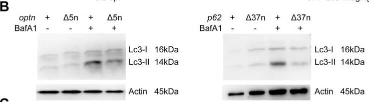
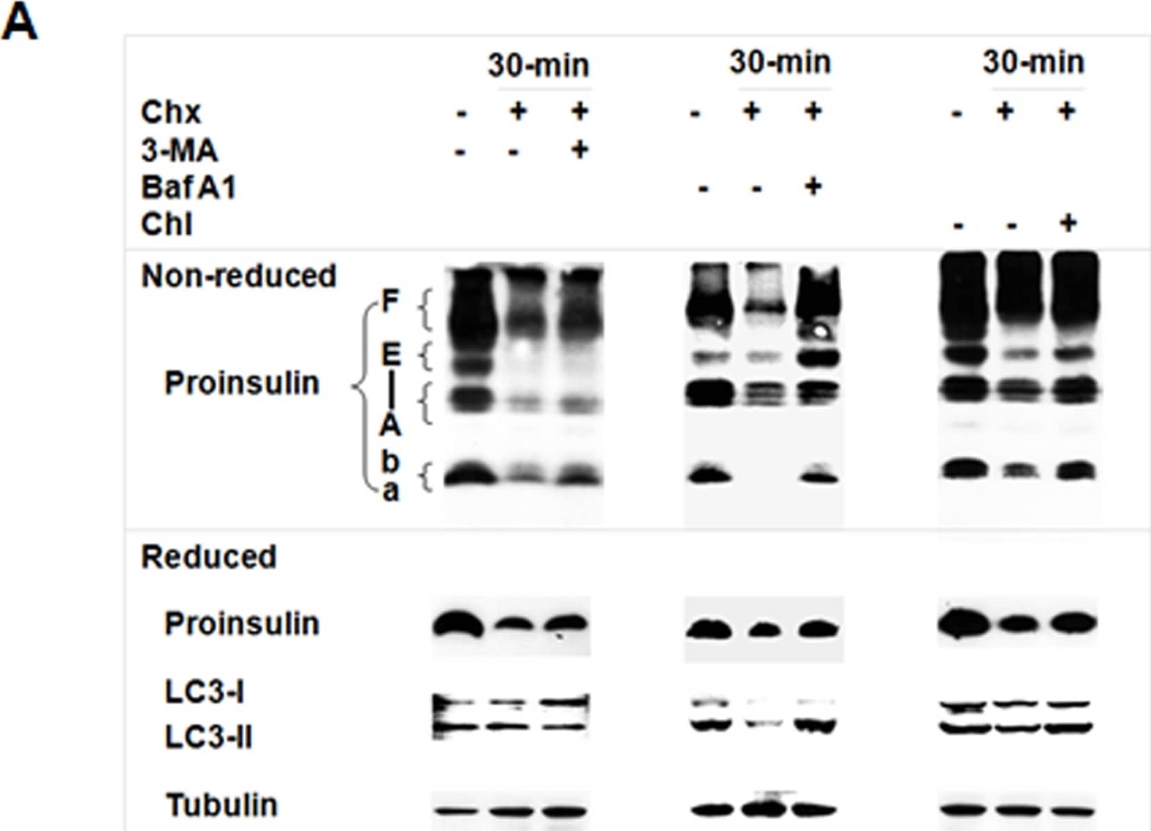


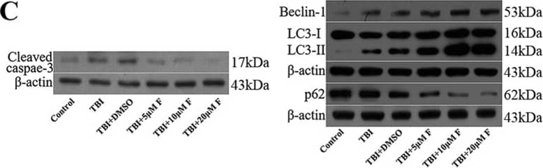
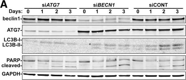

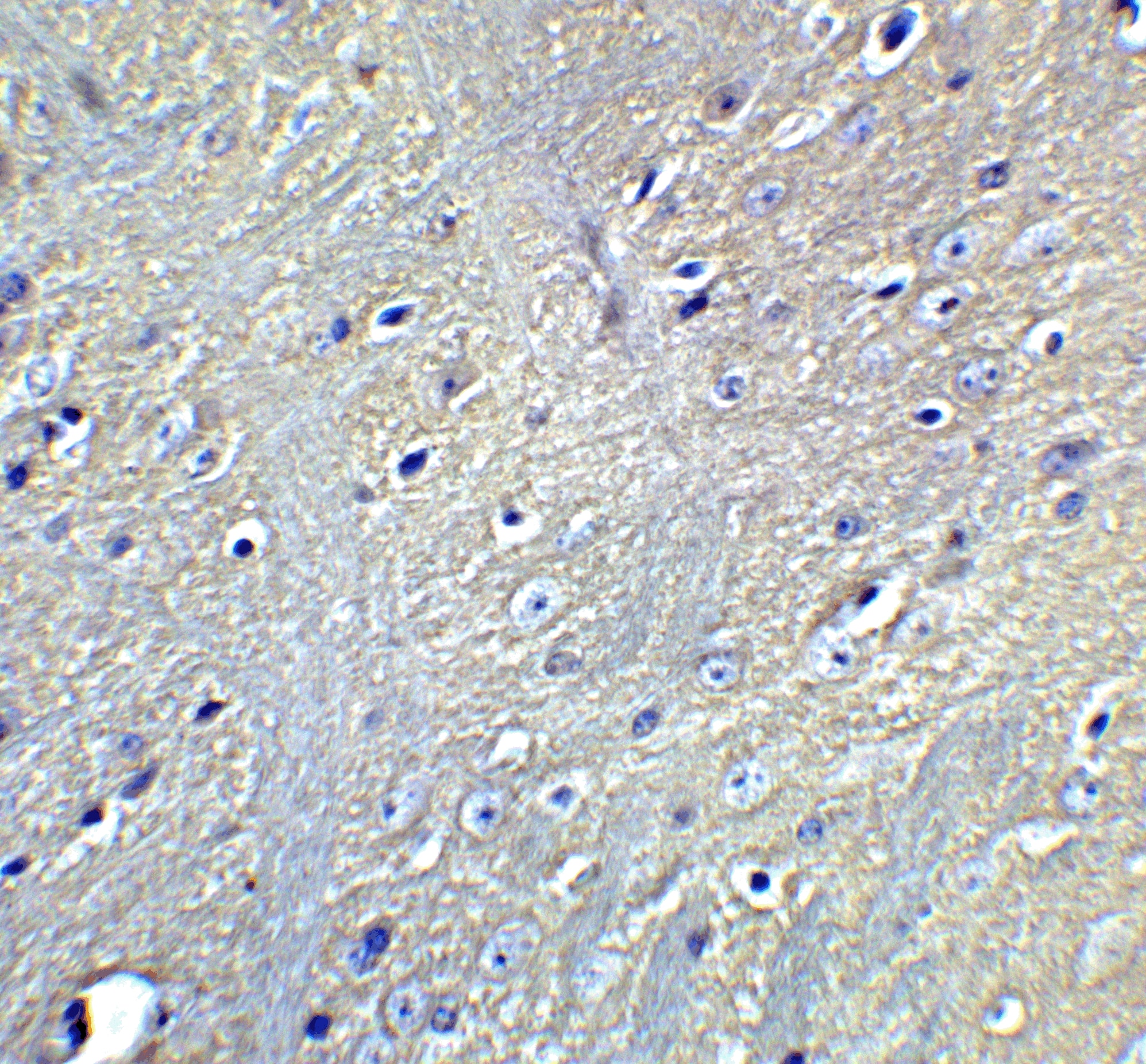
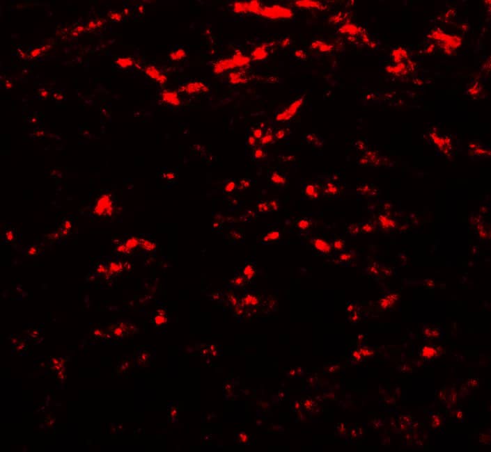
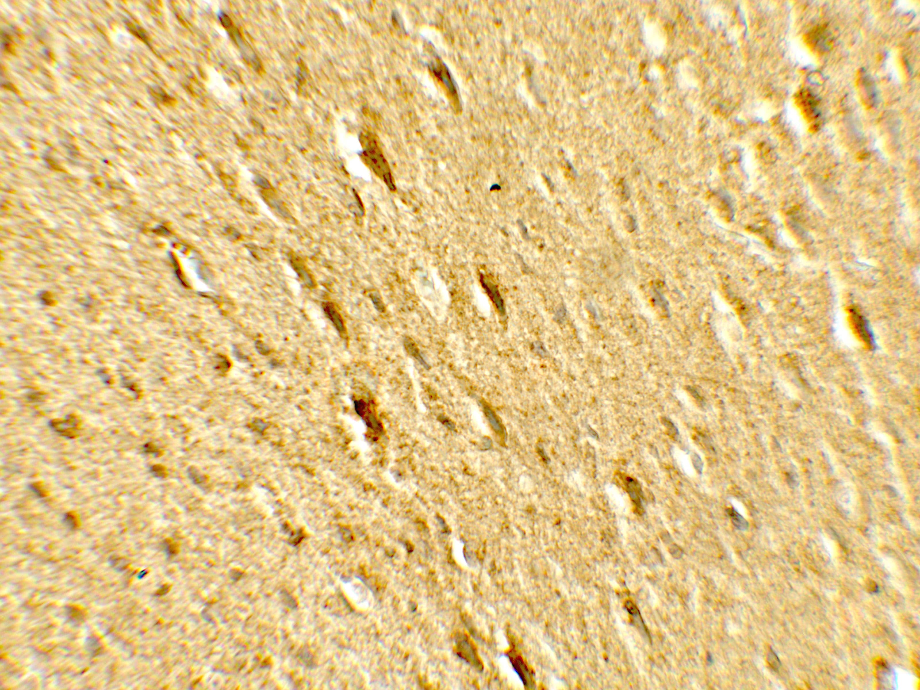


![Immunohistochemistry ATG7 Antibody (683906) [Unconjugated]](https://images.novusbio.com/images/antibody/ATG7_MAB6608_Immunohistochemistry_10631.jpg)
![Simple Western ATG7 Antibody (683906) [Unconjugated]](https://images.novusbio.com/images/antibody/ATG7_MAB6608_Simple_Western_16409.jpg)
![Intracellular Staining by Flow Cytometry AKT [p Ser473] Antibody [Unconjugated] - Pan Specific](https://images.novusbio.com/images/antibody/Akt3_AF887_Flow_Cytometry_8283.jpg)
![Western Blot AKT [p Ser473] Antibody [Unconjugated] - Pan Specific](https://images.novusbio.com/images/af887_phospho-akt-s473-pan-specific-affinity-purified-pab-41202410485440.jpg)
![Western Blot AKT [p Ser473] Antibody [Unconjugated] - Pan Specific](https://images.novusbio.com/images/af887_phospho-akt-s473-pan-specific-affinity-purified-pab-8120255552843.jpg)
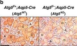
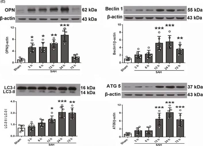
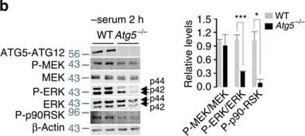


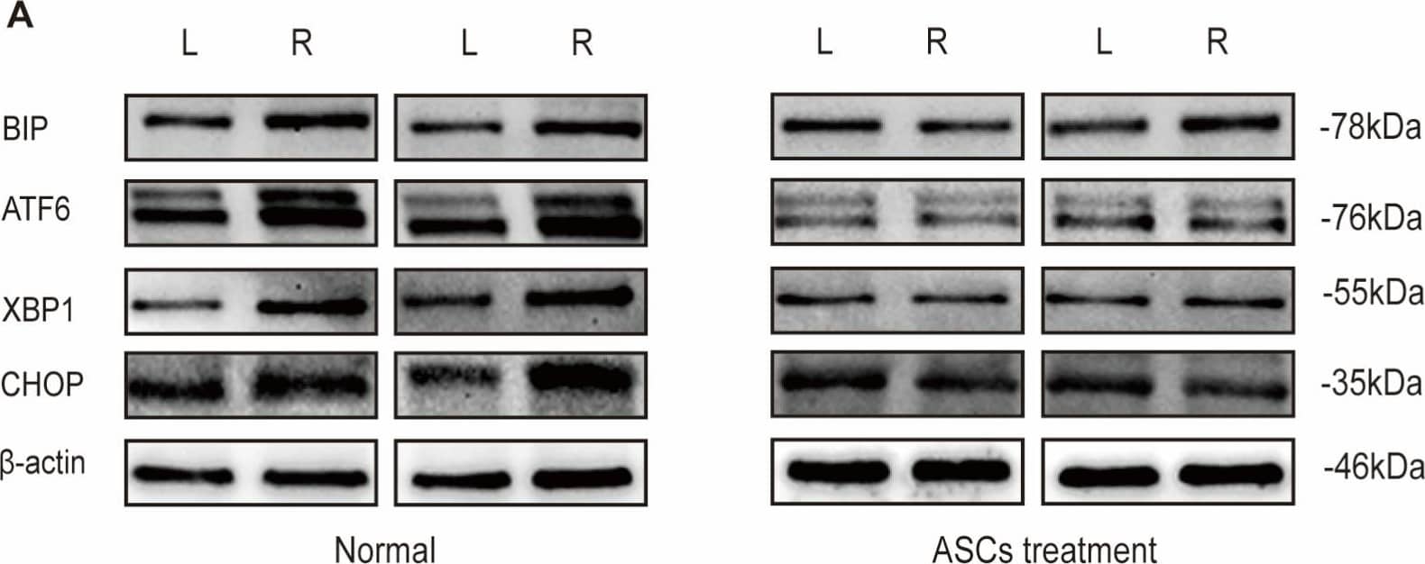
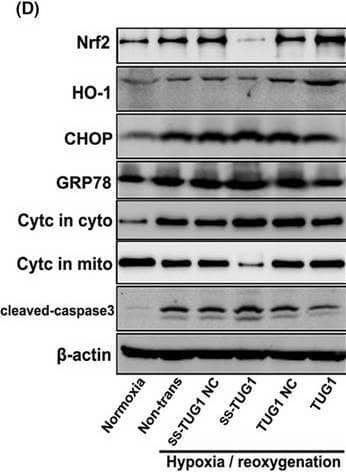
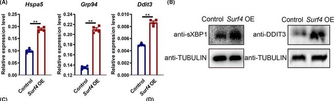


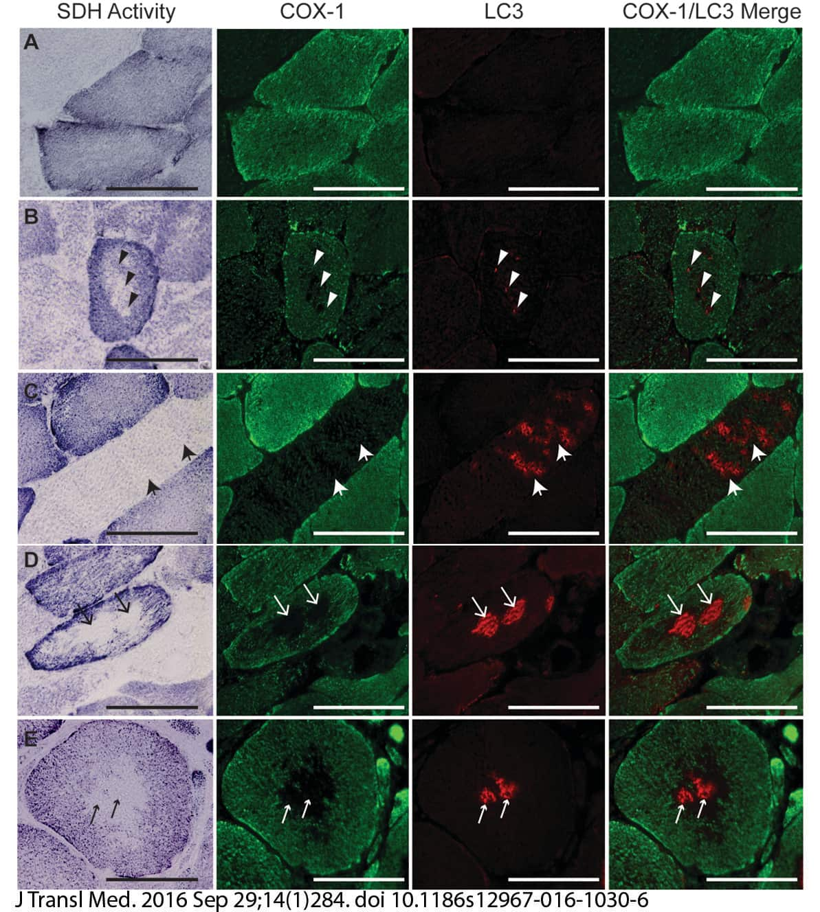


-(01-ml)_NB100-2220_8451.jpg)


 but was not detected in LC3B knockout HeLa cells (right) using rabbit anti-human LC3B polyclonal antibody (Catalog #NB100-2220) at 0.3 ug/mL for 3 hours at room temperature. Cells were stained using the NorthernLights(TM) 557-conjugated anti-Rabbit IgG Secondary Antibody (red; Catalog # NL004) and counterstained with DAPI (blue). Specific staining was localized to cytoplasm.")


. Normal fibers from a Non-PAD sample (row A). Scale bar = 100 uM. Image from verified customer review.")

![Detection of LC3B in mouse ES cell lysates using Rabbit anti- [Catalog # NB100-2220]. LC3 detected at a molecular weight of approximately 17 kDa. Atg5-/- ES cells from Dr. Noboru Mizushima [Mizushima, N. et al. J. Cell Biol. 152 (2001)] Photo courtesy of Dr. Beth Levine, UT SW Medical Center.](http://images.novusbio.com/fullsize/LC3B-Antibody-Western-Blot-NB100-2220-img0047.jpg "Detection of LC3B in mouse ES cell lysates using Rabbit anti- [Catalog # NB100-2220]. LC3 detected at a molecular weight of approximately 17 kDa. Atg5-/- ES cells from Dr. Noboru Mizushima [Mizushima, N. et al. J. Cell Biol. 152 (2001)] Photo courtesy of Dr. Beth Levine, UT SW Medical Center.")

 or treated (+) with CQ. PVDF (Polyvinylidene difluoride) membrane was probed with 0.5 ug/mL rabbit anti-LC3B polyclonal Antibody (Catalog # NB100-2220, Novus Biologicals), followed by 1:2000 dilution of goat anti-rabbit IgG secondary antibody. LC3 detected at a molecular weight of approximately 15 kDa in treated NIH3T3 and PC-12 cells.")
![Detection of LC3I and LC3II in mouse cochlea cell line SV-K1 using Rabbit anti- [Catalog # NB100-2220]. Cells were treated with CQ (1 uM), and As2O3 (1 uM) for 24 hrs. Image from verified customer review.](http://images.novusbio.com/fullsize/LC3B-Antibody-Western-Blot-NB100-2220-img0056.jpg "Detection of LC3I and LC3II in mouse cochlea cell line SV-K1 using Rabbit anti- [Catalog # NB100-2220]. Cells were treated with CQ (1 uM), and As2O3 (1 uM) for 24 hrs. Image from verified customer review.")
 Representative WBs (Western Blots) of LC3B-I, LC3B-II and GAPDH loading control in DANs derived from 4 iPD patients and 4 healthy controls. (B) WBs from three independent experiments were quantified by densitometry and LC3B-II normalized to a loading control. Grouping of data by healthy vs iPD as well as iPD stratified for T/T and C/C genotype. (C) Ratio of LC3B-II (normalized to loading control) following bafilomycin treatment/untreated. Citation: Marrone L, Bus C, Schondorf D, Fitzgerald JC, Kubler M, Schmid B, et al. (2018) Generation of iPSCs carrying a common LRRK2 risk allele for in vitro modeling of idiopathic Parkinson's disease. PLoS ONE 13(3): e0192497. //doi.org/10.1371/journal.pone.0192497")


![Staining of rat brain tissue section. Analysis using Rabbit anti-LC3B Antibody [Catalog # NB100-2220]. Image from verified customer review.](http://images.novusbio.com/fullsize/LC3B-Antibody-Immunohistochemistry-NB100-2220-img0037.jpg "Staining of rat brain tissue section. Analysis using Rabbit anti-LC3B Antibody [Catalog # NB100-2220]. Image from verified customer review.")

![FFPE (Formalin-Fixed Paraffin-Embedded) tissue section of mouse brain using 1:200 dilution of Rabbit anti-LC3B antibody [Catalog # NB100-2220]. The specific signal of LC3 was detected using HRP-conjugated secondary antibody with DAB (3, 3 -diaminobenzidine) reagent, and nuclei of cells were counterstained using hematoxylin. This LC3B antibody generated a low to moderate levels of cytoplasmic staining in the glial cells. The neurons depicted a moderate to strong staining for LC3 in their cytoplasm.](http://images.novusbio.com/fullsize/LC3B-Antibody-Immunohistochemistry-Paraffin-NB100-2220-img0048.jpg "FFPE (Formalin-Fixed Paraffin-Embedded) tissue section of mouse brain using 1:200 dilution of Rabbit anti-LC3B antibody [Catalog # NB100-2220]. The specific signal of LC3 was detected using HRP-conjugated secondary antibody with DAB (3, 3 -diaminobenzidine) reagent, and nuclei of cells were counterstained using hematoxylin. This LC3B antibody generated a low to moderate levels of cytoplasmic staining in the glial cells. The neurons depicted a moderate to strong staining for LC3 in their cytoplasm.")
![Analysis using the Biotin conjugate of Rabbit anti-LC3B Antibody [Catalog # NB100-2220]. Staining of brain, cerebral cortex, neurons with cell processes.](http://images.novusbio.com/fullsize/LC3B-Antibody-Immunohistochemistry-NB100-2220-img0053.jpg "Analysis using the Biotin conjugate of Rabbit anti-LC3B Antibody [Catalog # NB100-2220]. Staining of brain, cerebral cortex, neurons with cell processes.")

, LC3-2/LC3-1 (a,c, N = 8), p62 (a,d, N = 12), TIM23 (a,e, N = 10), TOM20 (a,f, N = 9), HSP60 (a,g, N = 16), optineurin (a,h, N = 9) & NDP52 (a,i, N = 8) protein levels were analyzed in control (p53+/+) or TP53-deficient (p53−/−) HAP1 cells as described in the Methods. Bars represent the means ± SEM of 3-4 independent experiments performed in triplicate & are expressed as percent of control (p53+/+) cells. Actin expression is provided as a representative gel loading control in a. Statistical analyses were performed with GraphPad Prism software by using unpaired Student’s t-test. Significant differences are: ** p 50%; & staining (E) absent, (F) weak, (G) moderate, (H) strong. (upper panel, x200; lower panel, x400). Image collected & cropped by CiteAb from the following publication (//pubmed.ncbi.nlm.nih.gov/29190884), licensed under a CC-BY license. Not internally tested by Novus Biologicals.")
![Western Blot: LC3B Antibody - BSA Free [NB100-2220] - P53 mediates topotecan-induced autophagy through the activation of sestrin 2 & AMPK in colon cancer cells with wild-type p53.A. HCT116 & LS-174T cells were treated with various concentrations of TPT for 24 h. The levels of P53, sestrin2 & p-AMPK were analysed by immunoblotting. B. HCT116 cells were transfected with p53 or sestrin 2 siRNAs for 24 h, treated with or without 1 µg/mL TPT for an additional 24 h, & the indicated proteins were then analysed by immunoblotting. C. HCT116 cells were transfected with LKB1 or CaMKK beta siRNAs, treated with or without 1 µg/mL TPT for an additional 24 h, & the indicated proteins were detected by immunoblotting. Image collected & cropped by CiteAb from the following publication (//pubmed.ncbi.nlm.nih.gov/23024792), licensed under a CC-BY license. Not internally tested by Novus Biologicals.](http://images.novusbio.com/fullsize/nb100-2220_rabbit-polyclonal-lc3b-antibody-310202415334948.jpg "Western Blot: LC3B Antibody - BSA Free [NB100-2220] - P53 mediates topotecan-induced autophagy through the activation of sestrin 2 & AMPK in colon cancer cells with wild-type p53.A. HCT116 & LS-174T cells were treated with various concentrations of TPT for 24 h. The levels of P53, sestrin2 & p-AMPK were analysed by immunoblotting. B. HCT116 cells were transfected with p53 or sestrin 2 siRNAs for 24 h, treated with or without 1 µg/mL TPT for an additional 24 h, & the indicated proteins were then analysed by immunoblotting. C. HCT116 cells were transfected with LKB1 or CaMKK beta siRNAs, treated with or without 1 µg/mL TPT for an additional 24 h, & the indicated proteins were detected by immunoblotting. Image collected & cropped by CiteAb from the following publication (//pubmed.ncbi.nlm.nih.gov/23024792), licensed under a CC-BY license. Not internally tested by Novus Biologicals.")
![Western Blot: LC3B Antibody - BSA Free [NB100-2220] - Specific inhibitor SP600125 or siRNA directed JNK blocked ceramide-induced autophagy & up-regulation of LC3 expression. (A) SUNE1 cells were treated with 20 μM ceramide for 24 h in the absence or presence of SP600125 or JNK1/2 siRNA. Lysates were analyzed by immunoblotting. (B) Autophagosome formation was visualized using YFP-LC3 expressing & observed under a confocal microscope. Representative immunofluorescence pictures are shown at the original magnification × 1000. (C) The expression of LC3 mRNA was examined by RT-PCR analysis. Image collected & cropped by CiteAb from the following publication (//pubmed.ncbi.nlm.nih.gov/21943220), licensed under a CC-BY license. Not internally tested by Novus Biologicals.](http://images.novusbio.com/fullsize/nb100-2220_rabbit-polyclonal-lc3b-antibody-31020241535643.jpg "Western Blot: LC3B Antibody - BSA Free [NB100-2220] - Specific inhibitor SP600125 or siRNA directed JNK blocked ceramide-induced autophagy & up-regulation of LC3 expression. (A) SUNE1 cells were treated with 20 μM ceramide for 24 h in the absence or presence of SP600125 or JNK1/2 siRNA. Lysates were analyzed by immunoblotting. (B) Autophagosome formation was visualized using YFP-LC3 expressing & observed under a confocal microscope. Representative immunofluorescence pictures are shown at the original magnification × 1000. (C) The expression of LC3 mRNA was examined by RT-PCR analysis. Image collected & cropped by CiteAb from the following publication (//pubmed.ncbi.nlm.nih.gov/21943220), licensed under a CC-BY license. Not internally tested by Novus Biologicals.")
![Western Blot: LC3B Antibody - BSA Free [NB100-2220] - Glutamine deprivation induces IL-8 secretion.(a) RT–PCR analysis of LC3, IL-8, CHOP & GADD34 in U2OS cells subjected to glutamine deprivation (−Q) or exposed to TPG (1 μM). (b) Immunoblot analysis of ER stress- & autophagy-related proteins in U2OS cells subjected to glutamine deprivation or TPG treatment. Samples are run in triplicate. (c) Cytokine array analysis of conditioned media from U2OS & A549 cells grown in the presence (+Q) or absence (−Q) of glutamine. (d) IL-8 enzyme-linked immunosorbent assay of conditioned media from U2OS & A549 cells grown in the presence (+Q) or absence (−Q) of glutamine. Error bars in all figures represent s.d. of three biological replicates. Image collected & cropped by CiteAb from the following publication (//www.nature.com/articles/ncomms5900), licensed under a CC-BY license. Not internally tested by Novus Biologicals.](http://images.novusbio.com/fullsize/nb100-2220_rabbit-polyclonal-lc3b-antibody-310202415395934.jpg "Western Blot: LC3B Antibody - BSA Free [NB100-2220] - Glutamine deprivation induces IL-8 secretion.(a) RT–PCR analysis of LC3, IL-8, CHOP & GADD34 in U2OS cells subjected to glutamine deprivation (−Q) or exposed to TPG (1 μM). (b) Immunoblot analysis of ER stress- & autophagy-related proteins in U2OS cells subjected to glutamine deprivation or TPG treatment. Samples are run in triplicate. (c) Cytokine array analysis of conditioned media from U2OS & A549 cells grown in the presence (+Q) or absence (−Q) of glutamine. (d) IL-8 enzyme-linked immunosorbent assay of conditioned media from U2OS & A549 cells grown in the presence (+Q) or absence (−Q) of glutamine. Error bars in all figures represent s.d. of three biological replicates. Image collected & cropped by CiteAb from the following publication (//www.nature.com/articles/ncomms5900), licensed under a CC-BY license. Not internally tested by Novus Biologicals.")
![Western Blot: LC3B Antibody - BSA Free [NB100-2220] - NDs API improves ATO-based therapy in vivo. HepG2 tumor-bearing nude mice administered intravenously (i.v.) w/ NS (200 μL), NDs (5 mg kg−1), ATO (0.75 mg kg−1), ATO (1.5 mg kg−1), NDs–0.75 mg kg−1 ATO mixture,/NDs–1.5 mg kg−1 ATO mixture daily for 5 consecutive days every week, w/ 13-week cycles c Immunoblots of autophagy-related proteins LC3-II, p62 (left); semi-quantified analysis (n = 3) in tumor tissues from mice after treatment (right). GAPDH used as loading control. Normalized band densities shown below each band. Image collected & cropped by CiteAb from following publication (//pubmed.ncbi.nlm.nih.gov/30341298), licensed under a CC-BY license. Not internally tested by Novus Biologicals.](http://images.novusbio.com/fullsize/nb100-2220_rabbit-polyclonal-lc3b-antibody-31020241539454.jpg "Western Blot: LC3B Antibody - BSA Free [NB100-2220] - NDs API improves ATO-based therapy in vivo. HepG2 tumor-bearing nude mice administered intravenously (i.v.) w/ NS (200 μL), NDs (5 mg kg−1), ATO (0.75 mg kg−1), ATO (1.5 mg kg−1), NDs–0.75 mg kg−1 ATO mixture,/NDs–1.5 mg kg−1 ATO mixture daily for 5 consecutive days every week, w/ 13-week cycles c Immunoblots of autophagy-related proteins LC3-II, p62 (left); semi-quantified analysis (n = 3) in tumor tissues from mice after treatment (right). GAPDH used as loading control. Normalized band densities shown below each band. Image collected & cropped by CiteAb from following publication (//pubmed.ncbi.nlm.nih.gov/30341298), licensed under a CC-BY license. Not internally tested by Novus Biologicals.")
![Immunocytochemistry/ Immunofluorescence: LC3B Antibody - BSA Free [NB100-2220] - NDs API improves the ATO-based therapy in vivo. HepG2 tumor-bearing nude mice administered intravenously (i.v.) w/ NS (200 μL), NDs (5 mg kg−1), ATO (0.75 mg kg−1), ATO (1.5 mg kg−1), NDs–0.75 mg kg−1 ATO mixture,/NDs–1.5 mg kg−1 ATO mixture daily for 5 consecutive days every week, w/ 13-week cycles. g Immunostaining of LC3 & ATG13 in tumor tissues of NDs–1.5 mg kg−1 ATO-treated mice. Schematic showing (left) & imaging (right) indicate that LC3/Atg13 puncta in periphery & around the blood vessels inside the tumors. Scale bars: 20 μm. Error bars s.d. Image collected & cropped by CiteAb from the following publication (//pubmed.ncbi.nlm.nih.gov/30341298), licensed under a CC-BY license. Not internally tested by Novus Biologicals.](http://images.novusbio.com/fullsize/nb100-2220_rabbit-polyclonal-lc3b-antibody-310202415382444.jpg "Immunocytochemistry/ Immunofluorescence: LC3B Antibody - BSA Free [NB100-2220] - NDs API improves the ATO-based therapy in vivo. HepG2 tumor-bearing nude mice administered intravenously (i.v.) w/ NS (200 μL), NDs (5 mg kg−1), ATO (0.75 mg kg−1), ATO (1.5 mg kg−1), NDs–0.75 mg kg−1 ATO mixture,/NDs–1.5 mg kg−1 ATO mixture daily for 5 consecutive days every week, w/ 13-week cycles. g Immunostaining of LC3 & ATG13 in tumor tissues of NDs–1.5 mg kg−1 ATO-treated mice. Schematic showing (left) & imaging (right) indicate that LC3/Atg13 puncta in periphery & around the blood vessels inside the tumors. Scale bars: 20 μm. Error bars s.d. Image collected & cropped by CiteAb from the following publication (//pubmed.ncbi.nlm.nih.gov/30341298), licensed under a CC-BY license. Not internally tested by Novus Biologicals.")
![Immunohistochemistry: LC3B Antibody - BSA Free [NB100-2220] - LC3, CRF, & HIF-1 alpha expression. (a) for each antibody the spatial distribution of immunohistochemical staining was the same in cases with spontaneous & induced labor. (b) The analysis of data from optical density values of Western blotting bands shows no differences of LC3-II & HIF-1 alpha expression between groups. On the contrary CRF is higher in spontaneous labor. LC3-II, HIF-1 alpha , & CRF are normalized onto alpha -tubulin. (c) No correlation between LC3-II & CRF expression was found. SP: spontaneous labor; PG: induction with prostaglandin; PO: induction with prostaglandin & oxytocin; OX: induction with oxytocin. Image collected & cropped by CiteAb from the following publication (//pubmed.ncbi.nlm.nih.gov/23956998), licensed under a CC-BY license. Not internally tested by Novus Biologicals.](http://images.novusbio.com/fullsize/nb100-2220_rabbit-polyclonal-lc3b-antibody-310202415392554.jpg "Immunohistochemistry: LC3B Antibody - BSA Free [NB100-2220] - LC3, CRF, & HIF-1 alpha expression. (a) for each antibody the spatial distribution of immunohistochemical staining was the same in cases with spontaneous & induced labor. (b) The analysis of data from optical density values of Western blotting bands shows no differences of LC3-II & HIF-1 alpha expression between groups. On the contrary CRF is higher in spontaneous labor. LC3-II, HIF-1 alpha , & CRF are normalized onto alpha -tubulin. (c) No correlation between LC3-II & CRF expression was found. SP: spontaneous labor; PG: induction with prostaglandin; PO: induction with prostaglandin & oxytocin; OX: induction with oxytocin. Image collected & cropped by CiteAb from the following publication (//pubmed.ncbi.nlm.nih.gov/23956998), licensed under a CC-BY license. Not internally tested by Novus Biologicals.")
![Immunocytochemistry/ Immunofluorescence: LC3B Antibody - BSA Free [NB100-2220] - The time-dependent effects of melatonin on the LC3-II & Beclin 1 expression in U87MG (a, b) & A172 (c, d) cells. The cells were treated with 1 mM melatonin for 4, 12, 24 or 36 h. PBS was used as a negative control, & rapamycin (Rap 200 nM) served as a positive control. The levels of LC3-II & Beclin-1 expression were determined by a Western blot analysis. The protein bands for each regimen were quantified by densitometry, & their differences are presented in the graph. The values represent the mean ± S.E.M. for three separate determinations. Statistical significance * at P 10 puncta per cell from three independent experiments is depicted. Bars represent mean±s.d. from three independent experiments (>50 cells per experiment). The statistical significance (P value) was determined by a two-tailed, paired Student’s t-test. *P<0.05. (c) Immunoblot analysis of U2OS cells subjected to glutamine deprivation with or without 400 nM BafA1. Cells were pretreated with BafA1 for 1 h before & during exposure to glutamine-deficient medium. Autophagic activity was monitored by detection of p62 & LC3-II proteins. (d) U2OS mCherry-GFP-LC3 cells were cultured in the presence (+Q) or absence (−Q) of glutamine for 18 h. Red vesicles denote autolysosomes, whereas yellow vesicles represent autophagosomes. Bars indicate numbers of yellow vesicles (autophagosomes) or red vesicles (autolysosomes) per cell±s.d. (e) Images of U2OS mCherry-GFP-LC3 cells cultured for 18 h in the presence (+Q) or absence (−Q) of glutamine. Scale bar, 10 μm. (f) Phagophore formation in mCherry-ULK1. Scale bar, 10 μm. (g) mCherry-ATG5-expressing U2OS cells after 24 h in the presence (+Q) or absence (−Q) of glutamine. Scale bar, 10 μm. Image collected & cropped by CiteAb from the following publication (//www.nature.com/articles/ncomms5900), licensed under a CC-BY license. Not internally tested by Novus Biologicals.](http://images.novusbio.com/fullsize/nb100-2220_rabbit-polyclonal-lc3b-antibody-31020241554193.jpg "Immunocytochemistry/ Immunofluorescence: LC3B Antibody - BSA Free [NB100-2220] - The time-dependent effects of melatonin on the LC3-II & Beclin 1 expression in U87MG (a, b) & A172 (c, d) cells. The cells were treated with 1 mM melatonin for 4, 12, 24 or 36 h. PBS was used as a negative control, & rapamycin (Rap 200 nM) served as a positive control. The levels of LC3-II & Beclin-1 expression were determined by a Western blot analysis. The protein bands for each regimen were quantified by densitometry, & their differences are presented in the graph. The values represent the mean ± S.E.M. for three separate determinations. Statistical significance * at P 10 puncta per cell from three independent experiments is depicted. Bars represent mean±s.d. from three independent experiments (>50 cells per experiment). The statistical significance (P value) was determined by a two-tailed, paired Student’s t-test. *P<0.05. (c) Immunoblot analysis of U2OS cells subjected to glutamine deprivation with or without 400 nM BafA1. Cells were pretreated with BafA1 for 1 h before & during exposure to glutamine-deficient medium. Autophagic activity was monitored by detection of p62 & LC3-II proteins. (d) U2OS mCherry-GFP-LC3 cells were cultured in the presence (+Q) or absence (−Q) of glutamine for 18 h. Red vesicles denote autolysosomes, whereas yellow vesicles represent autophagosomes. Bars indicate numbers of yellow vesicles (autophagosomes) or red vesicles (autolysosomes) per cell±s.d. (e) Images of U2OS mCherry-GFP-LC3 cells cultured for 18 h in the presence (+Q) or absence (−Q) of glutamine. Scale bar, 10 μm. (f) Phagophore formation in mCherry-ULK1. Scale bar, 10 μm. (g) mCherry-ATG5-expressing U2OS cells after 24 h in the presence (+Q) or absence (−Q) of glutamine. Scale bar, 10 μm. Image collected & cropped by CiteAb from the following publication (//www.nature.com/articles/ncomms5900), licensed under a CC-BY license. Not internally tested by Novus Biologicals.")
![Western Blot: LC3B Antibody - BSA Free [NB100-2220] - Glucocorticoids promote BCG survival in macrophages by inhibiting autophagy. (a) RAW264.7 cells were pretreated with dexamethasone (DEX) (1 μM) or vehicle control ethanol (mock) for 6 hr & then challenged with M. bovis BCG (MOI 5) or treated with the vehicle control DMSO or rapamycin (4 μM). LC3 level was detected with Western blot analysis. & GAPDH was used as the loading control. Full-length blots are presented in Supplementary Fig. 12. Data are representative of three independent experiments with similar results. (b,c) RAW264.7 cells were treated as described in (a), & viable bacilli were determined by CFU assays at 6 hpi (b). Survival rate was calculated compared with that of mock-treated cells (c). Data are shown as the mean ± SEM of three independent experiments. *p < 0.05. Image collected & cropped by CiteAb from the following publication (//www.nature.com/articles/s41598-017-01174-9), licensed under a CC-BY license. Not internally tested by Novus Biologicals.](http://images.novusbio.com/fullsize/nb100-2220_rabbit-polyclonal-lc3b-antibody-310202415532059.jpg "Western Blot: LC3B Antibody - BSA Free [NB100-2220] - Glucocorticoids promote BCG survival in macrophages by inhibiting autophagy. (a) RAW264.7 cells were pretreated with dexamethasone (DEX) (1 μM) or vehicle control ethanol (mock) for 6 hr & then challenged with M. bovis BCG (MOI 5) or treated with the vehicle control DMSO or rapamycin (4 μM). LC3 level was detected with Western blot analysis. & GAPDH was used as the loading control. Full-length blots are presented in Supplementary Fig. 12. Data are representative of three independent experiments with similar results. (b,c) RAW264.7 cells were treated as described in (a), & viable bacilli were determined by CFU assays at 6 hpi (b). Survival rate was calculated compared with that of mock-treated cells (c). Data are shown as the mean ± SEM of three independent experiments. *p < 0.05. Image collected & cropped by CiteAb from the following publication (//www.nature.com/articles/s41598-017-01174-9), licensed under a CC-BY license. Not internally tested by Novus Biologicals.")
![Western Blot: LC3B Antibody - BSA Free [NB100-2220] - CysC regulates AMPK activity during the induction of autophagy. (a) Immunoblotting analysis of autophagy regulators. N2a cells expressing SOD1 treated with or without CysC (1 μM) for 6 h. The lysates analyzed by immunoblotting using antibodies for phosphorylated AMPK (pAMPK), phosphorylated mTOR (pmTOR), phosphorylated PKCδ (pPKCδ), Myc & beta -actin. (b) Inactivation of AMPK by mutant SOD1 expression. Each relative pAMPK level normalized by beta -actin in (a) is quantified. *P<0.05, **P<0.01 versus mock. (c) Activation of AMPK by CysC treatment. Relative levels of pAMPK for CysC-treated samples normalized by that of PBS-treated control, which is shown as the broken line, in (a) quantified. *P<0.05 versus PBS-treated controls. (d) CysC induced autophagy through the AMPK activation. N2a cells treated with CysC (1 μM), CC (5 μM) or AICA-riboside (AICAR, 5 mM) for 12 h. The lysates analyzed by immunoblotting using antibodies against pAMPK, LC3 & Tubulin (left panel). Quantification of immunoblots was plotted (right panel). (e) The effect of pAMPK activation on neuroprotection by CysC. N2a cells expressing G85R SOD1 mutant treated with CysC (0.2 μM), CC (5 μM) or AICAR (5 mM). Cell viability was measured by the MTS assay. Data expressed as means±S.E.M. from 3 independent experiments. Each experiment was performed in triplicate. *P<0.01 compared to non-treated control, #P<0.01 compared to CysC-treated one. (f) Immunoblotting analysis of pAMPK in SOD1 transgenic mouse spinal cords. The spinal cord lysates from the transgenic mice at indicated ages analyzed for the levels of pAMPK, SOD1 & beta -actin (upper panel). Expression levels of pAMPK normalized by beta -actin (lower panel). All data expressed as means±S.E.M. from 3 independent experiments Image collected & cropped by CiteAb from the following publication (//www.nature.com/articles/cddis2014459), licensed under a CC-BY license. Not internally tested by Novus Biologicals.](http://images.novusbio.com/fullsize/nb100-2220_rabbit-polyclonal-lc3b-antibody-310202415535189.jpg "Western Blot: LC3B Antibody - BSA Free [NB100-2220] - CysC regulates AMPK activity during the induction of autophagy. (a) Immunoblotting analysis of autophagy regulators. N2a cells expressing SOD1 treated with or without CysC (1 μM) for 6 h. The lysates analyzed by immunoblotting using antibodies for phosphorylated AMPK (pAMPK), phosphorylated mTOR (pmTOR), phosphorylated PKCδ (pPKCδ), Myc & beta -actin. (b) Inactivation of AMPK by mutant SOD1 expression. Each relative pAMPK level normalized by beta -actin in (a) is quantified. *P<0.05, **P<0.01 versus mock. (c) Activation of AMPK by CysC treatment. Relative levels of pAMPK for CysC-treated samples normalized by that of PBS-treated control, which is shown as the broken line, in (a) quantified. *P<0.05 versus PBS-treated controls. (d) CysC induced autophagy through the AMPK activation. N2a cells treated with CysC (1 μM), CC (5 μM) or AICA-riboside (AICAR, 5 mM) for 12 h. The lysates analyzed by immunoblotting using antibodies against pAMPK, LC3 & Tubulin (left panel). Quantification of immunoblots was plotted (right panel). (e) The effect of pAMPK activation on neuroprotection by CysC. N2a cells expressing G85R SOD1 mutant treated with CysC (0.2 μM), CC (5 μM) or AICAR (5 mM). Cell viability was measured by the MTS assay. Data expressed as means±S.E.M. from 3 independent experiments. Each experiment was performed in triplicate. *P<0.01 compared to non-treated control, #P<0.01 compared to CysC-treated one. (f) Immunoblotting analysis of pAMPK in SOD1 transgenic mouse spinal cords. The spinal cord lysates from the transgenic mice at indicated ages analyzed for the levels of pAMPK, SOD1 & beta -actin (upper panel). Expression levels of pAMPK normalized by beta -actin (lower panel). All data expressed as means±S.E.M. from 3 independent experiments Image collected & cropped by CiteAb from the following publication (//www.nature.com/articles/cddis2014459), licensed under a CC-BY license. Not internally tested by Novus Biologicals.")
![Immunocytochemistry/ Immunofluorescence: LC3B Antibody - BSA Free [NB100-2220] - Vps34 inhibition blocks autophagy initiation & causes accumulation of ubiquitin-positive & p62-positive structures. a Representative confocal images of cortical neurons treated with vehicle, Bafilomycin A1 (BafA1) at 50 nM, VPS34IN1 at 3 µM or cotreated for 3 h. Arrows highlight LC3 & p62 structures. Right panel, bar graphs denote average object intensity, per cell (mean ± SEM, N = 49-60 cells, from three independent experiments). Scale bar, 10 µm. ***p < 0.001 in one-way ANOVA, Holm–Sidak’s multiple comparisons test. b Representative confocal images of cortical neurons treated as in a & immunostained for LAMP-1, LC3, & p62. Airyscan insets highlight position of LC3 & p62 structures relative to LAMP-1-positive membranes. Scale bar, 10 µm. c Representative confocal images of cultured cortical neurons treated with vehicle or VPS34IN1 at 3 µM for 24 h. Arrows highlight p62 & ubiquitin colocalization. Scale bar, 10 µm Image collected & cropped by CiteAb from the following publication (//www.nature.com/articles/s41467-017-02533-w), licensed under a CC-BY license. Not internally tested by Novus Biologicals.](http://images.novusbio.com/fullsize/nb100-2220_rabbit-polyclonal-lc3b-antibody-310202415535128.jpg "Immunocytochemistry/ Immunofluorescence: LC3B Antibody - BSA Free [NB100-2220] - Vps34 inhibition blocks autophagy initiation & causes accumulation of ubiquitin-positive & p62-positive structures. a Representative confocal images of cortical neurons treated with vehicle, Bafilomycin A1 (BafA1) at 50 nM, VPS34IN1 at 3 µM or cotreated for 3 h. Arrows highlight LC3 & p62 structures. Right panel, bar graphs denote average object intensity, per cell (mean ± SEM, N = 49-60 cells, from three independent experiments). Scale bar, 10 µm. ***p < 0.001 in one-way ANOVA, Holm–Sidak’s multiple comparisons test. b Representative confocal images of cortical neurons treated as in a & immunostained for LAMP-1, LC3, & p62. Airyscan insets highlight position of LC3 & p62 structures relative to LAMP-1-positive membranes. Scale bar, 10 µm. c Representative confocal images of cultured cortical neurons treated with vehicle or VPS34IN1 at 3 µM for 24 h. Arrows highlight p62 & ubiquitin colocalization. Scale bar, 10 µm Image collected & cropped by CiteAb from the following publication (//www.nature.com/articles/s41467-017-02533-w), licensed under a CC-BY license. Not internally tested by Novus Biologicals.")
![Immunocytochemistry/ Immunofluorescence: LC3B Antibody - BSA Free [NB100-2220] - ORP1L & VAP-A form cholesterol-dependent ER-AV contact sites(a) MelJuSo cells expressing GFP or GFP-tagged ORP1L mutants were fixed & stained for LC3 & Calnexin. Y477 & D478 in the FFAT motif were mutated to alanines (A) in the ORP1L ydaa mutant. Scale bar, 10 μm. Right: co-immunoprecipitation for ORP1L (mutants) with VAP-A. GFP-ORP1L mutants or GFP were isolated from lysates of HEK293T cells co-overexpressing HA-VAPA using GFP-Trap beads. Western blot filters were probed for isolated GFP-tagged proteins, the associated HA-VAP-A & the input HA-VAP-A, as indicated. (b) Cryo-immuno-EM on HeLa cells expressing HA-LC3 & GFP-ORP1L delta ORD, as detected by HA10 nm & GFP15 nm gold antibodies. Insets show ORP1L labelling in the membrane contact site between ER & autophagosome. The membranes of the ER are depicted in the bottom inset. Scale bar, 50 nm. (c) Three-colour super-resolution image of an autophagosomal vesicle labelled by LC3 (green), ORP1L (blue) & the ER protein VAP-A (red). Scale bar, 500 nm (d) MelJuSo cells cultured either in lipid depleted serum or control medium were fixed & stained for LC3 & ER marker Calnexin. Scale bar, 10 μm. Right panel: Manders coefficient for LC3 localization to the ER was calculated on at least 10 cells over three independent experiments. Bars indicate mean+s.d. Student's t-test statistical analysis (****P<0.0001). Image collected & cropped by CiteAb from the following publication (//pubmed.ncbi.nlm.nih.gov/27283760), licensed under a CC-BY license. Not internally tested by Novus Biologicals.](http://images.novusbio.com/fullsize/nb100-2220_rabbit-polyclonal-lc3b-antibody-31020241554525.jpg "Immunocytochemistry/ Immunofluorescence: LC3B Antibody - BSA Free [NB100-2220] - ORP1L & VAP-A form cholesterol-dependent ER-AV contact sites(a) MelJuSo cells expressing GFP or GFP-tagged ORP1L mutants were fixed & stained for LC3 & Calnexin. Y477 & D478 in the FFAT motif were mutated to alanines (A) in the ORP1L ydaa mutant. Scale bar, 10 μm. Right: co-immunoprecipitation for ORP1L (mutants) with VAP-A. GFP-ORP1L mutants or GFP were isolated from lysates of HEK293T cells co-overexpressing HA-VAPA using GFP-Trap beads. Western blot filters were probed for isolated GFP-tagged proteins, the associated HA-VAP-A & the input HA-VAP-A, as indicated. (b) Cryo-immuno-EM on HeLa cells expressing HA-LC3 & GFP-ORP1L delta ORD, as detected by HA10 nm & GFP15 nm gold antibodies. Insets show ORP1L labelling in the membrane contact site between ER & autophagosome. The membranes of the ER are depicted in the bottom inset. Scale bar, 50 nm. (c) Three-colour super-resolution image of an autophagosomal vesicle labelled by LC3 (green), ORP1L (blue) & the ER protein VAP-A (red). Scale bar, 500 nm (d) MelJuSo cells cultured either in lipid depleted serum or control medium were fixed & stained for LC3 & ER marker Calnexin. Scale bar, 10 μm. Right panel: Manders coefficient for LC3 localization to the ER was calculated on at least 10 cells over three independent experiments. Bars indicate mean+s.d. Student's t-test statistical analysis (****P<0.0001). Image collected & cropped by CiteAb from the following publication (//pubmed.ncbi.nlm.nih.gov/27283760), licensed under a CC-BY license. Not internally tested by Novus Biologicals.")
![Immunohistochemistry: LC3B Antibody - BSA Free [NB100-2220] - OSI induces autophagy through upregulation of MCT1 in CRC cells.a Immunoblotting analysis of MCT1 expression in CRC cells treated with or without 5 μM OSI for 24 h. b Immunoblotting analysis of MCT1 & phosphorylated AMPK in tumor xenografts obtained from vehicle- or OSI-treated mice. (Each protein of interest from each group was electrophoretically transferred onto a PVDF membrane, incubated with indicated primary & secondary antibodies, & developed as a digital image.) c Immunohistochemical analysis of MCT1 expression in tumor xenografts. Scale bar, 50 μm. d Relative intensity of MCT1 staining in (c). e CRC cells were transfected with siScramble or siMCT1 for 24 h, followed by treatment with or without 5 μM OSI for another 24 h. The protein levels of LC3, phosphorylated AMPK, phosphorylated LKB1 & MCT1 were analyzed by immunoblotting. f CRC cells were treated as in (e), the endogenous LC3 puncta were analyzed by immunofluorescence. Scale bar, 10 μm. g The number of LC3 puncta in (f). h CRC cells were transfected with empty vector or Flag-MCT1 plasmid for 48 h, the protein levels of MCT1 & phosphorylated AMPK were analyzed by immunoblotting. i CRC cells were treated as in (h), the endogenous LC3 puncta were analyzed by immunofluorescence. Scale bar, 10 μm. j The number of LC3 puncta in (i). k Immunoblotting analysis of LC3, MCT1 & phosphorylated AMPK levels in CRC cells co-transfected with Flag-MCT1 & DN-AMPK plasmids for 48 h. l CRC cells were treated as in (k), the endogenous LC3 puncta were analyzed by immunofluorescence. Scale bar, 10 μm. m The number of LC3 puncta per cell in (l). Data are presented as mean SEM, Student’s t-test, & are representative of three independent experiments. *P < 0.05; ***P < 0.001 Image collected & cropped by CiteAb from the following publication (//pubmed.ncbi.nlm.nih.gov/31409796), licensed under a CC-BY license. Not internally tested by Novus Biologicals.](http://images.novusbio.com/fullsize/nb100-2220_rabbit-polyclonal-lc3b-antibody-310202415532016.jpg "Immunohistochemistry: LC3B Antibody - BSA Free [NB100-2220] - OSI induces autophagy through upregulation of MCT1 in CRC cells.a Immunoblotting analysis of MCT1 expression in CRC cells treated with or without 5 μM OSI for 24 h. b Immunoblotting analysis of MCT1 & phosphorylated AMPK in tumor xenografts obtained from vehicle- or OSI-treated mice. (Each protein of interest from each group was electrophoretically transferred onto a PVDF membrane, incubated with indicated primary & secondary antibodies, & developed as a digital image.) c Immunohistochemical analysis of MCT1 expression in tumor xenografts. Scale bar, 50 μm. d Relative intensity of MCT1 staining in (c). e CRC cells were transfected with siScramble or siMCT1 for 24 h, followed by treatment with or without 5 μM OSI for another 24 h. The protein levels of LC3, phosphorylated AMPK, phosphorylated LKB1 & MCT1 were analyzed by immunoblotting. f CRC cells were treated as in (e), the endogenous LC3 puncta were analyzed by immunofluorescence. Scale bar, 10 μm. g The number of LC3 puncta in (f). h CRC cells were transfected with empty vector or Flag-MCT1 plasmid for 48 h, the protein levels of MCT1 & phosphorylated AMPK were analyzed by immunoblotting. i CRC cells were treated as in (h), the endogenous LC3 puncta were analyzed by immunofluorescence. Scale bar, 10 μm. j The number of LC3 puncta in (i). k Immunoblotting analysis of LC3, MCT1 & phosphorylated AMPK levels in CRC cells co-transfected with Flag-MCT1 & DN-AMPK plasmids for 48 h. l CRC cells were treated as in (k), the endogenous LC3 puncta were analyzed by immunofluorescence. Scale bar, 10 μm. m The number of LC3 puncta per cell in (l). Data are presented as mean SEM, Student’s t-test, & are representative of three independent experiments. *P < 0.05; ***P < 0.001 Image collected & cropped by CiteAb from the following publication (//pubmed.ncbi.nlm.nih.gov/31409796), licensed under a CC-BY license. Not internally tested by Novus Biologicals.")
![Western Blot: LC3B Antibody - BSA Free [NB100-2220] - Abrogation of autophagy by siRNAs targeting autophagy-related genes in gastric cancer cells induced tumor-intrinsic PD-L1 expression. a Inhibition of autophagy by knockdown of ATG5 in AGS & NCI-n87 gastric cancer cells induced the expression of PD-L1 in the presence & absence of INF-gamma (200 U/mL) as shown by flow cytometry analysis at 48 h post-transfection. b The induction of PD-L1 was confirmed by Western blots at 72 h post-transfection. The knockdown efficacies of ATG5 & ATG7 siRNA were verified. The conversion of LC3B-I to LC3B-II was reduced. Results were averaged & blots were representative of 4 independent experiments. The ratio of PD-L1 MFI minus isotype control was shown as mean ± S.D. relative to Ctrl from 4 independent experiments, *p < 0.05 Image collected & cropped by CiteAb from the following publication (//pubmed.ncbi.nlm.nih.gov/30925913), licensed under a CC-BY license. Not internally tested by Novus Biologicals.](http://images.novusbio.com/fullsize/nb100-2220_rabbit-polyclonal-lc3b-antibody-31020241682127.jpg "Western Blot: LC3B Antibody - BSA Free [NB100-2220] - Abrogation of autophagy by siRNAs targeting autophagy-related genes in gastric cancer cells induced tumor-intrinsic PD-L1 expression. a Inhibition of autophagy by knockdown of ATG5 in AGS & NCI-n87 gastric cancer cells induced the expression of PD-L1 in the presence & absence of INF-gamma (200 U/mL) as shown by flow cytometry analysis at 48 h post-transfection. b The induction of PD-L1 was confirmed by Western blots at 72 h post-transfection. The knockdown efficacies of ATG5 & ATG7 siRNA were verified. The conversion of LC3B-I to LC3B-II was reduced. Results were averaged & blots were representative of 4 independent experiments. The ratio of PD-L1 MFI minus isotype control was shown as mean ± S.D. relative to Ctrl from 4 independent experiments, *p < 0.05 Image collected & cropped by CiteAb from the following publication (//pubmed.ncbi.nlm.nih.gov/30925913), licensed under a CC-BY license. Not internally tested by Novus Biologicals.")
![Western Blot: LC3B Antibody - BSA Free [NB100-2220] - OSI induces autophagy through upregulation of MCT1 in CRC cells.a Immunoblotting analysis of MCT1 expression in CRC cells treated with or without 5 μM OSI for 24 h. b Immunoblotting analysis of MCT1 & phosphorylated AMPK in tumor xenografts obtained from vehicle- or OSI-treated mice. (Each protein of interest from each group was electrophoretically transferred onto a PVDF membrane, incubated with indicated primary & secondary antibodies, & developed as a digital image.) c Immunohistochemical analysis of MCT1 expression in tumor xenografts. Scale bar, 50 μm. d Relative intensity of MCT1 staining in (c). e CRC cells were transfected with siScramble or siMCT1 for 24 h, followed by treatment with or without 5 μM OSI for another 24 h. The protein levels of LC3, phosphorylated AMPK, phosphorylated LKB1 & MCT1 were analyzed by immunoblotting. f CRC cells were treated as in (e), the endogenous LC3 puncta were analyzed by immunofluorescence. Scale bar, 10 μm. g The number of LC3 puncta in (f). h CRC cells were transfected with empty vector or Flag-MCT1 plasmid for 48 h, the protein levels of MCT1 & phosphorylated AMPK were analyzed by immunoblotting. i CRC cells were treated as in (h), the endogenous LC3 puncta were analyzed by immunofluorescence. Scale bar, 10 μm. j The number of LC3 puncta in (i). k Immunoblotting analysis of LC3, MCT1 & phosphorylated AMPK levels in CRC cells co-transfected with Flag-MCT1 & DN-AMPK plasmids for 48 h. l CRC cells were treated as in (k), the endogenous LC3 puncta were analyzed by immunofluorescence. Scale bar, 10 μm. m The number of LC3 puncta per cell in (l). Data are presented as mean SEM, Student’s t-test, & are representative of three independent experiments. *P < 0.05; ***P < 0.001 Image collected & cropped by CiteAb from the following publication (//pubmed.ncbi.nlm.nih.gov/31409796), licensed under a CC-BY license. Not internally tested by Novus Biologicals.](http://images.novusbio.com/fullsize/nb100-2220_rabbit-polyclonal-lc3b-antibody-310202415541982.jpg "Western Blot: LC3B Antibody - BSA Free [NB100-2220] - OSI induces autophagy through upregulation of MCT1 in CRC cells.a Immunoblotting analysis of MCT1 expression in CRC cells treated with or without 5 μM OSI for 24 h. b Immunoblotting analysis of MCT1 & phosphorylated AMPK in tumor xenografts obtained from vehicle- or OSI-treated mice. (Each protein of interest from each group was electrophoretically transferred onto a PVDF membrane, incubated with indicated primary & secondary antibodies, & developed as a digital image.) c Immunohistochemical analysis of MCT1 expression in tumor xenografts. Scale bar, 50 μm. d Relative intensity of MCT1 staining in (c). e CRC cells were transfected with siScramble or siMCT1 for 24 h, followed by treatment with or without 5 μM OSI for another 24 h. The protein levels of LC3, phosphorylated AMPK, phosphorylated LKB1 & MCT1 were analyzed by immunoblotting. f CRC cells were treated as in (e), the endogenous LC3 puncta were analyzed by immunofluorescence. Scale bar, 10 μm. g The number of LC3 puncta in (f). h CRC cells were transfected with empty vector or Flag-MCT1 plasmid for 48 h, the protein levels of MCT1 & phosphorylated AMPK were analyzed by immunoblotting. i CRC cells were treated as in (h), the endogenous LC3 puncta were analyzed by immunofluorescence. Scale bar, 10 μm. j The number of LC3 puncta in (i). k Immunoblotting analysis of LC3, MCT1 & phosphorylated AMPK levels in CRC cells co-transfected with Flag-MCT1 & DN-AMPK plasmids for 48 h. l CRC cells were treated as in (k), the endogenous LC3 puncta were analyzed by immunofluorescence. Scale bar, 10 μm. m The number of LC3 puncta per cell in (l). Data are presented as mean SEM, Student’s t-test, & are representative of three independent experiments. *P < 0.05; ***P < 0.001 Image collected & cropped by CiteAb from the following publication (//pubmed.ncbi.nlm.nih.gov/31409796), licensed under a CC-BY license. Not internally tested by Novus Biologicals.")
![Immunocytochemistry/ Immunofluorescence: LC3B Antibody - BSA Free [NB100-2220] - Screening of potent NAPIs in HepG2. a Immunoblots for autophagy-related proteins LC3-II, p62 (left); semi-quantified analysis (n = 3) in various nanoparticles treated cells (right). CQ & Rapamycin (Rapa) were used as positive controls for autophagy inhibition & autophagy activation, respectively. Glyceraldehyde 3-phosphate dehydrogenase (GAPDH) was used as the loading control. Normalized band densities were shown below each band. b Fluorescence images of mCherry-GFP-LC3 cells after incubation with CQ or NDs for 48 h (autophagosomes: mCherry+/GFP+ yellow puncta; autolysosomes: mCherry+/GFP) & quantification of the number of LC3 puncta per cell in cells (10 cells per group). Scale bar: 10 μm. c Immunoblots for autophagy-related proteins LC3-II, p62 (left); semi-quantified analysis (n = 3) in CQ, NDs, or CQ–NDs-treated cells (right). &P < 0.05, significantly different from NDs. GAPDH was used as the loading control. Normalized band densities were shown below each band. d Left: Cell viability after incubation with ATO or various NAPIs–ATO mixture for 48 h (n = 3). ##P < 0.01 by t-test, significantly different from ATO. Right: Cell viability after 48 h NDs–ATO treatment with RNAi of autophagy proteins ATG5 & ATG7 (n = 3). e, f Immunoblots for autophagy-related protein LC3-II & autolysosomal process-related protein NUPR1, SNAP25, VAMP8 in NDs-treated cells. g Immunoblots for autolysosomal process-related protein NUPR1 after NDs treatment with RNAi of autophagy proteins ATG5 & ATG7. GAPDH was used as the loading control. Error bars are s.d. Image collected & cropped by CiteAb from the following publication (//pubmed.ncbi.nlm.nih.gov/30341298), licensed under a CC-BY license. Not internally tested by Novus Biologicals.](http://images.novusbio.com/fullsize/nb100-2220_rabbit-polyclonal-lc3b-antibody-310202415532088.jpg "Immunocytochemistry/ Immunofluorescence: LC3B Antibody - BSA Free [NB100-2220] - Screening of potent NAPIs in HepG2. a Immunoblots for autophagy-related proteins LC3-II, p62 (left); semi-quantified analysis (n = 3) in various nanoparticles treated cells (right). CQ & Rapamycin (Rapa) were used as positive controls for autophagy inhibition & autophagy activation, respectively. Glyceraldehyde 3-phosphate dehydrogenase (GAPDH) was used as the loading control. Normalized band densities were shown below each band. b Fluorescence images of mCherry-GFP-LC3 cells after incubation with CQ or NDs for 48 h (autophagosomes: mCherry+/GFP+ yellow puncta; autolysosomes: mCherry+/GFP) & quantification of the number of LC3 puncta per cell in cells (10 cells per group). Scale bar: 10 μm. c Immunoblots for autophagy-related proteins LC3-II, p62 (left); semi-quantified analysis (n = 3) in CQ, NDs, or CQ–NDs-treated cells (right). &P < 0.05, significantly different from NDs. GAPDH was used as the loading control. Normalized band densities were shown below each band. d Left: Cell viability after incubation with ATO or various NAPIs–ATO mixture for 48 h (n = 3). ##P < 0.01 by t-test, significantly different from ATO. Right: Cell viability after 48 h NDs–ATO treatment with RNAi of autophagy proteins ATG5 & ATG7 (n = 3). e, f Immunoblots for autophagy-related protein LC3-II & autolysosomal process-related protein NUPR1, SNAP25, VAMP8 in NDs-treated cells. g Immunoblots for autolysosomal process-related protein NUPR1 after NDs treatment with RNAi of autophagy proteins ATG5 & ATG7. GAPDH was used as the loading control. Error bars are s.d. Image collected & cropped by CiteAb from the following publication (//pubmed.ncbi.nlm.nih.gov/30341298), licensed under a CC-BY license. Not internally tested by Novus Biologicals.")
![Western Blot: LC3B Antibody - BSA Free [NB100-2220] - Screening of potent NAPIs in HepG2. a Immunoblots for autophagy-related proteins LC3-II, p62 (left); semi-quantified analysis (n = 3) in various nanoparticles treated cells (right). CQ & Rapamycin (Rapa) were used as positive controls for autophagy inhibition & autophagy activation, respectively. Glyceraldehyde 3-phosphate dehydrogenase (GAPDH) was used as the loading control. Normalized band densities were shown below each band. b Fluorescence images of mCherry-GFP-LC3 cells after incubation with CQ or NDs for 48 h (autophagosomes: mCherry+/GFP+ yellow puncta; autolysosomes: mCherry+/GFP) & quantification of the number of LC3 puncta per cell in cells (10 cells per group). Scale bar: 10 μm. c Immunoblots for autophagy-related proteins LC3-II, p62 (left); semi-quantified analysis (n = 3) in CQ, NDs, or CQ–NDs-treated cells (right). &P < 0.05, significantly different from NDs. GAPDH was used as the loading control. Normalized band densities were shown below each band. d Left: Cell viability after incubation with ATO or various NAPIs–ATO mixture for 48 h (n = 3). ##P < 0.01 by t-test, significantly different from ATO. Right: Cell viability after 48 h NDs–ATO treatment with RNAi of autophagy proteins ATG5 & ATG7 (n = 3). e, f Immunoblots for autophagy-related protein LC3-II & autolysosomal process-related protein NUPR1, SNAP25, VAMP8 in NDs-treated cells. g Immunoblots for autolysosomal process-related protein NUPR1 after NDs treatment with RNAi of autophagy proteins ATG5 & ATG7. GAPDH was used as the loading control. Error bars are s.d. Image collected & cropped by CiteAb from the following publication (//pubmed.ncbi.nlm.nih.gov/30341298), licensed under a CC-BY license. Not internally tested by Novus Biologicals.](http://images.novusbio.com/fullsize/nb100-2220_rabbit-polyclonal-lc3b-antibody-310202415523913.jpg "Western Blot: LC3B Antibody - BSA Free [NB100-2220] - Screening of potent NAPIs in HepG2. a Immunoblots for autophagy-related proteins LC3-II, p62 (left); semi-quantified analysis (n = 3) in various nanoparticles treated cells (right). CQ & Rapamycin (Rapa) were used as positive controls for autophagy inhibition & autophagy activation, respectively. Glyceraldehyde 3-phosphate dehydrogenase (GAPDH) was used as the loading control. Normalized band densities were shown below each band. b Fluorescence images of mCherry-GFP-LC3 cells after incubation with CQ or NDs for 48 h (autophagosomes: mCherry+/GFP+ yellow puncta; autolysosomes: mCherry+/GFP) & quantification of the number of LC3 puncta per cell in cells (10 cells per group). Scale bar: 10 μm. c Immunoblots for autophagy-related proteins LC3-II, p62 (left); semi-quantified analysis (n = 3) in CQ, NDs, or CQ–NDs-treated cells (right). &P < 0.05, significantly different from NDs. GAPDH was used as the loading control. Normalized band densities were shown below each band. d Left: Cell viability after incubation with ATO or various NAPIs–ATO mixture for 48 h (n = 3). ##P < 0.01 by t-test, significantly different from ATO. Right: Cell viability after 48 h NDs–ATO treatment with RNAi of autophagy proteins ATG5 & ATG7 (n = 3). e, f Immunoblots for autophagy-related protein LC3-II & autolysosomal process-related protein NUPR1, SNAP25, VAMP8 in NDs-treated cells. g Immunoblots for autolysosomal process-related protein NUPR1 after NDs treatment with RNAi of autophagy proteins ATG5 & ATG7. GAPDH was used as the loading control. Error bars are s.d. Image collected & cropped by CiteAb from the following publication (//pubmed.ncbi.nlm.nih.gov/30341298), licensed under a CC-BY license. Not internally tested by Novus Biologicals.")
![Western Blot: LC3B Antibody - BSA Free [NB100-2220] - OSI induces autophagy in CRC cells in vitro & in vivo.a Immunoblotting analysis of LC3, Atg5, & p62/SQSTM1 expression in CRC cells treated with indicated concentrations of OSI for 24 h. b The formation of endogenous LC3 puncta in cells treated with DMSO or 5 μM OSI for 24 h. c Total number of endogenous LC3 puncta per cell in (b). d, e LC3 expression in xenograft tissues was examined by IHC. Representative images were provided as indicated in (d) & relative intensity of LC3 staining was quantified in (e). f Immunoblotting analysis of LC3 & p62/SQSTM1 expression in tumor xenografts (Each protein of interest from each group was electrophoretically transferred onto a PVDF membrane, incubated with indicated primary & secondary antibodies, & developed as a digital image.) g Relative intensity of LC3 in (f). h Co-immunoprecipitation analysis of the interaction between Beclin 1 & Bcl-2 in CRC cells treated with or without 5 μM OSI for 24 h. i Immunoblotting analysis of LC3 expression in CRC cells treated with or without 5 μM OSI in the presence or absence of 5 mM 3-MA for 24 h. j CRC cells were treated as in (i), the LC3 puncta were analyzed by immunofluorescence. Scale bar, 10 μm. k, l Immunoblotting analysis of LC3 expression in CRC cells transfected with siScramble, siATG5 (k), or siBECN1 (l) for 24 h, followed by treatment with or without 5 μM OSI for another 24 h. m CRC cells were treated as in (k, l). The LC3 puncta were analyzed by immunofluorescence. Scale bar, 10 μm. Data are presented as mean SEM, Student’s t-test, & are representative of 3 independent experiments. **P < 0.01; ***P < 0.001 Image collected & cropped by CiteAb from the following publication (//pubmed.ncbi.nlm.nih.gov/31409796), licensed under a CC-BY license. Not internally tested by Novus Biologicals.](http://images.novusbio.com/fullsize/nb100-2220_rabbit-polyclonal-lc3b-antibody-310202415523911.jpg "Western Blot: LC3B Antibody - BSA Free [NB100-2220] - OSI induces autophagy in CRC cells in vitro & in vivo.a Immunoblotting analysis of LC3, Atg5, & p62/SQSTM1 expression in CRC cells treated with indicated concentrations of OSI for 24 h. b The formation of endogenous LC3 puncta in cells treated with DMSO or 5 μM OSI for 24 h. c Total number of endogenous LC3 puncta per cell in (b). d, e LC3 expression in xenograft tissues was examined by IHC. Representative images were provided as indicated in (d) & relative intensity of LC3 staining was quantified in (e). f Immunoblotting analysis of LC3 & p62/SQSTM1 expression in tumor xenografts (Each protein of interest from each group was electrophoretically transferred onto a PVDF membrane, incubated with indicated primary & secondary antibodies, & developed as a digital image.) g Relative intensity of LC3 in (f). h Co-immunoprecipitation analysis of the interaction between Beclin 1 & Bcl-2 in CRC cells treated with or without 5 μM OSI for 24 h. i Immunoblotting analysis of LC3 expression in CRC cells treated with or without 5 μM OSI in the presence or absence of 5 mM 3-MA for 24 h. j CRC cells were treated as in (i), the LC3 puncta were analyzed by immunofluorescence. Scale bar, 10 μm. k, l Immunoblotting analysis of LC3 expression in CRC cells transfected with siScramble, siATG5 (k), or siBECN1 (l) for 24 h, followed by treatment with or without 5 μM OSI for another 24 h. m CRC cells were treated as in (k, l). The LC3 puncta were analyzed by immunofluorescence. Scale bar, 10 μm. Data are presented as mean SEM, Student’s t-test, & are representative of 3 independent experiments. **P < 0.01; ***P < 0.001 Image collected & cropped by CiteAb from the following publication (//pubmed.ncbi.nlm.nih.gov/31409796), licensed under a CC-BY license. Not internally tested by Novus Biologicals.")
![Immunohistochemistry: LC3B Antibody - BSA Free [NB100-2220] - OSI induces autophagy in CRC cells in vitro & in vivo.a Immunoblotting analysis of LC3, Atg5, & p62/SQSTM1 expression in CRC cells treated with indicated concentrations of OSI for 24 h. b The formation of endogenous LC3 puncta in cells treated with DMSO or 5 μM OSI for 24 h. c Total number of endogenous LC3 puncta per cell in (b). d, e LC3 expression in xenograft tissues was examined by IHC. Representative images were provided as indicated in (d) & relative intensity of LC3 staining was quantified in (e). f Immunoblotting analysis of LC3 & p62/SQSTM1 expression in tumor xenografts (Each protein of interest from each group was electrophoretically transferred onto a PVDF membrane, incubated with indicated primary & secondary antibodies, & developed as a digital image.) g Relative intensity of LC3 in (f). h Co-immunoprecipitation analysis of the interaction between Beclin 1 & Bcl-2 in CRC cells treated with or without 5 μM OSI for 24 h. i Immunoblotting analysis of LC3 expression in CRC cells treated with or without 5 μM OSI in the presence or absence of 5 mM 3-MA for 24 h. j CRC cells were treated as in (i), the LC3 puncta were analyzed by immunofluorescence. Scale bar, 10 μm. k, l Immunoblotting analysis of LC3 expression in CRC cells transfected with siScramble, siATG5 (k), or siBECN1 (l) for 24 h, followed by treatment with or without 5 μM OSI for another 24 h. m CRC cells were treated as in (k, l). The LC3 puncta were analyzed by immunofluorescence. Scale bar, 10 μm. Data are presented as mean SEM, Student’s t-test, & are representative of 3 independent experiments. **P < 0.01; ***P < 0.001 Image collected & cropped by CiteAb from the following publication (//pubmed.ncbi.nlm.nih.gov/31409796), licensed under a CC-BY license. Not internally tested by Novus Biologicals.](http://images.novusbio.com/fullsize/nb100-2220_rabbit-polyclonal-lc3b-antibody-31020241553713.jpg "Immunohistochemistry: LC3B Antibody - BSA Free [NB100-2220] - OSI induces autophagy in CRC cells in vitro & in vivo.a Immunoblotting analysis of LC3, Atg5, & p62/SQSTM1 expression in CRC cells treated with indicated concentrations of OSI for 24 h. b The formation of endogenous LC3 puncta in cells treated with DMSO or 5 μM OSI for 24 h. c Total number of endogenous LC3 puncta per cell in (b). d, e LC3 expression in xenograft tissues was examined by IHC. Representative images were provided as indicated in (d) & relative intensity of LC3 staining was quantified in (e). f Immunoblotting analysis of LC3 & p62/SQSTM1 expression in tumor xenografts (Each protein of interest from each group was electrophoretically transferred onto a PVDF membrane, incubated with indicated primary & secondary antibodies, & developed as a digital image.) g Relative intensity of LC3 in (f). h Co-immunoprecipitation analysis of the interaction between Beclin 1 & Bcl-2 in CRC cells treated with or without 5 μM OSI for 24 h. i Immunoblotting analysis of LC3 expression in CRC cells treated with or without 5 μM OSI in the presence or absence of 5 mM 3-MA for 24 h. j CRC cells were treated as in (i), the LC3 puncta were analyzed by immunofluorescence. Scale bar, 10 μm. k, l Immunoblotting analysis of LC3 expression in CRC cells transfected with siScramble, siATG5 (k), or siBECN1 (l) for 24 h, followed by treatment with or without 5 μM OSI for another 24 h. m CRC cells were treated as in (k, l). The LC3 puncta were analyzed by immunofluorescence. Scale bar, 10 μm. Data are presented as mean SEM, Student’s t-test, & are representative of 3 independent experiments. **P < 0.01; ***P < 0.001 Image collected & cropped by CiteAb from the following publication (//pubmed.ncbi.nlm.nih.gov/31409796), licensed under a CC-BY license. Not internally tested by Novus Biologicals.")
![Western Blot: LC3B Antibody - BSA Free [NB100-2220] - Growth retardation & shortened lifespan of the mice with deletion of an autophagy-essential gene Atg7 in hematopoietic system. (A) Three genotypes for wild-type, heterozygote, & homozygote for Atg 7 deletion in hematopoietic system with representative images of the mice. The images were taken at age of 10 weeks. (B) PCR Genotyping analysis of the offsprings from Atg7f/f mice crossing Vav-iCre mice to screen Atg7f/f;Vav-iCre mice. The sequences for the primers used in PCR are given in the method section, & their PCR amplified bands representing specific genotypes were indicated in the agarose gel electrophoresis films. (C) Examination of Atg7 expression in wild-type & the Atg7-deleted mice. Upper panel, quantitative PCR analysis of Atg7 transcription normalized to Gapdh transcript in different organs; lower panel, western blotting analysis of autophagy-essential protein ATG7 & lipidation of LC3 in different organs. GAPDH used as a loading control. (D) Growth comparison between wild-type & Atg7-deleted mice. Wild-type mice progressively gain weight before age of 60 weeks (left panel), but Atg7-deleted mice cease weight gain at about age of 6 weeks (right panel). (E) Measurement of lifespan of wild-type & Atg7-deleted mice. (F) Immunohistological examination of heart, liver, lung & thymus from 10 weeks old wild-type & Atg7-deleted mice by HE staining. Image collected & cropped by CiteAb from the following publication (//pubmed.ncbi.nlm.nih.gov/31327762), licensed under a CC-BY license. Not internally tested by Novus Biologicals.](http://images.novusbio.com/fullsize/nb100-2220_rabbit-polyclonal-lc3b-antibody-310202415535185.jpg "Western Blot: LC3B Antibody - BSA Free [NB100-2220] - Growth retardation & shortened lifespan of the mice with deletion of an autophagy-essential gene Atg7 in hematopoietic system. (A) Three genotypes for wild-type, heterozygote, & homozygote for Atg 7 deletion in hematopoietic system with representative images of the mice. The images were taken at age of 10 weeks. (B) PCR Genotyping analysis of the offsprings from Atg7f/f mice crossing Vav-iCre mice to screen Atg7f/f;Vav-iCre mice. The sequences for the primers used in PCR are given in the method section, & their PCR amplified bands representing specific genotypes were indicated in the agarose gel electrophoresis films. (C) Examination of Atg7 expression in wild-type & the Atg7-deleted mice. Upper panel, quantitative PCR analysis of Atg7 transcription normalized to Gapdh transcript in different organs; lower panel, western blotting analysis of autophagy-essential protein ATG7 & lipidation of LC3 in different organs. GAPDH used as a loading control. (D) Growth comparison between wild-type & Atg7-deleted mice. Wild-type mice progressively gain weight before age of 60 weeks (left panel), but Atg7-deleted mice cease weight gain at about age of 6 weeks (right panel). (E) Measurement of lifespan of wild-type & Atg7-deleted mice. (F) Immunohistological examination of heart, liver, lung & thymus from 10 weeks old wild-type & Atg7-deleted mice by HE staining. Image collected & cropped by CiteAb from the following publication (//pubmed.ncbi.nlm.nih.gov/31327762), licensed under a CC-BY license. Not internally tested by Novus Biologicals.")
![Western Blot: LC3B Antibody - BSA Free [NB100-2220] - Overexpression of wild-type MAGEA6, but not mutant MAGEA6, suppresses autophagy in PDAC cell lines.(A) Immunoblot analysis of autophagy signaling in HPDE-iKRAS cells expressing GFP (Vec) & MAGEA6 variants. (B) Immunofluorescence staining of LC3B puncta in the transduced HPDE-iKRAS cells. Representative photos (left) & statistical analysis (mean ± standard deviation of counted cells, N=~100 per cohort) are shown. *p=0.002; two-tailed unpaired t-test. (C) Immunoblot analysis of autophagy substrate SQSTM1/p62 in the transduced HPDE-iKRAS cells treated with BafA1 for the indicated time points., Immunoblot analysis of (D) autophagy signaling & (E) SQSTM1/p62 accumulation in wild-type MAGEA6 expressing & (F) autophagy signaling in MAGEA6H305fs* expressing HPDE-iKRAS cells under nutrient-deficient conditions. (G) Immunoblot analysis of autophagy signaling in wild-type MAGEA6 expressing cells under prolonged nutrient-deficient conditions.Immunoblot analysis of autophagy signaling of transduced AsPC-1 & MIA PaCa-2.Immunoblot analysis of the accumulation of autophagy substrate SQSTM/p62 in the transduced HPDE-iKRAS cells under BafA1 for the indicated time points.Immunoblot analysis of autophagy activity in transduced HPDE-iKRAS cells under nutrient-depleted conditions as indicated. Image collected & cropped by CiteAb from the following publication (//pubmed.ncbi.nlm.nih.gov/32270762), licensed under a CC-BY license. Not internally tested by Novus Biologicals.](http://images.novusbio.com/fullsize/nb100-2220_rabbit-polyclonal-lc3b-antibody-310202415535165.jpg "Western Blot: LC3B Antibody - BSA Free [NB100-2220] - Overexpression of wild-type MAGEA6, but not mutant MAGEA6, suppresses autophagy in PDAC cell lines.(A) Immunoblot analysis of autophagy signaling in HPDE-iKRAS cells expressing GFP (Vec) & MAGEA6 variants. (B) Immunofluorescence staining of LC3B puncta in the transduced HPDE-iKRAS cells. Representative photos (left) & statistical analysis (mean ± standard deviation of counted cells, N=~100 per cohort) are shown. *p=0.002; two-tailed unpaired t-test. (C) Immunoblot analysis of autophagy substrate SQSTM1/p62 in the transduced HPDE-iKRAS cells treated with BafA1 for the indicated time points., Immunoblot analysis of (D) autophagy signaling & (E) SQSTM1/p62 accumulation in wild-type MAGEA6 expressing & (F) autophagy signaling in MAGEA6H305fs* expressing HPDE-iKRAS cells under nutrient-deficient conditions. (G) Immunoblot analysis of autophagy signaling in wild-type MAGEA6 expressing cells under prolonged nutrient-deficient conditions.Immunoblot analysis of autophagy signaling of transduced AsPC-1 & MIA PaCa-2.Immunoblot analysis of the accumulation of autophagy substrate SQSTM/p62 in the transduced HPDE-iKRAS cells under BafA1 for the indicated time points.Immunoblot analysis of autophagy activity in transduced HPDE-iKRAS cells under nutrient-depleted conditions as indicated. Image collected & cropped by CiteAb from the following publication (//pubmed.ncbi.nlm.nih.gov/32270762), licensed under a CC-BY license. Not internally tested by Novus Biologicals.")
![Western Blot: LC3B Antibody - BSA Free [NB100-2220] - Inhibition of LRRK2 alters the autophagy/lysosomal pathway in H4 neuroglioma cells. A) LC3-II levels increase upon LRRK2-in1 inhibitor treatment (1 μM, overnight treatment; quantification from 3 independent replicates, the plot shows mean & SD, * indicates significance (p < 0.05)). B) Dose dependent increase in LC3-II upon overnight treatment with LRRK2-in1. C) MTT assay showing no alteration of cell viability upon over night treatment with LRRK2-in1 from 1 to 5 μM. A small toxic effect appeared with the higher dose (10 μM). The plot shows mean & SD, * indicates significance (p < 0.05). D) LRRK2 knockdown cells display reduced response to LRRK2-in1. LRRK2 protein levels are decreased in shRNA stable line compared to wild type cells (right panel), & knockdown of LRRK2 reduces response to 1 μM LRRK2-in1 treatment compared to wild type or scrambled shRNA cells (left panel). E) Western blot analysis of H4 cells treated with DMSO & LRRK2-in1 (5 μM, 2.5 hours treatment) in the presence & absence of 40 nM bafilomycin added at the same time as the inhibitor. Quantification of three replicates is shown in the right hand panel, the plot shows mean & SD, * indicates significance (p < 0.05). F) LRRK2-in1 increases LC3-II levels independent of mTORC1 activity. P70S6K & phoshoThr389-P70S6K; S6 & phosphoSer235/236-S6 levels are shown in control, starvation & amino-acid stimulated conditions. LRRK2-in1 (1 μM over night) treatment does not alter phosphorylation of P70S6K & S6 in conditions that show increased levels of LC3-II. G) Neutral red staining. The plot shows mean & SEM. * indicates significance (p < 0.05). Image collected & cropped by CiteAb from the following publication (//linkinghub.elsevier.com/retrieve/pii/S0167488913002954), licensed under a CC-BY license. Not internally tested by Novus Biologicals.](http://images.novusbio.com/fullsize/nb100-2220_rabbit-polyclonal-lc3b-antibody-31020241553728.jpg "Western Blot: LC3B Antibody - BSA Free [NB100-2220] - Inhibition of LRRK2 alters the autophagy/lysosomal pathway in H4 neuroglioma cells. A) LC3-II levels increase upon LRRK2-in1 inhibitor treatment (1 μM, overnight treatment; quantification from 3 independent replicates, the plot shows mean & SD, * indicates significance (p < 0.05)). B) Dose dependent increase in LC3-II upon overnight treatment with LRRK2-in1. C) MTT assay showing no alteration of cell viability upon over night treatment with LRRK2-in1 from 1 to 5 μM. A small toxic effect appeared with the higher dose (10 μM). The plot shows mean & SD, * indicates significance (p < 0.05). D) LRRK2 knockdown cells display reduced response to LRRK2-in1. LRRK2 protein levels are decreased in shRNA stable line compared to wild type cells (right panel), & knockdown of LRRK2 reduces response to 1 μM LRRK2-in1 treatment compared to wild type or scrambled shRNA cells (left panel). E) Western blot analysis of H4 cells treated with DMSO & LRRK2-in1 (5 μM, 2.5 hours treatment) in the presence & absence of 40 nM bafilomycin added at the same time as the inhibitor. Quantification of three replicates is shown in the right hand panel, the plot shows mean & SD, * indicates significance (p < 0.05). F) LRRK2-in1 increases LC3-II levels independent of mTORC1 activity. P70S6K & phoshoThr389-P70S6K; S6 & phosphoSer235/236-S6 levels are shown in control, starvation & amino-acid stimulated conditions. LRRK2-in1 (1 μM over night) treatment does not alter phosphorylation of P70S6K & S6 in conditions that show increased levels of LC3-II. G) Neutral red staining. The plot shows mean & SEM. * indicates significance (p < 0.05). Image collected & cropped by CiteAb from the following publication (//linkinghub.elsevier.com/retrieve/pii/S0167488913002954), licensed under a CC-BY license. Not internally tested by Novus Biologicals.")
![Immunocytochemistry/ Immunofluorescence: LC3B Antibody - BSA Free [NB100-2220] - SQSTM1 recruited ARHGAP5-AS1 for autophagic degradation.a ARHGAP5-AS1 expression in resistant cells after transfecting siNC or SQSTM1 siRNAs was assessed using qRT-PCR. **p < 0.01. b ARHGAP5-AS1 expression in sensitive cells after SQSTM1 overexpression was detected by qRT-PCR. c The interaction of ARHGAP5-AS1 with SQSTM1 in SGC-R cells was analyzed by RIP assay followed with qRT-PCR. IgG was served as the negative control. d RNA pull down assay was performed to verify the binding of SQSTM1 to ARHGAP5-AS1 in SGC7901 or SGC-R cells. beta -actin was served as negative control. NC: no probe. The interaction of ARHGAP5-AS1 with SQSTM1 in SGC-R cells before & after EBSS treatment (4 h) were analyzed using RIP assay (e) & RNA pull down assay (f). g The interaction of various SQSTM1 constructs with ARHGAP5-AS1 were analyzed using RIP assay. h The interaction of various ARHGAP5-AS1 fragments with SQSTM1 was analyzed by RNA pull down assay. i Half-life of ARHGAP5-AS1 in SGC7901 & SGC-R cells with different SQSTM1 expression status were determined by qRT-PCR. The colocalization of ARHGAP5-AS with SQSTM1 (j) & LC3B (k) in SGC-R cells treated as indicated were analyzed by combined FISH & IFC assay (original magnification, ×100). Scale bar: 100 μm (j) & 10 μm (k) Image collected & cropped by CiteAb from the following publication (//pubmed.ncbi.nlm.nih.gov/31097692), licensed under a CC-BY license. Not internally tested by Novus Biologicals.](http://images.novusbio.com/fullsize/nb100-2220_rabbit-polyclonal-lc3b-antibody-31020241553716.jpg "Immunocytochemistry/ Immunofluorescence: LC3B Antibody - BSA Free [NB100-2220] - SQSTM1 recruited ARHGAP5-AS1 for autophagic degradation.a ARHGAP5-AS1 expression in resistant cells after transfecting siNC or SQSTM1 siRNAs was assessed using qRT-PCR. **p < 0.01. b ARHGAP5-AS1 expression in sensitive cells after SQSTM1 overexpression was detected by qRT-PCR. c The interaction of ARHGAP5-AS1 with SQSTM1 in SGC-R cells was analyzed by RIP assay followed with qRT-PCR. IgG was served as the negative control. d RNA pull down assay was performed to verify the binding of SQSTM1 to ARHGAP5-AS1 in SGC7901 or SGC-R cells. beta -actin was served as negative control. NC: no probe. The interaction of ARHGAP5-AS1 with SQSTM1 in SGC-R cells before & after EBSS treatment (4 h) were analyzed using RIP assay (e) & RNA pull down assay (f). g The interaction of various SQSTM1 constructs with ARHGAP5-AS1 were analyzed using RIP assay. h The interaction of various ARHGAP5-AS1 fragments with SQSTM1 was analyzed by RNA pull down assay. i Half-life of ARHGAP5-AS1 in SGC7901 & SGC-R cells with different SQSTM1 expression status were determined by qRT-PCR. The colocalization of ARHGAP5-AS with SQSTM1 (j) & LC3B (k) in SGC-R cells treated as indicated were analyzed by combined FISH & IFC assay (original magnification, ×100). Scale bar: 100 μm (j) & 10 μm (k) Image collected & cropped by CiteAb from the following publication (//pubmed.ncbi.nlm.nih.gov/31097692), licensed under a CC-BY license. Not internally tested by Novus Biologicals.")
![Western Blot: LC3B Antibody - BSA Free [NB100-2220] - Effects of autophagy & caspase inhibitors on survival of neurons after glucose reperfusion (GR).After 3 h GD, neuro2a cells were replaced by 20-Methyladenine (3MA); Bafilomycin A1 (BA1) & z-VAD-FMK (z-VAD) were added at final concentrations of 5 mM, 5 nM & 20 uM, respectively. (A) Cell viability was assessed using the MTT assay after treatment of autophagy & pan-caspase inhibitors. MTT mitochondrial reduction was shown as a relative percentage of the MTT values at the indicated time point (3 h GD/48 h GR) from three independent experiments ± SD (*p<0.05, **p<0.001). (B) Changes in protein levels by inhibitors of autophagy or caspase activation were analyzed by immunoblot. Proteins were blotted with anti-LC3, p62, cleaved-caspase 3 & PARP antibodies. Tubulin was used as loading control. (C) Quantification of immunoblot data in (B). The amount of each protein was normalized against the amount of tubulin. Data represent the mean ± SD (*p<0.05, n = 3). (D) Cells transiently expressing GFP-LC3 (green) were treated with 3MA, BA1 or z-VAD for 48 h of GR after 3h GD. Nuclei were stained with DAPI (blue). Scale bar represents 5 µm. Quantification shown on the right graph represents mean GFP-LC3 puncta per cell (n = 10 cells per condition) from three independent experiments ± SD. The asterisks (*) indicate significant differences in the values.*p<0.05, **p<0.001. Image collected & cropped by CiteAb from the following publication (//dx.plos.org/10.1371/journal.pone.0076466), licensed under a CC-BY license. Not internally tested by Novus Biologicals.](http://images.novusbio.com/fullsize/nb100-2220_rabbit-polyclonal-lc3b-antibody-31020241553717.jpg "Western Blot: LC3B Antibody - BSA Free [NB100-2220] - Effects of autophagy & caspase inhibitors on survival of neurons after glucose reperfusion (GR).After 3 h GD, neuro2a cells were replaced by 20-Methyladenine (3MA); Bafilomycin A1 (BA1) & z-VAD-FMK (z-VAD) were added at final concentrations of 5 mM, 5 nM & 20 uM, respectively. (A) Cell viability was assessed using the MTT assay after treatment of autophagy & pan-caspase inhibitors. MTT mitochondrial reduction was shown as a relative percentage of the MTT values at the indicated time point (3 h GD/48 h GR) from three independent experiments ± SD (*p<0.05, **p<0.001). (B) Changes in protein levels by inhibitors of autophagy or caspase activation were analyzed by immunoblot. Proteins were blotted with anti-LC3, p62, cleaved-caspase 3 & PARP antibodies. Tubulin was used as loading control. (C) Quantification of immunoblot data in (B). The amount of each protein was normalized against the amount of tubulin. Data represent the mean ± SD (*p<0.05, n = 3). (D) Cells transiently expressing GFP-LC3 (green) were treated with 3MA, BA1 or z-VAD for 48 h of GR after 3h GD. Nuclei were stained with DAPI (blue). Scale bar represents 5 µm. Quantification shown on the right graph represents mean GFP-LC3 puncta per cell (n = 10 cells per condition) from three independent experiments ± SD. The asterisks (*) indicate significant differences in the values.*p<0.05, **p<0.001. Image collected & cropped by CiteAb from the following publication (//dx.plos.org/10.1371/journal.pone.0076466), licensed under a CC-BY license. Not internally tested by Novus Biologicals.")
![Immunocytochemistry/ Immunofluorescence: LC3B Antibody - BSA Free [NB100-2220] - Exposure of microglia to cART resulted in blockade of autophagosome–lysosome fusion. (A) rPMs were seeded into a 12-well plate followed by tandem fluorescent-tagged MAP1LC3B plasmid. Next, cells were exposed to cART (5 µM each of TDF, FTC, & DTG) for an additional 24 h & observed by confocal imaging. The results showed that cART exposure significantly increased the formation of autophagosomes (yellow puncta). (B) Representative bar graph showing the number of autophagosome (yellow puncta) per cell. (C) Representative bar graph showing the number of autolysosome (red puncta) per cell. (D) rPMs were seeded into 12-well plates followed with cART exposure for 24 h. Cells were then double immunostained with MAP1LC3B & LAMP2 antibody & observed by immunofluorescent microscopy. (E,F) Representative bar graphs showing cART-mediated decreased LAMP2 puncta & decreased colocalization of MAP1LC3B & LAMP2. BAF—autophagosome fusion inhibitor, & rapamycin (RAP—autophagy inducer) were used as controls for autophagy flux. Data is from three independent experiments & is expressed as means ± SEM & were analyzed using one-way ANOVA. *, p < 0.05 vs. control. Image collected & cropped by CiteAb from the following publication (//pubmed.ncbi.nlm.nih.gov/31569373), licensed under a CC-BY license. Not internally tested by Novus Biologicals.](http://images.novusbio.com/fullsize/nb100-2220_rabbit-polyclonal-lc3b-antibody-31020241683936.jpg "Immunocytochemistry/ Immunofluorescence: LC3B Antibody - BSA Free [NB100-2220] - Exposure of microglia to cART resulted in blockade of autophagosome–lysosome fusion. (A) rPMs were seeded into a 12-well plate followed by tandem fluorescent-tagged MAP1LC3B plasmid. Next, cells were exposed to cART (5 µM each of TDF, FTC, & DTG) for an additional 24 h & observed by confocal imaging. The results showed that cART exposure significantly increased the formation of autophagosomes (yellow puncta). (B) Representative bar graph showing the number of autophagosome (yellow puncta) per cell. (C) Representative bar graph showing the number of autolysosome (red puncta) per cell. (D) rPMs were seeded into 12-well plates followed with cART exposure for 24 h. Cells were then double immunostained with MAP1LC3B & LAMP2 antibody & observed by immunofluorescent microscopy. (E,F) Representative bar graphs showing cART-mediated decreased LAMP2 puncta & decreased colocalization of MAP1LC3B & LAMP2. BAF—autophagosome fusion inhibitor, & rapamycin (RAP—autophagy inducer) were used as controls for autophagy flux. Data is from three independent experiments & is expressed as means ± SEM & were analyzed using one-way ANOVA. *, p < 0.05 vs. control. Image collected & cropped by CiteAb from the following publication (//pubmed.ncbi.nlm.nih.gov/31569373), licensed under a CC-BY license. Not internally tested by Novus Biologicals.")
![Western Blot: LC3B Antibody - BSA Free [NB100-2220] - The cytoprotection of in vitro sIPC is diminished by autophagy inhibitors in RPTC cells. RPTC cells were subjected to: (1) control; (2) CCCP-R; (3) sIPC + CCCP-R in the absence or presence of chloroquine (20 μM) & 3-methyladenine (10 mM). Both inhibitors were used for 1-h pretreatment & during 2-h recovery from prolonged CCCP treatment. Cells were collected for morphological & immunoblot analyses. (a) Representative images of phase contrast & fluorescence microscopy showing cellular & nuclear morphology of apoptosis. Scale bar: 200 μm. (b) Quantification of cell apoptosis. Data are expressed as mean ± SD. *, P < 0.05, significantly different from the control group; #, P < 0.05, significantly different from CCCP-R group. (c) Analysis of apoptosis inhibitory efficiency by sIPC. Data are expressed as mean ± SD. *, P < 0.05, significantly different from the group without inhibitors. (d & e) Immunoblots of LC3B & cleaved CASP3. ACTB was used as a loading control. The molecular mass marker lanes were labelled as kDa. For densitometric analysis of cleaved CASP3, after normalization with ACTB, the protein signals of the control were arbitrarily set as 1, & the signals of other conditions were normalized to the control to calculate fold changes. Data are expressed as mean ± SD. *, P < 0.05, significantly different from the control group; #, P < 0.05, significantly different from CCCP-R group. Image collected & cropped by CiteAb from the following publication (//pubmed.ncbi.nlm.nih.gov/31066324), licensed under a CC-BY license. Not internally tested by Novus Biologicals.](http://images.novusbio.com/fullsize/nb100-2220_rabbit-polyclonal-lc3b-antibody-310202415541971.jpg "Western Blot: LC3B Antibody - BSA Free [NB100-2220] - The cytoprotection of in vitro sIPC is diminished by autophagy inhibitors in RPTC cells. RPTC cells were subjected to: (1) control; (2) CCCP-R; (3) sIPC + CCCP-R in the absence or presence of chloroquine (20 μM) & 3-methyladenine (10 mM). Both inhibitors were used for 1-h pretreatment & during 2-h recovery from prolonged CCCP treatment. Cells were collected for morphological & immunoblot analyses. (a) Representative images of phase contrast & fluorescence microscopy showing cellular & nuclear morphology of apoptosis. Scale bar: 200 μm. (b) Quantification of cell apoptosis. Data are expressed as mean ± SD. *, P < 0.05, significantly different from the control group; #, P < 0.05, significantly different from CCCP-R group. (c) Analysis of apoptosis inhibitory efficiency by sIPC. Data are expressed as mean ± SD. *, P < 0.05, significantly different from the group without inhibitors. (d & e) Immunoblots of LC3B & cleaved CASP3. ACTB was used as a loading control. The molecular mass marker lanes were labelled as kDa. For densitometric analysis of cleaved CASP3, after normalization with ACTB, the protein signals of the control were arbitrarily set as 1, & the signals of other conditions were normalized to the control to calculate fold changes. Data are expressed as mean ± SD. *, P < 0.05, significantly different from the control group; #, P < 0.05, significantly different from CCCP-R group. Image collected & cropped by CiteAb from the following publication (//pubmed.ncbi.nlm.nih.gov/31066324), licensed under a CC-BY license. Not internally tested by Novus Biologicals.")
![Immunocytochemistry/ Immunofluorescence: LC3B Antibody - BSA Free [NB100-2220] - ORP1L & VAP-A form cholesterol-dependent ER-AV contact sites(a) MelJuSo cells expressing GFP or GFP-tagged ORP1L mutants were fixed & stained for LC3 & Calnexin. Y477 & D478 in the FFAT motif were mutated to alanines (A) in the ORP1L ydaa mutant. Scale bar, 10 μm. Right: co-immunoprecipitation for ORP1L (mutants) with VAP-A. GFP-ORP1L mutants or GFP were isolated from lysates of HEK293T cells co-overexpressing HA-VAPA using GFP-Trap beads. Western blot filters were probed for isolated GFP-tagged proteins, the associated HA-VAP-A & the input HA-VAP-A, as indicated. (b) Cryo-immuno-EM on HeLa cells expressing HA-LC3 & GFP-ORP1L delta ORD, as detected by HA10 nm & GFP15 nm gold antibodies. Insets show ORP1L labelling in the membrane contact site between ER & autophagosome. The membranes of the ER are depicted in the bottom inset. Scale bar, 50 nm. (c) Three-colour super-resolution image of an autophagosomal vesicle labelled by LC3 (green), ORP1L (blue) & the ER protein VAP-A (red). Scale bar, 500 nm (d) MelJuSo cells cultured either in lipid depleted serum or control medium were fixed & stained for LC3 & ER marker Calnexin. Scale bar, 10 μm. Right panel: Manders coefficient for LC3 localization to the ER was calculated on at least 10 cells over three independent experiments. Bars indicate mean+s.d. Student's t-test statistical analysis (****P<0.0001). Image collected & cropped by CiteAb from the following publication (//pubmed.ncbi.nlm.nih.gov/27283760), licensed under a CC-BY license. Not internally tested by Novus Biologicals.](http://images.novusbio.com/fullsize/nb100-2220_rabbit-polyclonal-lc3b-antibody-310202415541949.jpg "Immunocytochemistry/ Immunofluorescence: LC3B Antibody - BSA Free [NB100-2220] - ORP1L & VAP-A form cholesterol-dependent ER-AV contact sites(a) MelJuSo cells expressing GFP or GFP-tagged ORP1L mutants were fixed & stained for LC3 & Calnexin. Y477 & D478 in the FFAT motif were mutated to alanines (A) in the ORP1L ydaa mutant. Scale bar, 10 μm. Right: co-immunoprecipitation for ORP1L (mutants) with VAP-A. GFP-ORP1L mutants or GFP were isolated from lysates of HEK293T cells co-overexpressing HA-VAPA using GFP-Trap beads. Western blot filters were probed for isolated GFP-tagged proteins, the associated HA-VAP-A & the input HA-VAP-A, as indicated. (b) Cryo-immuno-EM on HeLa cells expressing HA-LC3 & GFP-ORP1L delta ORD, as detected by HA10 nm & GFP15 nm gold antibodies. Insets show ORP1L labelling in the membrane contact site between ER & autophagosome. The membranes of the ER are depicted in the bottom inset. Scale bar, 50 nm. (c) Three-colour super-resolution image of an autophagosomal vesicle labelled by LC3 (green), ORP1L (blue) & the ER protein VAP-A (red). Scale bar, 500 nm (d) MelJuSo cells cultured either in lipid depleted serum or control medium were fixed & stained for LC3 & ER marker Calnexin. Scale bar, 10 μm. Right panel: Manders coefficient for LC3 localization to the ER was calculated on at least 10 cells over three independent experiments. Bars indicate mean+s.d. Student's t-test statistical analysis (****P<0.0001). Image collected & cropped by CiteAb from the following publication (//pubmed.ncbi.nlm.nih.gov/27283760), licensed under a CC-BY license. Not internally tested by Novus Biologicals.")
![Western Blot: LC3B Antibody - BSA Free [NB100-2220] - Inhibition of LRRK2 alters the autophagy/lysosomal pathway in H4 neuroglioma cells. A) LC3-II levels increase upon LRRK2-in1 inhibitor treatment (1 μM, overnight treatment; quantification from 3 independent replicates, the plot shows mean & SD, * indicates significance (p < 0.05)). B) Dose dependent increase in LC3-II upon overnight treatment with LRRK2-in1. C) MTT assay showing no alteration of cell viability upon over night treatment with LRRK2-in1 from 1 to 5 μM. A small toxic effect appeared with the higher dose (10 μM). The plot shows mean & SD, * indicates significance (p < 0.05). D) LRRK2 knockdown cells display reduced response to LRRK2-in1. LRRK2 protein levels are decreased in shRNA stable line compared to wild type cells (right panel), & knockdown of LRRK2 reduces response to 1 μM LRRK2-in1 treatment compared to wild type or scrambled shRNA cells (left panel). E) Western blot analysis of H4 cells treated with DMSO & LRRK2-in1 (5 μM, 2.5 hours treatment) in the presence & absence of 40 nM bafilomycin added at the same time as the inhibitor. Quantification of three replicates is shown in the right hand panel, the plot shows mean & SD, * indicates significance (p < 0.05). F) LRRK2-in1 increases LC3-II levels independent of mTORC1 activity. P70S6K & phoshoThr389-P70S6K; S6 & phosphoSer235/236-S6 levels are shown in control, starvation & amino-acid stimulated conditions. LRRK2-in1 (1 μM over night) treatment does not alter phosphorylation of P70S6K & S6 in conditions that show increased levels of LC3-II. G) Neutral red staining. The plot shows mean & SEM. * indicates significance (p < 0.05). Image collected & cropped by CiteAb from the following publication (//linkinghub.elsevier.com/retrieve/pii/S0167488913002954), licensed under a CC-BY license. Not internally tested by Novus Biologicals.](http://images.novusbio.com/fullsize/nb100-2220_rabbit-polyclonal-lc3b-antibody-3102024165723.jpg "Western Blot: LC3B Antibody - BSA Free [NB100-2220] - Inhibition of LRRK2 alters the autophagy/lysosomal pathway in H4 neuroglioma cells. A) LC3-II levels increase upon LRRK2-in1 inhibitor treatment (1 μM, overnight treatment; quantification from 3 independent replicates, the plot shows mean & SD, * indicates significance (p < 0.05)). B) Dose dependent increase in LC3-II upon overnight treatment with LRRK2-in1. C) MTT assay showing no alteration of cell viability upon over night treatment with LRRK2-in1 from 1 to 5 μM. A small toxic effect appeared with the higher dose (10 μM). The plot shows mean & SD, * indicates significance (p < 0.05). D) LRRK2 knockdown cells display reduced response to LRRK2-in1. LRRK2 protein levels are decreased in shRNA stable line compared to wild type cells (right panel), & knockdown of LRRK2 reduces response to 1 μM LRRK2-in1 treatment compared to wild type or scrambled shRNA cells (left panel). E) Western blot analysis of H4 cells treated with DMSO & LRRK2-in1 (5 μM, 2.5 hours treatment) in the presence & absence of 40 nM bafilomycin added at the same time as the inhibitor. Quantification of three replicates is shown in the right hand panel, the plot shows mean & SD, * indicates significance (p < 0.05). F) LRRK2-in1 increases LC3-II levels independent of mTORC1 activity. P70S6K & phoshoThr389-P70S6K; S6 & phosphoSer235/236-S6 levels are shown in control, starvation & amino-acid stimulated conditions. LRRK2-in1 (1 μM over night) treatment does not alter phosphorylation of P70S6K & S6 in conditions that show increased levels of LC3-II. G) Neutral red staining. The plot shows mean & SEM. * indicates significance (p < 0.05). Image collected & cropped by CiteAb from the following publication (//linkinghub.elsevier.com/retrieve/pii/S0167488913002954), licensed under a CC-BY license. Not internally tested by Novus Biologicals.")
![Immunocytochemistry/ Immunofluorescence: LC3B Antibody - BSA Free [NB100-2220] - TFEB overexpression in macrophages induces the autophagy markers LC3 & p62 & restores their co-localization in atherosclerotic aortic roots.(a,b) Representative immunofluorescence images of atherosclerotic aortic roots (2 months' western diet) from control & mφTFEB-TG mice (ApoE-null background) stained with antibodies against TFEB (a), TFEB & MOMA-2 (b; scale bar, 50 μm). (c) Quantification of the average TFEB intensity & co-localization with nuclear marker DAPI (n=4-5 mice per group). (d) Representative immunofluorescence images of atherosclerotic aortic roots from control & mφTFEB-TG mice stained with p62 & LC3 (scale bar, 50 μm). (e) Quantification of the p62 & LC3 average intensity from control & mφTFEB-TG-stained roots (n=13–14 mice per group). (f) Representative pseudocolour image of these p62/LC3 images (green represents co-localization) & graph depicting the increased p62/LC3 correlation seen in a representative mφTFEB-TG as compared to a control lesion (scale bar, 50 μm). (g) Quantification of the p62/LC3 co-localization from control & mφTFEB-TG-stained roots shown (n=13–14 mice per group). (h,i) FACS analysis of aortic macrophages isolated from atherosclerotic aortas of Control or mφTFEB-TG mice (western diet-fed ApoE-KO background, n=3–4 pooled aortas) & stained for either (h) p62 & LC3, or (i) Lamp2 & LC3 antibodies (per cent of macrophages expressing each marker is shown below plots). For all graphs, data are presented as mean±s.e.m. *P<0.05, ***P<0.001, two-tailed unpaired t-test. Image collected & cropped by CiteAb from the following publication (//www.nature.com/articles/ncomms15750), licensed under a CC-BY license. Not internally tested by Novus Biologicals.](http://images.novusbio.com/fullsize/nb100-2220_rabbit-polyclonal-lc3b-antibody-31020241553207.jpg "Immunocytochemistry/ Immunofluorescence: LC3B Antibody - BSA Free [NB100-2220] - TFEB overexpression in macrophages induces the autophagy markers LC3 & p62 & restores their co-localization in atherosclerotic aortic roots.(a,b) Representative immunofluorescence images of atherosclerotic aortic roots (2 months' western diet) from control & mφTFEB-TG mice (ApoE-null background) stained with antibodies against TFEB (a), TFEB & MOMA-2 (b; scale bar, 50 μm). (c) Quantification of the average TFEB intensity & co-localization with nuclear marker DAPI (n=4-5 mice per group). (d) Representative immunofluorescence images of atherosclerotic aortic roots from control & mφTFEB-TG mice stained with p62 & LC3 (scale bar, 50 μm). (e) Quantification of the p62 & LC3 average intensity from control & mφTFEB-TG-stained roots (n=13–14 mice per group). (f) Representative pseudocolour image of these p62/LC3 images (green represents co-localization) & graph depicting the increased p62/LC3 correlation seen in a representative mφTFEB-TG as compared to a control lesion (scale bar, 50 μm). (g) Quantification of the p62/LC3 co-localization from control & mφTFEB-TG-stained roots shown (n=13–14 mice per group). (h,i) FACS analysis of aortic macrophages isolated from atherosclerotic aortas of Control or mφTFEB-TG mice (western diet-fed ApoE-KO background, n=3–4 pooled aortas) & stained for either (h) p62 & LC3, or (i) Lamp2 & LC3 antibodies (per cent of macrophages expressing each marker is shown below plots). For all graphs, data are presented as mean±s.e.m. *P<0.05, ***P<0.001, two-tailed unpaired t-test. Image collected & cropped by CiteAb from the following publication (//www.nature.com/articles/ncomms15750), licensed under a CC-BY license. Not internally tested by Novus Biologicals.")
![Immunohistochemistry: LC3B Antibody - BSA Free [NB100-2220] - Keap1 co-localizes with SQSTM1-immunopositive sarcoplasmic aggregates in AVM muscle. a-c. Only focal & minimal co-localization of LC3 (a) & Keap1 immunofluorescence (b) is seen in a representative toxic AVM sample (colchicine-treated subject #31); merged panel is shown in (c) (LC3, red; Keap1, green). d-f. In the same specimen, there is extensive co-localization of SQSTM1 (d) & Keap1 immunofluorescence (e); merged panel is shown in (f) (SQSTM1, red; Keap1, green). g-i. Similar to toxic AVM specimens, essentially no co-localization of LC3 (g) & Keap1 immunofluorescence (h) is seen in a representative IBM muscle biopsy (subject #46); merged panel is shown in (i) (LC3, red; Keap1, green). j-l. In the same specimen, there is extensive co-localization of SQSTM1 (j) & Keap1 immunofluorescence (k); merged panel is shown in (l) (SQSTM1, red; Keap1, green). Scale bar, 25 μm Image collected & cropped by CiteAb from the following publication (//actaneurocomms.biomedcentral.com/articles/10.1186/s40478-016-0384-6), licensed under a CC-BY license. Not internally tested by Novus Biologicals.](http://images.novusbio.com/fullsize/nb100-2220_rabbit-polyclonal-lc3b-antibody-310202415535125.jpg "Immunohistochemistry: LC3B Antibody - BSA Free [NB100-2220] - Keap1 co-localizes with SQSTM1-immunopositive sarcoplasmic aggregates in AVM muscle. a-c. Only focal & minimal co-localization of LC3 (a) & Keap1 immunofluorescence (b) is seen in a representative toxic AVM sample (colchicine-treated subject #31); merged panel is shown in (c) (LC3, red; Keap1, green). d-f. In the same specimen, there is extensive co-localization of SQSTM1 (d) & Keap1 immunofluorescence (e); merged panel is shown in (f) (SQSTM1, red; Keap1, green). g-i. Similar to toxic AVM specimens, essentially no co-localization of LC3 (g) & Keap1 immunofluorescence (h) is seen in a representative IBM muscle biopsy (subject #46); merged panel is shown in (i) (LC3, red; Keap1, green). j-l. In the same specimen, there is extensive co-localization of SQSTM1 (j) & Keap1 immunofluorescence (k); merged panel is shown in (l) (SQSTM1, red; Keap1, green). Scale bar, 25 μm Image collected & cropped by CiteAb from the following publication (//actaneurocomms.biomedcentral.com/articles/10.1186/s40478-016-0384-6), licensed under a CC-BY license. Not internally tested by Novus Biologicals.")
![Western Blot: LC3B Antibody - BSA Free [NB100-2220] - Glucose deprivation (GD) induces neuronal cell death.Time course of cell viability & cell death after GD was estimated by MTT & LDH assays, respectively. Bars depict rates of MTT reduction (A) & LDH release (B) time dependent manner after GD. MTT data represent relative percentages compared with controls (*p<0.05, **p<0.001, n = 3). LDH release from neuro2a cells was calculated as fold increase from the LDH release in controls (*p<0.01, **p<0.001, n = 3). (C) Levels of LC3 & p62 expression, cleaved-caspase 3 & PARP cleavage were determined at indicated times after GD by immunoblot analysis. Tubulin was used as loading control. (D) Quantitative analyses of the immunoblots are shown in (C). The amount of each protein was normalized against the amount of tubulin. Data represent the mean ± SD for each condition (*p<0.05, **p<0.001, n = 3). Image collected & cropped by CiteAb from the following publication (//dx.plos.org/10.1371/journal.pone.0076466), licensed under a CC-BY license. Not internally tested by Novus Biologicals.](http://images.novusbio.com/fullsize/nb100-2220_rabbit-polyclonal-lc3b-antibody-31020241552390.jpg "Western Blot: LC3B Antibody - BSA Free [NB100-2220] - Glucose deprivation (GD) induces neuronal cell death.Time course of cell viability & cell death after GD was estimated by MTT & LDH assays, respectively. Bars depict rates of MTT reduction (A) & LDH release (B) time dependent manner after GD. MTT data represent relative percentages compared with controls (*p<0.05, **p<0.001, n = 3). LDH release from neuro2a cells was calculated as fold increase from the LDH release in controls (*p<0.01, **p<0.001, n = 3). (C) Levels of LC3 & p62 expression, cleaved-caspase 3 & PARP cleavage were determined at indicated times after GD by immunoblot analysis. Tubulin was used as loading control. (D) Quantitative analyses of the immunoblots are shown in (C). The amount of each protein was normalized against the amount of tubulin. Data represent the mean ± SD for each condition (*p<0.05, **p<0.001, n = 3). Image collected & cropped by CiteAb from the following publication (//dx.plos.org/10.1371/journal.pone.0076466), licensed under a CC-BY license. Not internally tested by Novus Biologicals.")
![Immunohistochemistry: LC3B Antibody - BSA Free [NB100-2220] - OSI induces autophagy through upregulation of MCT1 in CRC cells.a Immunoblotting analysis of MCT1 expression in CRC cells treated with or without 5 μM OSI for 24 h. b Immunoblotting analysis of MCT1 & phosphorylated AMPK in tumor xenografts obtained from vehicle- or OSI-treated mice. (Each protein of interest from each group was electrophoretically transferred onto a PVDF membrane, incubated with indicated primary & secondary antibodies, & developed as a digital image.) c Immunohistochemical analysis of MCT1 expression in tumor xenografts. Scale bar, 50 μm. d Relative intensity of MCT1 staining in (c). e CRC cells were transfected with siScramble or siMCT1 for 24 h, followed by treatment with or without 5 μM OSI for another 24 h. The protein levels of LC3, phosphorylated AMPK, phosphorylated LKB1 & MCT1 were analyzed by immunoblotting. f CRC cells were treated as in (e), the endogenous LC3 puncta were analyzed by immunofluorescence. Scale bar, 10 μm. g The number of LC3 puncta in (f). h CRC cells were transfected with empty vector or Flag-MCT1 plasmid for 48 h, the protein levels of MCT1 & phosphorylated AMPK were analyzed by immunoblotting. i CRC cells were treated as in (h), the endogenous LC3 puncta were analyzed by immunofluorescence. Scale bar, 10 μm. j The number of LC3 puncta in (i). k Immunoblotting analysis of LC3, MCT1 & phosphorylated AMPK levels in CRC cells co-transfected with Flag-MCT1 & DN-AMPK plasmids for 48 h. l CRC cells were treated as in (k), the endogenous LC3 puncta were analyzed by immunofluorescence. Scale bar, 10 μm. m The number of LC3 puncta per cell in (l). Data are presented as mean SEM, Student’s t-test, & are representative of three independent experiments. *P < 0.05; ***P < 0.001 Image collected & cropped by CiteAb from the following publication (//pubmed.ncbi.nlm.nih.gov/31409796), licensed under a CC-BY license. Not internally tested by Novus Biologicals.](http://images.novusbio.com/fullsize/nb100-2220_rabbit-polyclonal-lc3b-antibody-310202415525226.jpg "Immunohistochemistry: LC3B Antibody - BSA Free [NB100-2220] - OSI induces autophagy through upregulation of MCT1 in CRC cells.a Immunoblotting analysis of MCT1 expression in CRC cells treated with or without 5 μM OSI for 24 h. b Immunoblotting analysis of MCT1 & phosphorylated AMPK in tumor xenografts obtained from vehicle- or OSI-treated mice. (Each protein of interest from each group was electrophoretically transferred onto a PVDF membrane, incubated with indicated primary & secondary antibodies, & developed as a digital image.) c Immunohistochemical analysis of MCT1 expression in tumor xenografts. Scale bar, 50 μm. d Relative intensity of MCT1 staining in (c). e CRC cells were transfected with siScramble or siMCT1 for 24 h, followed by treatment with or without 5 μM OSI for another 24 h. The protein levels of LC3, phosphorylated AMPK, phosphorylated LKB1 & MCT1 were analyzed by immunoblotting. f CRC cells were treated as in (e), the endogenous LC3 puncta were analyzed by immunofluorescence. Scale bar, 10 μm. g The number of LC3 puncta in (f). h CRC cells were transfected with empty vector or Flag-MCT1 plasmid for 48 h, the protein levels of MCT1 & phosphorylated AMPK were analyzed by immunoblotting. i CRC cells were treated as in (h), the endogenous LC3 puncta were analyzed by immunofluorescence. Scale bar, 10 μm. j The number of LC3 puncta in (i). k Immunoblotting analysis of LC3, MCT1 & phosphorylated AMPK levels in CRC cells co-transfected with Flag-MCT1 & DN-AMPK plasmids for 48 h. l CRC cells were treated as in (k), the endogenous LC3 puncta were analyzed by immunofluorescence. Scale bar, 10 μm. m The number of LC3 puncta per cell in (l). Data are presented as mean SEM, Student’s t-test, & are representative of three independent experiments. *P < 0.05; ***P < 0.001 Image collected & cropped by CiteAb from the following publication (//pubmed.ncbi.nlm.nih.gov/31409796), licensed under a CC-BY license. Not internally tested by Novus Biologicals.")
![Immunohistochemistry: LC3B Antibody - BSA Free [NB100-2220] - OSI induces autophagy in CRC cells in vitro & in vivo.a Immunoblotting analysis of LC3, Atg5, & p62/SQSTM1 expression in CRC cells treated with indicated concentrations of OSI for 24 h. b The formation of endogenous LC3 puncta in cells treated with DMSO or 5 μM OSI for 24 h. c Total number of endogenous LC3 puncta per cell in (b). d, e LC3 expression in xenograft tissues was examined by IHC. Representative images were provided as indicated in (d) & relative intensity of LC3 staining was quantified in (e). f Immunoblotting analysis of LC3 & p62/SQSTM1 expression in tumor xenografts (Each protein of interest from each group was electrophoretically transferred onto a PVDF membrane, incubated with indicated primary & secondary antibodies, & developed as a digital image.) g Relative intensity of LC3 in (f). h Co-immunoprecipitation analysis of the interaction between Beclin 1 & Bcl-2 in CRC cells treated with or without 5 μM OSI for 24 h. i Immunoblotting analysis of LC3 expression in CRC cells treated with or without 5 μM OSI in the presence or absence of 5 mM 3-MA for 24 h. j CRC cells were treated as in (i), the LC3 puncta were analyzed by immunofluorescence. Scale bar, 10 μm. k, l Immunoblotting analysis of LC3 expression in CRC cells transfected with siScramble, siATG5 (k), or siBECN1 (l) for 24 h, followed by treatment with or without 5 μM OSI for another 24 h. m CRC cells were treated as in (k, l). The LC3 puncta were analyzed by immunofluorescence. Scale bar, 10 μm. Data are presented as mean SEM, Student’s t-test, & are representative of 3 independent experiments. **P < 0.01; ***P < 0.001 Image collected & cropped by CiteAb from the following publication (//pubmed.ncbi.nlm.nih.gov/31409796), licensed under a CC-BY license. Not internally tested by Novus Biologicals.](http://images.novusbio.com/fullsize/nb100-2220_rabbit-polyclonal-lc3b-antibody-310202415535133.jpg "Immunohistochemistry: LC3B Antibody - BSA Free [NB100-2220] - OSI induces autophagy in CRC cells in vitro & in vivo.a Immunoblotting analysis of LC3, Atg5, & p62/SQSTM1 expression in CRC cells treated with indicated concentrations of OSI for 24 h. b The formation of endogenous LC3 puncta in cells treated with DMSO or 5 μM OSI for 24 h. c Total number of endogenous LC3 puncta per cell in (b). d, e LC3 expression in xenograft tissues was examined by IHC. Representative images were provided as indicated in (d) & relative intensity of LC3 staining was quantified in (e). f Immunoblotting analysis of LC3 & p62/SQSTM1 expression in tumor xenografts (Each protein of interest from each group was electrophoretically transferred onto a PVDF membrane, incubated with indicated primary & secondary antibodies, & developed as a digital image.) g Relative intensity of LC3 in (f). h Co-immunoprecipitation analysis of the interaction between Beclin 1 & Bcl-2 in CRC cells treated with or without 5 μM OSI for 24 h. i Immunoblotting analysis of LC3 expression in CRC cells treated with or without 5 μM OSI in the presence or absence of 5 mM 3-MA for 24 h. j CRC cells were treated as in (i), the LC3 puncta were analyzed by immunofluorescence. Scale bar, 10 μm. k, l Immunoblotting analysis of LC3 expression in CRC cells transfected with siScramble, siATG5 (k), or siBECN1 (l) for 24 h, followed by treatment with or without 5 μM OSI for another 24 h. m CRC cells were treated as in (k, l). The LC3 puncta were analyzed by immunofluorescence. Scale bar, 10 μm. Data are presented as mean SEM, Student’s t-test, & are representative of 3 independent experiments. **P < 0.01; ***P < 0.001 Image collected & cropped by CiteAb from the following publication (//pubmed.ncbi.nlm.nih.gov/31409796), licensed under a CC-BY license. Not internally tested by Novus Biologicals.")
![Immunocytochemistry/ Immunofluorescence: LC3B Antibody - BSA Free [NB100-2220] - Measurement of dysregulated autophagy by fluorescent microscopyOVCAR3 cells were treated with the indicated drugs for 24 hours & then fixed & immunostained for p62 (red) & LC3 (green), with a DAPI costain (blue). Confocal z-stacks were flattened for image analysis to capture all punctate area. A. Doses were: chloroquine (10 μM), nelfinavir (10 μM), rapamycin (10 nM), dasatinib (50 nM), & all were combined for Necramed. B. Similar immunostaining as in (A) Punctae were quantified for size & number per cell by ImageJ with at least 40 cells per condition. LC3 channel was analyzed for autophagosomes, & the p62 channel for sequestosomes. Doses were same as in (A), but with chloroquine & nelfinavir reduced to 5 μM. *p < 0.05, ***p < 0.001 by t-test. All error bars are s.e.m. C. OVCAR3 cells with a virally integrated mCherry-GFP-LC3 construct were studied by live microscopy, for the number of hours indicated following Necramed (doses as in (A)) or control treatment. Image collected & cropped by CiteAb from the following publication (//pubmed.ncbi.nlm.nih.gov/26418751), licensed under a CC-BY license. Not internally tested by Novus Biologicals.](http://images.novusbio.com/fullsize/nb100-2220_rabbit-polyclonal-lc3b-antibody-3102024165734.jpg "Immunocytochemistry/ Immunofluorescence: LC3B Antibody - BSA Free [NB100-2220] - Measurement of dysregulated autophagy by fluorescent microscopyOVCAR3 cells were treated with the indicated drugs for 24 hours & then fixed & immunostained for p62 (red) & LC3 (green), with a DAPI costain (blue). Confocal z-stacks were flattened for image analysis to capture all punctate area. A. Doses were: chloroquine (10 μM), nelfinavir (10 μM), rapamycin (10 nM), dasatinib (50 nM), & all were combined for Necramed. B. Similar immunostaining as in (A) Punctae were quantified for size & number per cell by ImageJ with at least 40 cells per condition. LC3 channel was analyzed for autophagosomes, & the p62 channel for sequestosomes. Doses were same as in (A), but with chloroquine & nelfinavir reduced to 5 μM. *p < 0.05, ***p < 0.001 by t-test. All error bars are s.e.m. C. OVCAR3 cells with a virally integrated mCherry-GFP-LC3 construct were studied by live microscopy, for the number of hours indicated following Necramed (doses as in (A)) or control treatment. Image collected & cropped by CiteAb from the following publication (//pubmed.ncbi.nlm.nih.gov/26418751), licensed under a CC-BY license. Not internally tested by Novus Biologicals.")
![Western Blot: LC3B Antibody - BSA Free [NB100-2220] - Rop-DPRL combined with CR inhibits tumor growth. (A) Fluorescent imaging of major organs & tumor. Tumor-bearing mice were injected with cy5-labeled liposome or unlabelled liposome. The tumor & major organs from control or drug-treated mice were excised at 12 h or 24 h. (B) Fluorescent image of tumor sections. Tumor from control or Rop-DPRL treated mice were excised at 12 h. Tumor frozen sections were observed under confocal microscope. Scale bar = 20 µm. (C) Immunofluorescence staining of tumor sections for the autophagosome marker LC3, P62 & LAMP1. (D) Image of representative tumors. (E) Change in tumor volume. (F) Tumor weight. (G) TUNEL staining of tumor sections. Nuclei were stained with Hoechst. Scale bar = 20µm. (H) Body weight changes of mice under different treatments. CR: Calorie restriction. Arrows: Enlarged lysosomes. Data are presented as the mean ± SEM, ** p < 0.01. Image collected & cropped by CiteAb from the following publication (//pubmed.ncbi.nlm.nih.gov/32308756), licensed under a CC-BY license. Not internally tested by Novus Biologicals.](http://images.novusbio.com/fullsize/nb100-2220_rabbit-polyclonal-lc3b-antibody-31020241555242.jpg "Western Blot: LC3B Antibody - BSA Free [NB100-2220] - Rop-DPRL combined with CR inhibits tumor growth. (A) Fluorescent imaging of major organs & tumor. Tumor-bearing mice were injected with cy5-labeled liposome or unlabelled liposome. The tumor & major organs from control or drug-treated mice were excised at 12 h or 24 h. (B) Fluorescent image of tumor sections. Tumor from control or Rop-DPRL treated mice were excised at 12 h. Tumor frozen sections were observed under confocal microscope. Scale bar = 20 µm. (C) Immunofluorescence staining of tumor sections for the autophagosome marker LC3, P62 & LAMP1. (D) Image of representative tumors. (E) Change in tumor volume. (F) Tumor weight. (G) TUNEL staining of tumor sections. Nuclei were stained with Hoechst. Scale bar = 20µm. (H) Body weight changes of mice under different treatments. CR: Calorie restriction. Arrows: Enlarged lysosomes. Data are presented as the mean ± SEM, ** p < 0.01. Image collected & cropped by CiteAb from the following publication (//pubmed.ncbi.nlm.nih.gov/32308756), licensed under a CC-BY license. Not internally tested by Novus Biologicals.")
![Western Blot: LC3B Antibody - BSA Free [NB100-2220] - SsD & SsA inhibit autophagy in HeLa cells. a Structures of SsA, SsC, & SsD. b SsD (15 μM) & SsA (30 μM), but not SsC, significantly increased LC3-II yellow puncta formation but did not significantly affect LC3-II red-only puncta in RFP-GFP-LC3-expressing HeLa cells. Scale bar = 10 μm. Quantification of LC3 yellow puncta/red puncta (%) is presented as the mean ± S.E., n = ~80 cells from 3 independent experiments. c Treatment of HeLa cells with SsD for 6 h induced the accumulation of both LC3-II & SQSTM1 in a dose-dependent manner. d SsD (15 μM) induced the accumulation of both LC3-II & SQSTM1 in HeLa cells in a time-dependent manner. e SsA (30 μM) induced the accumulation of both LC3-II & SQSTM1 in HeLa cells in a time-dependent manner. f Treatment with different concentrations of SsC for 6 h failed to induce the accumulation of either LC3-II or SQSTM1 in HeLa cells. g SsD (15 μM) reversibly inhibited autophagy Image collected & cropped by CiteAb from the following publication (//pubmed.ncbi.nlm.nih.gov/30820356), licensed under a CC-BY license. Not internally tested by Novus Biologicals.](http://images.novusbio.com/fullsize/nb100-2220_rabbit-polyclonal-lc3b-antibody-31020241553736.jpg "Western Blot: LC3B Antibody - BSA Free [NB100-2220] - SsD & SsA inhibit autophagy in HeLa cells. a Structures of SsA, SsC, & SsD. b SsD (15 μM) & SsA (30 μM), but not SsC, significantly increased LC3-II yellow puncta formation but did not significantly affect LC3-II red-only puncta in RFP-GFP-LC3-expressing HeLa cells. Scale bar = 10 μm. Quantification of LC3 yellow puncta/red puncta (%) is presented as the mean ± S.E., n = ~80 cells from 3 independent experiments. c Treatment of HeLa cells with SsD for 6 h induced the accumulation of both LC3-II & SQSTM1 in a dose-dependent manner. d SsD (15 μM) induced the accumulation of both LC3-II & SQSTM1 in HeLa cells in a time-dependent manner. e SsA (30 μM) induced the accumulation of both LC3-II & SQSTM1 in HeLa cells in a time-dependent manner. f Treatment with different concentrations of SsC for 6 h failed to induce the accumulation of either LC3-II or SQSTM1 in HeLa cells. g SsD (15 μM) reversibly inhibited autophagy Image collected & cropped by CiteAb from the following publication (//pubmed.ncbi.nlm.nih.gov/30820356), licensed under a CC-BY license. Not internally tested by Novus Biologicals.")
![Immunocytochemistry/ Immunofluorescence: LC3B Antibody - BSA Free [NB100-2220] - PERK pathway-independent induction of prosurvival autophagy in KG1 LSC-like cells upon G9a inhibition. a, LC3-I/II & p62 expression, analyzed by western blotting, after cell treatment with BIX-01294 (15 μmol/L) in the absence or presence of bafilomycin A1 (BafA1) for 24 h. b, Confocal microscopy images of GFP-LC3-transfected cells after treatment with BIX-01294 (5 μmol/L). c, Apoptotic fractions, measured using flow cytometry, after cell treatment with BIX-01294 (15 μmol/L) in the absence or presence of the autophagy inhibitor 3-MA (5 mmol/L) or BafA1 (2 nmol/L) for 48 h. d, Protein expression, analyzed by western blotting with the indicated antibodies, after treatment of PERK siRNA-transfected & non-transfected cells with BIX-01294 (15 μmol/L) for 48 h. e, Protein expression, analyzed by western blotting with the indicated antibodies, after cell treatment with BIX-01294 (10 μmol/L) in the absence or presence of GSK2606414 (20 μmol/L) for 48 h. f, Protein expression, analyzed by western blotting with the indicated antibodies, after treatment of NRF2 siRNA-transfected & non-transfected cells with BIX-01294 (10 μmol/L) for 48 h. g, Levels of apoptosis, evaluated by flow cytometry, after cell treatment for 48 h with BIX-01294 (10 μmol/L) in the absence or presence of the PERK inhibitor GSK2606414 (10 μmol/L) or the autophagy inhibitor 3-MA (5 mmol/L), or BafA1 (2 nmol/L). h, Levels of apoptosis, evaluated by flow cytometry, after treatment of PERK siRNA- or control siRNA-transfected cells with BIX-01294 (10 μmol/L) in the absence or presence of 3-MA (5 mmol/L) or BafA1 (2 nmol/L) Image collected & cropped by CiteAb from the following publication (//pubmed.ncbi.nlm.nih.gov/32293500), licensed under a CC-BY license. Not internally tested by Novus Biologicals.](http://images.novusbio.com/fullsize/nb100-2220_rabbit-polyclonal-lc3b-antibody-31020241682153.jpg "Immunocytochemistry/ Immunofluorescence: LC3B Antibody - BSA Free [NB100-2220] - PERK pathway-independent induction of prosurvival autophagy in KG1 LSC-like cells upon G9a inhibition. a, LC3-I/II & p62 expression, analyzed by western blotting, after cell treatment with BIX-01294 (15 μmol/L) in the absence or presence of bafilomycin A1 (BafA1) for 24 h. b, Confocal microscopy images of GFP-LC3-transfected cells after treatment with BIX-01294 (5 μmol/L). c, Apoptotic fractions, measured using flow cytometry, after cell treatment with BIX-01294 (15 μmol/L) in the absence or presence of the autophagy inhibitor 3-MA (5 mmol/L) or BafA1 (2 nmol/L) for 48 h. d, Protein expression, analyzed by western blotting with the indicated antibodies, after treatment of PERK siRNA-transfected & non-transfected cells with BIX-01294 (15 μmol/L) for 48 h. e, Protein expression, analyzed by western blotting with the indicated antibodies, after cell treatment with BIX-01294 (10 μmol/L) in the absence or presence of GSK2606414 (20 μmol/L) for 48 h. f, Protein expression, analyzed by western blotting with the indicated antibodies, after treatment of NRF2 siRNA-transfected & non-transfected cells with BIX-01294 (10 μmol/L) for 48 h. g, Levels of apoptosis, evaluated by flow cytometry, after cell treatment for 48 h with BIX-01294 (10 μmol/L) in the absence or presence of the PERK inhibitor GSK2606414 (10 μmol/L) or the autophagy inhibitor 3-MA (5 mmol/L), or BafA1 (2 nmol/L). h, Levels of apoptosis, evaluated by flow cytometry, after treatment of PERK siRNA- or control siRNA-transfected cells with BIX-01294 (10 μmol/L) in the absence or presence of 3-MA (5 mmol/L) or BafA1 (2 nmol/L) Image collected & cropped by CiteAb from the following publication (//pubmed.ncbi.nlm.nih.gov/32293500), licensed under a CC-BY license. Not internally tested by Novus Biologicals.")
![Immunohistochemistry: LC3B Antibody - BSA Free [NB100-2220] - OSI induces autophagy in CRC cells in vitro & in vivo.a Immunoblotting analysis of LC3, Atg5, & p62/SQSTM1 expression in CRC cells treated with indicated concentrations of OSI for 24 h. b The formation of endogenous LC3 puncta in cells treated with DMSO or 5 μM OSI for 24 h. c Total number of endogenous LC3 puncta per cell in (b). d, e LC3 expression in xenograft tissues was examined by IHC. Representative images were provided as indicated in (d) & relative intensity of LC3 staining was quantified in (e). f Immunoblotting analysis of LC3 & p62/SQSTM1 expression in tumor xenografts (Each protein of interest from each group was electrophoretically transferred onto a PVDF membrane, incubated with indicated primary & secondary antibodies, & developed as a digital image.) g Relative intensity of LC3 in (f). h Co-immunoprecipitation analysis of the interaction between Beclin 1 & Bcl-2 in CRC cells treated with or without 5 μM OSI for 24 h. i Immunoblotting analysis of LC3 expression in CRC cells treated with or without 5 μM OSI in the presence or absence of 5 mM 3-MA for 24 h. j CRC cells were treated as in (i), the LC3 puncta were analyzed by immunofluorescence. Scale bar, 10 μm. k, l Immunoblotting analysis of LC3 expression in CRC cells transfected with siScramble, siATG5 (k), or siBECN1 (l) for 24 h, followed by treatment with or without 5 μM OSI for another 24 h. m CRC cells were treated as in (k, l). The LC3 puncta were analyzed by immunofluorescence. Scale bar, 10 μm. Data are presented as mean SEM, Student’s t-test, & are representative of 3 independent experiments. **P < 0.01; ***P < 0.001 Image collected & cropped by CiteAb from the following publication (//pubmed.ncbi.nlm.nih.gov/31409796), licensed under a CC-BY license. Not internally tested by Novus Biologicals.](http://images.novusbio.com/fullsize/nb100-2220_rabbit-polyclonal-lc3b-antibody-31020241555224.jpg "Immunohistochemistry: LC3B Antibody - BSA Free [NB100-2220] - OSI induces autophagy in CRC cells in vitro & in vivo.a Immunoblotting analysis of LC3, Atg5, & p62/SQSTM1 expression in CRC cells treated with indicated concentrations of OSI for 24 h. b The formation of endogenous LC3 puncta in cells treated with DMSO or 5 μM OSI for 24 h. c Total number of endogenous LC3 puncta per cell in (b). d, e LC3 expression in xenograft tissues was examined by IHC. Representative images were provided as indicated in (d) & relative intensity of LC3 staining was quantified in (e). f Immunoblotting analysis of LC3 & p62/SQSTM1 expression in tumor xenografts (Each protein of interest from each group was electrophoretically transferred onto a PVDF membrane, incubated with indicated primary & secondary antibodies, & developed as a digital image.) g Relative intensity of LC3 in (f). h Co-immunoprecipitation analysis of the interaction between Beclin 1 & Bcl-2 in CRC cells treated with or without 5 μM OSI for 24 h. i Immunoblotting analysis of LC3 expression in CRC cells treated with or without 5 μM OSI in the presence or absence of 5 mM 3-MA for 24 h. j CRC cells were treated as in (i), the LC3 puncta were analyzed by immunofluorescence. Scale bar, 10 μm. k, l Immunoblotting analysis of LC3 expression in CRC cells transfected with siScramble, siATG5 (k), or siBECN1 (l) for 24 h, followed by treatment with or without 5 μM OSI for another 24 h. m CRC cells were treated as in (k, l). The LC3 puncta were analyzed by immunofluorescence. Scale bar, 10 μm. Data are presented as mean SEM, Student’s t-test, & are representative of 3 independent experiments. **P < 0.01; ***P < 0.001 Image collected & cropped by CiteAb from the following publication (//pubmed.ncbi.nlm.nih.gov/31409796), licensed under a CC-BY license. Not internally tested by Novus Biologicals.")
![Western Blot: LC3B Antibody - BSA Free [NB100-2220] - Metabolic profiling & biochemical assay. (A) Relative abundance of the substrates in the glycolytic pathway & TCA cycle in ORP compared to OCL by targeted metabolic profiling. When compared with OCL heart, ORP hearts have significantly lower glucose-6-phosphate & fructose-6-phosphate (both are glycolytic metabolites), & significantly higher a-ketoglutarate, fumarate, malate, & citrate (all are TCA cycle metabolites). *P < 0.05 compared with OCL. See Table S6 for numerical data. (B) A schematic diagram summarizing the changes in metabolism by rapamycin in old heart. (C) Western blots of autophagic markers show no significant change of LC3 II/I, p62, or beclin-1 in cardiac aging. However, OCR has significantly lower p62 than that in OCL. #P < 0.05 compared with OCL. (D) Both CR & RP significantly reduce the age-dependent increase in protein carbonyls (nmol mL−1). #P < 0.05 compared with OCL. (E). Both CR & RP significantly reduce the age-dependent increase in protein ubiquitination.*P < 0.05 compared with YCL & #P < 0.05 compared with OCL. n = 3–8. G6P: glucose 6-phosphate; G1P: glucose 1-phosphate; F6P: fructose 6-phosphate; F1P: fructose 1-phosphate; F16BP: fructose 1,6-bisphosphate; F26BP: fructose 2,6-biphosphate; G3P: glyceraldehyde 3-phosphate; DHAP:dihydroxyacetone phosphate; 2(3)-PGA: 2- or 3-phosphoglycerate; & PEP: phosphoenolpyruvate. Isomers of same molecular weight, that is, G6P versus G1P, F6P versus F1P, & F16BP versus F26BP, were not distinguishable by the LC-MS/MS-based metabolic profiling method. Image collected & cropped by CiteAb from the following publication (//pubmed.ncbi.nlm.nih.gov/24612461), licensed under a CC-BY license. Not internally tested by Novus Biologicals.](http://images.novusbio.com/fullsize/nb100-2220_rabbit-polyclonal-lc3b-antibody-310202415535191.jpg "Western Blot: LC3B Antibody - BSA Free [NB100-2220] - Metabolic profiling & biochemical assay. (A) Relative abundance of the substrates in the glycolytic pathway & TCA cycle in ORP compared to OCL by targeted metabolic profiling. When compared with OCL heart, ORP hearts have significantly lower glucose-6-phosphate & fructose-6-phosphate (both are glycolytic metabolites), & significantly higher a-ketoglutarate, fumarate, malate, & citrate (all are TCA cycle metabolites). *P < 0.05 compared with OCL. See Table S6 for numerical data. (B) A schematic diagram summarizing the changes in metabolism by rapamycin in old heart. (C) Western blots of autophagic markers show no significant change of LC3 II/I, p62, or beclin-1 in cardiac aging. However, OCR has significantly lower p62 than that in OCL. #P < 0.05 compared with OCL. (D) Both CR & RP significantly reduce the age-dependent increase in protein carbonyls (nmol mL−1). #P < 0.05 compared with OCL. (E). Both CR & RP significantly reduce the age-dependent increase in protein ubiquitination.*P < 0.05 compared with YCL & #P < 0.05 compared with OCL. n = 3–8. G6P: glucose 6-phosphate; G1P: glucose 1-phosphate; F6P: fructose 6-phosphate; F1P: fructose 1-phosphate; F16BP: fructose 1,6-bisphosphate; F26BP: fructose 2,6-biphosphate; G3P: glyceraldehyde 3-phosphate; DHAP:dihydroxyacetone phosphate; 2(3)-PGA: 2- or 3-phosphoglycerate; & PEP: phosphoenolpyruvate. Isomers of same molecular weight, that is, G6P versus G1P, F6P versus F1P, & F16BP versus F26BP, were not distinguishable by the LC-MS/MS-based metabolic profiling method. Image collected & cropped by CiteAb from the following publication (//pubmed.ncbi.nlm.nih.gov/24612461), licensed under a CC-BY license. Not internally tested by Novus Biologicals.")
![Western Blot: LC3B Antibody - BSA Free [NB100-2220] - iNOS level is regulated by autophagy during LPS-stimulated condition. (A) Raw 264.7 cells were treated with LPS (200 ng/mL) for 12 h first, followed by removing the LPS & added CQ (30 µM), SAR (1 µM), Torin (1 µM), Rap (1 µM) for another 12 h. CTRL is blank control group with DMSO treatment. (B) Western blot analysis of iNOS in Raw 264.7 cells under LPS treatment. (C) Western blot analysis of p62 in Raw 264.7 cells under LPS treatment. (D) Raw 264.7 cells were treated LPS (200 ng/mL) for 12 h, then removed LPS added CHX (1 µg/mL) & autophagy related drugs for another 12 h. (E) Level of iNOS in Raw 264.7 cell after CHX treatment. (B–E) Values were analyzed from three individual experiments by GraphPad Prism, each experiment conducted three times. * p < 0.05, ** p < 0.01, *** p < 0.001. Error bars (mean ± SEM). One-way ANOVA with Turkeys as post hoc tests. Image collected & cropped by CiteAb from the following publication (//pubmed.ncbi.nlm.nih.gov/31618870), licensed under a CC-BY license. Not internally tested by Novus Biologicals.](http://images.novusbio.com/fullsize/nb100-2220_rabbit-polyclonal-lc3b-antibody-31020241682195.jpg "Western Blot: LC3B Antibody - BSA Free [NB100-2220] - iNOS level is regulated by autophagy during LPS-stimulated condition. (A) Raw 264.7 cells were treated with LPS (200 ng/mL) for 12 h first, followed by removing the LPS & added CQ (30 µM), SAR (1 µM), Torin (1 µM), Rap (1 µM) for another 12 h. CTRL is blank control group with DMSO treatment. (B) Western blot analysis of iNOS in Raw 264.7 cells under LPS treatment. (C) Western blot analysis of p62 in Raw 264.7 cells under LPS treatment. (D) Raw 264.7 cells were treated LPS (200 ng/mL) for 12 h, then removed LPS added CHX (1 µg/mL) & autophagy related drugs for another 12 h. (E) Level of iNOS in Raw 264.7 cell after CHX treatment. (B–E) Values were analyzed from three individual experiments by GraphPad Prism, each experiment conducted three times. * p < 0.05, ** p < 0.01, *** p < 0.001. Error bars (mean ± SEM). One-way ANOVA with Turkeys as post hoc tests. Image collected & cropped by CiteAb from the following publication (//pubmed.ncbi.nlm.nih.gov/31618870), licensed under a CC-BY license. Not internally tested by Novus Biologicals.")
![Immunocytochemistry/ Immunofluorescence: LC3B Antibody - BSA Free [NB100-2220] - Autophagic pathway regulates inflammasome activation in blood-derived monocytes stimulated with M. leprae. (A–C) Monocytes were purified from healthy donors’ peripheral blood mononuclear cells by 2-h adherence & stimulated with 10 µg/mL M. leprae (ML) in the presence or absence of autophagy regulators for 18 h. Autophagy was triggered with 200 ng/mL rapamycin (RP), & inhibited with 100 µM chloroquine (CQ, autophagic flux blocker) & 10 µM 3-methyladenine (3-MA). (A) Monocytes stimulated with M. leprae, RP, CQ, & 3-MA were fixed & stained with anti-NLRP3 (red), anti-LC3B (green) antibodies & DAPI (blue). Monocytes stimulated with M. leprae increase the number of LC3-II-decorated autophagosomes (puncta) per cell, which is reforced by RP addition, & reverted by 3-MA. Autophagy blocking with CQ & 3-MA was able to increase NLRP3 specks numbers per cell in relation to the well stimulated with M. leprae & RP. Immunofluorescence images were quantified & bars represent the mean values of the number of LC3 puncta & NLRP3 specks per cell ± SEM (n = 3). *(in relation to NS) or #(indicated by the dashes) p < 0.05. Scale bar: 10 µm. (B) Purified mRNAs from monocyte cultures stimulated with M. leprae, the autophagy blocker 3-MA, & RP, as a control of autophagy activation, were analyzed by RT-qPCR for LC3B & IL1B. Bars represent the mean of the fold change values in relation to the non-stimulated controls of three independent experiments with similar results. (C) The IL-1 beta , IL-6, & TNF levels in the monocyte cultures supernatants were assessed by ELISA. Bars represent the fold change values in relation to the non-stimulated controls ± SEM (n = 3). Image collected & cropped by CiteAb from the following publication (//pubmed.ncbi.nlm.nih.gov/29915584), licensed under a CC-BY license. Not internally tested by Novus Biologicals.](http://images.novusbio.com/fullsize/nb100-2220_rabbit-polyclonal-lc3b-antibody-31020241554517.jpg "Immunocytochemistry/ Immunofluorescence: LC3B Antibody - BSA Free [NB100-2220] - Autophagic pathway regulates inflammasome activation in blood-derived monocytes stimulated with M. leprae. (A–C) Monocytes were purified from healthy donors’ peripheral blood mononuclear cells by 2-h adherence & stimulated with 10 µg/mL M. leprae (ML) in the presence or absence of autophagy regulators for 18 h. Autophagy was triggered with 200 ng/mL rapamycin (RP), & inhibited with 100 µM chloroquine (CQ, autophagic flux blocker) & 10 µM 3-methyladenine (3-MA). (A) Monocytes stimulated with M. leprae, RP, CQ, & 3-MA were fixed & stained with anti-NLRP3 (red), anti-LC3B (green) antibodies & DAPI (blue). Monocytes stimulated with M. leprae increase the number of LC3-II-decorated autophagosomes (puncta) per cell, which is reforced by RP addition, & reverted by 3-MA. Autophagy blocking with CQ & 3-MA was able to increase NLRP3 specks numbers per cell in relation to the well stimulated with M. leprae & RP. Immunofluorescence images were quantified & bars represent the mean values of the number of LC3 puncta & NLRP3 specks per cell ± SEM (n = 3). *(in relation to NS) or #(indicated by the dashes) p < 0.05. Scale bar: 10 µm. (B) Purified mRNAs from monocyte cultures stimulated with M. leprae, the autophagy blocker 3-MA, & RP, as a control of autophagy activation, were analyzed by RT-qPCR for LC3B & IL1B. Bars represent the mean of the fold change values in relation to the non-stimulated controls of three independent experiments with similar results. (C) The IL-1 beta , IL-6, & TNF levels in the monocyte cultures supernatants were assessed by ELISA. Bars represent the fold change values in relation to the non-stimulated controls ± SEM (n = 3). Image collected & cropped by CiteAb from the following publication (//pubmed.ncbi.nlm.nih.gov/29915584), licensed under a CC-BY license. Not internally tested by Novus Biologicals.")
![Immunocytochemistry/ Immunofluorescence: LC3B Antibody - BSA Free [NB100-2220] - Melatonin induced LC3 aggregation & conversion. a GFP-LC3 aggregation & conversion in U87MG cells stably expressing an exogenous fusion protein, green fluorescent protein (GFP) & microtubule-associated light chain 3 (LC3) (U87-GFP-LC3), after treatment with DMSO (control), rapamycin (RAP 200 nM), melatonin (MEL 1 mM) or agomelatine (AGO 2 μM) for 24 h (×40); b U87MG cells & c A172 cells treated with DMSO (control), rapamycin (RAP 200 nM), melatonin (MEL 1 mM), agomelatine (AGO 2 μM) & luzindole (LUZ 5 μM/L) for 24 h were fixed & analyzed for endogenous LC3 dots by immunofluorescence using anti-LC3 antibodies (green), DAPI (blue) staining shows the nuclei (×40). d Stable U87-GFP-LC3 cell lines & GFP in vector control. U87MG cells stably expressing an exogenous fusion protein, green fluorescent protein (GFP) & microtubule-associated light chain 3 (LC3) (U87-GFP-LC3) (×40) Image collected & cropped by CiteAb from the following publication (//pubmed.ncbi.nlm.nih.gov/31870319), licensed under a CC-BY license. Not internally tested by Novus Biologicals.](http://images.novusbio.com/fullsize/nb100-2220_rabbit-polyclonal-lc3b-antibody-31020241612627.jpg "Immunocytochemistry/ Immunofluorescence: LC3B Antibody - BSA Free [NB100-2220] - Melatonin induced LC3 aggregation & conversion. a GFP-LC3 aggregation & conversion in U87MG cells stably expressing an exogenous fusion protein, green fluorescent protein (GFP) & microtubule-associated light chain 3 (LC3) (U87-GFP-LC3), after treatment with DMSO (control), rapamycin (RAP 200 nM), melatonin (MEL 1 mM) or agomelatine (AGO 2 μM) for 24 h (×40); b U87MG cells & c A172 cells treated with DMSO (control), rapamycin (RAP 200 nM), melatonin (MEL 1 mM), agomelatine (AGO 2 μM) & luzindole (LUZ 5 μM/L) for 24 h were fixed & analyzed for endogenous LC3 dots by immunofluorescence using anti-LC3 antibodies (green), DAPI (blue) staining shows the nuclei (×40). d Stable U87-GFP-LC3 cell lines & GFP in vector control. U87MG cells stably expressing an exogenous fusion protein, green fluorescent protein (GFP) & microtubule-associated light chain 3 (LC3) (U87-GFP-LC3) (×40) Image collected & cropped by CiteAb from the following publication (//pubmed.ncbi.nlm.nih.gov/31870319), licensed under a CC-BY license. Not internally tested by Novus Biologicals.")
![Immunocytochemistry/ Immunofluorescence: LC3B Antibody - BSA Free [NB100-2220] - Autophagy induction by atorvastatin in T24 human bladder cancer cells. (A) Immunocytochemistry for the co-localization of LC3 puncta (green) & Lysotracker (red, demarcation for the lysosome) after atorvastatin treatments (30 μM) for 24 h in T24 cells. Rapamycin treated T24 cells were the positive control of autophagy induction. 4′,6-diamidino-2-phenylindole (DAPI) was used for nucleus staining. Scale bar = 100 μm; (B) High magnification view of immunocytochemistry for the co-localization LC3 puncta (green) & Lysotracker after atorvastatin treatments (30 μM) for 24 h in T24 cells at the single-cell level. Scale bar = 50 μm; (C) Western blot analysis of autophagosome formation markers p62/SQSTM1, LC3-I & LC3-II in untreated (control) & atorvastatin (30 μM) treated T24 cells. Image collected & cropped by CiteAb from the following publication (//www.mdpi.com/1422-0067/15/5/8106), licensed under a CC-BY license. Not internally tested by Novus Biologicals.](http://images.novusbio.com/fullsize/nb100-2220_rabbit-polyclonal-lc3b-antibody-310202415541929.jpg "Immunocytochemistry/ Immunofluorescence: LC3B Antibody - BSA Free [NB100-2220] - Autophagy induction by atorvastatin in T24 human bladder cancer cells. (A) Immunocytochemistry for the co-localization of LC3 puncta (green) & Lysotracker (red, demarcation for the lysosome) after atorvastatin treatments (30 μM) for 24 h in T24 cells. Rapamycin treated T24 cells were the positive control of autophagy induction. 4′,6-diamidino-2-phenylindole (DAPI) was used for nucleus staining. Scale bar = 100 μm; (B) High magnification view of immunocytochemistry for the co-localization LC3 puncta (green) & Lysotracker after atorvastatin treatments (30 μM) for 24 h in T24 cells at the single-cell level. Scale bar = 50 μm; (C) Western blot analysis of autophagosome formation markers p62/SQSTM1, LC3-I & LC3-II in untreated (control) & atorvastatin (30 μM) treated T24 cells. Image collected & cropped by CiteAb from the following publication (//www.mdpi.com/1422-0067/15/5/8106), licensed under a CC-BY license. Not internally tested by Novus Biologicals.")
![Immunocytochemistry/ Immunofluorescence: LC3B Antibody - BSA Free [NB100-2220] - Increased LC3B-positive puncta correlated positively with vascular remodeling during AngII-induced hypertension. (A) Western blot analysis of LC3B-II expression in aortas from sham or AngII-infused mice at different time points after operation. **P < 0.01 vs. the corresponding sham group, Student's t-test. n = 10 mice/group. (B) Representative immunofluorescence staining of LC3B (green) & alpha -SMA (red) in thoracic aorta section. Nuclei were stained with DAPI. Scale bars, 20 µm. (C) Quantification of LC3B puncta. Five random fields (2,500 µm2/filed) were measured in one section. Statistical significance was determined by one-way ANOVA. **P < 0.01 vs. sham, n = 12 sections from six mice per group. (D-G) Structural parameters of mouse aorta from sham & hypertensive mice. WD, wall diameter; LD, lumen diameter; CSA, cross-sectional area. *P < 0.05, **P < 0.01 vs. the corresponding sham group, Student's t-test. n = 10 mice/group. (H) Correlation between changes in LC3B puncta & CSA values. Image collected & cropped by CiteAb from the following publication (//pubmed.ncbi.nlm.nih.gov/32226533), licensed under a CC-BY license. Not internally tested by Novus Biologicals.](http://images.novusbio.com/fullsize/nb100-2220_rabbit-polyclonal-lc3b-antibody-31020241555241.jpg "Immunocytochemistry/ Immunofluorescence: LC3B Antibody - BSA Free [NB100-2220] - Increased LC3B-positive puncta correlated positively with vascular remodeling during AngII-induced hypertension. (A) Western blot analysis of LC3B-II expression in aortas from sham or AngII-infused mice at different time points after operation. **P < 0.01 vs. the corresponding sham group, Student's t-test. n = 10 mice/group. (B) Representative immunofluorescence staining of LC3B (green) & alpha -SMA (red) in thoracic aorta section. Nuclei were stained with DAPI. Scale bars, 20 µm. (C) Quantification of LC3B puncta. Five random fields (2,500 µm2/filed) were measured in one section. Statistical significance was determined by one-way ANOVA. **P < 0.01 vs. sham, n = 12 sections from six mice per group. (D-G) Structural parameters of mouse aorta from sham & hypertensive mice. WD, wall diameter; LD, lumen diameter; CSA, cross-sectional area. *P < 0.05, **P < 0.01 vs. the corresponding sham group, Student's t-test. n = 10 mice/group. (H) Correlation between changes in LC3B puncta & CSA values. Image collected & cropped by CiteAb from the following publication (//pubmed.ncbi.nlm.nih.gov/32226533), licensed under a CC-BY license. Not internally tested by Novus Biologicals.")
![Immunohistochemistry: LC3B Antibody - BSA Free [NB100-2220] - Representative images of the variation of LC3 staining associated with cavities in SDH activity & mitochondrial COX-1 staining in PAD gastrocnemius muscle sections. First column shows succinate dehydrogenase (SDH) activity (dark fibers are type I, intermediate fibers are type IIa, & light fibers are type IIa/x), second column shows mitochondrial COX-1 protein (green), third column shows LC3 (red) & fourth column shows COX-1 & LC3 merged. Examples of normal fibers with no LC3 staining or COX-1 cavities (rowA), punctate LC3 staining where a COX-1 cavitiy is forming (row B), elevated LC3 staining with low COX-1 staining (rowC), LC3 accumulation in areas lacking COX-1 staining (row D), & LC3 plaque in the center of an SDH/COX1 cavity (COX-1; rowE). Arrows within the same row point to areas of LC3 accumulation in same fiber. Scale bar = 100 µM Image collected & cropped by CiteAb from the following publication (//pubmed.ncbi.nlm.nih.gov/27687713), licensed under a CC-BY license. Not internally tested by Novus Biologicals.](http://images.novusbio.com/fullsize/nb100-2220_rabbit-polyclonal-lc3b-antibody-3102024168218.jpg "Immunohistochemistry: LC3B Antibody - BSA Free [NB100-2220] - Representative images of the variation of LC3 staining associated with cavities in SDH activity & mitochondrial COX-1 staining in PAD gastrocnemius muscle sections. First column shows succinate dehydrogenase (SDH) activity (dark fibers are type I, intermediate fibers are type IIa, & light fibers are type IIa/x), second column shows mitochondrial COX-1 protein (green), third column shows LC3 (red) & fourth column shows COX-1 & LC3 merged. Examples of normal fibers with no LC3 staining or COX-1 cavities (rowA), punctate LC3 staining where a COX-1 cavitiy is forming (row B), elevated LC3 staining with low COX-1 staining (rowC), LC3 accumulation in areas lacking COX-1 staining (row D), & LC3 plaque in the center of an SDH/COX1 cavity (COX-1; rowE). Arrows within the same row point to areas of LC3 accumulation in same fiber. Scale bar = 100 µM Image collected & cropped by CiteAb from the following publication (//pubmed.ncbi.nlm.nih.gov/27687713), licensed under a CC-BY license. Not internally tested by Novus Biologicals.")
![Immunocytochemistry/ Immunofluorescence: LC3B Antibody - BSA Free [NB100-2220] - Vps34 inhibition blocks autophagy initiation & causes accumulation of ubiquitin-positive & p62-positive structures. a Representative confocal images of cortical neurons treated with vehicle, Bafilomycin A1 (BafA1) at 50 nM, VPS34IN1 at 3 µM or cotreated for 3 h. Arrows highlight LC3 & p62 structures. Right panel, bar graphs denote average object intensity, per cell (mean ± SEM, N = 49-60 cells, from three independent experiments). Scale bar, 10 µm. ***p < 0.001 in one-way ANOVA, Holm–Sidak’s multiple comparisons test. b Representative confocal images of cortical neurons treated as in a & immunostained for LAMP-1, LC3, & p62. Airyscan insets highlight position of LC3 & p62 structures relative to LAMP-1-positive membranes. Scale bar, 10 µm. c Representative confocal images of cultured cortical neurons treated with vehicle or VPS34IN1 at 3 µM for 24 h. Arrows highlight p62 & ubiquitin colocalization. Scale bar, 10 µm Image collected & cropped by CiteAb from the following publication (//www.nature.com/articles/s41467-017-02533-w), licensed under a CC-BY license. Not internally tested by Novus Biologicals.](http://images.novusbio.com/fullsize/nb100-2220_rabbit-polyclonal-lc3b-antibody-3102024155521.jpg "Immunocytochemistry/ Immunofluorescence: LC3B Antibody - BSA Free [NB100-2220] - Vps34 inhibition blocks autophagy initiation & causes accumulation of ubiquitin-positive & p62-positive structures. a Representative confocal images of cortical neurons treated with vehicle, Bafilomycin A1 (BafA1) at 50 nM, VPS34IN1 at 3 µM or cotreated for 3 h. Arrows highlight LC3 & p62 structures. Right panel, bar graphs denote average object intensity, per cell (mean ± SEM, N = 49-60 cells, from three independent experiments). Scale bar, 10 µm. ***p < 0.001 in one-way ANOVA, Holm–Sidak’s multiple comparisons test. b Representative confocal images of cortical neurons treated as in a & immunostained for LAMP-1, LC3, & p62. Airyscan insets highlight position of LC3 & p62 structures relative to LAMP-1-positive membranes. Scale bar, 10 µm. c Representative confocal images of cultured cortical neurons treated with vehicle or VPS34IN1 at 3 µM for 24 h. Arrows highlight p62 & ubiquitin colocalization. Scale bar, 10 µm Image collected & cropped by CiteAb from the following publication (//www.nature.com/articles/s41467-017-02533-w), licensed under a CC-BY license. Not internally tested by Novus Biologicals.")
![Western Blot: LC3B Antibody - BSA Free [NB100-2220] - Mechanistic analysis of autophagic inhibition in ATO-treated HepG2. a The viability of HepG2 & NB4 cells after incubation with ATO at various concentrations for 48 h (n = 3; error bars are s.d.). Upper: Confocal images of NB4 or HepG2 cells after being stained with calcein AM. Scale bars: 10 μm. b The viability of HepG2 & NB4 cells after incubation with CQ, ATO, or CQ–ATO mixture for 48 h (n = 3). ##P < 0.01 by t-test, significantly different from ATO. ns not significant. c Immunoblots of autophagy-related proteins LC3-II, p62 (left); semi-quantified analysis (n = 3) in NDs, ATO, or NDs–ATO mixture-treated cells (right). GAPDH was used as the loading control. Normalized band densities were shown below each band. d Cell viability & Caspase-3 activity after incubation with ATO or NDs–ATO mixture for 48 h (n = 3). ##P < 0.01 by t-test, significantly different from ATO. e Immunostaining of LC3 in NDs, ATO, or NDs–ATO mixture-treated cells (left). Scale bar: 20 μm. TEM images of ATO or NDs–ATO mixture-treated cells (right). Scale bars: 1 μm. Typical structures of autophagosomes induced by ATO & autolysosome containing NDs induced by NDs–ATO mixture are indicated with green or blue arrows, respectively (magnification). Vacuoles inside cells are indicated with red arrows (magnification). Scale bars: 500 nm. Error bars are s.d. Image collected & cropped by CiteAb from the following publication (//pubmed.ncbi.nlm.nih.gov/30341298), licensed under a CC-BY license. Not internally tested by Novus Biologicals.](http://images.novusbio.com/fullsize/nb100-2220_rabbit-polyclonal-lc3b-antibody-310202415525276.jpg "Western Blot: LC3B Antibody - BSA Free [NB100-2220] - Mechanistic analysis of autophagic inhibition in ATO-treated HepG2. a The viability of HepG2 & NB4 cells after incubation with ATO at various concentrations for 48 h (n = 3; error bars are s.d.). Upper: Confocal images of NB4 or HepG2 cells after being stained with calcein AM. Scale bars: 10 μm. b The viability of HepG2 & NB4 cells after incubation with CQ, ATO, or CQ–ATO mixture for 48 h (n = 3). ##P < 0.01 by t-test, significantly different from ATO. ns not significant. c Immunoblots of autophagy-related proteins LC3-II, p62 (left); semi-quantified analysis (n = 3) in NDs, ATO, or NDs–ATO mixture-treated cells (right). GAPDH was used as the loading control. Normalized band densities were shown below each band. d Cell viability & Caspase-3 activity after incubation with ATO or NDs–ATO mixture for 48 h (n = 3). ##P < 0.01 by t-test, significantly different from ATO. e Immunostaining of LC3 in NDs, ATO, or NDs–ATO mixture-treated cells (left). Scale bar: 20 μm. TEM images of ATO or NDs–ATO mixture-treated cells (right). Scale bars: 1 μm. Typical structures of autophagosomes induced by ATO & autolysosome containing NDs induced by NDs–ATO mixture are indicated with green or blue arrows, respectively (magnification). Vacuoles inside cells are indicated with red arrows (magnification). Scale bars: 500 nm. Error bars are s.d. Image collected & cropped by CiteAb from the following publication (//pubmed.ncbi.nlm.nih.gov/30341298), licensed under a CC-BY license. Not internally tested by Novus Biologicals.")
![Immunocytochemistry/ Immunofluorescence: LC3B Antibody - BSA Free [NB100-2220] - Rop-DPRL impairs autophagy. (A) Fluorescent images of GFP-LC3-HeLa cells treated with 1.5 mM ropivacaine for 0-24 h, or starvation for 0-24 h, & stained with 75 nM LysoTracker Red. Scale bar = 5 µm. (B) Western blot & (C) statistical results of P62, immature & mature cathepsin D (CathD), LC3I, LC3II, & beta -actin. HeLa cells were treated with ropivacaine or starvation for 0-24 h. (D) Western blot & (E) statistical results of P62, LC3I, LC3II, & beta -actin. HeLa cells were treated with PBS, CQ (50 µM), ropivacaine, or CQ+Rop for 24 h. (F) Fluorescent images of GFP-LC3-HeLa cells treated with empty liposome or Rop-DPRL for 24 h & stained with 75 nM LysoTracker Red. Scale bar = 5 μm. (G,I, K & M) Western blot & (H,J,L & N) statistical results for P62, LC3I, LC3II & beta -actin. Cells were treated with empty liposome, CQ, Rop-DPRL, starvation or indicated combination for 24 h. Data are presented as the mean ± SEM, *p < 0.05, ** p < 0.01, *** p < 0.001, ns = no significant difference, Rop: ropivacaine, starv: starvation. Image collected & cropped by CiteAb from the following publication (//pubmed.ncbi.nlm.nih.gov/32308756), licensed under a CC-BY license. Not internally tested by Novus Biologicals.](http://images.novusbio.com/fullsize/nb100-2220_rabbit-polyclonal-lc3b-antibody-31020241682123.jpg "Immunocytochemistry/ Immunofluorescence: LC3B Antibody - BSA Free [NB100-2220] - Rop-DPRL impairs autophagy. (A) Fluorescent images of GFP-LC3-HeLa cells treated with 1.5 mM ropivacaine for 0-24 h, or starvation for 0-24 h, & stained with 75 nM LysoTracker Red. Scale bar = 5 µm. (B) Western blot & (C) statistical results of P62, immature & mature cathepsin D (CathD), LC3I, LC3II, & beta -actin. HeLa cells were treated with ropivacaine or starvation for 0-24 h. (D) Western blot & (E) statistical results of P62, LC3I, LC3II, & beta -actin. HeLa cells were treated with PBS, CQ (50 µM), ropivacaine, or CQ+Rop for 24 h. (F) Fluorescent images of GFP-LC3-HeLa cells treated with empty liposome or Rop-DPRL for 24 h & stained with 75 nM LysoTracker Red. Scale bar = 5 μm. (G,I, K & M) Western blot & (H,J,L & N) statistical results for P62, LC3I, LC3II & beta -actin. Cells were treated with empty liposome, CQ, Rop-DPRL, starvation or indicated combination for 24 h. Data are presented as the mean ± SEM, *p < 0.05, ** p < 0.01, *** p < 0.001, ns = no significant difference, Rop: ropivacaine, starv: starvation. Image collected & cropped by CiteAb from the following publication (//pubmed.ncbi.nlm.nih.gov/32308756), licensed under a CC-BY license. Not internally tested by Novus Biologicals.")
![Western Blot: LC3B Antibody - BSA Free [NB100-2220] - PERK pathway-independent induction of prosurvival autophagy in KG1 LSC-like cells upon G9a inhibition. a, LC3-I/II & p62 expression, analyzed by western blotting, after cell treatment with BIX-01294 (15 μmol/L) in the absence or presence of bafilomycin A1 (BafA1) for 24 h. b, Confocal microscopy images of GFP-LC3-transfected cells after treatment with BIX-01294 (5 μmol/L). c, Apoptotic fractions, measured using flow cytometry, after cell treatment with BIX-01294 (15 μmol/L) in the absence or presence of the autophagy inhibitor 3-MA (5 mmol/L) or BafA1 (2 nmol/L) for 48 h. d, Protein expression, analyzed by western blotting with the indicated antibodies, after treatment of PERK siRNA-transfected & non-transfected cells with BIX-01294 (15 μmol/L) for 48 h. e, Protein expression, analyzed by western blotting with the indicated antibodies, after cell treatment with BIX-01294 (10 μmol/L) in the absence or presence of GSK2606414 (20 μmol/L) for 48 h. f, Protein expression, analyzed by western blotting with the indicated antibodies, after treatment of NRF2 siRNA-transfected & non-transfected cells with BIX-01294 (10 μmol/L) for 48 h. g, Levels of apoptosis, evaluated by flow cytometry, after cell treatment for 48 h with BIX-01294 (10 μmol/L) in the absence or presence of the PERK inhibitor GSK2606414 (10 μmol/L) or the autophagy inhibitor 3-MA (5 mmol/L), or BafA1 (2 nmol/L). h, Levels of apoptosis, evaluated by flow cytometry, after treatment of PERK siRNA- or control siRNA-transfected cells with BIX-01294 (10 μmol/L) in the absence or presence of 3-MA (5 mmol/L) or BafA1 (2 nmol/L) Image collected & cropped by CiteAb from the following publication (//pubmed.ncbi.nlm.nih.gov/32293500), licensed under a CC-BY license. Not internally tested by Novus Biologicals.](http://images.novusbio.com/fullsize/nb100-2220_rabbit-polyclonal-lc3b-antibody-310202415525287.jpg "Western Blot: LC3B Antibody - BSA Free [NB100-2220] - PERK pathway-independent induction of prosurvival autophagy in KG1 LSC-like cells upon G9a inhibition. a, LC3-I/II & p62 expression, analyzed by western blotting, after cell treatment with BIX-01294 (15 μmol/L) in the absence or presence of bafilomycin A1 (BafA1) for 24 h. b, Confocal microscopy images of GFP-LC3-transfected cells after treatment with BIX-01294 (5 μmol/L). c, Apoptotic fractions, measured using flow cytometry, after cell treatment with BIX-01294 (15 μmol/L) in the absence or presence of the autophagy inhibitor 3-MA (5 mmol/L) or BafA1 (2 nmol/L) for 48 h. d, Protein expression, analyzed by western blotting with the indicated antibodies, after treatment of PERK siRNA-transfected & non-transfected cells with BIX-01294 (15 μmol/L) for 48 h. e, Protein expression, analyzed by western blotting with the indicated antibodies, after cell treatment with BIX-01294 (10 μmol/L) in the absence or presence of GSK2606414 (20 μmol/L) for 48 h. f, Protein expression, analyzed by western blotting with the indicated antibodies, after treatment of NRF2 siRNA-transfected & non-transfected cells with BIX-01294 (10 μmol/L) for 48 h. g, Levels of apoptosis, evaluated by flow cytometry, after cell treatment for 48 h with BIX-01294 (10 μmol/L) in the absence or presence of the PERK inhibitor GSK2606414 (10 μmol/L) or the autophagy inhibitor 3-MA (5 mmol/L), or BafA1 (2 nmol/L). h, Levels of apoptosis, evaluated by flow cytometry, after treatment of PERK siRNA- or control siRNA-transfected cells with BIX-01294 (10 μmol/L) in the absence or presence of 3-MA (5 mmol/L) or BafA1 (2 nmol/L) Image collected & cropped by CiteAb from the following publication (//pubmed.ncbi.nlm.nih.gov/32293500), licensed under a CC-BY license. Not internally tested by Novus Biologicals.")
![Western Blot: LC3B Antibody - BSA Free [NB100-2220] - TLR4 mediates Hsp70/Hsp90-induced catabolic response in myotubes. a Hsp70 & Hsp90-induced catabolic response in myotubes is dependent on TLR4. C2C12 myoblasts were transfected with siRNA as indicated. After differentiation, myotubes were treated with rHsp70 & rHsp90 & analyzed for catabolic response as described above. b LLC-released EVs induce muscle catabolism in C2C12 myotubes through TLR4. C2C12 myotubes with either TLR2 or TLR4 knockdown were treated with EVs isolated from LCM (AchE 6 mU) & analyzed for catabolic response. c CD9-positive EVs activate TLR4 in reporter cells. EVs isolated from LCM were subjected to immunoprecipitation (IP) using an antibody against CD9 with pre-immune IgG as control. Resulting pellet & supernatant were used to treat TLR4 reporter cell line HEK-Blue hTLR4 for 24 h, & compared with original EVs. TLR4 activation was measured as SEAP activity. Data (n = 3) were analyzed by analysis of variance. * denotes a difference (P < 0.05) Image collected & cropped by CiteAb from the following publication (//www.nature.com/articles/s41467-017-00726-x), licensed under a CC-BY license. Not internally tested by Novus Biologicals.](http://images.novusbio.com/fullsize/nb100-2220_rabbit-polyclonal-lc3b-antibody-310202415533814.jpg "Western Blot: LC3B Antibody - BSA Free [NB100-2220] - TLR4 mediates Hsp70/Hsp90-induced catabolic response in myotubes. a Hsp70 & Hsp90-induced catabolic response in myotubes is dependent on TLR4. C2C12 myoblasts were transfected with siRNA as indicated. After differentiation, myotubes were treated with rHsp70 & rHsp90 & analyzed for catabolic response as described above. b LLC-released EVs induce muscle catabolism in C2C12 myotubes through TLR4. C2C12 myotubes with either TLR2 or TLR4 knockdown were treated with EVs isolated from LCM (AchE 6 mU) & analyzed for catabolic response. c CD9-positive EVs activate TLR4 in reporter cells. EVs isolated from LCM were subjected to immunoprecipitation (IP) using an antibody against CD9 with pre-immune IgG as control. Resulting pellet & supernatant were used to treat TLR4 reporter cell line HEK-Blue hTLR4 for 24 h, & compared with original EVs. TLR4 activation was measured as SEAP activity. Data (n = 3) were analyzed by analysis of variance. * denotes a difference (P < 0.05) Image collected & cropped by CiteAb from the following publication (//www.nature.com/articles/s41467-017-00726-x), licensed under a CC-BY license. Not internally tested by Novus Biologicals.")
![Immunocytochemistry/ Immunofluorescence: LC3B Antibody - BSA Free [NB100-2220] - cIAP2 & XIAP regulate autophagosome fusion, but not cIAP1.a MEFs expressing mCherry-GFP-LC3b were transfected with siRNA against either cIAP1, cIAP2 or XIAP. Cells were analysed with the microscope & the number of mCherry+, & GFP + puncta/cell were calculated & the ratio of GFP + /mCherry + puncta is indicated. Shown are the means & the error bars represent the SEM of at least three independent experiments. Westerns show efficient knockdown of cIAP1 & XIAP expression. cIAP2 siRNA efficiency was determined by real time PCR as shown in the graph below the westerns. b Wild type & cIAP2−/− XIAP−/− dermal fibroblasts expressing mCherry-GFP-LC3b were treated analysed on the microscope & mCherry+, & GFP + puncta/cell were calculated. The ratio of GFP+/mCherry + puncta is indicated. c Wild type & cIAP2−/− XIAP−/− dermal fibroblasts were left in complete media (CM) or starved for 2 h in EBSS. Cells were lysed & proteins analysed by western blot for XIAP, LC3, & Actin. cIAP2−/− was confirmed by PCR due to lack of effective antibodies for mouse cIAP2 (see supplemental Fig. 2). d Immunofluoresence showing accumulation of LC3 in starved cIAP2−/− XIAP−/− dermal fibroblasts. Wild type & cIAP2−/− XIAP−/− dermal fibroblasts were incubated in complete media or starved in EBSS for 2 h, then fixed & stained for LC3 (green channel) & LAMP2 (red channel). Nuclei are stained blue with Hoechst. Shown in upper panels are overviews. Lower panels show zoomed in regions indicated in the upper panels Image collected & cropped by CiteAb from the following publication (//pubmed.ncbi.nlm.nih.gov/29743550), licensed under a CC-BY license. Not internally tested by Novus Biologicals.](http://images.novusbio.com/fullsize/nb100-2220_rabbit-polyclonal-lc3b-antibody-310202415541921.jpg "Immunocytochemistry/ Immunofluorescence: LC3B Antibody - BSA Free [NB100-2220] - cIAP2 & XIAP regulate autophagosome fusion, but not cIAP1.a MEFs expressing mCherry-GFP-LC3b were transfected with siRNA against either cIAP1, cIAP2 or XIAP. Cells were analysed with the microscope & the number of mCherry+, & GFP + puncta/cell were calculated & the ratio of GFP + /mCherry + puncta is indicated. Shown are the means & the error bars represent the SEM of at least three independent experiments. Westerns show efficient knockdown of cIAP1 & XIAP expression. cIAP2 siRNA efficiency was determined by real time PCR as shown in the graph below the westerns. b Wild type & cIAP2−/− XIAP−/− dermal fibroblasts expressing mCherry-GFP-LC3b were treated analysed on the microscope & mCherry+, & GFP + puncta/cell were calculated. The ratio of GFP+/mCherry + puncta is indicated. c Wild type & cIAP2−/− XIAP−/− dermal fibroblasts were left in complete media (CM) or starved for 2 h in EBSS. Cells were lysed & proteins analysed by western blot for XIAP, LC3, & Actin. cIAP2−/− was confirmed by PCR due to lack of effective antibodies for mouse cIAP2 (see supplemental Fig. 2). d Immunofluoresence showing accumulation of LC3 in starved cIAP2−/− XIAP−/− dermal fibroblasts. Wild type & cIAP2−/− XIAP−/− dermal fibroblasts were incubated in complete media or starved in EBSS for 2 h, then fixed & stained for LC3 (green channel) & LAMP2 (red channel). Nuclei are stained blue with Hoechst. Shown in upper panels are overviews. Lower panels show zoomed in regions indicated in the upper panels Image collected & cropped by CiteAb from the following publication (//pubmed.ncbi.nlm.nih.gov/29743550), licensed under a CC-BY license. Not internally tested by Novus Biologicals.")
![Western Blot: LC3B Antibody - BSA Free [NB100-2220] - Enhanced mTORC2 signaling is sufficient to protect MSC from calcification. Flag-tagged rictor or green fluorescent protein (GFP) as a control were introduced into MSC by lentiviral transfer. Cells were cultured for 21 days under control conditions, with osteoblast induction medium (OM) & vehicle, or OM & 20 nM Rapa as indicated. (A) Western blot analyses of rictor & flag confirming overexpression of rictor due to gene transfer, downstream targets of the two mTOR complexes (mTORC1: pp70-S6KSer389; mTORC2: pAKTSer473), inhibition of autophagy (LC3B II, p62), cellular senescence (p16INK4a), & apoptosis-related proteins (Bcl-2: negative regulator of apoptosis; cleaved caspase 3 (cl casp 3): executioner caspase). Representative western blots from six independent experiments are shown. For densitometric quantification, band intensities were normalized to GAPDH as a loading control. Control (no OM) was set to 1. (B) Lactate dehydrogenase (LDH) activity in cell culture supernatants normalized to total protein concentrations was determined to quantify cell death. Control was set to 1. (C) Alkaline phosphatase (ALP) activity normalized to total protein concentrations was measured after 7 days of incubation. (D) Calcium deposition was quantified with the ortho-cresolphthalein method & normalized to total protein concentrations. Bars represent mean + SEM, *P < 0.05, **P < 0.01, ***P < 0.001. Numbers of independent experiments are indicated at the bottom of each graph. (E) Alizarin red S staining of calcium deposits. Representative experiment out of six, phase contrast microscopy, original magnification x200, scale bar = 100 µm. Image collected & cropped by CiteAb from the following publication (//pubmed.ncbi.nlm.nih.gov/31882658), licensed under a CC-BY license. Not internally tested by Novus Biologicals.](http://images.novusbio.com/fullsize/nb100-2220_rabbit-polyclonal-lc3b-antibody-310202415523919.jpg "Western Blot: LC3B Antibody - BSA Free [NB100-2220] - Enhanced mTORC2 signaling is sufficient to protect MSC from calcification. Flag-tagged rictor or green fluorescent protein (GFP) as a control were introduced into MSC by lentiviral transfer. Cells were cultured for 21 days under control conditions, with osteoblast induction medium (OM) & vehicle, or OM & 20 nM Rapa as indicated. (A) Western blot analyses of rictor & flag confirming overexpression of rictor due to gene transfer, downstream targets of the two mTOR complexes (mTORC1: pp70-S6KSer389; mTORC2: pAKTSer473), inhibition of autophagy (LC3B II, p62), cellular senescence (p16INK4a), & apoptosis-related proteins (Bcl-2: negative regulator of apoptosis; cleaved caspase 3 (cl casp 3): executioner caspase). Representative western blots from six independent experiments are shown. For densitometric quantification, band intensities were normalized to GAPDH as a loading control. Control (no OM) was set to 1. (B) Lactate dehydrogenase (LDH) activity in cell culture supernatants normalized to total protein concentrations was determined to quantify cell death. Control was set to 1. (C) Alkaline phosphatase (ALP) activity normalized to total protein concentrations was measured after 7 days of incubation. (D) Calcium deposition was quantified with the ortho-cresolphthalein method & normalized to total protein concentrations. Bars represent mean + SEM, *P < 0.05, **P < 0.01, ***P < 0.001. Numbers of independent experiments are indicated at the bottom of each graph. (E) Alizarin red S staining of calcium deposits. Representative experiment out of six, phase contrast microscopy, original magnification x200, scale bar = 100 µm. Image collected & cropped by CiteAb from the following publication (//pubmed.ncbi.nlm.nih.gov/31882658), licensed under a CC-BY license. Not internally tested by Novus Biologicals.")
![Immunocytochemistry/ Immunofluorescence: LC3B Antibody - BSA Free [NB100-2220] - Autophagy induction by atorvastatin in T24 human bladder cancer cells. (A) Immunocytochemistry for the co-localization of LC3 puncta (green) & Lysotracker (red, demarcation for the lysosome) after atorvastatin treatments (30 μM) for 24 h in T24 cells. Rapamycin treated T24 cells were the positive control of autophagy induction. 4′,6-diamidino-2-phenylindole (DAPI) was used for nucleus staining. Scale bar = 100 μm; (B) High magnification view of immunocytochemistry for the co-localization LC3 puncta (green) & Lysotracker after atorvastatin treatments (30 μM) for 24 h in T24 cells at the single-cell level. Scale bar = 50 μm; (C) Western blot analysis of autophagosome formation markers p62/SQSTM1, LC3-I & LC3-II in untreated (control) & atorvastatin (30 μM) treated T24 cells. Image collected & cropped by CiteAb from the following publication (//www.mdpi.com/1422-0067/15/5/8106), licensed under a CC-BY license. Not internally tested by Novus Biologicals.](http://images.novusbio.com/fullsize/nb100-2220_rabbit-polyclonal-lc3b-antibody-310202415532040.jpg "Immunocytochemistry/ Immunofluorescence: LC3B Antibody - BSA Free [NB100-2220] - Autophagy induction by atorvastatin in T24 human bladder cancer cells. (A) Immunocytochemistry for the co-localization of LC3 puncta (green) & Lysotracker (red, demarcation for the lysosome) after atorvastatin treatments (30 μM) for 24 h in T24 cells. Rapamycin treated T24 cells were the positive control of autophagy induction. 4′,6-diamidino-2-phenylindole (DAPI) was used for nucleus staining. Scale bar = 100 μm; (B) High magnification view of immunocytochemistry for the co-localization LC3 puncta (green) & Lysotracker after atorvastatin treatments (30 μM) for 24 h in T24 cells at the single-cell level. Scale bar = 50 μm; (C) Western blot analysis of autophagosome formation markers p62/SQSTM1, LC3-I & LC3-II in untreated (control) & atorvastatin (30 μM) treated T24 cells. Image collected & cropped by CiteAb from the following publication (//www.mdpi.com/1422-0067/15/5/8106), licensed under a CC-BY license. Not internally tested by Novus Biologicals.")
![Western Blot: LC3B Antibody - BSA Free [NB100-2220] - OSI induces autophagy by activating LKB1/AMPK signaling in CRC cells.a Immunoblotting analysis of AMPK, phosphorylated AMPK (Thr172) & LC3 levels in CRC cells treated with indicated concentration of OSI for 24 h. b Immunohistochemistry analysis of AMPK phosphorylation levels in xenograft tissues. Scale bar, 50 μm. c Relative intensity of phosphorylated AMPK staining in (b). d CRC cells were transfected with siScramble or siAMPK for 24 h & followed by treatment with or without 5 μM for another 24 h. LC3 & phosphorylated AMPK (Thr172) levels were detected by immunoblotting. e CRC cells were transfected with empty vector or DN-AMPK plasmid for 24 h, followed by treatment with or without 5 μM OSI for another 24 h. LC3 & AMPK phosphorylation levels were determined by immunoblotting. f CRC cells were treated as in (d), the endogenous LC3 puncta in CRC cells were assessed by immunofluorescence. Scale bar, 10 μm. g The number of LC3 puncta per cell in (f). h CRC cells were treated as in (e), the endogenous LC3 puncta in CRC cells were assessed by immunofluorescence, Scale bar, 10 μm. i The number of LC3 puncta per cell in (h). j Immunoblotting analysis of phosphorylated LKB1 & phosphorylated CaMKK beta levels in CRC cells treated with or without 5 μM OSI. Data are presented as mean SEM, Student’s t-test, & are representative of three independent experiments. ***P < 0.001 Image collected & cropped by CiteAb from the following publication (//pubmed.ncbi.nlm.nih.gov/31409796), licensed under a CC-BY license. Not internally tested by Novus Biologicals.](http://images.novusbio.com/fullsize/nb100-2220_rabbit-polyclonal-lc3b-antibody-310202415532067.jpg "Western Blot: LC3B Antibody - BSA Free [NB100-2220] - OSI induces autophagy by activating LKB1/AMPK signaling in CRC cells.a Immunoblotting analysis of AMPK, phosphorylated AMPK (Thr172) & LC3 levels in CRC cells treated with indicated concentration of OSI for 24 h. b Immunohistochemistry analysis of AMPK phosphorylation levels in xenograft tissues. Scale bar, 50 μm. c Relative intensity of phosphorylated AMPK staining in (b). d CRC cells were transfected with siScramble or siAMPK for 24 h & followed by treatment with or without 5 μM for another 24 h. LC3 & phosphorylated AMPK (Thr172) levels were detected by immunoblotting. e CRC cells were transfected with empty vector or DN-AMPK plasmid for 24 h, followed by treatment with or without 5 μM OSI for another 24 h. LC3 & AMPK phosphorylation levels were determined by immunoblotting. f CRC cells were treated as in (d), the endogenous LC3 puncta in CRC cells were assessed by immunofluorescence. Scale bar, 10 μm. g The number of LC3 puncta per cell in (f). h CRC cells were treated as in (e), the endogenous LC3 puncta in CRC cells were assessed by immunofluorescence, Scale bar, 10 μm. i The number of LC3 puncta per cell in (h). j Immunoblotting analysis of phosphorylated LKB1 & phosphorylated CaMKK beta levels in CRC cells treated with or without 5 μM OSI. Data are presented as mean SEM, Student’s t-test, & are representative of three independent experiments. ***P < 0.001 Image collected & cropped by CiteAb from the following publication (//pubmed.ncbi.nlm.nih.gov/31409796), licensed under a CC-BY license. Not internally tested by Novus Biologicals.")
![Western Blot: LC3B Antibody - BSA Free [NB100-2220] - Cathepsin inhibitors promote neuronal survival after glucose reperfusion (GR).After 3 h GD, neuro2a cells were replaced by 20; pepstatin A (PA, 10 uM), leupeptin (Leu, 10 uM) or both (PA+Leu). (A) Cell viability was assessed using the MTT assay. MTT mitochondrial reduction was shown as a relative percentage of the MTT values at the indicated time point (3 h GD/48 h GR) from three independent experiments ± SD (*p<0.05, **p<0.001). (B) Changes of protein level by cathepsin inhibitors were analyzed by immunoblot analysis. Proteins from cell lysates were fractionated on SDS-PAGE; blots were probed with anti-LC3, anti-p62, anti-cleaved caspase-3, & anti-PARP antibodies. Tubulin was used as loading control. (C) The graphs represent relative band density of each protein shown in (B). The amount of each protein was normalized against the amount of tubulin. Data represent the mean ± SD (*p<0.05, n = 3). Image collected & cropped by CiteAb from the following publication (//dx.plos.org/10.1371/journal.pone.0076466), licensed under a CC-BY license. Not internally tested by Novus Biologicals.](http://images.novusbio.com/fullsize/nb100-2220_rabbit-polyclonal-lc3b-antibody-31020241553712.jpg "Western Blot: LC3B Antibody - BSA Free [NB100-2220] - Cathepsin inhibitors promote neuronal survival after glucose reperfusion (GR).After 3 h GD, neuro2a cells were replaced by 20; pepstatin A (PA, 10 uM), leupeptin (Leu, 10 uM) or both (PA+Leu). (A) Cell viability was assessed using the MTT assay. MTT mitochondrial reduction was shown as a relative percentage of the MTT values at the indicated time point (3 h GD/48 h GR) from three independent experiments ± SD (*p<0.05, **p<0.001). (B) Changes of protein level by cathepsin inhibitors were analyzed by immunoblot analysis. Proteins from cell lysates were fractionated on SDS-PAGE; blots were probed with anti-LC3, anti-p62, anti-cleaved caspase-3, & anti-PARP antibodies. Tubulin was used as loading control. (C) The graphs represent relative band density of each protein shown in (B). The amount of each protein was normalized against the amount of tubulin. Data represent the mean ± SD (*p<0.05, n = 3). Image collected & cropped by CiteAb from the following publication (//dx.plos.org/10.1371/journal.pone.0076466), licensed under a CC-BY license. Not internally tested by Novus Biologicals.")


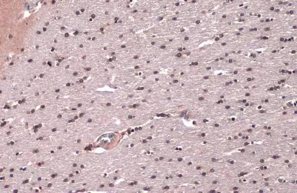


![Immunohistochemistry TOR/mTOR [p Ser2448] Antibody - BSA Free](https://images.novusbio.com/images/nb600-607_rabbit-polyclonal-tor-mtor-p-ser2448-antibody-235202318143142.jpg)
![Western Blot TOR/mTOR [p Ser2448] Antibody - BSA Free](https://images.novusbio.com/images/nb600-607_rabbit-polyclonal-tor-mtor-p-ser2448-antibody-31020241535647.jpg)
![Data TOR/mTOR [p Ser2448] Antibody - BSA Free](https://images.novusbio.com/images/TOR-mTOR-[p-Ser2448]-Antibody-N-A-NB600-607-img0008.jpg)
![Immunohistochemistry Caspase-3 Antibody [Unconjugated] - Active](https://images.novusbio.com/images/af835_human-mouse-active-caspase-3-affinity-purified-pab-41202410331943.jpg)
![Western Blot Caspase-3 Antibody [Unconjugated] - Active](https://images.novusbio.com/images/af835_human-mouse-active-caspase-3-affinity-purified-pab-812025554170.jpg)
![Western Blot Caspase-3 Antibody [Unconjugated] - Active](https://images.novusbio.com/images/af835_human-mouse-active-caspase-3-affinity-purified-pab-8120255534731.jpg)


![Western Blot: Human Brain Whole Tissue Lysate (Adult Normal) [NB820-59177] - Beta Actin expression on human brain whole tissue lysate using anti-Beta Actin antibody (Cat. # NBP1-47423). Image from verified customer review.](https://images.novusbio.com/images/Human-Brain-Whole-Tissue-Lysate-Adult-Normal-Western-Blot-NB820-59177-img0001.jpg "Western Blot: Human Brain Whole Tissue Lysate (Adult Normal) [NB820-59177] - Beta Actin expression on human brain whole tissue lysate using anti-Beta Actin antibody (Cat. # NBP1-47423). Image from verified customer review.")
![Western Blot: Goat anti-Rabbit IgG (H+L) Secondary Antibody [HRP] [NB7160] - Western blot showing vemurafenib treatment in BRAFV600E CRC cells inhibits fission mediator DRP1 with no significant effect on fusion proteins (Mfn1 & 2) using MFN-1 antibody (NBP1-51841) and corresponding secondary antibody, goat anti-rabbit IgG-HRP (NB7160). Image collected and cropped by CiteAb from the following publication (https://pubmed.ncbi.nlm.nih.gov/33738242).](https://images.novusbio.com/images/Goat-anti-Rabbit-IgG-H+L-Secondary-Antibody-HRP-Western-Blot-NB7160-img0001.jpg "Western Blot: Goat anti-Rabbit IgG (H+L) Secondary Antibody [HRP] [NB7160] - Western blot showing vemurafenib treatment in BRAFV600E CRC cells inhibits fission mediator DRP1 with no significant effect on fusion proteins (Mfn1 & 2) using MFN-1 antibody (NBP1-51841) and corresponding secondary antibody, goat anti-rabbit IgG-HRP (NB7160). Image collected and cropped by CiteAb from the following publication (https://pubmed.ncbi.nlm.nih.gov/33738242).")
 followed by 30 min incubation with Goat anti Rabbit HRP conjugated secondary antibodies (Catalog # HAF008) at 1:20 dilution + DAB chromogen (brown). The tissue was counterstained with Hematoxylin (blue). Control was done by omitting primary antibody.")
![Flow Cytometry: Rabbit IgG Isotype Control [NBP2-24891] - An intracellular stain was performed on Raji cells with Adiponectin antibody NB100-65810 (blue) and a matched isotype control NBP2-24893 (orange). Cells were fixed with 4% PFA and then permeablized with 0.1% saponin. Cells were incubated in an antibody dilution of 1 ug/mL for 30 minutes at room temperature, followed by Dylight488-conjugated anti-rabbit secondary antibody. Image using the Azide Free form of this antibody.](https://images.novusbio.com/images/Rabbit--Mouse-IgG-Isotype-Control-Flow-Cytometry-NBP2-24891-img0006.jpg "Flow Cytometry: Rabbit IgG Isotype Control [NBP2-24891] - An intracellular stain was performed on Raji cells with Adiponectin antibody NB100-65810 (blue) and a matched isotype control NBP2-24893 (orange). Cells were fixed with 4% PFA and then permeablized with 0.1% saponin. Cells were incubated in an antibody dilution of 1 ug/mL for 30 minutes at room temperature, followed by Dylight488-conjugated anti-rabbit secondary antibody. Image using the Azide Free form of this antibody.")


