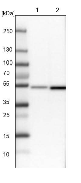
 HAF017). A specific band was detected for Prolactin at approximately 23 kDa (as indicated). This experiment was conducted under reducing conditions and using Immunoblot Buffer Group 1." title="Western blot shows lysates of mouse pituitary tissue and rat pituitary tissue. PVDF membrane was probed with 0.25 µg/mL of Goat Anti-Mouse/Rat Prolactin Antigen Affinity-purified Polyclonal Antibody (Catalog # AF1445) followed by HRP-conjugated Anti-Goat IgG Secondary Antibody (HAF017). A specific band was detected for Prolactin at approximately 23 kDa (as indicated). This experiment was conducted under reducing conditions and using Immunoblot Buffer Group 1." />
| Reactivity | Mu, RtSpecies Glossary |
| Applications | WB, Simple Western, IHC, B/N, ELISA(Cap) |
| Clonality | Polyclonal |
| Host | Goat |
| Conjugate | Unconjugated |
| Concentration | LYOPH |
| Immunogen | E. coli-derived recombinant mouse Prolactin Leu32-Cys228 Accession # NP_035294 |
| Specificity | Detects mouse and rat Prolactin in Western blots. In sandwich immunoassays, less than 1% cross-reactivity with recombinant human Prolactin is observed. |
| Source | N/A |
| Isotype | IgG |
| Clonality | Polyclonal |
| Host | Goat |
| Gene | PRL |
| Purity Statement | Antigen Affinity-purified |
| Endotoxin Note | <0.10 EU per 1 μg of the antibody by the LAL method. |
| Innovator's Reward | Test in a species/application not listed above to receive a full credit towards a future purchase. |
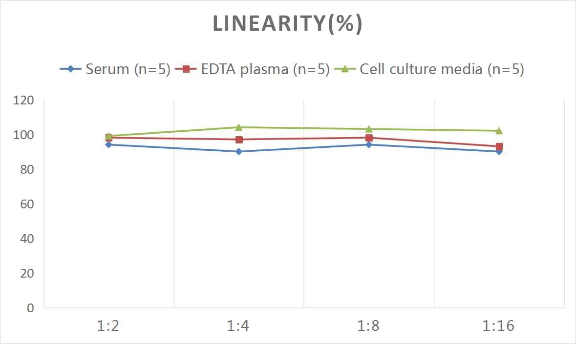
| Dilutions |
|
|
| Application Notes | ELISA Capture: Mouse Prolactin Antibody (Catalog # AF1445) ELISA Detection: Mouse Prolactin Biotinylated Antibody (Catalog # BAF1445) Standard: Recombinant Mouse Prolactin (Catalog # 1445-PL) |
|
| Publications |
|
| Storage | Use a manual defrost freezer and avoid repeated freeze-thaw cycles.
|
| Buffer | Lyophilized from a 0.2 μm filtered solution in PBS with Trehalose. See Certificate of Analysis for details. *Small pack size (-SP) is supplied either lyophilized or as a 0.2 µm filtered solution in PBS. |
| Preservative | No Preservative |
| Concentration | LYOPH |
| Reconstitution Instructions | Reconstitute at 0.2 mg/mL in sterile PBS. For liquid material, refer to CoA for concentration. |
Prolactin (PRL) is a neuroendocrine pituitary hormone. Prolactin is synthesized by the anterior pituitary, placenta, brain, uterus, dermal fibroblasts, decidua, B cells, T cells, NK cells and breast cancer cells. Originally characterized as a lactogenic hormone, further studies have demonstrated broader roles in breast cancer development, regulation of reproductive function, and immunoregulation. In the immune system, Prolactin has been shown to be secreted by human PBMC and to act as a proliferative growth factor. Additionally, Prolactin treatment of human PBMC has been shown to enhance IFN-gamma production. In the breast, Prolactin-induced morphogenesis of the mammary cells is mediated through IGF-2, which in turn upregulates cyclin D1. Prolactin has several molecular forms. The predominant form is a monomer; the non-glycosylated form is 23 kDa and the glycosylated form is 25 kDa. Glycosylated Prolactin is removed from the circulation faster and has been reported to have lower biological potency. Mouse Prolactin cDNA encodes a 228 amino acid (aa) residue protein with a putative 31 aa residue signal peptide. The Prolactin receptor is a transmembrane type I glycoprotein that belongs to the cytokine hematopoietic receptor family. B cells, T cells, macrophages, NK cells, monocytes, CD34+ progenitor cells, neutrophils, mammary gland, liver, kidney, adrenals, ovaries, testis, prostrate, seminal vesicles, and hypothalamus have all been shown to express the Prolactin receptor. Three forms of the receptor, generated by differential splicing, have been identified. These isoforms differ in the length of their cytoplasmic domains. It is believed that the short cytoplasmic form is non-functional. Prolactin signal transduction involves the JAK/STAT families and Src kinase family (1‑9).


![Immunoprecipitation STAT5b Antibody [Unconjugated]](https://images.novusbio.com/images/af1584_human-mouse-stat5b-affinity-purified-pab-immunoprecipitation-2211202212563.jpg)
![Knockout Validated STAT5b Antibody [Unconjugated]](https://images.novusbio.com/images/antibody/STAT5b_AF1584_Knockout_Validated_21968.jpg)
![Knockout Validated STAT5b Antibody [Unconjugated]](https://images.novusbio.com/images/af1584_human-mouse-stat5b-affinity-purified-pab-knockout-validated-22112022124011.jpg)


Secondary Antibodies |
Isotype Controls |
|
Application Focus: New Methods for iPSC Differentiation, Inducing a Mammary Fate Discovery of the Key to PluripotencyInduced pluripotent stem cells (iPSCs) may be generated from a wide range of fully differentiated cells, and under optimal conditions may be prompted to differentiate into virtu... Read full blog post. |
The concentration calculator allows you to quickly calculate the volume, mass or concentration of your vial. Simply enter your mass, volume, or concentration values for your reagent and the calculator will determine the rest.


 1445-PL) stimulates proliferation in the Nb2-11 rat lymphoma cell line in a dose-dependent manner (orange line). Proliferation elicited by Recombinant Mouse Prolactin (10 ng/mL) is neutralized (green line) by increasing concentrations of Goat Anti-Mouse/Rat Prolactin Antigen Affinity-purified Polyclonal Antibody (Catalog # AF1445). The ND50 is typically 0.25-1.0 µg/mL." title="Recombinant Mouse Prolactin (
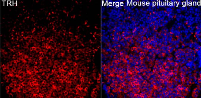
 VC004) and counterstained with hematoxylin (blue). Specific staining was localized to cytoplasm in lactotroph cells. View our protocol for
  using 2.5 µg/mL of Goat Anti-Mouse/Rat Prolactin Antigen Affinity-purified Polyclonal Antibody (Catalog # af1445). This experiment was conducted under reducing conditions and using the 12-230kDa separation system.")
 The protein expression level of ER alpha was detected by western blotting. GAPDH was used as loading control. (B) The protein expression level of PRL was detected by western blotting. GAPDH was used as loading control. (C) Quantitation of ER alpha protein level. Data are presented as mean ± S.E.M of n = 4 samples per group (P < 0.05 by ANOVA). (D) Quantitation of PRL protein level. Data are presented as mean ± S.E.M of n = 4 samples per group (P < 0.05 by ANOVA). Image collected and cropped by CiteAb from the following open publication (//pubmed.ncbi.nlm.nih.gov/32194503), licensed under a CC-BY license. Not internally tested by R&D Systems.")
 The expression level of miR-130a-3p was detected by quantitative real-time PCR (qRT-PCR). U6 snRNA was used to normalize the miRNA expression. Data are presented as mean ± S.E.M of n = 6 samples per group (*P < 0.05 by t-test). (B) The expression level of PRL mRNA was detected by qRT-PCR. GAPDH was used to normalize each gene expression. Data are presented as mean ± S.E.M of n = 6 samples per group (*P < 0.05 by t-test). (C) The protein level of PRL in GH3 cells was analyzed by western blotting. GAPDH was used as loading control. (D) Quantitation of the PRL protein level. Data are presented as mean ± S.E.M of n = 4 samples per group (*P < 0.05 by t-test). (E) The protein level of PRL was detected by ICC after GH3 cells transfected with miR-130a-3p mimic and NC. Scale bar, 50 μM. **P < 0.01. Image collected and cropped by CiteAb from the following open publication (//pubmed.ncbi.nlm.nih.gov/32194503), licensed under a CC-BY license. Not internally tested by R&D Systems.")
 The expression level of miR-130a-3p was detected by quantitative real-time PCR (qRT-PCR). U6 snRNA was used to normalize the miRNA expression. Data are presented as mean ± S.E.M of n = 4 samples per group (*P < 0.05 by t-test). (B) The expression levels of HSP70, ER alpha , and PRL mRNAs in GH3 cells were detected by qRT-PCR. GAPDH was used to normalize each gene expression. Data are presented as mean ± S.E.M of n = 4 samples per group (*P < 0.05, ns, not significant by t-test). (C) The protein levels of HSP70, ER alpha , and PRL in GH3 cells were analyzed by western blotting. GAPDH was used as loading control. (D) Quantitation of HSP70, ER alpha , and PRL protein levels. Data are presented as mean ± S.E.M of n = 4 samples per group (*P < 0.05 by t-test). **P < 0.01. Image collected and cropped by CiteAb from the following open publication (//pubmed.ncbi.nlm.nih.gov/32194503), licensed under a CC-BY license. Not internally tested by R&D Systems.")
 The mRNA levels of PRL (A) and ER alpha (B) in the pituitary gland of three groups were analyzed by quantitative real-time PCR (qRT-PCR). GAPDH was used to normalize each gene expression. Data are presented as mean ± S.E.M of n = 5 animals per group. Bars that share different letter are significantly different (P < 0.05 by ANOVA). (C) The serum PRL concentration was detected by Elisa assay. Data are presented as mean ± S.E.M of n = 5 animals per group. Bars that share different letter are significantly different (P < 0.05 by ANOVA). (D) The protein levels of PRL and ER alpha in the pituitary gland were analyzed by western blotting. beta -actin was used as loading control. (E) Quantitation of PRL and ER alpha protein levels. Data are presented as mean ± SD of n = 3 animals per group. Bars that share different letter are significantly different (P < 0.05 by ANOVA). The expression levels of miR-130a-3p (F), miR-130b-3p (G), miR-301a-3p (H), and miR-301b-3p (I) were detected by qRT-PCR. U6 snRNA was used to normalize each miRNA expression. Data are presented as mean ± S.E.M of n = 5 animals per group. Bars that share different letter are significantly different (P < 0.05 by ANOVA). Image collected and cropped by CiteAb from the following open publication (//pubmed.ncbi.nlm.nih.gov/32194503), licensed under a CC-BY license. Not internally tested by R&D Systems.")
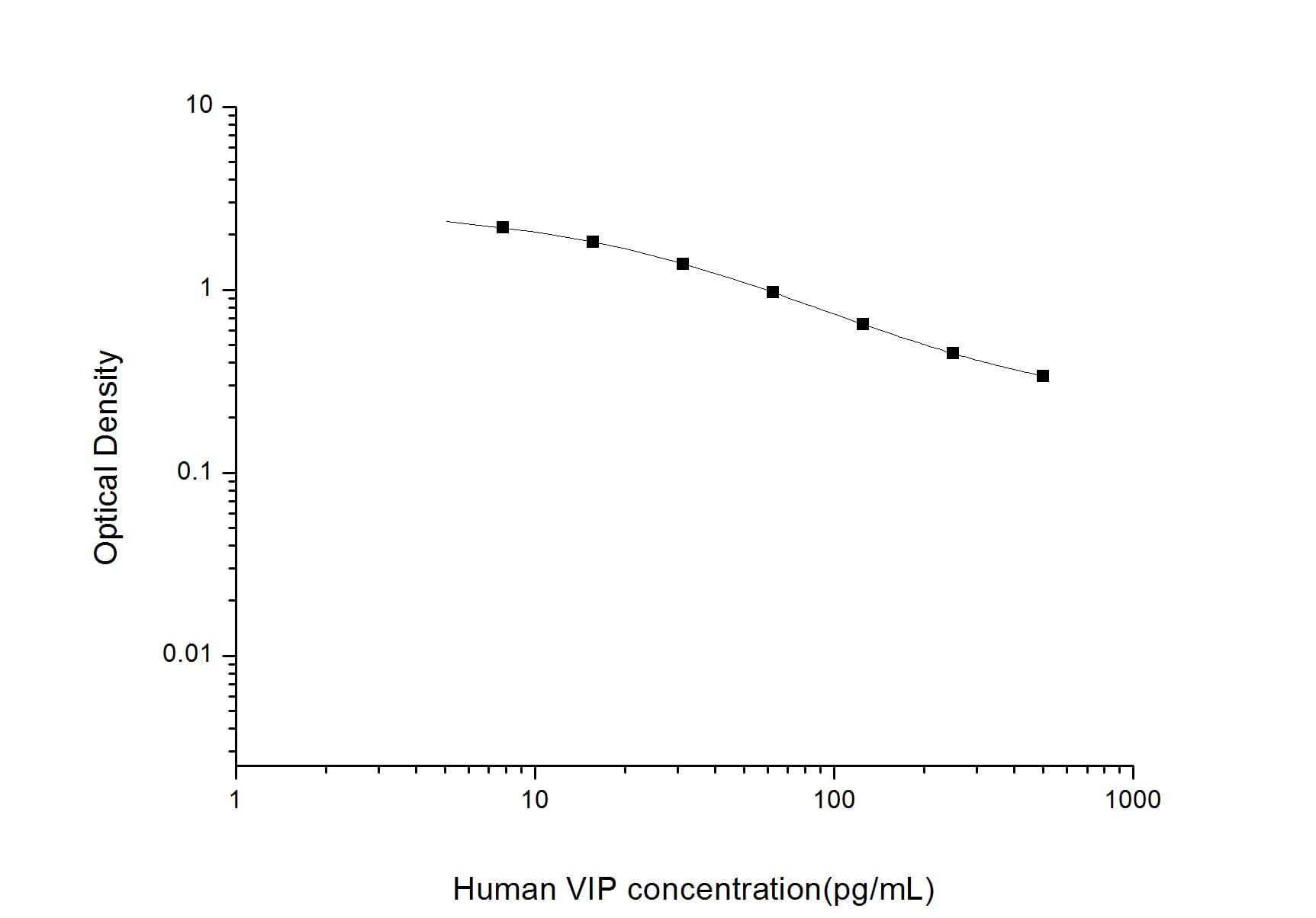
 1445-PL) was serially diluted and captured by Goat Anti-Mouse/Rat Prolactin Antigen Affinity-purified Polyclonal Antibody (Catalog # AF1445) coated on a Clear Polystyrene Microplate (Catalog #
  1445-PL) was serially diluted and captured by Goat Anti-Mouse/Rat Prolactin Antigen Affinity-purified Polyclonal Antibody (Catalog #
 ![Western Blot Growth Hormone Antibody [Unconjugated]](https://images.novusbio.com/images/antibody/Growth_Hormone_AF1067_Western_Blot_20961.jpg)


![Immunohistochemistry Insulin Antibody (182410) [Unconjugated]](https://images.novusbio.com/images/antibody/mab1417_human-bovine-mouse-insulin-mab-clone-182410-immunohistochemistry-308202115145.jpg)
![Immunocytochemistry Insulin Antibody (182410) [Unconjugated]](https://images.novusbio.com/images/antibody/Insulin_MAB1417_Immunocytochemistry_9376.jpg)
![Bioactivity IGF-I/IGF-1 [Unconjugated]](https://images.novusbio.com/images/protein/IGF-I_291-G1_41.jpg)
![Mass Spectrometry IGF-I/IGF-1 [Unconjugated]](https://images.novusbio.com/images/protein/IGF-I_291-G1_42.jpg)
![SEC-MALS IGF-I/IGF-1 [Unconjugated]](https://images.novusbio.com/images/291-g1_recombinant-human-igf-i-igf-1-protein-cf-sec-mals-224202691859.jpg)


![Simple Western STAT5a/b Antibody [Unconjugated] - Pan Specific](https://images.novusbio.com/images/antibody/16339.jpg)
![Chromatin Immunoprecipitation (ChIP) STAT5a/b Antibody [Unconjugated] - Pan Specific](https://images.novusbio.com/images/antibody/STAT5b_AF2168_Chromatin_Immunoprecipitation_11246.jpg)
![Immunohistochemistry SHBG Antibody [Unconjugated]](https://images.novusbio.com/images/antibody/SHBG_AF2656_Immunohistochemistry_7324.jpg)
![Western Blot SHBG Antibody [Unconjugated]](https://images.novusbio.com/images/af2656_human-shbg-affinity-purified-pab-western-blot-2642023152834..jpg)
![Immunohistochemistry SHBG Antibody [Unconjugated]](https://images.novusbio.com/images/af2656_human-shbg-affinity-purified-pab-8120255541789.jpg)
 or Normal Goat IgG Isotype Control Antibody (Catalog # AB-108-C, open histogram), followed by Phycoerythrin-conjugated Anti-Goat IgG Secondary Antibody (Catalog # F0107).")