![Western Blot: LC3A Antibody [NBP1-78964] - Analysis of LC3I in Neuro2A cell lysate.](http://images.novusbio.com/fullsize/LC3A-Antibody-Western-Blot-NBP1-78964-img0015.jpg "Western Blot: LC3A Antibody [NBP1-78964] - Analysis of LC3I in Neuro2A cell lysate.")
| Reactivity | Hu, Mu, Rt, Po, Bv, PmSpecies Glossary |
| Applications | WB, ICC/IF, IHC |
| Clonality | Polyclonal |
| Host | Rabbit |
| Conjugate | Unconjugated |
| Format | BSA Free |
| Concentration | 1.09 mg/ml |
| Immunogen | This LC3A Antibody was prepared from a synthetic peptide made to a C-terminal region of the human LC3 protein (within residues 50-120). [Swiss-Prot Q9H492]. |
| Localization | Cytoplasm. |
| Specificity | This LC3I antibody specifically detects the cytosolic form of LC3 before it gets converted into LC3II during autophagy. |
| Isotype | IgG |
| Clonality | Polyclonal |
| Host | Rabbit |
| Gene | MAP1LC3A |
| Purity | Immunogen affinity purified |
| Innovator's Reward | Test in a species/application not listed above to receive a full credit towards a future purchase. |
| Dilutions |
|
||||||
| Application Notes | This LC3I antibody is useful for Western Blot, Immunocytochemistry/Immunofluorescence, and IHC-paraffin embedded sections. In Western Blot, a band is seen ~15kDa representing LC3I. In ICC/IF, observed staining showed inactivated LC3 throughout the cytoplasm of Neuro2a cells. In IHC-P, staining was observed in the cytoplasm of mouse testes tissue. Prior to immunostaining paraffin tissues, antigen retrieval with sodium citrate buffer (pH 6.0) is recommended. |
||||||
| Control |
|
||||||
| Reviewed Applications |
|
| Storage | Store at 4C short term. Aliquot and store at -20C long term. Avoid freeze-thaw cycles. |
| Buffer | PBS and 30% Glycerol |
| Preservative | 0.05% Sodium Azide |
| Concentration | 1.09 mg/ml |
| Purity | Immunogen affinity purified |


![Intracellular Staining by Flow Cytometry AKT [p Ser473] Antibody [Unconjugated] - Pan Specific](https://images.novusbio.com/images/antibody/Akt3_AF887_Flow_Cytometry_8283.jpg)
![Western Blot AKT [p Ser473] Antibody [Unconjugated] - Pan Specific](https://images.novusbio.com/images/af887_phospho-akt-s473-pan-specific-affinity-purified-pab-41202410485440.jpg)
![Western Blot AKT [p Ser473] Antibody [Unconjugated] - Pan Specific](https://images.novusbio.com/images/af887_phospho-akt-s473-pan-specific-affinity-purified-pab-8120255552843.jpg)
![Immunohistochemistry ATG7 Antibody (683906) [Unconjugated]](https://images.novusbio.com/images/antibody/ATG7_MAB6608_Immunohistochemistry_10631.jpg)
![Simple Western ATG7 Antibody (683906) [Unconjugated]](https://images.novusbio.com/images/antibody/ATG7_MAB6608_Simple_Western_16409.jpg)


| Images | Ratings | Applications | Species | Date | Details | ||||
|---|---|---|---|---|---|---|---|---|---|
-(01-ml)_NBP1-78964_8641.bmp)
Enlarge |
reviewed by:
Bryan Tinsley |
ICC | Human | 07/01/2014 |
Summary
|
| Human Brain Whole Tissue Lysate (Adult Whole Normal) | |
| Neuro2a Chloroquine Treated / Untreated Cell Lysate | |
| HeLa Chloroquine Treated / Untreated Cell Lysate |
Secondary Antibodies |
Isotype Controls |
Research Areas for LC3A Antibody (NBP1-78964)Find related products by research area.
|
|
Read full blog post. |
|
Losing memory: Toxicity from mutant APP and amyloid beta explain the hippocampal neuronal damage in Alzheimer's disease By Jamshed Arslan Pharm.D. Alzheimer's disease (AD) is an irreversible brain disorder that destroys memory and thinking skills. The telltale signs of AD brains are extracellular deposits of amy... Read full blog post. |
|
Nuclear LC3: Why is it there and what is it doing? By Christina Towers, PhD. Cells use the complex process of autophagy to degrade and recycle cytoplasmic material. There are over 20 proteins that have been implicated in this process and appropriately named core ... Read full blog post. |
|
Why LC3B Antibodies Make Ideal Autophagosomes Membrane Markers The human form of microtubule-associated protein light chain 3 (LC3) is expressed as 3 splice variants LC3A, LC3B, and LC3C.1 LC3B is a subunit of the MAP1A and MAP1B microtubule-binding proteins and plays a central role in autophagosome membrane stru... Read full blog post. |
The concentration calculator allows you to quickly calculate the volume, mass or concentration of your vial. Simply enter your mass, volume, or concentration values for your reagent and the calculator will determine the rest.


![Immunocytochemistry/Immunofluorescence: LC3A Antibody [NBP1-78964] - IF Confocal analysis of A549 cells using LC3I antibody (NBP1-78964, 1:5). An Alexa Fluor 488-conjugated Goat to rabbit IgG was used as secondary antibody (green). Actin filaments were labeled with Alexa Fluor 568 phalloidin (red). DAPI was used to stain the cell nuclei (blue).](http://images.novusbio.com/fullsize/LC3A-Antibody-Immunocytochemistry-Immunofluorescence-NBP1-78964-img0017.jpg "Immunocytochemistry/Immunofluorescence: LC3A Antibody [NBP1-78964] - IF Confocal analysis of A549 cells using LC3I antibody (NBP1-78964, 1:5). An Alexa Fluor 488-conjugated Goat to rabbit IgG was used as secondary antibody (green). Actin filaments were labeled with Alexa Fluor 568 phalloidin (red). DAPI was used to stain the cell nuclei (blue).")
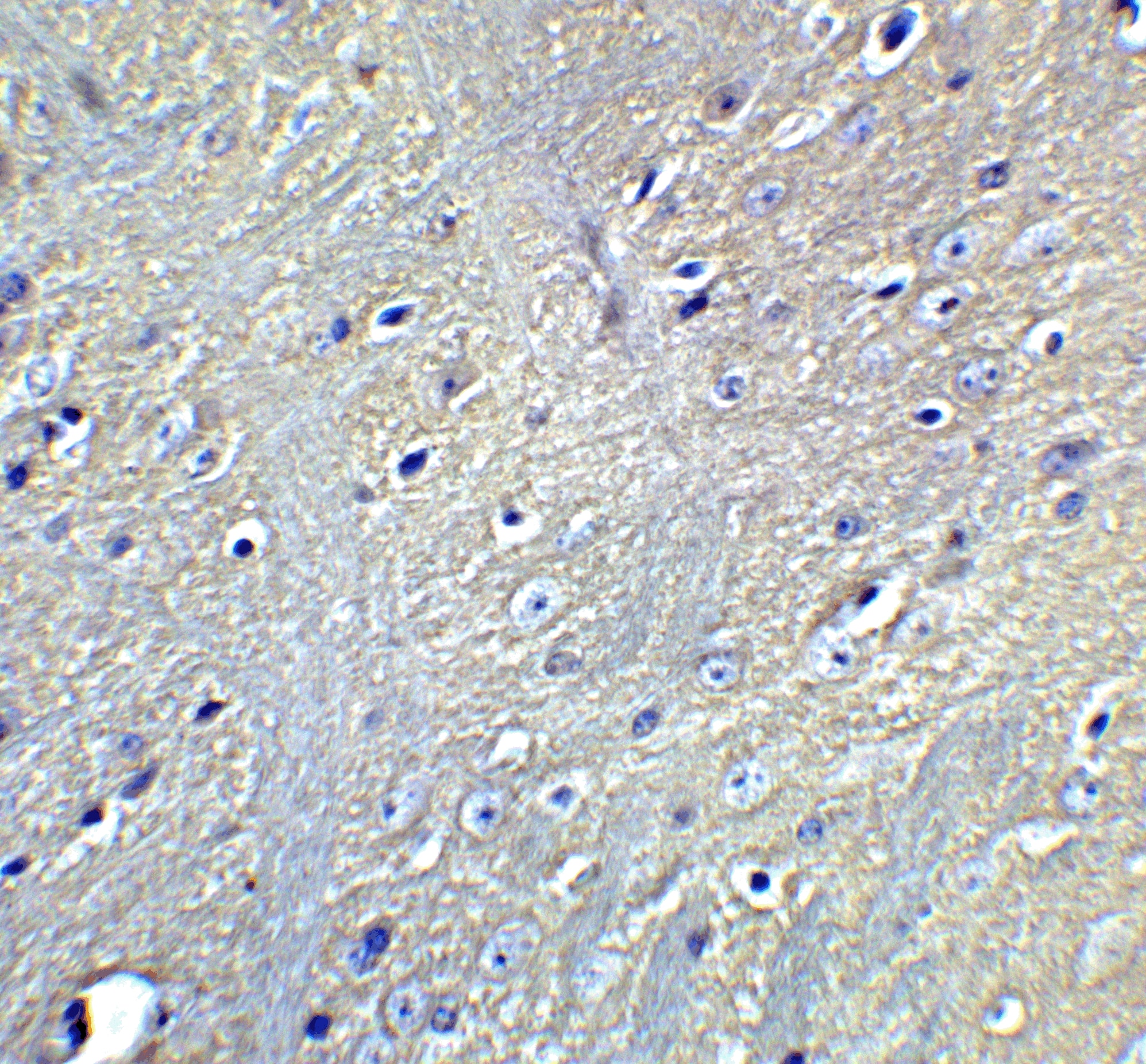
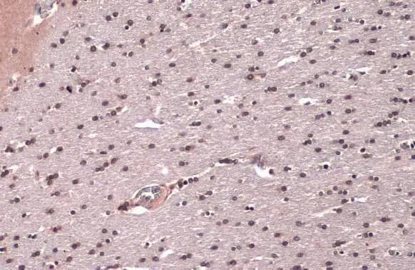
![Immunohistochemistry: LC3A Antibody [NBP1-78964] - Analysis of LC3I in mouse testis using DAB with hematoxylin counterstain.](http://images.novusbio.com/fullsize/LC3A-Antibody-Immunohistochemistry-NBP1-78964-img0016.jpg "Immunohistochemistry: LC3A Antibody [NBP1-78964] - Analysis of LC3I in mouse testis using DAB with hematoxylin counterstain.")


![Immunohistochemistry TOR/mTOR [p Ser2448] Antibody - BSA Free](https://images.novusbio.com/images/nb600-607_rabbit-polyclonal-tor-mtor-p-ser2448-antibody-235202318143142.jpg)
![Western Blot TOR/mTOR [p Ser2448] Antibody - BSA Free](https://images.novusbio.com/images/nb600-607_rabbit-polyclonal-tor-mtor-p-ser2448-antibody-31020241535647.jpg)
![Data TOR/mTOR [p Ser2448] Antibody - BSA Free](https://images.novusbio.com/images/TOR-mTOR-[p-Ser2448]-Antibody-N-A-NB600-607-img0008.jpg)
![Immunohistochemistry Caspase-3 Antibody [Unconjugated] - Active](https://images.novusbio.com/images/af835_human-mouse-active-caspase-3-affinity-purified-pab-41202410331943.jpg)
![Western Blot Caspase-3 Antibody [Unconjugated] - Active](https://images.novusbio.com/images/af835_human-mouse-active-caspase-3-affinity-purified-pab-812025554170.jpg)
![Western Blot Caspase-3 Antibody [Unconjugated] - Active](https://images.novusbio.com/images/af835_human-mouse-active-caspase-3-affinity-purified-pab-8120255534731.jpg)


![Western Blot ERK2 Antibody [Unconjugated]](https://images.novusbio.com/images/antibody/ERK2_AF1230_Western_Blot_5097.jpg)
![Knockout Validated ERK2 Antibody [Unconjugated]](https://images.novusbio.com/images/antibody/ERK2_AF1230_Knockout_Validated_22864.jpg)
![Immunohistochemistry ERK2 Antibody [Unconjugated]](https://images.novusbio.com/images/antibody/ERK2_AF1230_Immunohistochemistry_20696.jpg)


![Western Blot: Human Brain Whole Tissue Lysate (Adult Normal) [NB820-59177] - Beta Actin expression on human brain whole tissue lysate using anti-Beta Actin antibody (Cat. # NBP1-47423). Image from verified customer review.](https://images.novusbio.com/images/Human-Brain-Whole-Tissue-Lysate-Adult-Normal-Western-Blot-NB820-59177-img0001.jpg "Western Blot: Human Brain Whole Tissue Lysate (Adult Normal) [NB820-59177] - Beta Actin expression on human brain whole tissue lysate using anti-Beta Actin antibody (Cat. # NBP1-47423). Image from verified customer review.")
![Western Blot: Goat anti-Rabbit IgG (H+L) Secondary Antibody [HRP] [NB7160] - Western blot showing vemurafenib treatment in BRAFV600E CRC cells inhibits fission mediator DRP1 with no significant effect on fusion proteins (Mfn1 & 2) using MFN-1 antibody (NBP1-51841) and corresponding secondary antibody, goat anti-rabbit IgG-HRP (NB7160). Image collected and cropped by CiteAb from the following publication (https://pubmed.ncbi.nlm.nih.gov/33738242).](https://images.novusbio.com/images/Goat-anti-Rabbit-IgG-H+L-Secondary-Antibody-HRP-Western-Blot-NB7160-img0001.jpg "Western Blot: Goat anti-Rabbit IgG (H+L) Secondary Antibody [HRP] [NB7160] - Western blot showing vemurafenib treatment in BRAFV600E CRC cells inhibits fission mediator DRP1 with no significant effect on fusion proteins (Mfn1 & 2) using MFN-1 antibody (NBP1-51841) and corresponding secondary antibody, goat anti-rabbit IgG-HRP (NB7160). Image collected and cropped by CiteAb from the following publication (https://pubmed.ncbi.nlm.nih.gov/33738242).")
 followed by 30 min incubation with Goat anti Rabbit HRP conjugated secondary antibodies (Catalog # HAF008) at 1:20 dilution + DAB chromogen (brown). The tissue was counterstained with Hematoxylin (blue). Control was done by omitting primary antibody.")
![Flow Cytometry: Rabbit IgG Isotype Control [NBP2-24891] - An intracellular stain was performed on Raji cells with Adiponectin antibody NB100-65810 (blue) and a matched isotype control NBP2-24893 (orange). Cells were fixed with 4% PFA and then permeablized with 0.1% saponin. Cells were incubated in an antibody dilution of 1 ug/mL for 30 minutes at room temperature, followed by Dylight488-conjugated anti-rabbit secondary antibody. Image using the Azide Free form of this antibody.](https://images.novusbio.com/images/Rabbit--Mouse-IgG-Isotype-Control-Flow-Cytometry-NBP2-24891-img0006.jpg "Flow Cytometry: Rabbit IgG Isotype Control [NBP2-24891] - An intracellular stain was performed on Raji cells with Adiponectin antibody NB100-65810 (blue) and a matched isotype control NBP2-24893 (orange). Cells were fixed with 4% PFA and then permeablized with 0.1% saponin. Cells were incubated in an antibody dilution of 1 ug/mL for 30 minutes at room temperature, followed by Dylight488-conjugated anti-rabbit secondary antibody. Image using the Azide Free form of this antibody.")
-(01-ml)_NBP1-78964_8641.bmp)