| Xie S, Zhang S, Medeiros G et al. The G1/S transition in mammalian stem cells in vivo is autonomously regulated by cell size Nature Communications 2025-10-13 [PMID: 41083493] |
|
|
| Zuccoli E, Sawaf H, Tuzza M et al. Reproducibility of PD patient-specific midbrain organoid data for in vitro disease modeling iScience 2025-10-17 [PMID: 41050933] |
|
|
| Sánchez-Vázquez R, García-Oteyza S, Serrano R et al. Mice carrying the homologous human shelterin POT1-L259S mutation linked to pulmonary fibrosis show a telomerase deficiency-like phenotype with telomere shortening with increasing mouse generations. Genes & development 2025-09-15 [PMID: 40954016] |
|
|
| Peng M, Lee S, Nair H et al. RAD51 is chromatin enriched and targetable in BRCA1-deficient cells. Molecular Cell 2025-09-10 [PMID: 40934926] |
|
|
| Rivadeneira D, Thosar S, Quann K et al. Oxidative-stress-induced telomere instability drives T cell dysfunction in cancer. Immunity 2025-09-03 [PMID: 40930086] |
|
|
| Janovič T, Perez G, Boelting G et al. TRF1 and TRF2 form distinct shelterin subcomplexes at telomeres. Cell reports 2025-08-23 [PMID: 40849907] |
|
|
| Wang Z, Zhang X, Zhou Y et al. Energy Deficiency-Induced ATG4B Nuclear Translocation Inhibits PRMT1-Mediated DNA Repair and Promotes Leukemia Progression. Advanced science (Weinheim, Baden-Wurttemberg, Germany) 2025-08-11 [PMID: 40788108] |
|
|
| Marques M, Hayashide L, Amorim P et al. Doxorubicin Induces a Senescent Phenotype in Murine and Human Astrocytes Journal of neurochemistry 2025-08-05 [PMID: 40762103] |
|
|
| Azeroglu B, Wu W, Pavani R et al. Conserved and unique features of terminal telomeric sequences in ALT-positive cancer cells eLife 2025-08-01 [PMID: 40748043] |
|
|
| Galarreta A, Pasti V, Vornberger J et al. Identification of CNOT1-CCR4-NOT as a suppressor of 53BP1-p53-p21 signaling. Cell reports 2025-07-30 [PMID: 40742806] |
|
|
| Hernandez A, Puppala A, Hedlich-Dwyer J et al. Short-term consumption of the modified standard American diet perturbed the metabolic balance and altered DNA damage in MMTV-PyMT transgenic mice Breast Cancer Research : BCR 2025-07-25 [PMID: 40713647] |
|
|
| Mennerich D, Ashok Y, Vela-Rodríguez C et al. The E3 ubiquitin ligase DTX3L and the deubiquitinase USP28 fine-tune DNA repair through mutual regulation of their protein levels iScience 2025-08-15 [PMID: 40703443] |
|
|
| Xavier Raj, I;Kyun Koh, W;Harrison, J;Zhang, CR;Soares, B;Amato, R;Krishnan, A;O'Leary, DR;Bjeije, H;Parsons, TM;Han, W;Young, AL;Wang, T;Batista, LFZ;Challen, GA; Non-canonical functions of DNMT3A in hematopoietic stem cells regulate telomerase activity and genome integrity Cell stem cell 2025-08-07 [PMID: 40680747] |
|
|
| In S, Nunes P, Fernandes R et al. TERRA R-loops trigger a switch in telomere maintenance towards break-induced replication and PRIMPOL-dependent repair The EMBO Journal 2025-07-07 [PMID: 40624280] |
|
|
| Sobajima T, Fulcher L, Batley C et al. WIP1 mutations suppress DNA damage triggered bypass of the mitotic timer The EMBO Journal 2025-06-23 [PMID: 40551011] |
|
|
| Tang M, Li S, Zhu Z et al. USP37 counteracts HLTF to protect damaged replication forks and promote survival of BRCA1-deficient cells and PARP inhibitor resistance Nucleic acids research 2025-07-08 [PMID: 40548939] |
|
|
| Chabaytah N, Dumančić M, Asante E et al. Assessing radiosensitivity through sublethal damage recovery: a comparison of survival-based and molecular repair kinetics. Physics in medicine and biology 2025-06-16 [PMID: 40480255] |
|
|
| Heemskerk T, Rovituso M, Wal E et al. A Novel Live-Cell Microscopy Platform for Real-Time Visualization of 53BP1 Foci Dynamics and Accurate Dosimetry in Proton Therapy. Physica medica : PM : an international journal devoted to the applications of physics to medicine and biology : official journal of the Italian Association of Biomedical Physics (AIFB) 2025-06-05 [PMID: 40480095] |
|
|
| Ingham A, Ramaswami M, Ramangoudr-Bhojappa R et al. Loss of SLX4IP leads to common fragile site instability and compromises DNA interstrand crosslink repair in vivo The Journal of Biological Chemistry 2025-06-01 [PMID: 40383148] |
|
|
| Giroud, J;Delvaux, P;Carlier, L;De Schutter, C;Martin, N;Rouget, R;Bolouki, A;De Glas, V;Bouriez, I;Bourdoux, F;Burteau, S;Théry, J;Decanter, G;Penel, N;de Launoit, Y;Ledoux, B;Abbadie, C;Poumay, Y;Pluquet, O;Debacq-Chainiaux, F; Targeting ATF6? Attenuates UVB-Induced Senescence and Improves Skin Homeostasis by Regulating IL8 Expression Aging cell 2025-04-16 [PMID: 40241256] |
|
|
| Khalife M, Jia T, Caron P et al. SRSF2 overexpression induces transcription-/replication-dependent DNA double-strand breaks and interferes with DNA repair pathways to promote lung tumor progression. NAR cancer 2025-04-04 [PMID: 40181846] |
|
|
| Hervé S, Scelfo A, Bersano Marchisio G et Al. Chromosome mis-segregation triggers cell cycle arrest through a mechanosensitive nuclear envelope checkpoint Nat Cell Biol 2025-01-08 [PMID: 39779939] |
|
|
| Tosello, V;Rompietti, C;Papathanassiu, AE;Arrigoni, G;Piovan, E; BCAT1 Associates with DNA Repair Proteins KU70 and KU80 and Contributes to Regulate DNA Repair in T-Cell Acute Lymphoblastic Leukemia (T-ALL) International journal of molecular sciences 2024-12-18 [PMID: 39769333] |
|
|
| Zhao H, Fu X, Gu W et Al. 53BP1, a known chromatin-associated factor that promotes DNA damage repair, is differentially modulated during bovine herpesvirus 1 infection in vitro and in vivo Vet Microbiol 2024-12-04 [PMID: 39653013] |
|
|
| Tang, J;Weiser, NE;Wang, G;Chowdhry, S;Curtis, EJ;Zhao, Y;Wong, IT;Marinov, GK;Li, R;Hanoian, P;Tse, E;Mojica, SG;Hansen, R;Plum, J;Steffy, A;Milutinovic, S;Meyer, ST;Luebeck, J;Wang, Y;Zhang, S;Altemose, N;Curtis, C;Greenleaf, WJ;Bafna, V;Benkovic, SJ;Pinkerton, AB;Kasibhatla, S;Hassig, CA;Mischel, PS;Chang, HY; Enhancing transcription-replication conflict targets ecDNA-positive cancers Nature 2024-11-01 [PMID: 39506153] |
|
|
| Herrera-Moyano E, Porreca RM, Ranjha L et al. Human SKI component SKIV2L regulates telomeric DNA-RNA hybrids and prevents telomere fragility iScience 2024-10-04 [PMID: 39493885] |
|
|
| Chen JK, Merrick KA, Kong YW et al. An RNA damage response network mediates the lethality of 5-FU in colorectal cancer. Cell reports. Medicine 2024-10-16 [PMID: 39378883] |
|
|
| Lee H, Niida H, Sung S et Al. Haplotype-resolved de novo assembly revealed unique characteristics of alternative lengthening of telomeres in mouse embryonic stem cells Nucleic Acids Res 2024-11-11 [PMID: 39351882] |
|
|
| Iwasaki K, Tojo A, Kobayashi H et Al. Dose-dependent effects of histone methyltransferase NSD2 on site-specific double-strand break repair Genes Cells 2024-11-06 [PMID: 39245559] |
|
|
| Tejwani V, Carroll T, Macartney T et Al. PROTAC-mediated conditional degradation of the WRN helicase as a potential strategy for selective killing of cancer cells with microsatellite instability Sci Rep 2024-09-06 [PMID: 39242638] |
|
|
| Krzystyniak A, Gluchowska A, Pyty? A et Al. 2-Bromopalmitate treatment attenuates senescence phenotype in human adult cells - possible role of palmitoylation Aging (Albany NY) 2024-09-06 [PMID: 39181690] |
|
|
| Técher H, Gopaul D, Heuzé J et Al. MRE11 and TREX1 control senescence by coordinating replication stress and interferon signaling Nat Commun 2024-06-26 [PMID: 38926338] |
|
|
| J. Tyson McDonald, JangKeun Kim, Lily Farmerie, Meghan L. Johnson, Nidia S. Trovao, Shehbeel Arif, Keith Siew, Sergey Tsoy, Yaron Bram, Jiwoon Park, Eliah Overbey, Krista Ryon, Jeffrey Haltom, Urminder Singh, Francisco J. Enguita, Victoria Zaksas, Joseph W. Guarnieri, Michael Topper, Douglas C. Wallace, Cem Meydan, Stephen Baylin, Robert Meller, Masafumi Muratani, D. Marshall Porterfield, Brett Kaufman, Marcelo A. Mori, Stephen B. Walsh, Dominique Sigaudo-Roussel, Saida Mebarek, Massimo Bottini, Christophe A. Marquette, Eve Syrkin Wurtele, Robert E. Schwartz, Diego Galeano, Christopher E. Mason, Peter Grabham, Afshin Beheshti Space radiation damage rescued by inhibition of key spaceflight associated miRNAs Nature Communications 2024-06-11 [PMID: 38862542] |
|
|
| Paola Sanese, Katia De Marco, Martina Lepore Signorile, Francesca La Rocca, Giovanna Forte, Marialaura Latrofa, Candida Fasano, Vittoria Disciglio, Elisabetta Di Nicola, Antonino Pantaleo, Giusy Bianco, Vito Spilotro, Claudia Ferroni, Matilde Tubertini, Nicoletta Labarile, Lucia De Marinis, Raffaele Armentano, Gianluigi Gigante, Valerio Lantone, Giuliano Lantone, Marina Naldi, Manuela Bartolini, Greta Varchi, Alberto Del Rio, Valentina Grossi, Cristiano Simone The novel SMYD3 inhibitor EM127 impairs DNA repair response to chemotherapy-induced DNA damage and reverses cancer chemoresistance Journal of Experimental & Clinical Cancer Research : CR 2024-05-30 [PMID: 38812026] |
|
|
| Yunhui Li, Lei Li, Xinshu Wang, Fei Zhao, Yuntong Yang, Yujuan Zhou, Jiyuan Zhang, Li Wang, Zeshan Jiang, Yuanyuan Zhang, Yuping Chen, Chenming Wu, Ke Li, Tingting Zhang, Ping Wang, Zhiyong Mao, Weiguo Zhu, Xingzhi Xu, Shikang Liang, Zhenkun Lou, Jian Yuan USP25 Elevates SHLD2-Mediated DNA Double-Strand Break Repair and Regulates Chemoresponse in Cancer. Advanced science (Weinheim, Baden-Wurttemberg, Germany) 2024-05-27 [PMID: 38803048] |
|
|
| Hill BR, Ozgencil M, Buckley-Benbow L, Skingsley SLP et Al. Loss of POLE3-POLE4 unleashes replicative gap accumulation upon treatment with PARP inhibitors Cell Rep 2024-05-16 [PMID: 38753485] |
|
|
| Athanasios Siametis, Kalliopi Stratigi, Despoina Giamaki, Georgia Chatzinikolaou, Alexia Akalestou-Clocher, Evi Goulielmaki, Brian Luke, Björn Schumacher, George A. Garinis Transcription stress at telomeres leads to cytosolic DNA release and paracrine senescence Nature Communications 2024-05-14 [PMID: 38744897] |
|
|
| Néstor García-Rodríguez, Iria Domínguez-García, María del Carmen Domínguez-Pérez, Pablo Huertas EXO1 and DNA2-mediated ssDNA gap expansion is essential for ATR activation and to maintain viability in BRCA1-deficient cells Nucleic Acids Research 2024-06-24 [PMID: 38721777] |
|
|
| Yahui Wang, Xinyi Liu, Xianbo Zuo, Cuiling Wang, Zheng Zhang, Haitao Zhang, Tao Zeng, Shunqi Chen, Mengyu Liu, Hongxia Chen, Qingfeng Song, Qi Li, Chenning Yang, Yi Le, Jinliang Xing, Hongxin Zhang, Jiaze An, Weihua Jia, Longli Kang, Hongxing Zhang, Hui Xie, Jiazhou Ye, Tianzhun Wu, Fuchu He, Xuejun Zhang, Yuanfeng Li, Gangqiao Zhou NRDE2 deficiency impairs homologous recombination repair and sensitizes hepatocellular carcinoma to PARP inhibitors Cell Genomics 2024-05-01 [PMID: 38697125] |
|
|
| Victoria L Dunne, Timothy C Wright, Francisco D C Guerra Liberal, Joe M O'Sullivan, Kevin M Prise Synergistic Activity of DNA Damage Response Inhibitors in Combination with Radium-223 in Prostate Cancer. Cancers 2024-04-15 [PMID: 38672592] |
|
|
| Ubieto-Capella P, Ximénez-Embún P, Giménez-Llorente D et Al. A rewiring of DNA replication mediated by MRE11 exonuclease underlies primed-to-naive cell de-differentiation Cell Rep 2024-04-05 [PMID: 38581679] |
|
|
| Aurora Campos-Díaz, Patricia Morejón-García, Eva Monte-Serrano, David Ros-Pardo, Iñigo Marcos-Alcalde, Paulino Gómez-Puertas, Pedro A. Lazo Pathogenic effects of Leu200Pro and Arg387His VRK1 protein variants on phosphorylation targets and H4K16 acetylation in distal hereditary motor neuropathy Journal of Molecular Medicine (Berlin, Germany) 2024-03-30 [PMID: 38554151] |
|
|
| Meitinger F, Belal H, Davis RL, Martinez MB et Al. Control of cell proliferation by memories of mitosis Science 2024-03-28 [PMID: 38547292] |
|
|
| Michalis Petropoulos, Angeliki Karamichali, Giacomo G. Rossetti, Alena Freudenmann, Luca G. Iacovino, Vasilis S. Dionellis, Sotirios K. Sotiriou, Thanos D. Halazonetis Transcription–replication conflicts underlie sensitivity to PARP inhibitors Nature 2024-03-20 [PMID: 38509368] |
|
|
| Mattheus Xing Rong Foo, Peh Fern Ong, Zi Xuan Yap, Martina Maric, Christopher Jue Shi Bong, Peter Dröge, Brian Burke, Oliver Dreesen Genetic and pharmacological modulation of lamin A farnesylation determines its function and turnover Aging Cell 2024-03-19 [PMID: 38504487] |
|
|
| Caroline C Escoubas, Leah C Dorman, Phi T Nguyen, Christian Lagares-Linares, Haruna Nakajo, Sarah R Anderson, Jerika J Barron, Sarah D Wade, Beatriz Cuevas, Ilia D Vainchtein, Nicholas J Silva, Ricardo Guajardo, Yinghong Xiao, Peter V Lidsky, Ellen Y Wang, Brianna M Rivera, Sunrae E Taloma, Dong Kyu Kim, Elizaveta Kaminskaya, Hiromi Nakao-Inoue, Bjoern Schwer, Thomas D Arnold, Ari B Molofsky, Carlo Condello, Raul Andino, Tomasz J Nowakowski, Anna V Molofsky Type-I-interferon-responsive microglia shape cortical development and behavior. Cell 2024-04-15 [PMID: 38490196] |
|
|
| Marloes C M Jonkhout, Tijs Vanhessche, Mónica Ferreira, Iris Verbinnen, Fabienne Withof, Gerd Van der Hoeven, Kathelijne Szekér, Zahra Azhir, Wen-Hui Lien, Aleyde Van Eynde, Mathieu Bollen Embryonic NIPP1 Depletion in Keratinocytes Triggers a Cell Cycle Arrest and Premature Senescence in Adult Mice. The Journal of investigative dermatology 2024-02-29 [PMID: 38431220] |
|
|
| Bao K, Ma Y, Li Y, Shen X et Al. A di-acetyl-decorated chromatin signature couples liquid condensation to suppress DNA end synapsis Mol Cell 2024-02-29 [PMID: 38423014] |
|
|
| Carolline Ascenção, Jennie R Sims, Alexis Dziubek, William Comstock, Elizabeth A Fogarty, Jumana Badar, Raimundo Freire, Andrew Grimson, Robert S Weiss, Paula E Cohen, Marcus B Smolka, Akira Shinohara, Wei Yan A TOPBP1 allele causing male infertility uncouples XY silencing dynamics from sex body formation eLife 2024-02-23 [PMID: 38391183] |
|
|
| Scelfo A, Angrisani A, Grillo M, Barnes BM et Al. Specialized replication mechanisms maintain genome stability at human centromeres Mol Cell 2024-02-15 [PMID: 38359824] |
|
|
| Muñoz, S;Barroso, S;Badra-Fajardo, N;Marqueta-Gracia, JJ;García-Rubio, ML;Ubieto-Capella, P;Méndez, J;Aguilera, A; SIN3A histone deacetylase action counteracts MUS81 to promote stalled fork stability Cell reports 2024-02-09 [PMID: 38341854] |
|
|
| Imanol Duran, Joaquim Pombo, Bin Sun, Suchira Gallage, Hiromi Kudo, Domhnall McHugh, Laura Bousset, Jose Efren Barragan Avila, Roberta Forlano, Pinelopi Manousou, Mathias Heikenwalder, Dominic J Withers, Santiago Vernia, Robert D Goldin, Jesús Gil Detection of senescence using machine learning algorithms based on nuclear features. Nature communications 2024-02-05 [PMID: 38310113] |
|
|
| Korovina I, Elser M, Borodins O et Al. ?1 integrin mediates unresponsiveness to PI3K? inhibition for radiochemosensitization of 3D HNSCC models Biomed Pharmacother 2024-11-18 [PMID: 38286037] |
|
|
| Irene Sanchez-Lopez, Yolanda Orantos-Aguilera, Eulalia Pozo-Guisado, Alberto Alvarez-Barrientos, Sergio Lilla, Sara Zanivan, Christophe Lachaud, Francisco Javier Martin-Romero STIM1 translocation to the nucleus protects cells from DNA damage Nucleic Acids Research 2024-03-21 [PMID: 38224453] |
|
|
| Buyun Ma, Paula Martínez, Raúl Sánchez-Vázquez, Maria A Blasco Telomere dynamics in human pluripotent stem cells. Cell cycle (Georgetown, Tex.) 2024-03-08 [PMID: 38219218] |
|
|
| Smoczer C, Park Y, Herrington J et al. A Potential Intracanal Medicament, 2-Hydroxyisocaproic Acid (HICA): Cytotoxicity, Genotoxicity, and Its Effect on SCAP Differentiation Dentistry Journal 2023-11-27 [PMID: 38132408] (ICC/IF) |
ICC/IF |
|
| Braun H, Xu Z, Chang F et Al. ZNF524 directly interacts with telomeric DNA and supports telomere integrity Nat Commun 2023-12-12 [PMID: 38086788] |
|
|
| Stefan Kurtenbach, Margaret I. Sanchez, Jeffim Kuznetsoff, Daniel A. Rodriguez, Natalia Weich, James J. Dollar, Anthony Cruz, Sarah Kurtenbach, Matthew G. Field, Michael A. Durante, Christina Decatur, Mahsa Sorouri, Fan Lai, Gulum Yenisehirli, Bin Fang, Ramin Shiekhattar, Daniel Pelaez, Zelia M. Correa, Ramiro E. Verdun, J. William Harbour PRAME induces genomic instability in uveal melanoma Oncogene 2023-11-29 [PMID: 38030788] |
|
|
| So?tysiak M, Papli?ska-Goryca M, Misiukiewicz-St?pie? P et al. ?-escin activates ALDH and prevents cigarette smoke-induced cell death Biomedicine & pharmacotherapy = Biomedecine & pharmacotherapie 2023-11-27 [PMID: 38016364] (ICC/IF, Human) |
ICC/IF |
Human |
| Sutcu HH, Rassinoux P, Donnio LM et al. Decline of DNA damage response along with myogenic differentiation Life science alliance 2024-02-01 [PMID: 37993260] (ICC/IF, Mouse) |
ICC/IF |
Mouse |
| Stroik S, Carvajal-Garcia J, Gupta D et Al. Stepwise requirements for polymerases ? and ? in theta-mediated end joining Nature 2023-11-28 [PMID: 37968395] |
|
|
| Chatzinikolaou G, Stratigi K, Siametis A et al. XPF interacts with TOP2B for R-loop processing and DNA looping on actively transcribed genes Science advances 2023-11-10 [PMID: 37939182] |
|
|
| Yajun Wang, Wei Zhu, Yumi Jang, Joshua A Sommers, Gong Yi, Chandrakala Puligilla, Deborah L Croteau, Yibin Yang, Mihoko Kai, Yie Liu The RNA-binding motif protein 14 regulates telomere integrity at the interface of TERRA and telomeric R-loops Nucleic Acids Research 2023-12-11 [PMID: 37930826] |
|
|
| Liu X, Lu R, Yang Q et al. USP7 reduces the level of nuclear DICER, impairing DNA damage response and promoting cancer progression Molecular oncology 2023-10-22 [PMID: 37867415] (ICC/IF, Human) |
ICC/IF |
Human |
| Emori C, Boucher Z, Bolcun-Filas E CHEK2 signaling is the key regulator of oocyte survival after chemotherapy Science advances 2023-10-20 [PMID: 37862420] (Immunohistochemistry, Mouse) |
Immunohistochemistry |
Mouse |
| Tiffany S Ma, Katja R Worth, Conor Maher, Natalie Ng, Chiara Beghè, Natalia Gromak, Anna M Rose, Ester M Hammond Hypoxia-induced transcriptional stress is mediated by ROS-induced R-loops Nucleic Acids Research 2023-11-27 [PMID: 37843099] |
|
|
Qiu C, Shen X, Lu H et al. Combination therapy with HSP90 inhibitors and piperlongumine promotes ROS-mediated ER stress in colon cancer cells Cell death discovery 2023-10-13 [PMID: 37833257] (WB, Human)
Details:
1:800 dilution |
WB |
Human |
| ElGhazaly M, Collins MO, Ibler AEM, Humphreys D Typhoid toxin hijacks Wnt5a to establish host senescence and Salmonella infection Cell reports 2023-10-31 [PMID: 37792529] (ICC/IF, Human) |
ICC/IF |
Human |
| Bloom SI, Tucker JR, Machin DR et al. Reduction of double-strand DNA break repair exacerbates vascular aging Aging 2023-10-02 [PMID: 37787989] (Immunocytochemistry/ Immunofluorescence, Human, Mouse) |
Immunocytochemistry/ Immunofluorescence |
Human, Mouse |
| Handwerk L, Schreier HK, Kraft D et al. Simulating Space Conditions Evokes Different DNA Damage Responses in Immature and Mature Cells of the Human Hematopoietic System Int J Mol Sci 2023-09-06 [PMID: 37762064] (Immunoprecipitation) |
Immunoprecipitation |
|
| Liu J, Zheng T, Chen D et al. RBMX involves in telomere stability maintenance by regulating TERRA expression PLoS Genet 2023-09-27 [PMID: 37756323] (Immunoprecipitation) |
Immunoprecipitation |
|
| Guerra Liberal FDC, Parsons JL, McMahon SJ Most DNA repair defects do not modify the relationship between relative biological effectiveness and linear energy transfer in CRISPR-edited cells Medical physics 2023-09-27 [PMID: 37753877] (IF/ICC, Human) |
IF/ICC |
Human |
| Hu Q, Espejo Valle-Inclan J, Dahiya R et al. Non-homologous end joining shapes the genomic rearrangement landscape of chromothripsis from mitotic errors bioRxiv : the preprint server for biology 2023-08-11 [PMID: 37609143] (ICC/IF, Human) |
ICC/IF |
Human |
| Nie, L;Wang, C;Liu, X;Huang, M;Feng, X;Tang, M;Li, S;Hang, Q;Teng, H;Shen, X;Ma, L;Gan, B;Chen, J; DePARylation is critical for S phase progression and cell survival bioRxiv : the preprint server for biology 2023-08-02 [PMID: 37577639] |
|
|
| Qiu L, Xu W, Lu X et al. The HDAC6-RNF168 axis regulates H2A/H2A.X ubiquitination to enable double-strand break repair Nucleic acids research 2023-07-28 [PMID: 37503842] |
|
|
| Sung S, Kim E, Niida H et al. Distinct characteristics of two types of alternative lengthening of telomeres in mouse embryonic stem cells Nucleic acids research 2023-07-27 [PMID: 37496110] |
|
|
| Lee SO, Kelliher JL, Song W et al. UBA80 and UBA52 fine-tune RNF168-dependent histone ubiquitination and DNA repair The Journal of biological chemistry 2023-07-12 [PMID: 37451480] (ICC/IF, Human) |
ICC/IF |
Human |
| Guerra Liberal FDC, Thompson SJ, Prise KM, McMahon SJ. High-LET radiation induces large amounts of rapidly-repaired sublethal damage Scientific Reports 2023-07-11 [PMID: 37433844] (Immunoprecipitation) |
Immunoprecipitation |
|
| Min J, Zhao J, Zagelbaum J et al. Mechanisms of insertions at a DNA double-strand break Molecular cell 2023-07-20 [PMID: 37402370] (ICC/IF, Human) |
ICC/IF |
Human |
| Oropeza E, Seker S, Carrel S et al. Molecular portraits of cell cycle checkpoint kinases in cancer evolution, progression, and treatment responsiveness Science Advances 2023-06-30 [PMID: 37390209] (Immunohistochemistry, Immunocytochemistry/ Immunofluorescence) |
Immunohistochemistry, Immunocytochemistry/ Immunofluorescence |
|
Göder A, Quinlan A, Rainey M et al. PTBP1 enforces ATR-CHK1 signalling determining the potency of CDC7 inhibitors iScience 2023-05-01 [PMID: 37378325] (Flow Cytometry, Human)
Details:
1:200 Flow dilution |
Flow Cytometry |
Human |
| Heyza JR, Mikhova M, Bahl A et al. Systematic analysis of the molecular and biophysical properties of key DNA damage response factors eLife 2023-06-21 [PMID: 37341699] (Immunoprecipitation) |
Immunoprecipitation |
|
| Chen B, Ge T, Jian M et al. Transmembrane nuclease NUMEN/ENDOD1 regulates DNA repair pathway choice at the nuclear periphery Nature cell biology 2023-06-15 [PMID: 37322289] |
|
|
| Zhang X, Duan J, Li Y et al. NKAP acts with HDAC3 to prevent R-loop associated genome instability Cell death and differentiation 2023-07-01 [PMID: 37322264] (KD, IP, WB, ICC/IF, Human) |
KD, IP, WB, ICC/IF |
Human |
| Choi YJ, Kim MS, Rhoades JH et al. Patient-Induced Pluripotent Stem Cell-Derived Hepatostellate Organoids Establish a Basis for Liver Pathologies in Telomeropathies Cellular and molecular gastroenterology and hepatology 2023-06-09 [PMID: 37302654] (Human) |
|
Human |
| Bloom SI, Liu Y, Tucker JR et al. Endothelial cell telomere dysfunction induces senescence and results in vascular and metabolic impairments Aging cell 2023-05-31 [PMID: 37259606] (IF-FISH, Mouse) |
IF-FISH |
Mouse |
| Sachsenweger J, Jansche R, Merk T et al. ABRAXAS1 orchestrates BRCA1 activities to counter genome destabilizing repair pathways-lessons from breast cancer patients Cell death & disease 2023-05-17 [PMID: 37198153] (ICC/IF) |
ICC/IF |
|
Liberal F, McMahon S Characterization of Intrinsic Radiation Sensitivity in a Diverse Panel of Normal, Cancerous and CRISPR-Modified Cell Lines International Journal of Molecular Sciences 2023-04-26 [PMID: 37175568] (ICC/IF, Human)
Details:
Supplementary Figure S4 |
ICC/IF |
Human |
| Lin YF, Hu Q, Mazzagatti A et al. Mitotic clustering of pulverized chromosomes from micronuclei Nature 2023-06-29 [PMID: 37165191] (Immunoprecipitation) |
Immunoprecipitation |
|
Barreto-Galvez A, Niljikar M, Gagliardi J et al. Acetyl transferase EP300 deficiency leads to chronic replication stress mediated by defective fork protection at stalled replication forks bioRxiv : the preprint server for biology 2023-04-29 [PMID: 37163075] (Human)
Details:
Used in Mitotic DNA Synthesis (MiDAS); Dilution: 1:1000 |
|
Human |
| Harbour JW, Kurtenbach S, Sanchez M et al. PRAME induces genomic instability in uveal melanoma Research square 2023-04-26 [PMID: 37162820] (ICC/IF, Human) |
ICC/IF |
Human |
| Heckenbach I, Mkrtchyan G, Ezra M et al. Nuclear morphology is a deep learning biomarker of cellular senescence Nature Aging 2022-08-15 [PMID: 37118134] (ICC/IF, Human) |
ICC/IF |
Human |
| Li Z, Jiao X, Robertson AG et al. The DACH1 gene is frequently deleted in prostate cancer, restrains prostatic intraepithelial neoplasia, decreases DNA damage repair, and predicts therapy responses Oncogene 2023-04-24 [PMID: 37095257] (IHC, Mouse) |
IHC |
Mouse |
| Rosa M, Barnes RP, Nyalapatla PR et al. OGG1 and MUTYH repair activities promote telomeric 8-oxoguanine induced cellular senescence bioRxiv : the preprint server for biology 2023-04-11 [PMID: 37090589] (ICC/IF, ISH, Human) |
ICC/IF, ISH |
Human |
| Bao G, Zhou H, Zou S et al. Inhibition of Poly(ADP-ribose) Polymerase Sensitizes [177Lu]Lu-DOTAGA.(SA.FAPi)2-Mediated Radiotherapy in Triple-Negative Breast Cancer Molecular pharmaceutics 2023-05-01 [PMID: 37067162] |
|
|
| Jaworski D, Gzil A, Antosik P et al. Expression differences between proteins responsible for DNA damage repair according to the Gleason grade as a new heterogeneity marker in prostate cancer Archives of Medical Science 2023-03-01 [PMID: 37034507] (Immunoprecipitation) |
Immunoprecipitation |
|
| Bery A, Etienne O, Mouton L et al. XLF/Cernunnos loss impairs mouse brain development by altering symmetric proliferative divisions of neural progenitors Cell reports 2023-04-05 [PMID: 37027298] |
|
|
| Velazquez C, Orhan E, Tabet I et al. BRCA1-methylated triple negative breast cancers previously exposed to neoadjuvant chemotherapy form RAD51 foci and respond poorly to olaparib Frontiers in Oncology 2023-03-17 [PMID: 37007122] (Immunoprecipitation) |
Immunoprecipitation |
|
| Brambati A, Sacco O, Porcella S et al. RHINO restricts MMEJ activity to mitosis bioRxiv : the preprint server for biology 2023-03-16 [PMID: 36993461] (ISH, Mouse) |
ISH |
Mouse |
Debaugnies M, Rodr�guez-Acebes S, Blondeau J et al. RHOJ controls EMT-associated resistance to chemotherapy Nature 2023-03-22 [PMID: 36949199] (ICC/IF, Mouse)
Details:
Dilution used in ICC/IF 1:200 |
ICC/IF |
Mouse |
| AM Refaat, M Nakata, A Husain, H Kosako, T Honjo, NA Begum HNRNPU facilitates antibody class-switch recombination through C-NHEJ promotion and R-loop suppression Cell Reports, 2023-03-20;42(3):112284. 2023-03-20 [PMID: 36943867] |
|
|
| U Gioia, S Tavella, P Martínez-O, G Cicio, A Colliva, M Ceccon, M Cabrini, AC Henriques, V Fumagalli, A Paldino, E Presot, S Rajasekhar, N Iacomino, F Pisati, V Matti, S Sepe, MI Conte, S Barozzi, Z Lavagnino, T Carletti, MC Volpe, P Cavalcante, M Iannacone, C Rampazzo, R Bussani, C Tripodo, S Zacchigna, A Marcello, F d'Adda di SARS-CoV-2 infection induces DNA damage, through CHK1 degradation and impaired 53BP1 recruitment, and cellular senescence Nature Cell Biology, 2023-03-09;0(0):. 2023-03-09 [PMID: 36894671] |
|
|
| Huang J, Wu C, Kloeber JA et al. SLFN5-mediated chromatin dynamics sculpt higher-order DNA repair topology Molecular cell 2023-02-25 [PMID: 36854302] (WB, ICC/IF, Human) |
WB, ICC/IF |
Human |
| Sreekumar S, Zhou D, Mpoy C et al. Preclinical Efficacy of a PARP-1 Targeted Auger-Emitting Radionuclide in Prostate Cancer International journal of molecular sciences 2023-02-04 [PMID: 36834491] (ICC/IF, Human) |
ICC/IF |
Human |
| Stock AJ, Ayyar S, Kashyap A et al. Boosting NAD ameliorates hematopoietic impairment linked to short telomeres in vivo GeroScience 2023-02-24 [PMID: 36826621] (ICC/IF, Mouse) |
ICC/IF |
Mouse |
| Alagia A, Ketley RF, Gullerova M Detecting RNA-protein proximity at DNA double-strand breaks using combined fluorescence in situ hybridization with proximity ligation assay STAR protocols 2023-02-03 [PMID: 36825808] |
|
|
| Han Z, Andr� M, Madhavan BK et al. The importance of nuclear RAGE-Mcm2 axis in diabetes or cancer-associated replication stress Nucleic acids research 2023-03-21 [PMID: 36807739] |
|
|
| Su B, Lim D, Qi C et al. VPA mediates bidirectional regulation of cell cycle progression through the PPP2R2A-Chk1 signaling axis in response to HU Cell death & disease 2023-02-13 [PMID: 36781846] (IHC-P, Rat) |
IHC-P |
Rat |
| Benitez A, Sebald M, Kanagaraj R, Rodrigo-Brenni MC et Al. GEN1 promotes common fragile site expression Cell Rep 2023-02-02 [PMID: 36729836] |
|
|
| Udden SMN, Baek G, Pandey K et al. Towards precision radiation oncology: endocrine therapy response as a biomarker for personalization of breast radiotherapy npj Precision Oncology 2023-01-24 [PMID: 36693944] (Immunoprecipitation) |
Immunoprecipitation |
|
| Zeng F, Parker K, Zhan Y et al. Upregulated DNA Damage-Linked Biomarkers in Parkinson's Disease Model Mice ASN Neuro 2023-01-23 [PMID: 36683340] (Immunoprecipitation) |
Immunoprecipitation |
|
| Tham CY, Poon L, Yan T et al. High-throughput telomere length measurement at nucleotide resolution using the PacBio high fidelity sequencing platform Nature Communications 2023-01-17 [PMID: 36650155] (Immunoprecipitation) |
Immunoprecipitation |
|
| Zhao S, Huang C, Yang Y et al. DNA repair protein FANCD2 has both ubiquitination-dependent and ubiquitination-independent functions during germ cell development Journal of Biological Chemistry 2023-03-01 [PMID: 36642183] (Immunoprecipitation) |
Immunoprecipitation |
|
Yang JH, Hayano M, Griffin PT et al. Loss of epigenetic information as a cause of mammalian aging Cell 2023-01-19 [PMID: 36638792] (ICC/IF, Mouse)
Details:
See supplemental figure S1 |
ICC/IF |
Mouse |
| Petrosius V, Benada J, Nielsen O et al. Temporal phosphoproteomics reveals WEE1-dependent control of 53BP1 pathway iScience 2023-01-01 [PMID: 36632060] (ICC/IF, Human) |
ICC/IF |
Human |
| Sigismondo G, Arseni L, Palacio-Escat N et al. Multi-layered chromatin proteomics identifies cell vulnerabilities in DNA repair Nucleic acids research 2023-01-25 [PMID: 36629267] (ChIP, Human) |
ChIP |
Human |
| Schuhwerk H, Kleemann J, Gupta P et al. The EMT transcription factor ZEB1 governs a fitness-promoting but vulnerable DNA replication stress response Cell reports 2022-12-13 [PMID: 36516781] |
|
|
| Born E, Lipskaia L, Breau M et al. Eliminating Senescent Cells Can Promote Pulmonary Hypertension Development and Progression Circulation 2023-02-21 [PMID: 36515093] (IHC, Mouse) |
IHC |
Mouse |
| Sampadi B, Vermeulen S, Mi�ovic B et al. Divergent Molecular and Cellular Responses to Low and High-Dose Ionizing Radiation Cells 2022-11-27 [PMID: 36497055] (Immunoprecipitation) |
Immunoprecipitation |
|
| Dabrowska M, K?pczy?ska A, Go?dzik K et al. Novel Role of Mammalian Cell Senescence-Sustenance of Muscle Larvae of Trichinella spp Oxidative Medicine and Cellular Longevity 2022-11-28 [PMID: 36478989] (Immunoprecipitation) |
Immunoprecipitation |
|
| Kriger D, Novitskaya K, Vasileva G et al. Alpha-actnin-4 (ACTN4) selectively affects the DNA double-strand breaks repair in non-small lung carcinoma cells Biology direct 2022-12-07 [PMID: 36476259] (ICC/IF, Human) |
ICC/IF |
Human |
| Guerrero A, Innes AJ, Roux PF et al. 3-deazaadenosine (3DA) alleviates senescence to promote cellular fitness and cell therapy efficiency in mice Nature Aging 2022-11-28 [PMID: 36438588] (Immunoprecipitation) |
Immunoprecipitation |
|
| Schwab N, Taskina D, Leung E et al. Neurons and glial cells acquire a senescent signature after repeated mild traumatic brain injury in a sex-dependent manner Frontiers in neuroscience 2022-11-03 [PMID: 36408415] |
|
|
| Madden-Hennessey K, Gupta D, Radecki AA et al. Loss of mismatch repair promotes a direct selective advantage in human stem cells Stem cell reports 2022-10-27 [PMID: 36368329] (ICC/IF, Human) |
ICC/IF |
Human |
Bloom SI, Tucker JR, Lim J et al. Aging results in DNA damage and telomere dysfunction that is greater in endothelial versus vascular smooth muscle cells and is exacerbated in atheroprone regions GeroScience 2022-11-09 [PMID: 36350415] (ISH, Mouse)
Details:
Dilution used for ISH 1:500 |
ISH |
Mouse |
Mota A, Berezicki S, Wernersson E et al. FRET-FISH probes chromatin compaction at individual genomic loci in single cells Nature communications 2022-11-05 [PMID: 36335096] (ICC/IF, Mouse)
Details:
Dilution used in ICC 1:1000 |
ICC/IF |
Mouse |
Grange LJ, Reynolds JJ, Ullah F et al. Pathogenic variants in SLF2 and SMC5 cause segmented chromosomes and mosaic variegated hyperploidy Nature communications 2022-11-04 [PMID: 36333305] (ICC/IF, Human)
Details:
Dilution used in ICC 1:1000 |
ICC/IF |
Human |
Evans AC, Martin KA, Saxena M et al. Curcumin Nanodiscs Improve Solubility and Serve as Radiological Protectants against Ionizing Radiation Exposures in a Cell-Cycle Dependent Manner Nanomaterials (Basel, Switzerland) 2022-10-15 [PMID: 36296810] (ICC/IF, Human)
Details:
Dilution used in ICC 1:250 |
ICC/IF |
Human |
| Ketley RF, Battistini F, Alagia A et al. DNA double-strand break-derived RNA drives TIRR/53BP1 complex dissociation Cell reports 2022-10-25 [PMID: 36288694] |
|
|
| GarcIa-GonzAlez R, Monte-Serrano E, MorejOn-GarcIa P et al. The VRK1 chromatin kinase regulates the acetyltransferase activity of Tip60/KAT5 by sequential phosphorylations in response to DNA damage Biochimica et biophysica acta. Gene regulatory mechanisms 2022-10-21 [PMID: 36280132] |
|
|
| Sanz-Ros J, Romero-GarcIa N, Mas-Bargues C et al. Small extracellular vesicles from young adipose-derived stem cells prevent frailty, improve health span, and decrease epigenetic age in old mice Science advances 2022-10-21 [PMID: 36260670] (ISH, Mouse) |
ISH |
Mouse |
| Bouffard NA, Lee K, DeLance NM et al. Novel post-acquisition image processing to attenuate red blood cell autofluorescence for quantitative image analysis Histochemistry and cell biology 2022-10-19 [PMID: 36260111] |
|
|
| Liu Y, Betori RC, Pagacz J et al. Targeting telomerase reverse transcriptase with the covalent inhibitor NU-1 confers immunogenic radiation sensitization Cell chemical biology 2022-10-20 [PMID: 36206753] |
|
|
| Bader AS, Luessing J, Hawley BR et Al. DDX17 is required for efficient DSB repair at DNA:RNA hybrid deficient loci Nucleic Acids Res 2022-10-14 [PMID: 36200807] (Western Blot) |
Western Blot |
|
| Xu Q, Mojiri A, Boulahouache L et al. Vascular senescence in progeria: role of endothelial dysfunction European Heart Journal Open 2022-07-28 [PMID: 36117952] (Immunoprecipitation) |
Immunoprecipitation |
|
| van de Kooij B, Kruswick A, van Attikum H, Yaffe MB. Multi-pathway DNA-repair reporters reveal competition between end-joining, single-strand annealing and homologous recombination at Cas9-induced DNA double-strand breaks Nature Communications 2022-09-08 [PMID: 36075911] (Immunoprecipitation) |
Immunoprecipitation |
|
| Chen J, Lu Z, Gong W et al. Epstein-Barr virus protein BKRF4 restricts nucleosome assembly to suppress host antiviral responses Proceedings of the National Academy of Sciences 2022-09-13 [PMID: 36067323] (Immunoprecipitation) |
Immunoprecipitation |
|
| Zou RS, Marin-Gonzalez A, Liu Y et al. Massively parallel genomic perturbations with multi-target CRISPR interrogates Cas9 activity and DNA repair at endogenous sites Nature Cell Biology 2022-09-05 [PMID: 36064968] (Immunoprecipitation) |
Immunoprecipitation |
|
| M García-Pug, A Saenz-Anto, G Gerenu, A Arrieta, R Fernandez-, M Zulaica, A Saenz, J Elizazu, G Nogales-Ga, SM Gadalla, MJ Araúzo-Bra, A Lopez de M, A Matheu Senescence plays a role in Myotonic Dystrophy type 1 JCI Insight, 2022-10-10;0(0):. 2022-10-10 [PMID: 36040809] (Immunoprecipitation) |
Immunoprecipitation |
|
Wang Y, Ferrucci L, Seidman MM, Liu Y An optimized proximity ligation assay to detect telomere dysfunction induced foci in human and mouse cells STAR protocols 2022-09-16 [PMID: 36035793] (PLA, Human)
Details:
Dilution used in PLA 1:1000 |
PLA |
Human |
| Catarinella G, Nicoletti C, Bracaglia A et al. SerpinE1 drives a cell-autonomous pathogenic signaling in Hutchinson-Gilford progeria syndrome Cell death & disease 2022-08-26 [PMID: 36028501] (ICC/IF, Human) |
ICC/IF |
Human |
| Groelly FJ, Dagg RA, Petropoulos M et al. Mitotic DNA synthesis is caused by transcription-replication conflicts in BRCA2-deficient cells Molecular cell 2022-08-17 [PMID: 36002001] |
|
|
| Lanz MC, Zatulovskiy E, Swaffer MP et al. Increasing cell size remodels the proteome and promotes senescence Molecular Cell 2022-09-01 [PMID: 35987199] (Immunoprecipitation) |
Immunoprecipitation |
|
| Shi DD, Savani MR, Levitt MM et al. De novo pyrimidine synthesis is a targetable vulnerability in IDH mutant glioma Cancer cell 2022-08-12 [PMID: 35985343] (ICC/IF, Human) |
ICC/IF |
Human |
| Guerra Liberal FDC, Moreira H, Redmond KM et al. Differential responses to (223)Ra and Alpha-particles exposure in prostate cancer driven by mitotic catastrophe Frontiers in Oncology 2022-07-28 [PMID: 35965568] (Immunoprecipitation) |
Immunoprecipitation |
|
Wong WK, Guerra Liberal FDC, McMahon SJ DNA Repair Inhibitors Potentiate Fractionated Radiotherapy More Than Single-Dose Radiotherapy in Breast Cancer Cells Cancers 2022-08-04 [PMID: 35954456] (ICC/IF, Human)
Details:
Dilution used 1:5000 |
ICC/IF |
Human |
| Wang X, Mi S, Zhao M et al. Quantitative Analysis of the Protein Methylome Reveals PARP1 Methylation is involved in DNA Damage Response Frontiers in Molecular Biosciences 2022-06-29 [PMID: 35847980] (Immunoprecipitation) |
Immunoprecipitation |
|
| Ballarino R, Bouwman BAM, Agostini F et al. An atlas of endogenous DNA double-strand breaks arising during human neural cell fate determination Scientific Data 2022-07-12 [PMID: 35821502] (Immunoprecipitation) |
Immunoprecipitation |
|
| Donne R, Saroul-Ainama M, Cordier P et al. Replication stress triggered by nucleotide pool imbalance drives DNA damage and cGAS-STING pathway activation in NAFLD Developmental cell 2022-06-23 [PMID: 35768000] |
|
|
Vincendeau E, Wei W, Zhang X et al. SHLD1 is dispensable for 53BP1-dependent V(D)J recombination but critical for productive class switch recombination Nature communications 2022-06-28 [PMID: 35764636] (WB, Mouse)
Details:
1:1000 dilution |
WB |
Mouse |
Tsaridou S, Velimezi G, Willenbrock F et al. 53BP1-mediated recruitment of RASSF1A to ribosomal DNA breaks promotes local ATM signaling EMBO reports 2022-08-03 [PMID: 35758159] (ICC/IF, WB, Human)
Details:
1:200 Dilution |
ICC/IF, WB |
Human |
| Guo Y, Chomiak AA, Hong Y et al. Histone H2A ubiquitination resulting from Brap loss of function connects multiple aging hallmarks and accelerates neurodegeneration iScience 2022-07-15 [PMID: 35754718] (WB, Mouse) |
WB |
Mouse |
| Manandhar M, Bright SJ, Flint DB et al. Effect of boron compounds on the biological effectiveness of proton therapy Medical physics 2022-06-26 [PMID: 35754208] |
|
|
| MartInez P, SAnchez-VAzquez R, Ferrara-Romeo I et al. A mouse model for Li-Fraumeni-Like Syndrome with cardiac angiosarcomas associated to POT1 mutations PLoS genetics 2022-06-01 [PMID: 35727838] (IF/IHC, ICC/IF, Mouse) |
IF/IHC, ICC/IF |
Mouse |
| K�cher S, Zech HB, Krug L et al. A Lack of Effectiveness in the ATM-Orchestrated DNA Damage Response Contributes to the DNA Repair Defect of HPV-Positive Head and Neck Cancer Cells Frontiers in Oncology 2022-05-31 [PMID: 35719921] (Immunoprecipitation) |
Immunoprecipitation |
|
| Matsuzaki K, Kumatoriya K, Tando M et al. Polyphenols from persimmon fruit attenuate acetaldehyde-induced DNA double-strand breaks by scavenging acetaldehyde Scientific reports 2022-06-18 [PMID: 35717470] |
|
|
| Thomas GE, Egan G, García-Prat L et Al. The metabolic enzyme hexokinase 2 localizes to the nucleus in AML and normal haematopoietic stem and progenitor cells to maintain stemness Nat Cell Biol 2022-06-06 [PMID: 35668135] |
|
|
| Borel V, Boeing S, Van Wietmarschen N et al. Disrupted control of origin activation compromises genome integrity upon destabilization of Pol epsilon and dysfunction of the TRP53-CDKN1A/P21 axis Cell reports 2022-05-31 [PMID: 35649380] (ICC/IF, Mouse) |
ICC/IF |
Mouse |
| Manceau L, Richard Albert J, Lollini PL et al. Divergent transcriptional and transforming properties of PAX3-FOXO1 and PAX7-FOXO1 paralogs PLOS Genetics 2022-05-23 [PMID: 35604932] (Immunoprecipitation) |
Immunoprecipitation |
|
| Chomiak AA, Guo Y, Kopsidas CA et al. Nde1 is required for heterochromatin compaction and stability in neocortical neurons iScience 2022-06-17 [PMID: 35601919] (Immunoprecipitation) |
Immunoprecipitation |
|
| Fernandez RJ, Gardner ZJG, Slovik KJ et al. GSK3 inhibition rescues growth and telomere dysfunction in dyskeratosis congenita iPSC-derived type II alveolar epithelial cells eLife 2022-05-13 [PMID: 35559731] (Immunoprecipitation) |
Immunoprecipitation |
|
| Tang M, Chen Z, Wang C et al. Histone chaperone ASF1 acts with RIF1 to promote DNA end-joining in BRCA1-deficient cells The Journal of biological chemistry 2022-04-23 [PMID: 35472331] |
|
|
| Aminu B, Fux J, Mallette E et al. Targeted Degradation of 53BP1 Using Ubiquitin Variant Induced Proximity Biomolecules 2022-03-22 [PMID: 35454069] (WB, Human) |
WB |
Human |
| Mitrentsi I, Lou J, Kerjouan A et al. Heterochromatic repeat clustering imposes a physical barrier on homologous recombination to prevent chromosomal translocations Molecular cell 2022-04-13 [PMID: 35447083] (ICC/IF, Human) |
ICC/IF |
Human |
| Gallant K, Bektas A, Kaileh M et al. HiIDDD: a high-throughput imaging pipeline for the quantitative detection of DNA damage in primary human immune cells Scientific reports 2022-04-15 [PMID: 35428779] (ICC/IF, Human) |
ICC/IF |
Human |
| Gunther R, Pal A, Williams C et al. Alteration of Mitochondrial Integrity as Upstream Event in the Pathophysiology of SOD1-ALS Cells [PMID: 35406813] (ICC/IF, Human) |
ICC/IF |
Human |
| Fujita H, Ikeda M, Ui A et al. CHAMP1-POGZ counteracts the inhibitory effect of 53BP1 on homologous recombination and affects PARP inhibitor resistance Oncogene 2022-04-07 [PMID: 35393543] (WB, ICC/IF, Human) |
WB, ICC/IF |
Human |
| Gérald Lossaint, Anđela Horvat, Véronique Gire, Katarina Bačević, Karim Mrouj, Fabienne Charrier-Savournin, Virginie Georget, Daniel Fisher, Vjekoslav Dulić Reciprocal regulation of p21 and Chk1 controls the cyclin D1-RB pathway to mediate senescence onset after G2 arrest. Journal of cell science 2022-04-15 [PMID: 35343565] |
|
|
| Scherthan H, Wagner SQ, GrundhOfer J et al. Planar Proton Minibeam Irradiation Elicits Spatially Confined DNA Damage in a Human Epidermis Model Cancers 2022-03-17 [PMID: 35326696] (IF/IHC, Human) |
IF/IHC |
Human |
| Chardon F, Japaridze A, Witt H et al. CENP-B-mediated DNA loops regulate activity and stability of human centromeres Molecular cell 2022-03-17 [PMID: 35320753] |
|
|
| Goy E, Tomezak M, Facchin C et al. The out-of-field dose in radiation therapy induces delayed tumorigenesis by senescence evasion eLife 2022-03-18 [PMID: 35302491] (ICC/IF, Mouse) |
ICC/IF |
Mouse |
| Lee D, Apelt K, Lee SO et al. ZMYM2 restricts 53BP1 at DNA double-strand breaks to favor BRCA1 loading and homologous recombination Nucleic acids research 2022-03-07 [PMID: 35253893] (WB, ICC/IF, Human) |
WB, ICC/IF |
Human |
| Zhang T, Xu C, Zheng P et al. Glaucocalyxin B Attenuates Ovarian Cancer Cell Growth and Cisplatin Resistance In Vitro via Activating Oxidative Stress Oxidative Medicine and Cellular Longevity 2022-02-25 [PMID: 35251480] (ICC/IF, Human) |
ICC/IF |
Human |
| Dorman LC, Nguyen PT, Escoubas CC et al. A type I interferon response defines a conserved microglial state required for effective neuronal phagocytosis bioRxiv : the preprint server for biology 2022-02-22 [PMID: 35233577] (ICC/IF, Mouse) |
ICC/IF |
Mouse |
| Setiaputra D, Escribano-DIaz C, Reinert JK et al. RIF1 acts in DNA repair through phosphopeptide recognition of 53BP1 Molecular cell 2022-02-16 [PMID: 35216668] |
|
|
| Harhouri K, Cau P, Casey F et al. MG132 Induces Progerin Clearance and Improves Disease Phenotypes in HGPS-like Patients' Cells Cells 2022-02-10 [PMID: 35203262] (ICC/IF, Human) |
ICC/IF |
Human |
| Feng S, Ma S, Li K et al. RIF1-ASF1-mediated high-order chromatin structure safeguards genome integrity Nature Communications 2022-02-17 [PMID: 35177609] (Immunoprecipitation) |
Immunoprecipitation |
|
| Limas JC, Littlejohn AN, House AM et al. Quantitative profiling of adaptation to cyclin E overproduction Life Science Alliance 2022-05-01 [PMID: 35173014] (Immunoprecipitation) |
Immunoprecipitation |
|
| Lakiza O, Lutze J, Vogle A et al. Loss of MEN1 function impairs DNA repair capability of pancreatic neuroendocrine tumors Endocrine-Related Cancer 2022-04-01 [PMID: 35171113] (Immunoprecipitation) |
Immunoprecipitation |
|
| Osborne H, Larrosa I, Schmidt C Sesquiterpene Lactones Potentiate Olaparib-Induced DNA Damage in p53 Wildtype Cancer Cells International Journal of Molecular Sciences 2022-01-20 [PMID: 35163037] (ICC/IF, Human) |
ICC/IF |
Human |
| Ding M, Qing X, Zhang G et al. The Essential DNA Damage Response Complex MRN Is Dispensable for the Survival and Function of Purkinje Neurons Frontiers in Aging Neuroscience 2022-01-28 [PMID: 35153719] (Immunoprecipitation) |
Immunoprecipitation |
|
| Bosso G, Lanuza-Gracia P, PiNeiro-Hermida S et al. Early differential responses elicited by BRAFV600E in adult mouse models Cell death & disease 2022-02-10 [PMID: 35145078] (IF/IHC, Mouse) |
IF/IHC |
Mouse |
| Dutto I, Gerhards J, Herrera A et al. Pathway-specific effects of ADSL deficiency on neurodevelopment eLife 2022-02-08 [PMID: 35133277] (ICC/IF, Human) |
ICC/IF |
Human |
| Tamayo-Orrego L, Gallo D, Racicot F et al. Sonic hedgehog accelerates DNA replication to cause replication stress promoting cancer initiation in medulloblastoma Nat Cancer 2022-02-05 [PMID: 35122047] |
|
|
| Groelly FJ, Porru M, Zimmer J et al. Anti-tumoural activity of the G-quadruplex ligand pyridostatin against BRCA1/2-deficient tumours EMBO molecular medicine 2022-02-02 [PMID: 35107878] (ICC/IF, WB) |
ICC/IF, WB |
|
| Mei L, Kedziora KM, Song EA et al. The consequences of differential origin licensing dynamics in distinct chromatin environments Nucleic Acids Research 2022-09-23 [PMID: 35079814] (Immunoprecipitation) |
Immunoprecipitation |
|
| Datta S, Patel M, Kashyap S et al. Chimeric chromosome landscapes of human somatic cell cultures show dependence on stress and regulation of genomic repeats by CGGBP1 Oncotarget 2022-01-17 [PMID: 35070079] (Chemotaxis, Human) |
Chemotaxis |
Human |
| Zhang L, Geng X, Wang F et al. 53BP1 regulates heterochromatin through liquid phase separation Nature communications 2022-01-18 [PMID: 35042897] |
|
|
| Crozier L, Foy R, Mouery BL et al. CDK4/6 inhibitors induce replication stress to cause long-term cell cycle withdrawal The EMBO Journal 2022-03-15 [PMID: 35037284] (Immunoprecipitation) |
Immunoprecipitation |
|
| Mirman Z, Sasi NK, King A et al. 53BP1-shieldin-dependent DSB processing in BRCA1-deficient cells requires CST-Pol alpha-primase fill-in synthesis Nature cell biology 2022-01-01 [PMID: 35027730] (WB) |
WB |
|
| Lappin KM, Barros EM, Jhujh SS et al. Cancer-Associated SF3B1 Mutations Confer a BRCA-Like Cellular Phenotype and Synthetic Lethality to PARP Inhibitors Cancer Research 2022-03-01 [PMID: 35027467] (Immunoprecipitation) |
Immunoprecipitation |
|
| Zhou Z, Yin H, Suye S et al. The changes of DNA double-strand breaks and DNA repair during ovarian reserve formation in mice Reproductive biology 2022-01-10 [PMID: 35026551] (IF/IHC, Mouse) |
IF/IHC |
Mouse |
| Marty F, Bettamin L, Thouard A et al. Borna disease virus docks on neuronal DNA double-strand breaks to replicate and dampens neuronal activity iScience 2022-01-01 [PMID: 35024577] |
|
|
| Song S, Ma D, Xu L et al. Low-intensity pulsed ultrasound-generated singlet oxygen induces telomere damage leading to glioma stem cell awakening from quiescence iScience 2022-01-21 [PMID: 34988401] (ISH, Human) |
ISH |
Human |
| Bennett PV, Johnson AM, Ackerman SE et al. Dose-Rate Effects of Protons and Light Ions for DNA Damage Induction, Survival and Transformation in Apparently Normal Primary Human Fibroblasts Radiation research 2021-12-15 [PMID: 34910217] |
|
|
| Marsden CG, Das L, Nottoli TP Et al. Mouse Embryonic Fibroblasts Isolated From Nthl1 D227Y Knockin Mice Exhibit Defective DNA Repair and Increased Genome Instability DNA repair 2021-11-17 [PMID: 34826736] (ICC/IF, Mouse) |
ICC/IF |
Mouse |
| Wallis SS, Ventimiglia LN, Otigbah E Et al. The ESCRT machinery counteracts Nesprin-2G-mediated mechanical forces during nuclear envelope repair Developmental cell 2021-11-20 [PMID: 34818527] (ICC/IF, Human) |
ICC/IF |
Human |
Chatzidoukaki O, Stratigi K, Goulielmaki E Et al. R-loops trigger the release of cytoplasmic ssDNAs leading to chronic inflammation upon DNA damage Sci Adv 2021-11-19 [PMID: 34797720] (ICC/IF, Mouse)
Details:
Citation using the DyLight 550 version of this antibody. |
ICC/IF |
Mouse |
| Zhu Q, Huang J, Huang H Et al. RNF19A-mediated ubiquitination of BARD1 prevents BRCA1/BARD1-dependent homologous recombination Nature communications 2021-11-17 [PMID: 34789768] (WB, ICC/IF, Human) |
WB, ICC/IF |
Human |
| Singh JK, Smith R, Rother MB et al. Zinc finger protein ZNF384 is an adaptor of Ku to DNA during classical non-homologous end-joining Nature Communications 2021-11-12 [PMID: 34772923] (Immunoprecipitation) |
Immunoprecipitation |
|
| Wiegard A, Kuzin V, Cameron DP et Al. Topoisomerase 1 activity during mitotic transcription favors the transition from mitosis to G1 Mol Cell 2022-02-14 [PMID: 34767771] |
|
|
| Busatto FF, Mersaoui SY, Sun Y et al. Functions of the CSB Protein at Topoisomerase 2 Inhibitors-Induced DNA Lesions Frontiers in Cell and Developmental Biology 2021-10-22 [PMID: 34746125] (Immunoprecipitation) |
Immunoprecipitation |
|
| Gospodinova KO, Olsen D, Kaas M et al. Loss of SORCS2 is Associated with Neuronal DNA Double-Strand Breaks Cellular and Molecular Neurobiology 2023-01-01 [PMID: 34741697] (Immunoprecipitation) |
Immunoprecipitation |
|
| Wu J, Galvan KJ, Bogard RD Et al. DNA Double-strand Break Signaling Is a Therapeutic Target in Head and Neck Cancer Anticancer research 2021-11-01 [PMID: 34732408] |
|
|
| Sanchez-Bailon MP, Choi SY, Dufficy ER Et al. Arginine methylation and ubiquitylation crosstalk controls DNA end-resection and homologous recombination repair Nature communications 2021-11-02 [PMID: 34728620] (ICC/IF, Human) |
ICC/IF |
Human |
| Yamamura Y, Kawamura Y, Oiwa Y Et al. Isolation and characterization of neural stem/progenitor cells in the subventricular zone of the naked mole-rat brain Inflammation and regeneration 2021-11-01 [PMID: 34719407] (ICC/IF, Naked mole-rat, Mouse) |
ICC/IF |
Naked mole-rat, Mouse |
| Jiang H, Mei YF SARS-CoV-2 Spike Impairs DNA Damage Repair and Inhibits V(D)J Recombination In Vitro Viruses 2021-10-13 [PMID: 34696485] (WB, ICC/IF, Human) |
WB, ICC/IF |
Human |
| Li X, Li X, Xie C Et al. cGAS guards against chromosome end-to-end fusions during mitosis and facilitates replicative senescence Protein & cell 2021-10-22 [PMID: 34676498] (ICC/IF, Human) |
ICC/IF |
Human |
| Szebényi K, Wenger LMD, Sun Y et Al. Human ALS/FTD brain organoid slice cultures display distinct early astrocyte and targetable neuronal pathology Nat Neurosci 2021-10-21 [PMID: 34675437] |
|
|
| Uruski P, Sepetowska A, Konieczna C Et al. Primary high-grade serous ovarian cancer cells are sensitive to senescence induced by carboplatin and paclitaxel in vitro Cellular & molecular biology letters 2021-10-21 [PMID: 34674640] (ICC/IF, Human) |
ICC/IF |
Human |
| Tanaka H, Kondo K, Fujita K Et al. HMGB1 signaling phosphorylates Ku70 and impairs DNA damage repair in Alzheimer's disease pathology Communications biology 2021-10-11 [PMID: 34635772] (WB, Mouse) |
WB |
Mouse |
| Tirman S, Quinet A, Wood M Et al. Temporally distinct post-replicative repair mechanisms fill PRIMPOL-dependent ssDNA gaps in human cells Molecular cell 2021-10-07 [PMID: 34624216] (Human) |
|
Human |
| Pulver EM, Mukherjee C, van de Kamp G et al. A BRCA1 Coiled-Coil Domain Variant Disrupting PALB2 Interaction Promotes the Development of Mammary Tumors and Confers a Targetable Defect in Homologous Recombination Repair Cancer Research 2021-12-15 [PMID: 34548335] (Immunoprecipitation) |
Immunoprecipitation |
|
| Campillo-Marcos I, Monte-Serrano E Lysine Methyl Transferase Inhibitors Impair H4K20me2 and 53BP1 Foci in Response to DNA Damage in Sarcomas, A Synthetic Lethality Strategy Front Cell Dev Biol 2021-09-20 [PMID: 34540832] |
|
|
| Herviou L, Ovejero S, Izard F et al. Targeting the methyltransferase SETD8 impairs tumor cell survival and overcomes drug resistance independently of p53 status in multiple myeloma Clinical Epigenetics 2021-12-01 [PMID: 34530900] (Immunoprecipitation) |
Immunoprecipitation |
|
| F Napoletano, G Ferrari Br, IAP Voto, A Santin, L Celora, E Campaner, C Dezi, A Bertossi, E Valentino, M Santorsola, A Rustighi, V Fajner, E Maspero, F Ansaloni, V Cancila, CF Valenti, M Santo, OB Artimagnel, S Finaurini, U Gioia, S Polo, R Sanges, C Tripodo, A Mallamaci, S Gustincich, F d'Adda di, F Mantovani, V Specchia, G Del Sal The prolyl-isomerase PIN1 is essential for nuclear Lamin-B structure and function and protects heterochromatin under mechanical stress Cell Reports, 2021-09-14;36(11):109694. 2021-09-14 [PMID: 34525372] |
|
|
| Johnson AM, Bennett PV, Sanidad KZ et al. Evaluation of Histone Deacetylase Inhibitors as Radiosensitizers for Proton and Light Ion Radiotherapy Frontiers in Oncology 2021-08-26 [PMID: 34513712] (Immunoprecipitation) |
Immunoprecipitation |
|
| Rall-Scharpf M, Friedl TWP, Biechonski S et al. Sex-specific differences in DNA double-strand break repair of cycling human lymphocytes during aging Aging (Albany NY) 2021-09-15 [PMID: 34506302] (Immunoprecipitation) |
Immunoprecipitation |
|
| Arnaud LC, Gauthier T, Le Naour A et al. Short-Term and Long-Term Carcinogenic Effects of Food Contaminants (4-Hydroxynonenal and Pesticides) on Colorectal Human Cells: Involvement of Genotoxic and Non-Genomic Mechanisms Cancers (Basel) 2021-08-27 [PMID: 34503147] (Immunoprecipitation) |
Immunoprecipitation |
|
| Cheng YC, Snavely A, Barrett LB et al. Topoisomerase I inhibition and peripheral nerve injury induce DNA breaks and ATF3-associated axon regeneration in sensory neurons Cell Reports 2021-09-07 [PMID: 34496254] (Immunoprecipitation) |
Immunoprecipitation |
|
| Yang L, Wang B, Jiao X et al. TAZ maintains telomere length in TNBC cells by mediating Rad51C expression Breast Cancer Research 2021-12-01 [PMID: 34488828] (Immunoprecipitation) |
Immunoprecipitation |
|
| Moreira H, Guerra Liberal F, McMahon S, Prise K Characterization of a custom-made 241Am alpha-source for radiobiological studies Applied Radiation and Isotopes 2021-09-01 [PMID: 34488046] (ICC/IF, Human) |
ICC/IF |
Human |
| Dagg RA, Zonderland G, Lombardi EP Et al. A transcription-based mechanism for oncogenic beta-catenin-induced lethality in BRCA1/2-deficient cells Nature communications 2021-08-13 [PMID: 34389725] |
|
|
| Jin M, Jin X, Homma H et al. Prediction and verification of the AD-FTLD common pathomechanism based on dynamic molecular network analysis Communications Biology 2021-08-12 [PMID: 34385591] (Immunoprecipitation) |
Immunoprecipitation |
|
| Schimmel J, MuNoz-Subirana N, Kool H Et al. Small tandem DNA duplications result from CST-guided Pol alpha-primase action at DNA break termini Nature communications 2021-08-10 [PMID: 34376693] (WB, Mouse) |
WB |
Mouse |
| Wright TC, Dunne VL, Alshehri AHD et al. Abiraterone In Vitro Is Superior to Enzalutamide in Response to Ionizing Radiation Frontiers in Oncology 2021-07-21 [PMID: 34367984] (Immunoprecipitation) |
Immunoprecipitation |
|
| Suzuki Y, Wenwen W, Ohta T, Hayashi SI Molecular targeted drugs resistance impairs double-strand break repair and sensitizes ER-positive breast cancer to PARP inhibitors Breast cancer (Tokyo, Japan) 2021-08-03 [PMID: 34346034] (ICC/IF) |
ICC/IF |
|
| Su X, Wang J, Jiang L Et Al. PCNA inhibition enhances the cytotoxicity of beta-lapachone in NQO1-Positive cancer cells by augmentation of oxidative stress-induced DNA damage Cancer letters 2021-07-27 [PMID: 34329742] (ICC/IF) |
ICC/IF |
|
| Pal S, Nixon BR, Glennon MS Et al. Replication Stress Response Modifies Sarcomeric Cardiomyopathy Remodeling Journal of the American Heart Association 2021-08-03 [PMID: 34323119] (IHC-Fr, Mouse) |
IHC-Fr |
Mouse |
| Jordan R Becker, Gillian Clifford, Clara Bonnet, Anja Groth, Marcus D Wilson, J Ross Chapman BARD1 reads H2A lysine 15 ubiquitination to direct homologous recombination. Nature 2021-10-07 [PMID: 34321663] |
|
|
| Ovejero S, Soulet C, Moriel-Carretero M The Alkylating Agent Methyl Methanesulfonate Triggers Lipid Alterations at the Inner Nuclear Membrane That Are Independent from Its DNA-Damaging Ability International journal of molecular sciences 2021-07-12 [PMID: 34299079] |
|
|
| Isobe Sy, Hiraga Si, Nagao K Et Al. Protein phosphatase 1 acts as a RIF1 effector to suppress DSB resection prior to Shieldin action Cell reports 2021-07-13 [PMID: 34260925] (WB, ICC/IF) |
WB, ICC/IF |
|
| Yamamoto Y, Minami M, Yoshida K et al. Irradiation Accelerates Plaque Formation and Cellular Senescence in Flow-Altered Carotid Arteries of Apolipoprotein E Knock-Out Mice Journal of the American Heart Association 2021-07-20 [PMID: 34227406] (IHC-P) |
IHC-P |
|
| Cong K, Peng M, Kousholt An Et Al. Replication gaps are a key determinant of PARP inhibitor synthetic lethality with BRCA deficiency Molecular cell 2021-06-26 [PMID: 34216544] |
|
|
| Navarro-Carrasco E, Lazo PA. VRK1 Depletion Facilitates the Synthetic Lethality of Temozolomide and Olaparib in Glioblastoma Cells Frontiers in Cell and Developmental Biology 2021-06-14 [PMID: 34195200] (Immunoprecipitation) |
Immunoprecipitation |
|
| Luessing J, Sakhteh M, Sarai N et al. The nuclear kinesin KIF18B promotes 53BP1-mediated DNA double-strand break repair Cell reports 2021-06-29 [PMID: 34192545] |
|
|
| Hang Q, Zeng L, Wang L et al. Non-canonical function of DGCR8 in DNA double-strand break repair signaling and tumor radioresistance Nature Communications 2021-06-29 [PMID: 34188037] (Immunoprecipitation) |
Immunoprecipitation |
|
| Gaillard S, Charasson V, Ribeyre C et al. KDM5A and KDM5B histone-demethylases contribute to HU-induced replication stress response and tolerance Biology Open 2021-05-15 [PMID: 34184733] |
|
|
| Kosar M, Giannattasio M, Piccini D et al. The human nucleoporin Tpr protects cells from RNA-mediated replication stress Nature communications 2021-06-24 [PMID: 34168151] (ICC/IF, Human) |
ICC/IF |
Human |
| Ihle M, Biber S, Schroeder IS et al. Impact of the interplay between stemness features, p53 and pol iota on replication pathway choices Nucleic acids research 2021-06-24 [PMID: 34165573] |
|
|
| Lezaja A, Panagopoulos A, Wen Y et al. RPA shields inherited DNA lesions for post-mitotic DNA synthesis Nature communications 2021-06-22 [PMID: 34158486] (ICC/IF, Human) |
ICC/IF |
Human |
| Homma H, Tanaka H, Jin M et al. DNA damage in embryonic neural stem cell determines FTLDs' fate via early-stage neuronal necrosis Life Science Alliance 2021-07-01 [PMID: 34130995] (Immunoprecipitation) |
Immunoprecipitation |
|
| Jekaterina Vohhodina, Liana J. Goehring, Ben Liu, Qing Kong, Vladimir V. Botchkarev Jr., Mai Huynh, Zhiqi Liu, Fieda O. Abderazzaq, Allison P. Clark, Scott B. Ficarro, Jarrod A. Marto, Elodie Hatchi, David M. Livingston BRCA1 binds TERRA RNA and suppresses R-Loop-based telomeric DNA damage Nature Communications 2021-06-10 [PMID: 34112789] |
|
|
| Xu X, Ni K, He Y et al. The epigenetic regulator LSH maintains fork protection and genomic stability via MacroH2A deposition and RAD51 filament formation Nature communications 2021-06-10 [PMID: 34112784] (Chemotaxis, Human) |
Chemotaxis |
Human |
| Zastko L, Ra?kov� A, Petrovi?ov� P et al. Evaluation of Calyculin A Effect on ?H2AX/53BP1 Focus Formation and Apoptosis in Human Umbilical Cord Blood Lymphocytes International Journal of Molecular Sciences 2021-05-22 [PMID: 34067339] (Immunoprecipitation) |
Immunoprecipitation |
|
| Fortuna TR, Kour S, Anderson EN et al. DDX17 is involved in DNA damage repair and modifies FUS toxicity in an RGG-domain dependent manner Acta Neuropathologica 2021-06-01 [PMID: 34061233] (Immunoprecipitation) |
Immunoprecipitation |
|
| Peng H, Zhang S, Peng Y et al. Yeast Bromodomain Factor 1 and Its Human Homolog TAF1 Play Conserved Roles in Promoting Homologous Recombination Advanced science (Weinheim, Baden-Wurttemberg, Germany) 2021-05-30 [PMID: 34056863] (ICC/IF, Human) |
ICC/IF |
Human |
| Scaffa AM, Peterson AL, Carr JF et al. Hyperoxia causes senescence and increases glycolysis in cultured lung epithelial cells Physiological reports 2021-05-01 [PMID: 34042288] (ICC/IF, Mouse) |
ICC/IF |
Mouse |
| Olley G, Pradeepa MM, Grimes GR et al. Cornelia�de Lange syndrome-associated mutations cause a DNA damage signalling and repair defect Nature Communications 2021-05-25 [PMID: 34035299] (Immunoprecipitation) |
Immunoprecipitation |
|
| Uryga AK, Grootaert MOJ, Garrido AM et al. Telomere damage promotes vascular smooth muscle cell senescence and immune cell recruitment after vessel injury Communications biology 2021-05-21 [PMID: 34021256] (IF/IHC, WB, Human) |
IF/IHC, WB |
Human |
| O'Dea, R & Santocanale, C. Non-canonical regulation of homologous recombination DNA repair by the USP9X deubiquitinase. J Cell Sci 2020-01-01 [PMID: 34005198] |
|
|
| Lee K, Mann E, da Silva L et al. DNA damage measurements within tissue samples with Repair Assisted Damage Detection (RADD) Curr Res Biotechnol 2021-05-17 [PMID: 33997769] |
|
|
| Tichy ED, Ma N, Sidibe D et al. Persistent NF-kappa B activation in muscle stem cells induces proliferation-independent telomere shortening Cell reports 2021-05-11 [PMID: 33979621] (ICC/IF, Mouse) |
ICC/IF |
Mouse |
| Schwab N, Ju Y, Hazrati LN Early onset senescence and cognitive impairment in a murine model of repeated mTBI Acta neuropathologica communications 2021-05-08 [PMID: 33964983] (WB, Mouse) |
WB |
Mouse |
| Parnandi N, Rendo V, Cui G, Botuyan MV et Al. TIRR inhibits the 53BP1-p53 complex to alter cell-fate programs Mol Cell 2021-05-07 [PMID: 33961797] |
|
|
Ghodke I, Remisova M, Furst A et al. AHNAK controls 53BP1-mediated p53 response by restraining 53BP1 oligomerization and phase separation Molecular cell 2021-04-29 [PMID: 33961796] (WB, Human)
Details:
A wester blot was performed using the 53BP1 antibody in WT and independent AHNAK-/- U2OS human cell lines. The interactions shown on multiple western blots determined that AHNAK restrains 53BP1-mediated p53 activation. |
WB |
Human |
| PiNeiro-Hermida S, MartInez P, Blasco MA Short and dysfunctional telomeres protect from allergen-induced airway inflammation Aging cell 2021-05-01 [PMID: 33942458] (Mouse) |
|
Mouse |
| MartInez-Zamudio RI, Dewald HK, Vasilopoulos T et al. Senescence-associated beta-galactosidase reveals the abundance of senescent CD8+ T cells in aging humans Aging cell 2021-05-01 [PMID: 33939265] |
|
|
| Liu Y, Zhao X, Wang B et al. miR-376a Provokes Rectum Adenocarcinoma Via CTC1 Depletion-Induced Telomere Dysfunction Frontiers in Cell and Developmental Biology 2021-04-16 [PMID: 33937245] (IF/IHC, Human) |
IF/IHC |
Human |
| Jegadesan NK, Branzei D DDX11 loss causes replication stress and pharmacologically exploitable DNA repair defects Proceedings of the National Academy of Sciences of the United States of America 2021-04-27 [PMID: 33879618] (WB, Human) |
WB |
Human |
| Craig Davison, Roisin Morelli, Catherine Knowlson, Melanie McKechnie, Robbie Carson, Xanthi Stachtea, Kylie A. McLaughlin, Vivien E. Prise, Kienan Savage, Richard H. Wilson, Karl A. Mulligan, Peter M. Wilson, Robert D. Ladner, Melissa J. LaBonte Targeting nucleotide metabolism enhances the efficacy of anthracyclines and anti-metabolites in triple-negative breast cancer NPJ Breast Cancer 2021-04-06 [PMID: 33824328] |
|
|
| Grill S, Padmanaban S, Friedman A et al. TPP1 mutagenesis screens unravel shelterin interfaces and functions in hematopoiesis JCI insight 2021-04-06 [PMID: 33822766] (ICC/IF, Mouse) |
ICC/IF |
Mouse |
| Blakemore D, Vilaplana-Lopera N, Almaghrabi R et al. MYBL2 and ATM suppress replication stress in pluripotent stem cells EMBO reports 2021-03-28 [PMID: 33779025] (ICC/IF, Mouse) |
ICC/IF |
Mouse |
| Bui TM, Butin-Israeli V, Wiesolek HL et al. Neutrophils Alter DNA Repair Landscape to Impact Survival and Shape Distinct Therapeutic Phenotypes of Colorectal Cancer Gastroenterology 2021-03-19 [PMID: 33753103] (Mouse) |
|
Mouse |
| Sorimachi Y, Karigane D, Ootomo Y et al. p38alpha plays differential roles in hematopoietic stem cell activity dependent on aging contexts The Journal of biological chemistry 2021-03-18 [PMID: 33745970] (ICC/IF, Mouse) |
ICC/IF |
Mouse |
| Uruski P, Mikula-Pietrasik J, Drzewiecki M et al. Diverse functional responses to high glucose by primary and permanent hybrid endothelial cells in vitro Journal of molecular and cellular cardiology 2021-03-14 [PMID: 33731316] (ICC/IF, Human) |
ICC/IF |
Human |
| Belmans N, Gilles L, Welkenhuysen J et al. In vitro Assessment of the DNA Damage Response in Dental Mesenchymal Stromal Cells Following Low Dose X-ray Exposure Frontiers in Public Health 2021-02-15 [PMID: 33692980] (Immunoprecipitation) |
Immunoprecipitation |
|
| He W, Azzi-Martin L, Velasco V et al. The CDT of Helicobacter hepaticus induces pro-survival autophagy and nucleoplasmic reticulum formation concentrating the RNA binding proteins UNR/CSDE1 and P62/SQSTM1 PLoS pathogens 2021-03-01 [PMID: 33662035] (IHC-P, Human) |
IHC-P |
Human |
| Kaur A, Lim JYS, Sepramaniam S et al. WNT inhibition creates a BRCA-like state in Wnt-addicted cancer EMBO Molecular Medicine 2021-04-09 [PMID: 33660437] (Immunoprecipitation) |
Immunoprecipitation |
|
| Zastko L, Petrovicova P, Rackova A et al. DNA damage response and apoptosis induced by hyperthermia in human umbilical cord blood lymphocytes Toxicology in vitro : an international journal published in association with BIBRA 2021-02-27 [PMID: 33652125] (ICC/IF, Human) |
ICC/IF |
Human |
| Pal A, Kretner B, Abo-Rady M et al. Concomitant gain and loss of function pathomechanisms in C9ORF72 amyotrophic lateral sclerosis Life science alliance 2021-04-01 [PMID: 33619157] (ICC/IF, Human) |
ICC/IF |
Human |
| Raghunandan M, Geelen D, Majerova E, Decottignies A NHP2 downregulation counteracts hTR-mediated activation of the DNA damage response at ALT telomeres The EMBO journal 2021-02-17 [PMID: 33595114] |
|
|
| A Iftekhar, H Berger, N Bouznad, J Heuberger, F Boccellato, U Dobrindt, H Hermeking, M Sigal, TF Meyer Genomic aberrations after short-term exposure to colibactin-producing E. coli transform primary colon epithelial cells Nature Communications, 2021-02-12;12(1):1003. 2021-02-12 [PMID: 33579932] |
|
|
| Montrose DC, Saha S, Foronda M et al. Exogenous and Endogenous Sources of Serine Contribute to Colon Cancer Metabolism, Growth, and Resistance to 5-Fluorouracil Cancer Research 2021-05-01 [PMID: 33526512] (Immunoprecipitation) |
Immunoprecipitation |
|
| Hornsveld M, Feringa FM, Krenning L, et al. A FOXO-dependent replication checkpoint restricts proliferation of damaged cells Cell reports 2021-01-26 [PMID: 33503422] (ICC/IF, Human) |
ICC/IF |
Human |
| Kandhaya-Pillai R, Hou D, Zhang J, et al. SMAD4 mutations and cross-talk between TGF-beta /IFN gamma signaling accelerate rates of DNA damage and cellular senescence, resulting in a segmental progeroid syndrome-the Myhre syndrome GeroScience 2021-01-05 [PMID: 33428109] |
|
|
| Xiao C, Grzonka M, Meyer-Gerards C et al. Gradual centriole maturation associates with the mitotic surveillance pathway in mouse development EMBO reports 2021-02-03 [PMID: 33410253] (Immunoprecipitation) |
Immunoprecipitation |
|
| Gaballa A, Cheng RA, Harrand AS, et al. The Majority of Typhoid Toxin-Positive Salmonella Serovars Encode ArtB, an Alternate Binding Subunit mSphere 2021-01-06 [PMID: 33408236] (ICC/IF, Human) |
ICC/IF |
Human |
| Li Y, Rao X, Tang P et al. Bach2 Deficiency Promotes Intestinal Epithelial Regeneration by Accelerating DNA Repair in Intestinal Stem Cells Stem cell reports 2020-12-15 [PMID: 33382975] (ICC/IF, Mouse) |
ICC/IF |
Mouse |
| van den Berk P, Lancini C, Company C et al. USP15 Deubiquitinase Safeguards Hematopoiesis and Genome Integrity in Hematopoietic Stem Cells and Leukemia Cells Cell reports 2020-12-29 [PMID: 33378683] (ICC/IF, Human) |
ICC/IF |
Human |
| Chansel-Da Cruz M, Hohl M, Ceppi I et al. A Disease-Causing Single Amino Acid Deletion in the Coiled-Coil Domain of RAD50 Impairs MRE11 Complex Functions in Yeast and Humans Cell reports 2020-12-29 [PMID: 33378670] (ICC/IF, Human) |
ICC/IF |
Human |
| Hewitt G, Borel V, Segura-Bayona S et al. Defective ALC1 nucleosome remodeling confers PARPi sensitization and synthetic lethality with HRD Molecular cell 2020-12-15 [PMID: 33333017] (Immunoprecipitation) |
Immunoprecipitation |
|
| Briest F, Koziolek EJ, Albrecht J et Al. Does the proteasome inhibitor bortezomib sensitize to DNA-damaging therapy in gastroenteropancreatic neuroendocrine neoplasms? - A preclinical assessment in vitro and in vivo Neoplasia 2020-11-25 [PMID: 33246310] (Immunohistochemistry, Western Blot) |
Immunohistochemistry, Western Blot |
|
| Markiewicz-Potoczny M, Lobanova A, Loeb AM et al. TRF2-mediated telomere protection is dispensable in pluripotent stem cells Pathogens 2020-11-23 [PMID: 33239785] |
|
|
Mars JC, Tremblay MG, Valere M et al. The chemotherapeutic agent CX-5461 irreversibly blocks RNA polymerase I initiation and promoter release to cause nucleolar disruption, DNA damage and cell inviability NAR Cancer 2020-12-01 [PMID: 33196044] (ICC/IF, Human, Mouse)
Details:
NIH/3T3 cells and HEK292T cells (both treated with CX-5461 and untreated) underwent immunofluorescence microscopy |
ICC/IF |
Human, Mouse |
| Rother MB, Pellegrino S, Smith R et al. CHD7 and 53BP1 regulate distinct pathways for the re-ligation of DNA double-strand breaks Nat Commun 2020-11-13 [PMID: 33188175] (ICC/IF, Human) |
ICC/IF |
Human |
| Vancevska A, Pfeiffer V, Feretzaki M, Ahmed W SMCHD1 Promotes ATM-dependent DNA Damage Signaling and Repair of Uncapped Telomeres bioRxiv (ICC/IF, Human) |
ICC/IF |
Human |
| Luitel K Investigating the Effects of Particle Radiation Exposure on Lung Carcinogenesis Thesis (IHC-Fr, Mouse) |
IHC-Fr |
Mouse |
| Grill S, Bisht KK, Tesmer VM, Sifuentes CJ Two separation-of-function isoforms of human TPP1 and a novel intragenic noncoding RNA dictate telomerase regulation in somatic and germ cells. bioRxiv 2019-05-03 (ICC/IF, Human) |
ICC/IF |
Human |
| Calvo L, Cheng S, Skulimowski M et al. Cellular senescence is a central response to cytotoxic chemotherapy in high-grade serous ovarian cancer. bioRxiv 2018-09-25 (ICC/IF, Human) |
ICC/IF |
Human |
| Feng W, Jasin M. 53BP1 nuclear body-marked replication stress in a human mammary cell model of BRCA2 deficiency. J. Agric. Food Chem. 2018-11-27 (ICC/IF, Human) |
ICC/IF |
Human |
| Pinzaru AM, Kareh M, Lamm N et al. Met Mutation is a Potential Therapeutic Target for Advanced Endometrial Cancer Genes Dev 2020-09-28 [PMID: 33122293] (ISH, Human) |
ISH |
Human |
| Gilbreath C, Ma S, Yu L et al. The receptor for advanced glycation endproducts (RAGE) modulates T cell signaling Transl Oncol 2020-09-28 [PMID: 33096336] (IHC-P, Human) |
IHC-P |
Human |
| SAnchez-MorAn I, RodrIguez C, Lapresa R et al. Nuclear WRAP53 promotes neuronal survival and functional recovery after stroke Sci Adv 2020-10-01 [PMID: 33028529] (ICC/IF, Mouse) |
ICC/IF |
Mouse |
| Ostedgaard LS, Price MP, Whitworth KM et al. Lack of airway submucosal glands impairs respiratory host defenses Elife 2020-10-07 [PMID: 33026343] (ICC/IF, Human) |
ICC/IF |
Human |
| Yu Z, Mersaoui S, Guitton-Sert L et al. DDX5 resolves R-loops at DNA double-strand breaks to promote DNA repair and avoid chromosomal deletions NAR Cancer 2020-10-05 [PMID: 33015627] |
|
|
| Vinchure OS, Whittemore K, Kushwah D et al. miR-490 suppresses telomere maintenance program and associated hallmarks in glioblastoma Cell. Mol. Life Sci. 2020-09-24 [PMID: 32970185] |
|
|
| Kim JJ, Lee SY, Choi JH et al. PCAF-Mediated Histone Acetylation Promotes Replication Fork Degradation by MRE11 and EXO1 in BRCA-Deficient Cells Mol. Cell 2020-09-20 [PMID: 32966758] |
|
|
| Perez M, GarcIa-Heredia JM, Felipe-Abrio B et al. Sarcoma stratification by combined pH2AX and MAP17 (PDZK1IP1) levels for a better outcome on doxorubicin plus olaparib treatment Signal Transduct Target Ther 2020-09-23 [PMID: 32963243] (ICC/IF) |
ICC/IF |
|
| Deng M, Lin J, et al. Extracellular matrix stiffness determines DNA repair efficiency and cellular sensitivity to genotoxic agents. Sci Adv 2020-09-01 [PMID: 32917705] (ICC/IF, Human) |
ICC/IF |
Human |
| Britton S, Chanut P, Delteil C et al. ATM antagonizes NHEJ proteins assembly and DNA-ends synapsis at single-ended DNA double strand breaks Nucleic acids research 2020-09-25 [PMID: 32890395] (Human) |
|
Human |
| Rainey MD, Bennett D, O'Dea R et al. ATR Restrains DNA Synthesis and Mitotic Catastrophe in Response to CDC7 Inhibition Cell Rep 2020-09-01 [PMID: 32877678] (ICC/IF, Human) |
ICC/IF |
Human |
| Thongthip, S, Conti, B A Et al. Suppression of non-homologous end joining does not rescue DNA repair defects in Fanconi anemia patient cells. Cell Cycle 2020-10-01 [PMID: 32865112] (WB, Human) |
WB |
Human |
| Francica P, Mutlu M, Blomen VA et al. Functional Radiogenetic Profiling Implicates ERCC6L2 in Non-homologous End Joining Cell Rep 2020-08-25 [PMID: 32846126] (KD, WB, Mouse) |
KD, WB |
Mouse |
| Annunziato S Precision modeling of breast cancer in the CRISPR era Thesis 2020-01-01 [PMID: 32832592] |
|
|
| Ding W, Lim D, Wang Z et al. 2-hexyl-4-pentynoic acid, a potential therapeutic for breast carcinoma by influencing RPA2 hyperphosphorylation-mediated DNA repair DNA Repair 2020-07-01 [PMID: 32795962] (WB, ICC/IF, Human) |
WB, ICC/IF |
Human |
| Tang M, Feng X, Pei G et al. FOXK1 Participates in DNA Damage Response by Controlling 53BP1 Function Cell Rep 2020-08-11 [PMID: 32783940] |
|
|
| Oldrini B, Vaquero-Siguero N, Mu Q et al. MGMT genomic rearrangements contribute to chemotherapy resistance in gliomas Nat Commun 2020-08-04 [PMID: 32753598] (ICC/IF, Human) |
ICC/IF |
Human |
| Dibitetto D, Sims J, AscenCAo C et al. Intrinsic ATR signaling shapes DNA end resection and suppresses toxic DNA-PKcs signaling NAR Cancer 2020-08-04 [PMID: 32743550] |
|
|
| Alissafi T, Kalafati L, Lazari M et al. Mitochondrial Oxidative Damage Underlies Regulatory T Cell Defects in Autoimmunity Cell Metab. 2020-07-22 [PMID: 32738205] |
|
|
| Guo E, Ishii Y, Mueller J et al. FEN1 endonuclease as a therapeutic target for human cancers with defects in homologous recombination Proceedings of the National Academy of Sciences 2020-08-11 [PMID: 32719125] (Immunoprecipitation) |
Immunoprecipitation |
|
| Balan E, De Groote E, Bouillon M et al. No effect of the endurance training status on senescence despite reduced inflammation in skeletal muscle of older individuals Am. J. Physiol. Endocrinol. Metab. 2020-07-21 [PMID: 32691630] (Human) |
|
Human |
| Li T, Lin S, Li C et al. MiR-185 targets POT1 to induce telomere dysfunction and cellular senescence Aging (Albany NY) 2020-07-18 [PMID: 32687062] (ICC/IF, Human) |
ICC/IF |
Human |
| Smith S Persistent telomere cohesion protects aged cells from premature senescence Nat Commun 2020-07-05 [PMID: 32620872] (FLOW, Rat) |
FLOW |
Rat |
| Zhang C, Chen L, Peng D et al. METTL3 and N6-Methyladenosine Promote Homologous Recombination-Mediated Repair of DSBs by Modulating DNA-RNA Hybrid Accumulation Mol. Cell 2020-06-26 [PMID: 32615088] (ICC/IF, Human) |
ICC/IF |
Human |
| Saha C, Mohanraju P, Stubbs A et al. Guide-free Cas9 from pathogenic Campylobacter jejuni bacteria causes severe damage to DNA Sci Adv 2020-06-01 [PMID: 32596446] (ICC/IF, WB, Human) |
ICC/IF, WB |
Human |
| Ngo B, Kim E, Osorio-Vasquez V et al. Limited Environmental Serine and Glycine Confer Brain Metastasis Sensitivity to PHGDH Inhibition Cancer Discov 2020-06-22 [PMID: 32571778] (ICC/IF) |
ICC/IF |
|
| Qin B, Yu J, Nowsheen S et al. STK38 promotes ATM activation by acting as a reader of histone H4 ufmylation Sci Adv 2020-06-01 [PMID: 32537488] (Human) |
|
Human |
| Laprade H, Querido E, Smith MJ et al. Single-Molecule Imaging of Telomerase RNA Reveals a Recruitment-Retention Model for Telomere Elongation Mol. Cell 2020-05-29 [PMID: 32497497] |
|
|
| Wu J, Crowe DL Telomere DNA damage signaling regulates prostate cancer tumorigenesis Mol. Cancer Res. 2020-05-28 [PMID: 32467172] (ICC/IF, Mouse) |
ICC/IF |
Mouse |
| Bellelli R, Youds J, Borel V et al. Synthetic Lethality between DNA Polymerase Epsilon and RTEL1 in Metazoan DNA Replication Cell Rep 2020-05-26 [PMID: 32460026] (ICC/IF, Mouse) |
ICC/IF |
Mouse |
| Dan X, Babbar M, Moore A et al. DNA damage invokes mitophagy through a pathway involving Spata18 Nucleic Acids Res. 2020-05-26 [PMID: 32453416] (ICC/IF, Human) |
ICC/IF |
Human |
| Kelliher JL, West KL, Gong Q, Leung JWC Histone H2A variants alpha1-extension helix directs RNF168-mediated ubiquitination Nat Commun 2020-05-18 [PMID: 32424115] (ICC/IF, Human) |
ICC/IF |
Human |
| Hu Z, Mi S, Zhao T et al. BGL3 lncRNA mediates retention of the BRCA1/BARD1 complex at DNA damage sites EMBO J. 2020-04-29 [PMID: 32347575] (WB) |
WB |
|
| Wakita M, Takahashi A et al. A BET family protein degrader provokes senolysis by targeting NHEJ and autophagy in senescent cells. Nat Commun 2020-04-22 [PMID: 32321921] (IP, WB, Human) |
IP, WB |
Human |
| Schochter F, Werner K, KOstler C et al. 53BP1 Accumulation in Circulating Tumor Cells Identifies Chemotherapy-Responsive Metastatic Breast Cancer Patients Cancers (Basel) 2020-04-09 [PMID: 32283863] (WB, ICC/IF, Human) |
WB, ICC/IF |
Human |
| Gzil A, Jaworski D, Antosik P et al. The impact of TP53BP1 and MLH1 on metastatic capability in cases of locally advanced prostate cancer and their usefulness in clinical practice Urol. Oncol. 2020-04-10 [PMID: 32280038] (IHC-P, Human) |
IHC-P |
Human |
| Ramadan R, Vromans E, Anang DC et al. Connexin43 Hemichannel Targeting With TAT-Gap19 Alleviates Radiation-Induced Endothelial Cell Damage Front Pharmacol 2020-03-05 [PMID: 32210810] (ICC/IF, Human) |
ICC/IF |
Human |
| Ferrara-Romeo I, Martinez P, Saraswati S et Al. The mTOR pathway is necessary for survival of mice with short telomeres Nat Commun 2020-03-03 [PMID: 32127537] (ICC/IF, Mouse) |
ICC/IF |
Mouse |
| Salvador-Barbero B, Alvarez-FernAndez M, Zapatero-Solana E et al. CDK4/6 Inhibitors Impair Recovery from Cytotoxic Chemotherapy in Pancreatic Adenocarcinoma Cancer Cell 2020-03-16 [PMID: 32109375] |
|
|
| Gauthier T, Duarte-Hospital C, Vignard J et al. Versicolorin A, a precursor in aflatoxins biosynthesis, is a food contaminant toxic for human intestinal cells Environ Int 2020-02-24 [PMID: 32106047] (ICC/IF, Human) |
ICC/IF |
Human |
| Chojnowski A, Ong PF, Foo MXR et al. Heterochromatin loss as a determinant of progerin-induced DNA damage in Hutchinson-Gilford Progeria Aging Cell 2020-02-22 [PMID: 32087607] (ICC/IF, Human) |
ICC/IF |
Human |
| Vancevska, A, Ahmed, W Et al. SMCHD1 promotes ATM-dependent DNA damage signaling and repair of uncapped telomeres. EMBO J 2020-04-01 [PMID: 32080884] (WB, Porcine) |
WB |
Porcine |
| Struve N, Binder ZA, Stead LF et al. EGFRvIII upregulates DNA mismatch repair resulting in increased temozolomide sensitivity of MGMT promoter methylated glioblastoma Oncogene 2020-02-17 [PMID: 32066879] (ICC/IF, Human) |
ICC/IF |
Human |
| Popp HD, Kohl V, Naumann N et al. DNA Damage and DNA Damage Response in Chronic Myeloid Leukemia Int J Mol Sci 2020-02-11 [PMID: 32053969] (ICC/IF, Human) |
ICC/IF |
Human |
| Rimma Belotserkovskaya, Elisenda Raga Gil, Nicola Lawrence, Richard Butler, Gillian Clifford, Marcus D. Wilson, Stephen P. Jackson PALB2 chromatin recruitment restores homologous recombination in BRCA1-deficient cells depleted of 53BP1 Nature Communications 2020-02-10 [PMID: 32041954] |
|
|
| Belmans N, Gilles L, Vermeesen R et al. Quantification of DNA Double Strand Breaks and Oxidation Response in Children and Adults Undergoing Dental CBCT Scan Sci Rep 2020-02-07 [PMID: 32034200] (ICC/IF, Human) |
ICC/IF |
Human |
| O'Dea R, Santocanale C Non-canonical regulation of homologous recombination DNA repair by the USP9X deubiquitinase J. Cell. Sci. 2020-01-21 [PMID: 31964704] |
|
|
| Jakl L, Markova E et al. Biodosimetry of Low Dose Ionizing Radiation Using DNA Repair Foci in Human Lymphocytes. Genes (Basel) 2020-04-01 [PMID: 31947954] (ICC/IF, Human) |
ICC/IF |
Human |
| Zelensky AN, Schoonakker M, Brandsma I et al. Low dose ionizing radiation strongly stimulates insertional mutagenesis in a gamma H2AX dependent manner PLoS Genet. 2020-01-01 [PMID: 31945059] (IB, KO, Mouse) |
IB, KO |
Mouse |
| Riancho J, Castanedo-Vazquez D, Gil-Bea F et al. ALS-derived fibroblasts exhibit reduced proliferation rate, cytoplasmic TDP-43 aggregation and a higher susceptibility to DNA damage J. Neurol. 2020-01-14 [PMID: 31938860] |
|
|
| Current K, Meyer C, Magyar CE et al. Investigating PSMA-targeted radioligand therapy efficacy as a function of cellular PSMA levels and intra-tumoral PSMA heterogeneity Clin. Cancer Res. 2020-01-13 [PMID: 31932492] (IHC-P, Mouse) |
IHC-P |
Mouse |
| KOse-Vogel N, Stengel S, Gardey E, et al. Transcriptional Suppression of the NLRP3 Inflammasome and Cytokine Release in Primary Macrophages by Low-Dose Anthracyclines Cells 2019-12-28 [PMID: 31905600] (ICC/IF, Human) |
ICC/IF |
Human |
| Aaron D. Simpson, Ying Wei Jenetta Soo, Guillaume Rieunier, Tamara Aleksic, Olaf Ansorge, Chris Jones, Valentine M. Macaulay Type 1 IGF receptor associates with adverse outcome and cellular radioresistance in paediatric high-grade glioma British Journal of Cancer 2019-12-20 [PMID: 31857716] |
|
|
| Ashley J. Earle, Tyler J. Kirby, Gregory R. Fedorchak, Philipp Isermann, Jineet Patel, Sushruta Iruvanti, Steven A. Moore, Gisèle Bonne, Lori L. Wallrath, Jan Lammerding Mutant lamins cause nuclear envelope rupture and DNA damage in skeletal muscle cells Nature materials 2019-11-13 [PMID: 31844279] |
|
|
| Ding C, Su B, Li Q Et al. Histone deacetylase inhibitor 2-hexyl-4-pentynoic acid enhances hydroxyurea therapeutic effect in triple-negative breast cancer cells Mutation Research/Genetic Toxicology and Environmental Mutagenesis Jan 1 2022 (WB, Human) |
WB |
Human |
| El-Daher MT, Lemale J, Bruneau J et al. Chronic Intestinal Pseudo-Obstruction and Lymphoproliferative Syndrome as a Novel Phenotype Associated With Tetratricopeptide Repeat Domain 7A Deficiency Front Immunol 2019-11-07 [PMID: 31787977] (IB, ICC/IF, Mouse, Human) |
IB, ICC/IF |
Mouse, Human |
| Ihle M Connections between stem cell features, EMT, DNA Replication and DNA damage response: mechanisms and therapeutic implication Thesis 2021-01-01 (ICC/IF, PLA, WB, Rabbit) |
ICC/IF, PLA, WB |
Rabbit |
| Liu Y Tumor Cell Senescence in Cancer Therapy Thesis |
|
|
| Evans AC Characterizing the relative biologic effectiveness following clinical and space-relevant ionizing radiation exposures and exploring curcumin nanoparticles as effective radiological countermeasures Thesis Jan 1 2022 (ICC/IF, Human) |
ICC/IF |
Human |
| Crochemore C, FernAndez-Molina C, Montagne B, et al. CSB promoter downregulation via histone H3 hypoacetylation is an early determinant of replicative senescence Nat Commun 2019-12-06 [PMID: 31811121] (ICC/IF, Human) |
ICC/IF |
Human |
| Santos-Barriopedro I, Li Y, Bahl S, Seto E HDAC8 affects MGMT levels in glioblastoma cell lines via interaction with the proteasome receptor ADRM1 Genes Cancer 2019-12-05 [PMID: 31798765] (ICC/IF, Human) |
ICC/IF |
Human |
| Jin Z, Sharma A, Hendzel M Argonaute 1 Promotes Histone H3k9 Trimethylation to Facilitate Homologous Recombination SSRN Electronic Journal 2021-07-23 |
|
|
| Janoshazi AK, Horton JK, Zhao ML et al. Shining Light on the Response to Repair Intermediates in DNA of Living Cells DNA Repair (Amst.) 2019-11-12 [PMID: 31790865] (ICC/IF) |
ICC/IF |
|
| Henry E, Souissi-Sahraoui I, Deynoux M et al. Human hematopoietic stem/progenitor cells display ROS-dependent long-term hematopoietic defects after exposure to low dose of ionizing radiations Haematologica 2019-11-28 [PMID: 31780635] (ICC/IF, Human) |
ICC/IF |
Human |
| Scherthan H, Lee JH, Maus E et al. Nanostructure of Clustered DNA Damage in Leukocytes after In-Solution Irradiation with the Alpha Emitter Ra-223 Cancers (Basel) 2019-11-26 [PMID: 31779276] (ICC/IF, Human) |
ICC/IF |
Human |
| Aguado J, Sola-Carvajal A, Cancila V et al. Inhibition of DNA damage response at telomeres improves the detrimental phenotypes of Hutchinson-Gilford Progeria Syndrome Nat Commun 2019-11-18 [PMID: 31740672] (ICC/IF, Human) |
ICC/IF |
Human |
| Whittemore K, MartInez-Nevado E, Blasco MA Slower rates of accumulation of DNA damage in leukocytes correlate with longer lifespans across several species of birds and mammals Aging (Albany NY) 2019-11-15 [PMID: 31730540] (IHC-P, Reptile, Avian) |
IHC-P |
Reptile, Avian |
| Grzegorz Sarek, Panagiotis Kotsantis, Phil Ruis, David Van Ly, Pol Margalef, Valerie Borel, Xiao-Feng Zheng, Helen R. Flynn, Ambrosius P. Snijders, Dipanjan Chowdhury, Anthony J. Cesare, Simon J. Boulton CDK phosphorylation of TRF2 controls t-loop dynamics during the cell cycle Nature 2019-10-01 [PMID: 31723267] |
|
|
| Ochs, F, Karemore, G Et al. Stabilization of chromatin topology safeguards genome integrity. Nature 2019-10-01 [PMID: 31645724] (ICC/IF, Drosophila melanogaster) |
ICC/IF |
Drosophila melanogaster |
| Munoz-Lorente MA, Cano-Martin AC, Blasco MA Mice with hyper-long telomeres show less metabolic aging and longer lifespans Nat Commun 2019-10-17 [PMID: 31624261] (IHC-P, Mouse) |
IHC-P |
Mouse |
| Yang G, Komaki Y, Yoshida I, Ibuki Y Formaldehyde inhibits UV-induced phosphorylation of histone H2AX Toxicol In Vitro 2019-10-12 [PMID: 31614172] (ICC/IF, Human) |
ICC/IF |
Human |
| Bui TM Dissecting the Diverse Phenotypes and Pathological Impacts of Neutrophils in Colitis-to-CRC Progression Thesis 2022-01-01 |
|
|
| Yang J, Griffin P, Vera D et al. Erosion of the Epigenetic Landscape and Loss of Cellular Identity as a Cause of Aging in Mammals SSRN Journal 2019-10-03 (ICC/IF, Mouse) |
ICC/IF |
Mouse |
| Lu X, Tang M, Zhu Q et al. GLP-catalyzed H4K16me1 promotes 53BP1 recruitment to permit DNA damage repair and cell survival Nucleic Acids Res. 2019-10-15 [PMID: 31612207] (IP, KD, Human) |
IP, KD |
Human |
| Lee K J, Piett C G et al. Defective base excision repair in the response to DNA damaging agents in triple negative breast cancer. PLoS One 2019-10-10 [PMID: 31596905] (ICC/IF, Human) |
ICC/IF |
Human |
| Pessina F, Giavazzi F, Yin Y et al. Functional transcription promoters at DNA double-strand breaks mediate RNA-driven phase separation of damage-response factors. Nature cell biology 2020-02-11 [PMID: 31570834] |
|
|
| Bishop DK Non-enzymatic roles of human RAD51 at stalled replication forks. Mason JM, Chan YL, Weichselbaum RW et al. Nat Commun. [PMID: 31562309] (ICC/IF, Human) |
ICC/IF |
Human |
| Sambeat A, Ratajczak J, Joffraud M et al. Endogenous nicotinamide riboside metabolism protects against diet-induced liver damage Nat Commun 2019-09-20 [PMID: 31541116] (ICC/IF, Mouse) |
ICC/IF |
Mouse |
| Zheng XF, Acharya SS, Choe KN et Al. A mitotic CDK5-PP4 phospho-signaling cascade primes 53BP1 for DNA repair in G1 Nat Commun 2019-09-18 [PMID: 31534152] (Western Blot) |
Western Blot |
|
| Zatreanu D, Han Z, Mitter R et al. Elongation Factor TFIIS Prevents Transcription Stress and R-Loop Accumulation to Maintain Genome Stability Mol. Cell 2019-08-23 [PMID: 31519522] (ICC/IF, Human) |
ICC/IF |
Human |
| Parvin S, Ramirez-Labrada A, Aumann S et al. LMO2 Confers Synthetic Lethality to PARP Inhibition in DLBCL Cancer Cell 2019-08-12 [PMID: 31447348] (IP, Human) |
IP |
Human |
| Cameron BD, Traver G, Roland JT et al. Bcl2-Expressing Quiescent Type B Neural Stem Cells in the Ventricular-Subventricular Zone Are Resistant to Concurrent Temozolomide/X-Irradiation Stem Cells 2019-12-01 [PMID: 31430423] (IHC-P, Mouse) |
IHC-P |
Mouse |
| Sundaravinayagam Devakumar, Rahjouei Ali, Andreani Matteo et al. 53BP1 Supports Immunoglobulin Class Switch Recombination Independently of Its DNA Double-Strand Break End Protection Function. Cell Reports 2019-08-06 [PMID: 31390554] (WB, Mouse) |
WB |
Mouse |
| Hill RJ, Crossan GP DNA cross-link repair safeguards genomic stability during premeiotic germ cell development Nat. Genet. 2019-08-01 [PMID: 31367016] (ICC/IF, Mouse) |
ICC/IF |
Mouse |
| Fernandez-Antoran D, Piedrafita G, Murai K, Ong SH et Al. Outcompeting p53-Mutant Cells in the Normal Esophagus by Redox Manipulation Cell Stem Cell 2019-07-23 [PMID: 31327664] |
|
|
| Episkopou H, Diman A, Claude E et al. TSPYL5 Depletion Induces Specific Death of ALT Cells through USP7-Dependent Proteasomal Degradation of POT1 Molecular Cell 2019-07-01 [PMID: 31278054] |
|
|
| Jayavaradhan R, Pillis D. M, et al. CRISPR-Cas9 fusion to dominant-negative 53BP1 enhances HDR and inhibits NHEJ specifically at Cas9 target sites. Nat Commun 2019-06-28 [PMID: 31253785] (ICC/IF, Human) |
ICC/IF |
Human |
| Brandsma I, Sato K, van Rossum-Fikkert SE et al. HSF2BP Interacts with a Conserved Domain of BRCA2 and Is Required for Mouse Spermatogenesis Cell Rep 2019-06-25 [PMID: 31242413] (IF, Mouse, Human) |
IF |
Mouse, Human |
| Xia Y, Pfeifer CR, Zhu K et al. Rescue of DNA damage after constricted migration reveals a mechano-regulated threshold for cell cycle J. Cell Biol. 2019-06-25 [PMID: 31239284] |
|
|
| Peng J, Tang L, Cai M et al. RECQL5 plays an essential role in maintaining genome stability and viability of triple-negative breast cancer cells Cancer Med 2019-06-23 [PMID: 31231988] (ICC/IF, Human) |
ICC/IF |
Human |
| Grill S, Bisht K, Tesmer VM et al. Two Separation-of-Function Isoforms of Human TPP1 Dictate Telomerase Regulation in Somatic and Germ Cells bioRxiv 2019-06-18 [PMID: 31216472] (IP, Human) |
IP |
Human |
| Liu Y, Efimova EV, Ramamurthy AKron SJ Repair-independent functions of DNA-PKcs protect irradiated cells from mitotic slippage and accelerated senescence J. Cell. Sci. 2019-07-01 [PMID: 31189537] |
|
|
| Smith LR, Irianto J, Xia Y et al. Constricted migration modulates stem cell differentiation Mol. Biol. Cell 2019-07-22 [PMID: 31188712] (ICC/IF, Mouse) |
ICC/IF |
Mouse |
| Wen HJ, Gao S, Wang Y et al. Myeloid cell-derived HB-EGF Drives Tissue Recovery After Pancreatitis Cell Mol Gastroenterol Hepatol 2019-05-21 [PMID: 31125624] (IF, Mouse) |
IF |
Mouse |
| Ge Y, Wu S, Zhang Z et al. Inhibition of p53 and/or AKT as a new therapeutic approach specifically targeting ALT cancers Protein Cell 2019-05-21 [PMID: 31115790] (ICC/IF, Human) |
ICC/IF |
Human |
| Dorman LC A Multitude of Meals and Mechanisms: How Microglia Sculpt the Brain Thesis 2022-01-01 |
|
|
| Pladevall-Morera D, Munk S, Ingham A et al. Proteomic characterization of chromosomal common fragile site (CFS)-associated proteins uncovers ATRX as a regulator of CFS stability Nucleic Acids Res. 2019-06-10 [PMID: 31180492] (IF, Human) |
IF |
Human |
| Cho S, Vashisth M, Abbas A et al. Mechanosensing by the Lamina Protects against Nuclear Rupture, DNA Damage, and Cell-Cycle Arrest Dev. Cell 2019-05-08 [PMID: 31105008] (ICC/IF, Human) |
ICC/IF |
Human |
| Zeng X, Zhao F, Kloeber J Et Al. Elevated METTL16 expression in PDAC confers synthetic lethality to PARP inhibition by antagonizing MRE11-mediated DNA end resection Research Square Jun 25 2021 (WB) |
WB |
|
| Campillo-Marcos I, Lazo PA Olaparib and ionizing radiation trigger a cooperative DNA-damage repair response that is impaired by depletion of the VRK1 chromatin kinase J. Exp. Clin. Cancer Res. 2019-05-17 [PMID: 31101118] (ICC/IF, Human) |
ICC/IF |
Human |
| NM Shanbhag, MD Evans, W Mao, AL Nana, WW Seeley, A Adame, RA Rissman, E Masliah, L Mucke Early neuronal accumulation of DNA double strand breaks in Alzheimer's disease Acta Neuropathol Commun, 2019-05-17;7(1):77. 2019-05-17 [PMID: 31101070] |
|
|
| The autophagy Protein Atg5 Plays a Crucial Role in the Maintenance and Reconstitution Ability of Hematopoietic Stem Cells Jung HE, Shim YR, Oh JE Immune Netw. [PMID: 31089439] (FLOW, Mouse) |
FLOW |
Mouse |
| Huang J, Zhang J, Bellani MA et al. Remodeling of Interstrand Crosslink Proximal Replisomes Is Dependent on ATR, FANCM, and FANCD2 Cell Rep 2019-05-07 [PMID: 31067464] |
|
|
| Caron P, Pankotai T, Wiegant WW et al. WWP2 ubiquitylates RNA polymerase II for DNA-PK-dependent transcription arrest and repair at DNA breaks Genes Dev. 2019-05-02 [PMID: 31048545] (KD, ICC/IF) |
KD, ICC/IF |
|
| Lagunas AM, Francis M, Maniar NB et al. Paracrine Interaction of Cancer Stem Cell Populations Is Regulated by the Senescence-Associated Secretory Phenotype (SASP) Mol. Cancer Res. 2019-05-01 [PMID: 31043491] (IHC-P, Mouse) |
IHC-P |
Mouse |
| Prieto, I;Zambrano, A;Laso, J;Aranda, A;Samper, E;Monsalve, M; Early induction of senescence and immortalization in PGC-1 alpha-deficient mouse embryonic fibroblasts Free Radic. Biol. Med. 2019-04-25 [PMID: 31029787] (ICC/IF, Mouse) |
ICC/IF |
Mouse |
| Nadeau, S;Cheng, A;Colmegna, I;Rodier, F; Quantifying Senescence-Associated Phenotypes in Primary Multipotent Mesenchymal Stromal Cell Cultures Methods Mol. Biol. 2019-04-25 [PMID: 31020633] (ICC/IF, Mouse) |
ICC/IF |
Mouse |
| Galindo-Campos, MA;Bedora-Faure, M;Farres, J;Lescale, C;Moreno-Lama, L;Martinez, C;Martin-Caballero, J;Ampurdanes, C;Aparicio, P;Dantzer, F;Cerutti, A;Deriano, L;Yelamos, J; Coordinated signals from the DNA repair enzymes PARP-1 and PARP-2 promotes B-cell development and function Cell Death Differ. 2019-04-17 [PMID: 30996287] (ICC/IF, Mouse) |
ICC/IF |
Mouse |
| Gupta, R;Li, W;Yan, XJ;Barrientos, J;Kolitz, JE;Allen, SL;Rai, K;Chiorazzi, N;Mongini, PKA; Mechanism for IL-15-Driven B Cell Chronic Lymphocytic Leukemia Cycling: Roles for AKT and STAT5 in Modulating Cyclin D2 and DNA Damage Response Proteins J. Immunol. 2019-04-15 [PMID: 30988120] (FLOW, WB, Human) |
FLOW, WB |
Human |
| West, KL;Kelliher, JL;Xu, Z;An, L;Reed, MR;Eoff, RL;Wang, J;Huen, MSY;Leung, JWC; LC8/DYNLL1 is a 53BP1 effector and regulates checkpoint activation Nucleic Acids Res. 2019-04-15 [PMID: 30982887] (WB, Human) |
WB |
Human |
| Zhang, M;Wang, B;Li, T;Liu, R;Xiao, Y;Geng, X;Li, G;Liu, Q;Price, CM;Liu, Y;Wang, F; Mammalian CST averts replication failure by preventing G-quadruplex accumulation Nucleic Acids Res. 2019-04-12 [PMID: 30976812] (ICC/IF, Human) |
ICC/IF |
Human |
| Michelini, F;Rossiello, F;d'Adda di Fagagna, F;Francia, S; RNase A treatment and reconstitution with DNA damage response RNA in living cells as a tool to study the role of non-coding RNA in the formation of DNA damage response foci Nat Protoc 2019-05-01 [PMID: 30962605] (ICC/IF, Mouse) |
ICC/IF |
Mouse |
| Miska J, Lee-Chang C, Rashidi A et al. Miska J, Lee-Chang C, Rashidi A et al. Cell Rep [PMID: 30943404] (FLOW, Mouse) |
FLOW |
Mouse |
| Sekhar, KR;Wang, J;Freeman, ML;Kirschner, AN; Radiosensitization by enzalutamide for human prostate cancer is mediated through the DNA damage repair pathway PLoS ONE 2019-04-01 [PMID: 30933998] (ICC/IF, Human) |
ICC/IF |
Human |
| Gourzones C, Bellanger C, Lamure S et al. Antioxidant Defenses Confer Resistance to High Dose Melphalan in Multiple Myeloma Cells. Cancers (Basel). 2019-03-28 [PMID: 30925767] (ICC/IF, Human) |
ICC/IF |
Human |
| Li S, Su W, Zhang C. Linear Double-stranded DNAs as Innovative Biological Parts to Implement Genetic Circuits in Mammalian Cells FEBS J. 2019-03-26 [PMID: 30913358] (ICC/IF, Human) |
ICC/IF |
Human |
| Belmans N, Gilles L, Virag P et al. Method validation to assess in vivo cellular and subcellular changes in buccal mucosa cells and saliva following CBCT examinations Dentomaxillofac Radiol 2019-03-26 [PMID: 30912976] (ICC/IF, Human) |
ICC/IF |
Human |
| Schiroli G, Conti A, Ferrari S et al. Precise Gene Editing Preserves Hematopoietic Stem Cell Function following Transient p53-Mediated DNA Damage Response Cell Stem Cell 2019-03-14 [PMID: 30905619] (ICC/IF, Human) |
ICC/IF |
Human |
| Qin B, Yu J, Nowsheen S et al. UFL1 promotes histone H4 ufmylation and ATM activation Nat Commun 2019-03-18 [PMID: 30886146] (ICC/IF, Human) |
ICC/IF |
Human |
| Efimova EV, Appelbe OK, Ricco N et al. O-GlcNAcylation enhances double strand break repair, promotes cancer cell proliferation and prevents therapy-induced senescence in irradiated tumors Mol. Cancer Res. 2019-03-18 [PMID: 30885991] (ICC/IF, Human) |
ICC/IF |
Human |
| Selis D, Pande Y, Smoczer C et al. Cytotoxicity and Genotoxicity of a New Intracanal Medicament, 2-hydroxyisocaproic Acid-An InVitro Study J Endod 2019-03-14 [PMID: 30879773] (ICC/IF, Human) |
ICC/IF |
Human |
| Peng Y, Liao Q, Tan W et al. The deubiquitylating enzyme USP15 regulates homologous recombination repair and cancer cell response to PARP inhibitors Nat Commun 2019-03-15 [PMID: 30874560] (ICC/IF, Human) |
ICC/IF |
Human |
| Linder B, Wehle A, Hehlgans S et al. Arsenic Trioxide and (-)-Gossypol Synergistically Target Glioma Stem-Like Cells via Inhibition of Hedgehog and Notch Signaling Cancers (Basel) 2019-03-12 [PMID: 30871073] (ICC/IF, Human) |
ICC/IF |
Human |
| Ly P, Brunner SF, Shoshani O et al. Chromosome segregation errors generate a diverse spectrum of simple and complex genomic rearrangements Nat. Genet. 2019-04-01 [PMID: 30833795] (ICC/IF, Human) |
ICC/IF |
Human |
| Wang Y, Yang J, Wild AT et al. G-quadruplex DNA drives genomic instability and represents a targetable molecular abnormality in ATRX-deficient malignant glioma Nat Commun. 2019-02-25 [PMID: 30808951] (ICC/IF, Human) |
ICC/IF |
Human |
| Sen P, Lan Y, Li CY et al. Histone Acetyltransferase p300 Induces De Novo Super-Enhancers to Drive Cellular Senescence Mol. Cell 2019-02-21 [PMID: 30773298] (ICC/IF, Human) |
ICC/IF |
Human |
| Fichtman B, Zagairy F, Biran N et al. Combined loss of LAP1B and LAP1C results in an early onset multisystemic nuclear envelopathy Nat Commun 2019-02-05 [PMID: 30723199] (ICC/IF, Human) |
ICC/IF |
Human |
| Ogrodnik M, Zhu Y, Langhi LGP et al. Obesity-Induced Cellular Senescence Drives Anxiety and Impairs Neurogenesis. Cell Metab. 2019-01-03 [PMID: 30612898] (ICC/IF, Mouse) |
ICC/IF |
Mouse |
| Coluzzi E, Leone S, Sgura A. Oxidative Stress Induces Telomere Dysfunction and Senescence by Replication Fork Arrest. Cells 2019-01-03 [PMID: 30609792] (ICC/IF, Human) |
ICC/IF |
Human |
| Ovadya Y, Landsberger T, Leins H et al. Impaired immune surveillance accelerates accumulation of senescent cells and aging Nat Commun 2018-12-21 [PMID: 30575733] (IHC-P, Mouse) |
IHC-P |
Mouse |
| Guijarro T, Magro-Lopez E, Manso J et al. Detrimental pro-senescence effects of vitamin D on lung fibrosis Mol. Med. 2018-12-19 [PMID: 30567504] (ICC/IF, Human) |
ICC/IF |
Human |
| Becker JR, Cuella-Martin R, Barazas M et al. The ASCIZ-DYNLL1 axis promotes 53BP1-dependent non-homologous end joining and PARP inhibitor sensitivity Nat Commun 2018-12-17 [PMID: 30559443] (WB, Human) |
WB |
Human |
| Tumini E, Herrera-Moyano E, San Martin-Alonso M et al. The antitumor drugs trabectedin and lurbinectedin induce transcription-dependent replication stress and genome instability Mol. Cancer Res. 2018-12-14 [PMID: 30552231] (ICC/IF, Human) |
ICC/IF |
Human |
| Lafarga V, Sung HM, Haneke K et al. TIAR marks nuclear G2/M transition granules and restricts CDK1 activity under replication stress EMBO Rep. 2018-12-11 [PMID: 30538118] (ICC/IF, Human) |
ICC/IF |
Human |
| Luitel K, Bozeman R, Kaisani A et al. Proton radiation-induced cancer progression Life Sci Space Res (Amst) 2018-11-29 [PMID: 30482279] |
|
|
| Bodo S, Campagne C, Thin TH et al. Single-dose radiotherapy disables tumor cell homologous recombination via ischemia/reperfusion injury J. Clin. Invest. 2018-11-27 [PMID: 30480549] (IHC-P, Human) |
IHC-P |
Human |
| Fernandez-Vidal A, Arnaud LC, Maumus M et al. Exposure to the fungicide captan induces DNA base alterations and replicative stress in mammalian cells. Environ. Mol. Mutagen. 2018-11-24 [PMID: 30471166] (Human) |
|
Human |
| Majerska J, Feretzaki M, Glousker G, Lingner J. Transformation-induced stress at telomeres is counteracted through changes in the telomeric proteome including SAMHD1. Life Sci. Alliance 2018-08-01 [PMID: 30456372] (ICC/IF, Human) |
ICC/IF |
Human |
| El-Daher MT, Cagnard N, Gil M et al. Tetratricopeptide repeat domain 7A is a nuclear factor that modulates transcription and chromatin structure. Cell Discov. 2018-11-13 [PMID: 30455981] (ICC/IF, Human) |
ICC/IF |
Human |
| Wu J, Crowe DL. Molecular and cellular basis of mammary gland fibrosis and cancer risk. Int. J. Cancer. 2018-11-19 [PMID: 30450584] (Human) |
|
Human |
| Sung P, Yin T, Wallace C et al. Extracorporeal Shock Wave-Supported Adipose-Derived Fresh Stromal Vascular Fraction Preserved Left Ventricular (LV) Function and Inhibited LV Remodeling in Acute Myocardial Infarction in Rat Neurochem. Int. 2018-09-29 [PMID: 30416645] (IF/IHC, ICC/IF, Rat) |
IF/IHC, ICC/IF |
Rat |
| Sargolzaeiaval F, Zhang J, Schleit J et al. CTC1 mutations in a Brazilian family with progeroid features and recurrent bone fractures. Neurobiol. Dis. 2018-11-22 [PMID: 30393977] (ICC/IF, Mouse) |
ICC/IF |
Mouse |
| Bhatia V, Valdes-Sanchez L, Rodriguez-Martinez D et al. Formation of 53BP1 foci and ATM activation under oxidative stress is facilitated by RNA:DNA hybrids and loss of ATM-53BP1 expression promotes photoreceptor cell survival in mice F1000Res 2018-08-10 [PMID: 30345028] (ICC/IF, Mouse) |
ICC/IF |
Mouse |
| Piquet S, Le Parc F, Bai SK et al. The Histone Chaperone FACT Coordinates H2A.X-Dependent Signaling and Repair of DNA Damage Mol. Cell 2018-12-06 [PMID: 30344095] (WB, ICC/IF, Human) |
WB, ICC/IF |
Human |
| Lopez-Erauskin J, Tadokoro T, Baughn MW et al. ALS/FTD-Linked Mutation in FUS Suppresses Intra-axonal Protein Synthesis and Drives Disease Without Nuclear Loss-of-Function of FUS. Neuron. 2018-10-17 [PMID: 30344044] (Immunoprecipitation) |
Immunoprecipitation |
|
| Chakraborty P, Huang JTJ, Hiom K. DHX9 helicase promotes R-loop formation in cells with impaired RNA splicing Nat Commun 2018-10-19 [PMID: 30341290] (IP, Human) |
IP |
Human |
| Munoz-Galvan S, Lucena-Cacace A, Perez M et al. Tumor cell-secreted PLD increases tumor stemness by senescence-mediated communication with microenvironment. Oncogene. 2018-10-10 [PMID: 30305726] (ICC/IF, Human) |
ICC/IF |
Human |
| Tete A, Gallais I, Imran M et al. Mechanisms involved in the death of steatotic WIF-B9 hepatocytes co-exposed to benzo[a]pyrene and ethanol: a possible key role for xenobiotic metabolism and nitric oxide. Free Radic. Biol. Med. 2018-09-27 [PMID: 30268890] (ICC/IF, Human) |
ICC/IF |
Human |
| Razdan N, Vasilopoulos T, Herbig U. Telomere dysfunction promotes transdifferentiation of human fibroblasts into myofibroblasts. Aging Cell 2018-09-22 [PMID: 30244523] (Human) |
|
Human |
| Albers E, Sbroggio M, Pladevall-Morera D et al. Loss of PICH Results in Chromosomal Instability, p53 Activation, and Embryonic Lethality. Cell Rep 2018-09-18 [PMID: 30232008] (ICC/IF, Mouse) |
ICC/IF |
Mouse |
| Johnson TI, Costa ASH, Ferguson AN, Frezza C. Fumarate hydratase loss promotes mitotic entry in the presence of DNA damage after ionising radiation. Cell Death Dis 2018-09-06 [PMID: 30190474] (ICC/IF, Human) |
ICC/IF |
Human |
| Chow TT, Shi X, Wei JH et al. Local enrichment of HP1alpha at telomeres alters their structure and regulation of telomere protection. Nat Commun 2018-09-04 [PMID: 30181605] (ICC/IF, Human) |
ICC/IF |
Human |
| Xia Y, Ivanovska IL, Zhu K et al. Nuclear rupture at sites of high curvature compromises retention of DNA repair factors. J. Cell Biol. 2018-08-31 [PMID: 30171044] (ICC/IF, Human) |
ICC/IF |
Human |
| Petroni M, Sardina F, Infante P et al. MRE11 inhibition highlights a replication stress-dependent vulnerability of MYCN-driven tumors. Cell Death Dis 2018-08-30 [PMID: 30166519] (ICC/IF, IHC-P, Human) |
ICC/IF, IHC-P |
Human |
| Hehlgans S, Booms P, Gullulu O et al. Radiation Sensitization of Basal Cell and Head and Neck Squamous Cell Carcinoma by the Hedgehog Pathway Inhibitor Vismodegib Int J Mol Sci 2018-08-23 [PMID: 30142876] (ICC/IF, Human) |
ICC/IF |
Human |
| Qi Y, Toyooka T, Kashiwagi H et al. 2,4-Dimethylaniline generates phosphorylated histone H2AX in human urothelial and hepatic cells through reactive oxygen species produced by cytochrome P450 2E1 Arch. Toxicol. 2018-08-21 [PMID: 30132044] (ICC/IF, Human) |
ICC/IF |
Human |
| Muoio D, Berardinelli F, Leone S et al. Naphthalene diimide-derivatives G-quadruplex ligands induce cell proliferation inhibition, mild telomeric dysfunction and cell cycle perturbation in U251MG glioma cells FEBS J. 2018-08-10 [PMID: 30095224] (ICC/IF, Human) |
ICC/IF |
Human |
| de Carcer G, Venkateswaran SV, Salgueiro L et al. Plk1 overexpression induces chromosomal instability and suppresses tumor development Nat Commun 2018-08-01 [PMID: 30069007] (Mouse) |
|
Mouse |
| Evangelista FM, Maglott-Roth A, Stierle M et al. Transcription and mRNA export machineries SAGA and TREX-2 maintain monoubiquitinated H2B balance required for DNA repair J. Cell Biol. 2018-07-27 [PMID: 30054449] (ICC/IF, Human) |
ICC/IF |
Human |
| Genard G, Wera AC, Huart C et al. Proton irradiation orchestrates macrophage reprogramming through NFkB signaling Cell Death Dis 2018-06-27 [PMID: 29950610] (ICC/IF, Human) |
ICC/IF |
Human |
| Ramachandran S, Ma T, Ng N et al. Hypoxia-induced SETX links replication stress with the unfolded protein response bioRxiv Jan 1 2020 (ICC/IF, Human) |
ICC/IF |
Human |
| |
|
|
| Sadideen D, Chen B, Basili M, Shaheen M Ubiquitin-Specific-Processing Protease 47, A Novel 53BP1 regulator bioRxiv 2021-01-28 (WB) |
WB |
|
| Ghezraoui H, Oliveira C, Becker JR et al. 53BP1 cooperation with the REV7-shieldin complex underpins DNA structure-specific NHEJ Nature 2018-08-01 [PMID: 30046110] (WB, Mouse) |
WB |
Mouse |
| Zech HBAA Vergleich der DNA-Doppelstrangbruch-ReparaturkapazitAt HPV-positiver und negativer Oropharynxkarzinome im Ex-vivo-Assay Thesis 2020-01-01 |
|
|
| Georgilis A, Klotz S et al. PTBP1-Mediated Alternative Splicing Regulates the Inflammatory Secretome and the Pro-tumorigenic Effects of Senescent Cells. Cancer Cell 2018-09-07 [PMID: 29990503] (ICC/IF, Human) |
ICC/IF |
Human |
| Bellelli R, Borel V et al. Pol epislon Instability Drives Replication Stress, Abnormal Development, and Tumorigenesis. Mol Cell 2018-05-17 [PMID: 29754823] (ICC/IF, Mouse) |
ICC/IF |
Mouse |
| Mfossa A, Verslegers M, Verreet T et al. p53 drives premature neuronal differentiation in response to radiation-induced DNA damage during early neurogenesis bioRxiv Jun 27 2020 (IHC-P, Mouse) |
IHC-P |
Mouse |
| Becker J, Bonnet C, Clifford G et al. BARD1 links histone H2A Lysine-15 ubiquitination to initiation of BRCA1-dependent homologous recombination bioRxiv 2020-06-02 (WB, Human) |
WB |
Human |
| Chatzinikolaou G, Stratigi K, Agathangelou K ERCC1-XPF Interacts with Topoisomerase II beta to Facilitate the Repair of Activity-induced DNA Breaks bioRxiv Jan 1 2020 (ICC/IF, Mouse) |
ICC/IF |
Mouse |
| Borel V, Boeing S, Van Wietmarschen N et al. Disrupted Control of Origin Activation Promotes Genomic Instability Upon Loss of the Pole4 and Trp53 Tumour Suppressors SSRN Electronic Journal 2021-05-07 |
|
|
| Timm S, Lorat Y, Jakob B et al. Clustered DNA damage concentrated in particle trajectories causes persistent large-scale rearrangements in chromatin architecture. Radiother Oncol 2018-07-23 [PMID: 30049456] (ICC/IF, Human) |
ICC/IF |
Human |
| Mirman Z, Lottersberger F, Takai H et al. 53BP1-RIF1-shieldin counteracts DSB resection through CST-and Pola-dependent fill-in. Nature. 2018-07-18 [PMID: 30022158] (WB, Mouse) |
WB |
Mouse |
| Su W, Hong L, Xu X et al. miR-30 disrupts senescence and promotes cancer by targeting both p16INK4A and DNA damage pathways Oncogene 2018-10-01 [PMID: 29907771] (ICC/IF, Human) |
ICC/IF |
Human |
| Pecqueux C, Arslan A, Heller M et al. FGF-2 is a driving force for chromosomal instability and a stromal factor associated with adverse clinico-pathological features in prostate cancer Urol. Oncol. 2018-06-07 [PMID: 29887238] (Human) |
|
Human |
| Vallabhaneni H, Lynch PJ, Chen G et al. High Basal Levels of GH2AX in Human Induced Pluripotent Stem Cells are Linked to Replication-Associated DNA Damage and Repair Stem Cells 2018-06-06 [PMID: 29873142] (Human) |
|
Human |
| Canovas B, Igea A, Sartori AA et al. Targeting p38a Increases DNA Damage, Chromosome Instability, and the Anti-tumoral Response to Taxanes in Breast Cancer Cells Cancer Cell 2018-05-14 [PMID: 29805078] (Human) |
|
Human |
| Barazas M, Annunziato S, Pettitt SJ et Al. The CST Complex Mediates End Protection at Double-Strand Breaks and Promotes PARP Inhibitor Sensitivity in BRCA1-Deficient Cells Cell Rep 2018-05-15 [PMID: 29768208] |
|
|
| Le O, Palacio L, Bernier G et al. INK4a/ARF Expression Impairs Neurogenesis in the Brain of Irradiated Mice. Stem Cell Reports 2018-04-01 [PMID: 29706499] (ICC/IF, Mouse) |
ICC/IF |
Mouse |
| Biechonski S, Olender L, Zipin-Roitman A et al. Attenuated DNA damage responses and increased apoptosis characterize human hematopoietic stem cells exposed to irradiation. Sci Rep 2018-04-17 [PMID: 29666389] (ICC/IF, Human) |
ICC/IF |
Human |
| La Torre M, Merigliano C, Burla R et al. Mice with reduced expression of the telomere-associated protein Ft1 develop p53-sensitive progeroid traits. Aging Cell 2018-04-10 [PMID: 29635765] (Mouse) |
|
Mouse |
| Mitxelena J, Apraiz A, Vallejo-Rodriguez J et al. An E2F7-dependent transcriptional program modulates DNA damage repair and genomic stability. Nucleic Acids Res. 2018-03-24 [PMID: 29590434] (ICC/IF) |
ICC/IF |
|
| Weipert F. Studies on the interaction of the inhibitor of apoptosis protein Survivin with DNA-dependent protein kinase to modulate DNA double-strand break repair. Book. 2018-01-01 (WB, Human) |
WB |
Human |
| Bill M A, Srivastava K et al. Dual effects of radiation bystander signaling in urothelial cancer: purinergic-activation of apoptosis attenuates survival of urothelial cancer and normal urothelial cells. Oncotarget 2017-11-14 [PMID: 29228614] (ICC/IF, Human) |
ICC/IF |
Human |
| Lagunas A M, Wu J et al. Telomere DNA damage signaling regulates cancer stem cell evolution, epithelial mesenchymal transition, and metastasis. Oncotarget 2017-06-10 [PMID: 29113290] (IF/IHC, Mouse) |
IF/IHC |
Mouse |
| Ogrodnik M, Miwa S et al. Cellular senescence drives age-dependent hepatic steatosis. Nat Commun 2017-06-13 [PMID: 28608850] (ICC/IF, Mouse) |
ICC/IF |
Mouse |
| Di Marco S, Hasanova Z et al. RECQ5 Helicase Cooperates with MUS81 Endonuclease in Processing Stalled Replication Forks at Common Fragile Sites during Mitosis. Mol Cell 2017-01-06 [PMID: 28575661] (ICC/IF, Human) |
ICC/IF |
Human |
| Lin Y, Técher H, Gopaul D et al. MRE11 and TREX1 control senescence by coordinating replication stress and interferon signaling Research Square 2023-07-14 (ICC/IF, Human) |
ICC/IF |
Human |
| Liu G, Wang H et al. The Effect of VPA on Increasing Radiosensitivity in Osteosarcoma Cells and Primary-Culture Cells from Chemical Carcinogen-Induced Breast Cancer in Rats. Int J Mol Sci 2017-10-05 [PMID: 28489060] (ICC/IF, Human) |
ICC/IF |
Human |
| Farg M A, Konopka A et al. The DNA damage response (DDR) is induced by the C9orf72 repeat expansion in amyotrophic lateral sclerosis. Hum Mol Genet 2017-01-08 [PMID: 28481984] (WB, Human) |
WB |
Human |
| Meloche J, Lampron M C et al. Implication of Inflammation and Epigenetic Readers in Coronary Artery Remodeling in Patients With Pulmonary Arterial Hypertension. Arterioscler Thromb Vasc Biol 2017-01-08 [PMID: 28473439] (IF/IHC, Human) |
IF/IHC |
Human |
| Taniguchi J B, Kondo K et al. RpA1 ameliorates symptoms of mutant ataxin-1 knock-in mice and enhances DNA damage repair. Hum Mol Genet 2016-10-15 [PMID: 28173122] (WB, IF/IHC, Mouse) |
WB, IF/IHC |
Mouse |
| Tumini E, Barroso S, Aguilera A. Roles of human POLD1 and POLD3 in genome stability. Sci Rep. 2016-12-15 [PMID: 27974823] (ICC/IF, Human) |
ICC/IF |
Human |
| Wiedemann EM Peycheva M Pavri R. DNA Replication Origins in Immunoglobulin Switch Regions Regulate Class Switch Recombination in an R-Loop-Dependent Manner. Cell Rep. 2016-12-13 [PMID: 27974207] (Chemotaxis) |
Chemotaxis |
|
| Smirin-Yosef P, Zuckerman-Levin N, Tzur S et al. A biallelic mutation in the homologous recombination repair gene SPIDR is associated with human gonadal dysgenesis. J. Clin. Endocrinol. Metab. 2016-12-14 [PMID: 27967308] (Human) |
|
Human |
| Jia P, Chastain M, Zou Y et al. Human MLH1 suppresses the insertion of telomeric sequences at intra-chromosomal sites in telomerase-expressing cells. Nucleic Acids Res. 2016-11-29 [PMID: 27903905] (Human) |
|
Human |
| Nagelli S CIP2A IS A CRITICAL DNA DAMAGE RESPONSE PROTEIN THAT DRIVES BASAL-LIKE BREAST CANCER Thesis 2023-01-01 (ICC/IF) |
ICC/IF |
|
| Hess J, Unger K, Orth M et al. Genomic amplification of Fanconi anemia complementation group A (FancA) in head and neck squamous cell carcinoma (HNSCC): Cellular mechanisms of radioresistance and clinical relevance. Cancer Lett. 2016-11-17 [PMID: 27867017] (Human) |
|
Human |
| Hansen RK, Mund A, Poulsen SL et al. SCAI promotes DNA double-strand break repair in distinct chromosomal contexts. Nat. Cell Biol. 2016-12-01 [PMID: 27820601] (Mouse) |
|
Mouse |
| Li M, Yu X, Chen Q. Chemopreventive effects of ROS targeting in a murine model of BRCA1-deficient breast cancer. Cancer Res. 2016-11-04 [PMID: 27815389] (Human) |
|
Human |
| Toonen LJ, Schmidt I, Luijsterburg MS et al. Antisense oligonucleotide-mediated exon skipping as a strategy to reduce proteolytic cleavage of ataxin-3. Sci Rep 2016-10-12 [PMID: 27731380] |
|
|
| Sobiepanek A, Milner-Krawczyk M, Bobecka-Wesolowska K, Kobiela T The effect of delphinidin on the mechanical properties of keratinocytes exposed to UVB radiation. J. Photochem. Photobiol. B, Biol. 2016-11-01 [PMID: 27716574] (Human) |
|
Human |
| Imamura T, Fujita K, Tagawa K et al. Identification of hepta-histidine as a candidate drug for Huntington's disease by in silico-in vitro- in vivo-integrated screens of chemical libraries Sci Rep 2016-09-22 [PMID: 27653664] (IF/IHC, Mouse) |
IF/IHC |
Mouse |
| Lescale C, Lenden Hasse H, Blackford A et al. Specific Roles of XRCC4 Paralogs PAXX and XLF during V(D)J Recombination. Cell Rep 2016-09-13 [PMID: 27601299] (ICC/IF, Human) |
ICC/IF |
Human |
| Bhatt N, Ghosh R, Roy S et al. Robust reprogramming of Ataxia-Telangiectasia patient and carrier erythroid cells to induced pluripotent stem cells. Stem Cell Res. 2016-08-12 [PMID: 27596957] (ICC/IF) |
ICC/IF |
|
| Donato A, Bloom S, Tuday E et al. Analysis of Somatic Mutations in Senescent Cells Using Single-Cell Whole-Genome Sequencing Aging Biology 2023-06-29 (WB) |
WB |
|
| Croco E, Marchionni S, Bocchini M et al. DNA Damage Detection by 53BP1: Relationship to Species Longevity. J. Gerontol. A Biol. Sci. Med. Sci. 2016-08-29 [PMID: 27573809] |
|
|
| Nonnekens J, van Kranenburg M, Beerens CE et al. Potentiation of Peptide Receptor Radionuclide Therapy by the PARP Inhibitor Olaparib. Theranostics. 2016-08-29 [PMID: 27570553] (ICC/IF, Human) |
ICC/IF |
Human |
| Henry A, Nokin Mj, Leroi N et al. New role of osteopontin in DNA repair and impact on Human glioblastoma radiosensitivity. Oncotarget 2016-08-22 [PMID: 27563812] |
|
|
| Graciotti M, Fang Z, Johnsson K, Gonczy P. Chemical genetic screen identifies natural products modulating centriole number. Chembiochem. 2016-08-25 [PMID: 27558802] (Human) |
|
Human |
| Montero JJ, Lopez de Silanes I, Grana O, Blasco MA. Telomeric RNAs are essential to maintain telomeres. Nat Commun. 2016-08-17 [PMID: 27531349] (ICC/IF) |
ICC/IF |
|
| Taniguchi JB, Kondo K, Fujita K et al. RpA1 ameliorates symptoms of mutant ataxin-1 knock-in mice and enhances DNA damage repair. Hum. Mol. Genet. 2016-08-11 [PMID: 27516390] (IF/IHC, Mouse) |
IF/IHC |
Mouse |
| Patel PL, Suram A, Mirani N et al. Derepression of hTERT gene expression promotes escape from oncogene-induced cellular senescence. Proc. Natl. Acad. Sci. U.S.A. 2016-08-23 [PMID: 27503890] (ICC/IF, Human) |
ICC/IF |
Human |
| Chua S, Lee FY, Chiang HJ et al. The cardioprotective effect of melatonin and exendin-4 treatment in a rat model of cardiorenal syndrome. J. Pineal Res. 2016-07-28 [PMID: 27465663] (IF/IHC, ICC/IF, Rat) |
IF/IHC, ICC/IF |
Rat |
| Butterworth KT, Nicol JR, Ghita M et al. Preclinical evaluation of gold-DTDTPA nanoparticles as theranostic agents in prostate cancer radiotherapy. Nanomedicine (Lond). 2016-08-01 [PMID: 27463088] (ICC/IF, Human) |
ICC/IF |
Human |
| Lambrus BG, Daggubati V, Uetake Y et al. A USP28-53BP1-p53-p21 signaling axis arrests growth after centrosome loss or prolonged mitosis J. Cell Biol. 2016-07-18 [PMID: 27432896] (ICC/IF, WB) |
ICC/IF, WB |
|
| Mao P, Liu J, Zhang Z et al. Homologous recombination-dependent repair of telomeric DSBs in proliferating human cells Nat Commun 2016-07-11 [PMID: 27396625] (ICC/IF) |
ICC/IF |
|
| Fong CS, Mazo G, Das T et al. 53BP1 and USP28 mediate p53-dependent cell cycle arrest in response to centrosome loss and prolonged mitosis Elife 2016-07-16 [PMID: 27371829] |
|
|
| Parekh PR, Choudhuri R, Weyemi U et al. Evaluation of surrogate tissues as indicators of drug activity in a melanoma skin model. Cancer Med 2016-06-23 [PMID: 27339860] (IF/IHC) |
IF/IHC |
|
| Jullien D, Vignard J, Fedor Y et al. Chromatibody, a novel non-invasive molecular tool to explore and manipulate chromatin in living cells. J. Cell. Sci. 2016-05-20 [PMID: 27206857] (ICC/IF, Human) |
ICC/IF |
Human |
| Stankovic-Valentin N, Drzewicka K, Konig C et al. Redox regulation of SUMO enzymes is required for ATM activity and survival in oxidative stress. EMBO J. 2016-05-12 [PMID: 27174643] |
|
|
| Ho TLF, Guilbaud G, Blow JJ et al. The KRAB Zinc Finger Protein Roma/Zfp157 Is a Critical Regulator of Cell-Cycle Progression and Genomic Stability. Cell Reports. 2016-04-14 [PMID: 27149840] (IHC-P, Mouse) |
IHC-P |
Mouse |
| Kreienkamp R, Croke M, Neumann MA et al. Vitamin D receptor signaling improves Hutchinson-Gilford progeria syndrome cellular phenotypes. Oncotarget. 2016-04-27 [PMID: 27145372] (WB, Human) |
WB |
Human |
| Pauty J, Cote MF, Rodrigue A et al. Investigation of the DNA damage response to SFOM-0046, a new small-molecule drug inducing DNA double-strand breaks Sci Rep 2016-03-22 [PMID: 27001483] (ICC/IF, Human) |
ICC/IF |
Human |
| O'Hagan-Wong K, Nadeau S, Carrier-Leclerc A et al. Increased IL-6 secretion by aged human mesenchymal stromal cells disrupts hematopoietic stem and progenitor cells' homeostasis. Oncotarget 2016-03-22 [PMID: 26934440] (ICC/IF) |
ICC/IF |
|
| Francia S, Cabrini M, Matti V et al. DICER, DROSHA and DNA damage-response RNAs are necessary for the secondary recruitment of DNA damage response factors J. Cell. Sci. 2016-02-16 [PMID: 26906421] (ICC/IF, Human) |
ICC/IF |
Human |
| Cekan P, Hasegawa K, Pan Y et al. RCC1-dependent activation of Ran accelerates cell cycle and DNA repair, inhibiting DNA damage-induced cell senescence. Mol Biol Cell 2016-04-15 [PMID: 26864624] (WB) |
WB |
|
| Nagy Z, Kalousi A, Furst A et al. Tankyrases Promote Homologous Recombination and Check Point Activation in Response to DSBs. PLoS Genet 2016-02-01 [PMID: 26845027] (WB) |
WB |
|
| Lescale C, Abramowski V, Bedora-Faure M et al. RAG2 and XLF/Cernunnos interplay reveals a novel role for the RAG complex in DNA repair. Nat Commun. 2016-02-02 [PMID: 26833222] (ICC/IF, Mouse) |
ICC/IF |
Mouse |
| Zhang H, Liu H, Chen Y et al. A cell cycle-dependent BRCA1-UHRF1 cascade regulates DNA double-strand break repair pathway choice. Nat Commun. 2016-01-05 [PMID: 26727879] (WB, ICC/IF, Human) |
WB, ICC/IF |
Human |
| Chatalic KLS, Konijnenberg M, Nonnekens J. In Vivo Stabilization of a Gastrin-Releasing Peptide Re-ceptor Antagonist Enhances PET Imaging and Radionu-clide Therapy of Prostate Cancer in Preclinical Studies. Theranostics . 2016-01-01 [PMID: 26722377] (IHC-P, Mouse) |
IHC-P |
Mouse |
| Obermeier K, Sachsenweger J, Friedl TW et al. Heterozygous PALB2 c.1592delT mutation channels DNA double-strand break repair into error-prone pathways in breast cancer patients. Oncogene. 2015-12-07 [PMID: 26640152] (WB, ICC/IF, Human) |
WB, ICC/IF |
Human |
| Rall M, Kraft D, Volcic M et al. Impact of Charged Particle Exposure on Homologous DNA Double-Strand Break Repair in Human Blood-Derived Cells. Front Oncol 2015-01-01 [PMID: 26618143] (ICC/IF, Human) |
ICC/IF |
Human |
| Han X, Liu Z, Jo MC et al. CRISPR-Cas9 delivery to hard-to-transfect cells via membrane deformation. Sci Adv 2015-08-01 [PMID: 26601238] (ICC/IF) |
ICC/IF |
|
| Wang X, Chen S, Qiu Y et al. PARP inhibitor rescues hearing and hair cell impairment in Cx26?null mice VIEW 2023-08-27 (IHC-Fr, Mouse) |
IHC-Fr |
Mouse |
| Li M, Huang R, Jiang X et al. CRISPR/Cas9 Promotes Functional Study of Testis Specific X-Linked Gene In Vivo. PLoS ONE. 2015-11-25 [PMID: 26599493] (ICC/IF, Mouse) |
ICC/IF |
Mouse |
Werner E, Wang H, Doetsch PW. Role of Pro-inflammatory Cytokines in Radiation-Induced Genomic Instability in Human Bronchial Epithelial Cells. Radiat. Res. 2015-11-18 [PMID: 26579942] (ICC/IF, Human)
Details:
53BP1 antibody along with gamma-H2AX was used for the detection of DNA repair foci in irradiated Human Bronchial Epithelial Cells (HBEC3-KT) fixed with 4% paraformaldehyde and permeabilized with 0.2% Triton™ X-100. |
ICC/IF |
Human |
| Storch K, Cordes N. The impact of CDK9 on radiosensitivity, DNA damage repair and cell cycling of HNSCC cancer cells. Int. J. Oncol. 2015-11-13 [PMID: 26573875] |
|
|
| Djuzenova CS, Zimmermann M, Katzer A et al. A prospective study on histone Y-H2AX and 53BP1 foci expression in rectal carcinoma patients: correlation with radiation therapy-induced outcome. BMC Cancer. 2015-11-06 [PMID: 26541290] (ICC/IF, Human) |
ICC/IF |
Human |
| Markova E, Somsedikova A, Vasilyev S et al. DNA repair foci and late apoptosis/necrosis in peripheral blood lymphocytes of breast cancer patients undergoing radiotherapy. Int. J. Radiat. Biol. 2015-11-04 [PMID: 26537244] (ICC/IF, Human) |
ICC/IF |
Human |
| Perez M, Munoz-Galvan S, Jimenez-Garcia MP et al. Efficacy of CDK4 inhibition against sarcomas depends on their levels of CDK4 and p16ink4 mRNA. Oncotarget 2015-12-01 [PMID: 26528855] (WB) |
WB |
|
| Dalm SU, Nonnekens J, Doeswijk GN et al. Comparison of the Therapeutic Response to Treatment with a 177Lu-Labeled Somatostatin Receptor Agonist and Antagonist in Preclinical Models. J Nucl Med 2016-02-01 [PMID: 26514177] |
|
|
| Chaudhary P, Marshall TI, Currell FJ et al. Variations in the Processing of DNA Double-Strand Breaks Along 60-MeV Therapeutic Proton Beams. Int J Radiat Oncol Biol Phys 2015-07-29 [PMID: 26452569] |
|
|
| Hehlgans S, Oppermann J, Reichert S et al. The SMAC mimetic BV6 sensitizes colorectal cancer cells to ionizing radiation by interfering with DNA repair processes and enhancing apoptosis. Radiat Oncol 2015-09-19 [PMID: 26383618] (ICC/IF, Human) |
ICC/IF |
Human |
| Kubben N, Brimacombe KR, Donegan M et al. A high-content imaging-based screening pipeline for the systematic identification of anti-progeroid compounds. Methods 2015-09-01 [PMID: 26341717] (ICC/IF, Human) |
ICC/IF |
Human |
| Weaver AN, Cooper TS, Rodriguez M et al. DNA double strand break repair defect and sensitivity to poly ADP-ribose polymerase (PARP) inhibition in human papillomavirus 16-positive head and neck squamous cell carcinoma. Oncotarget 2015-09-29 [PMID: 26336991] (ICC/IF) |
ICC/IF |
|
| Chojnowski A, Ong PF, Wong ES et al. Progerin reduces LAP2alpha-telomere association in Hutchinson-Gilford progeria. Elife 2015-01-01 [PMID: 26312502] |
|
|
| Lin H, Ha K, Lu G et al. Cdc14A and Cdc14B Redundantly Regulate DNA Double-Strand Break Repair. Mol Cell Biol 2015-11-01 [PMID: 26283732] (Mouse) |
|
Mouse |
| Durdik M, Kosik P, Gursky J et al. Imaging flow cytometry as a sensitive tool to detect low-dose-induced DNA damage by analyzing 53BP1 and gammaH2AX foci in human lymphocytes. Cytometry A. 2015-08-04 [PMID: 26243567] (FLOW, ICC/IF, Human) |
FLOW, ICC/IF |
Human |
| Ortega-Martinez S. Dexamethasone acts as a radiosensitizer in three astrocytoma cell lines via oxidative stress. Redox Biol 2015-08-01 [PMID: 26160768] (ICC/IF) |
ICC/IF |
|
| Petroni M, Sardina F, Heil C et al. The MRN complex is transcriptionally regulated by MYCN during neural cell proliferation to control replication stress. Cell Death Differ 2016-02-01 [PMID: 26068589] (WB) |
WB |
|
Beyreuther E, Karsch L, Laschinsky L. Radiobiological response to ultra-short pulsed megavoltage electron beams of ultra-high pulse dose rate. Int. J. Radiat. Biol. 2015-05-13 [PMID: 25968557] (ICC/IF, Human)
Details:
53BP1 antibody was used for ICC-IF staining of cytospun cells of two human squamous cell carcinoma cell lines of different radiosensitivity -FaDu/undifferentiated HNSCC of moderate radiosensitivity and SKX/moderately well differentiated HNSCC of higher radiosensitivity than FaDu. Immunostaining was performed 24 h after irradiation and primary antibody was used at 1:3000 dilution with detection via AF488 conjugated secondary antibody. |
ICC/IF |
Human |
Markiewicz E, Barnard S, Haines J et al. Nonlinear ionizing radiation-induced changes in eye lens cell proliferation, cyclin D1 expression and lens shape. Open Biol. 2015-04-01 [PMID: 25924630] (WB, ICC/IF, Human)
Details:
53BP1 antibody was used for WB and ICC-IF applications on human lens epithgelial cell line FHL124 exposed or not to low-dose IR up to 2.28 Gy. See full text for protocol details and the data images (Figure 2 and 5). |
WB, ICC/IF |
Human |
Boucher D, Vu T, Bain AL et al. Ssb2/Nabp1 is dispensable for thymic maturation, male fertility, and DNA repair in mice. FASEB J. 2015-04-27 [PMID: 25917330] (ICC/IF, Mouse)
Details:
Vimentin antibody used for IHC on mouse cryosections from wounds that were untreated and treated with an in vitro-engineered adipose dressing (Fig 1A and 1E). |
ICC/IF |
Mouse |
| Zong D, Callen E, Pegoraro G et al. Ectopic expression of RNF168 and 53BP1 increases mutagenic but not physiological non-homologous end joining. Nucleic Acids Res. 2015-05-26 [PMID: 25916843] (WB, ICC/IF, Mouse) |
WB, ICC/IF |
Mouse |
| Meir M, Galanty Y, Kashani L et al. The COP9 signalosome is vital for timely repair of DNA double-strand breaks Nucleic Acids Res. 2015-04-08 [PMID: 25855810] (WB, Human) |
WB |
Human |
| Bol GM, Vesuna F, Xie M et al. Targeting DDX3 with a small molecule inhibitor for lung cancer therapy. EMBO Mol Med. 2015-03-27 [PMID: 25820276] (ICC/IF, Human) |
ICC/IF |
Human |
| Xu G, Chapman JR, Brandsma I et al. REV7 counteracts DNA double-strand break resection and affects PARP inhibition. Nature. 2015-03-23 [PMID: 25799992] (WB, Mouse) |
WB |
Mouse |
| Augello MA, Berman-Booty LD, Carr R et al. Consequence of the tumor-associated conversion to cyclin D1b. EMBO Mol Med. 2015-03-18 [PMID: 25787974] (ICC/IF, Human) |
ICC/IF |
Human |
| Papp SJ, Huber AL, Jordan SD et al. DNA damage shifts circadian clock time via Hausp-dependent Cry1 stabilization. Elife. 2015-03-11 [PMID: 25756610] (IP, ICC/IF, WB, Human, Mouse) |
IP, ICC/IF, WB |
Human, Mouse |
| Mar FA, Debnath J, Stohr BA. Autophagy-Independent Senescence and Genome Instability Driven by Targeted Telomere Dysfunction Autophagy 2015-03-09 [PMID: 25751002] (ICC/IF, Human) |
ICC/IF |
Human |
| Nakagawa Y, Sedukhina AS, Okamoto N et al. NF-kappaB signaling mediates acquired resistance after PARP inhibition. Oncotarget 2015-02-28 [PMID: 25686825] |
|
|
Sykora P, Misiak M, Wang Y et al. DNA polymerase b deficiency leads to neurodegeneration and exacerbates Alzheimer disease phenotypes. Nucleic Acids Res 2014-12-30 [PMID: 25552414] (IHC-P, Mouse)
Details:
Using the DyLight 550 conjugated version of NB100-304, catalog number NB100-304R. |
IHC-P |
Mouse |
| Herrera-Moyano Emilia, Moriel-Carretero Maria, Montelone Beth A, Aguilera A. The rem mutations in the ATP-binding groove of the Rad3/XPD helicase lead to Xeroderma pigmentosum-Cockayne syndrome-like phenotypes. PLoS Genet. 2014-12-11 [PMID: 25500814] (ICC/IF) |
ICC/IF |
|
| Uringa EJ, Baldeyron C, Odijk H et al. A mRad51-GFP antimorphic allele affects homologous recombination and DNA damage sensitivity. DNA Repair (Amst) 2015-01-01 [PMID: 25463395] |
|
|
| Frescas David, de Lange Titia. Binding of TPP1 protein to TIN2 protein is required for POT1a,b protein-mediated telomere protection. J Biol Chem. 2014-08-29 [PMID: 25056954] (Mouse) |
|
Mouse |
| Porro A, Feuerhahn S et al. TERRA-reinforced association of LSD1 with MRE11 promotes processing of uncapped telomeres. Cell Rep 2014-02-27 [PMID: 24529708] (ICC/IF, Human) |
ICC/IF |
Human |
| Lamkowski Andreas, Forcheron Fabien, Agay Diane et al. DNA damage focus analysis in blood samples of minipigs reveals acute partial body irradiation. PLoS One. 2014-02-03 [PMID: 24498326] |
|
|
| Chen B, Gilbert LA, Cimini BA et al. Dynamic imaging of genomic loci in living human cells by an optimized CRISPR/Cas system. Cell 2013-12-19 [PMID: 24360272] (Human) |
|
Human |
| Surface L. Functional analysis of the histone variant H2A.Z during lineage commitment. PLoS Genet 2013-08-31 [PMID: 23990805] |
|
|
| Ausborn NL, Wang T, Wentz SC et al. 53BP1 expression is a modifier of the prognostic value of lymph node ratio and CA 19--9 in pancreatic adenocarcinoma BMC Cancer 2013-03-26 [PMID: 23530749] (IHC-P, Human) |
IHC-P |
Human |
| Zhang Y, Chen LY, Han X et al. Phosphorylation of TPP1 regulates cell cycle-dependent telomerase recruitment Proc Natl Acad Sci U S A 2013-04-02 [PMID: 23509301] (ICC/IF, Human) |
ICC/IF |
Human |
| Ramsay AJ, Quesada V, Foronda M et al. POT1 mutations cause telomere dysfunction in chronic lymphocytic leukemia Nat Genet 2013-03-17 [PMID: 23502782] (ICC/IF, Human) |
ICC/IF |
Human |
| Grosso S, Doyen J, Parks SK et al. MiR-210 promotes a hypoxic phenotype and increases radioresistance in human lung cancer cell lines Cell Death Dis 2013-03-14 [PMID: 23492775] (ICC/IF, Human) |
ICC/IF |
Human |
| Korwek Z, Bielak-Zmijewska A, Mosieniak G et al. DNA damage-independent apoptosis induced by curcumin in normal resting human T cells and leukaemic Jurkat cells. Mutagenesis 2013-03-13 [PMID: 23486648] (ICC/IF, Human) |
ICC/IF |
Human |
| Jeyapalan JC, Sedivy JM. How to measure RNA expression in rare senescent cells expressing any specific protein such as p16Ink4a. Aging (Albany NY) 2013-02-01 [PMID: 23454889] (ICC/IF, Human) |
ICC/IF |
Human |
| Dreesen O, Chojnowski A, Ong PF et al. Lamin B1 fluctuations have differential effects on cellular proliferation and senescence. J Cell Biol 2013-03-04 [PMID: 23439683] (ICC/IF, Human) |
ICC/IF |
Human |
| Bohringer M, Obermeier K, Griner N et al. siRNA screening identifies differences in the Fanconi anemia pathway in BALB/c-Trp53+/- with susceptibility versus C57BL/6-Trp53+/- mice with resistance to mammary tumors. Oncogene 2013-02-25 [PMID: 23435420] (ICC/IF, Mouse) |
ICC/IF |
Mouse |
| Berti M, Chaudhuri AR, Thangavel S et al. Human RECQ1 promotes restart of replication forks reversed by DNA topoisomerase I inhibition. Nat Struct Mol Biol 2013-02-10 [PMID: 23396353] (ICC/IF, Human) |
ICC/IF |
Human |
| Okamoto K, Bartocci C, Ouzounov I et al. A two-step mechanism for TRF2-mediated chromosome-end protection. Nature 2013-02-06 [PMID: 23389450] (ICC/IF, Mouse) |
ICC/IF |
Mouse |
| Grotsky DA, Gonzalez-Suarez I, Novell A et al. BRCA1 loss activates cathepsin L-mediated degradation of 53BP1 in breast cancer cells. J Cell Biol 2013-01-21 [PMID: 23337117] (ICC/IF, IHC-P, WB, Human) |
ICC/IF, IHC-P, WB |
Human |
| Zimmermann M, Lottersberger F, Buonomo SB et al. 53BP1 regulates DSB repair using Rif1 to control 5' end resection. Science 2013-02-08 [PMID: 23306437] (Mouse) |
|
Mouse |
| Sotiropoulou PA, Karambelas AE, Debaugnies M et al. BRCA1 deficiency in skin epidermis leads to selective loss of hair follicle stem cells and their progeny Genes Dev 2012-12-27 [PMID: 23271346] (ICC/IF, IF/IHC, Mouse) |
ICC/IF, IF/IHC |
Mouse |
| Caretti V, Hiddingh L, Lagerweij T et al. WEE1 Kinase Inhibition Enhances the Radiation Response of Diffuse Intrinsic Pontine Gliomas. Mol Cancer Ther 2013-02-01 [PMID: 23270927] (ICC/IF, Human) |
ICC/IF |
Human |
| Scelfo A, Angrisani A, Barnes B et al. Specialized Replication Mechanisms Maintain Genome Stability at Human Centromeres papers.ssrn.com 2023-03-24 |
|
|
| Stout GJ, Blasco MA. Telomere length and telomerase activity impact the UV sensitivity syndrome xeroderma pigmentosum Cancer Res 2013-01-03 [PMID: 23288511] (Mouse) |
|
Mouse |
| Courilleau C, Chailleux C, Jauneau A et al. The chromatin remodeler p400 ATPase facilitates Rad51-mediated repair of DNA double-strand breaks J Cell Biol 2012-12-24 [PMID: 23266955] (ICC/IF, Human) |
ICC/IF |
Human |
| Técher H, Gopaul D, Heuzé J et al. Regulation of oncogene-induced senescence by the MRE11 and TREX1 nucleases bioRxiv 2023-03-31 (ICC/IF) |
ICC/IF |
|
| Minneker V Insights into the formation of oncogenic chromosome rearrangements Thesis 2022-01-01 (WB) |
WB |
|
| Pei H, Wu X, Liu T et al. The Histone Methyltransferase MMSET Regulates Class Switch Recombination J Immunol 2012-12-14 [PMID: 23241889] (WB, Mouse) |
WB |
Mouse |
| Hartlerode AJ, Guan Y, Rajendran A et al. Impact of Histone H4 Lysine 20 Methylation on 53BP1 Responses to Chromosomal Double Strand Breaks PLoS One 2012-01-01 [PMID: 23209566] (ICC/IF, Mouse) |
ICC/IF |
Mouse |
| Ziebarth AJ, Nowsheen S, Steg AD et al. Endoglin (CD105) contributes to platinum resistance and is a target for tumor-specific therapy in epithelial ovarian cancer. Clin Cancer Res 2013-01-01 [PMID: 23147994] (ICC/IF, IF/IHC, Mouse) |
ICC/IF, IF/IHC |
Mouse |
| Mokrani-Benhelli H, Gaillard L, Biasutto P et al. Primary Microcephaly, Impaired DNA Replication, and Genomic Instability Caused by Compound Heterozygous ATR Mutations Hum Mutat 2012-10-30 [PMID: 23111928] (ICC/IF, Human) |
ICC/IF |
Human |
| Nandakumar J, Bell CF, Weidenfeld I et al. The TEL patch of telomere protein TPP1 mediates telomerase recruitment and processivity. Nature. 2012-12-13 [PMID: 23103865] (ICC/IF, Human) |
ICC/IF |
Human |
| Popuri V, Croteau DL, Brosh RM Jr, Bohr VA. RECQ1 is required for cellular resistance to replication stress and catalyzes strand exchange on stalled replication fork structures Cell Cycle 2012-11-15 [PMID: 23095637] (ICC/IF, Human) |
ICC/IF |
Human |
| Popova T, Manie E, Rieunier G et al. Ploidy and Large-Scale Genomic Instability Consistently Identify Basal-like Breast Carcinomas with BRCA1/2 Inactivation Cancer Res 2012-10-22 [PMID: 22933060] (ICC/IF, Human) |
ICC/IF |
Human |
| Eke I, Storch K, Kastner I et al. Three-dimensional Invasion of Human Glioblastoma Cells Remains Unchanged by X-ray and Carbon Ion Irradiation In Vitro Int J Radiat Oncol Biol Phys 2012-08-14 [PMID: 22901381] (ICC/IF, Human) |
ICC/IF |
Human |
| Bosco N, de Lange T. A TRF1-controlled common fragile site containing interstitial telomeric sequences Chromosoma 2012-07-13 [PMID: 22790221] (ICC/IF, Human) |
ICC/IF |
Human |
| Francia S, Michelini F, Saxena A, Tang D, de Hoon M, Anelli V, Mione M, Carninci P, d’Adda di Fagagna F. Site-specific DICER and DROSHA RNA products control the DNA-damage response. Nature doi:101038/nature11179. 2012-05-23 [PMID: 22722852] (ICC/IF, Mouse, Human) |
ICC/IF |
Mouse, Human |
| Kok J Astrocyte-induced DNA damage as a mechanism of motor neuron death in amyotrophic lateral sclerosis Thesis 2022-01-01 |
|
|
| Lovejoy CA, Li W, Reisenweber S et al. Loss of ATRX, genome instability, and an altered DNA damage response are hallmarks of the alternative lengthening of telomeres pathway. PLoS Genet 2012-01-01 [PMID: 22829774] (ICC/IF, Human) |
ICC/IF |
Human |
| Paul M, Aaron J, Wait E et al. Distinct mobility patterns of BRCA2 molecules at DNA damage sites bioRxiv 2023-02-21 (ICC/IF, Mouse) |
ICC/IF |
Mouse |
| Trainor C, Butterworth KT, McGarry CK et al. DNA Damage Responses following Exposure to Modulated Radiation Fields PLoS One 2012-01-01 [PMID: 22912853] (ICC/IF, Human) |
ICC/IF |
Human |
| Bolderson E, Savage KI, Mahen R et al. Kruppel-associated Box (KRAB)-associated co-repressor (KAP-1) Ser-473 phosphorylation regulates heterochromatin protein 1beta (HP1-beta) mobilization and DNA repair in heterochromatin. J Biol Chem 2012-08-01 [PMID: 22715096] (ICC/IF, Human) |
ICC/IF |
Human |
| Ashok Y, Mennerich D, Vela-Rodr�guez C et al. DTX3L and USP28 fine-tune DNA double strand repair through mutual regulation of their protein levels bioRxiv 2023-01-31 (WB, Human) |
WB |
Human |
| Mund A, Schubert T, Staege H et al. SPOC1 modulates DNA repair by regulating key determinants of chromatin compaction and DNA damage response Nucleic Acids Res 2012-10-02 [PMID: 23034801] (ICC/IF, Human) |
ICC/IF |
Human |
| Squatrito M, Vanoli F, Schultz N et al. 53BP1 Is a Haploinsufficient Tumor Suppressor and Protects Cells from Radiation Response in Glioma Cancer Res 2012-10-15 [PMID: 22915756] (IF/IHC, Mouse) |
IF/IHC |
Mouse |
| Eke I, Dickreuter E, Cordes N. Enhanced radiosensitivity of head and neck squamous cell carcinoma cells by ?1 integrin inhibition Radiother Oncol 2012-06-28 [PMID: 22748391] (ICC/IF, Human) |
ICC/IF |
Human |
| Laschinsky L, Baumann M, Beyreuther E et al. Radiobiological effectiveness of laser accelerated electrons in comparison to electron beams from a conventional linear accelerator J Radiat Res 2012-06-01 [PMID: 22739009] (ICC/IF, Human) |
ICC/IF |
Human |
| Pires IM, Olcina MM, Anbalagan S et al. Targeting radiation-resistant hypoxic tumour cells through ATR inhibition. Br J Cancer 2012-07-01 [PMID: 22713662] (ICC/IF, Human) |
ICC/IF |
Human |
| Davoli T, de Lange T. Telomere-driven tetraploidization occurs in human cells undergoing crisis and promotes transformation of mouse cells. Cancer Cell 2012-06-01 [PMID: 22698402] (ICC/IF, Human) |
ICC/IF |
Human |
| Nishimaki N, Tsukimoto M, Kitami A, Kojima S. Autocrine regulation of ?-irradiation-induced DNA damage response via extracellular nucleotides-mediated activation of P2Y6 and P2Y12 receptors DNA Repair 2012-06-08 [PMID: 22682873] (ICC/IF, Human) |
ICC/IF |
Human |
| Turner R. Relationships between nuclear lamins and telomere biology in progeroid laminopathies and cancer. Thesis. 2015-01-01 (WB, ICC/IF, Human) |
WB, ICC/IF |
Human |
| Mirza S, Katafiasz BJ, Kumar R et al. Alteration/deficiency in activation-3 (Ada3) plays a critical role in maintaining genomic stability Cell Cycle 2012-11-15 [PMID: 23095635] (WB, Mouse) |
WB |
Mouse |
| Sato Y, Yamagata A, Goto-Ito S et al. Molecular Basis of Lys-63-linked Polyubiquitination Inhibition by the Interaction between Human Deubiquitinating Enzyme OTUB1 and Ubiquitin-conjugating Enzyme UBC13 J Biol Chem 2012-06-07 [PMID: 22679021] (ICC/IF, Human) |
ICC/IF |
Human |
| Pont AR, Sadri N, Hsiao SJ, Smith S, Schneider RJ. mRNA Decay Factor AUF1 Maintains Normal Aging, Telomere Maintenance, and Suppression of Senescence by Activation of Telomerase Transcription. Mol Cell. 2012-05-23 [PMID: 22633954] (ICC/IF, Mouse) |
ICC/IF |
Mouse |
| Luijsterburg MS, Acs K, Ackermann L, Wiegant WW, Bekker-Jensen S, Larsen DH, Khanna KK, van Attikum H, MailN, Dantuma NP. A new non-catalytic role for ubiquitin ligase RNF8 in unfolding higher-order chromatin structure. EMBO J. 2012-04-24 [PMID: 22531782] (ICC/IF, Mouse) |
ICC/IF |
Mouse |
| Wilsker D, Chung JH, Bunz F. Chk1 suppresses bypass of mitosis and tetraploidization in p53-deficient cancer cells Cell Cycle 2012-04-15 [PMID: 22433954] (WB, ICC/IF, Human) |
WB, ICC/IF |
Human |
| Fumagalli M, Rossiello F, Clerici M et al. Telomeric DNA damage is irreparable and causes persistent DNA-damage-response activation Nat Cell Biol 2012-03-18 [PMID: 22426077] (ICC/IF, Human) |
ICC/IF |
Human |
| Mallette FA, Mattiroli F, Cui G et al. RNF8- and RNF168-dependent degradation of KDM4A/JMJD2A triggers 53BP1 recruitment to DNA damage sites EMBO J. 2012-04-01 [PMID: 22373579] |
|
|
| Meng Y, Efimova EV, Hamzeh KW, Darga TE, Mauceri HJ, Fu YX, Kron SJ, Weichselbaum RR. Radiation-inducible Immunotherapy for Cancer: Senescent Tumor Cells as a Cancer Vaccine. Mol Ther. doi: 10.1038/mt.2012.19. 2012-02-14 [PMID: 22334019] |
|
|
| Kibe R, Zhang S, Guo D, Marrero L, Tsien F, Rodriguez P, Khan S, Zieske A, Huang J, Li W, Durum SK, Iwakuma T, Cui Y. IL-7R? deficiency in p53(null) mice exacerbates thymocyte telomere erosion and lymphomagenesis. Cell Death Differ. 2012-01-27 [PMID: 22281704] (ICC/IF, Mouse) |
ICC/IF |
Mouse |
| Dominguez-Sanchez MS, Barroso S, Gomez-Gonzalez B et al. Genome instability and transcription elongation impairment in human cells depleted of THO/TREX. PLoS Genet 2011-12-01 [PMID: 22144908] (WB) |
WB |
|
| Kizil C, Brand M. Cerebroventricular microinjection (CVMI) into adult zebrafish brain is an efficient misexpression method for forebrain ventricular cells. PLoS One 2011-01-01 [PMID: 22076157] (ICC/IF) |
ICC/IF |
|
| Le VT, Trilling M, Hengel H. The cytomegaloviral protein pUL138 acts as potentiator of tumor necrosis factor (TNF) receptor 1 surface density to enhance ULb'-encoded modulation of TNF-alpha signaling. J Virol 2011-12-01 [PMID: 21976655] (WB) |
WB |
|
| Beura LK, Dinh PX, Osorio FA, Pattnaik AK. Cellular poly(c) binding proteins 1 and 2 interact with porcine reproductive and respiratory syndrome virus nonstructural protein 1beta and support viral replication. J Virol 2011-12-01 [PMID: 21976648] (ICC/IF) |
ICC/IF |
|
| Fink LS, Roell M, Caiazza E et al. 53BP1 contributes to a robust genomic stability in human fibroblasts Aging 2011-09-01 [PMID: 21931182] |
|
|
| Zhang W, Peng G, Lin S-Y, Zhang P. DNA damage response is suppressed by high CDK1 activity in mitotic mammalian cells. J Biol Chem. 2011-08-30 [PMID: 21878640] |
|
|
| Fernandez-L A, Squatrito M, Northcott P et al. Oncogenic YAP promotes radioresistance and genomic instability in medulloblastoma through IGF2-mediated Akt activation. Oncogene. 2011-08-29 [PMID: 21874045] |
|
|
| Hara MR, Kovacs JJ, Whalen EJ et al. A stress response pathway regulates DNA damage through [beta](2)-adrenoreceptors and [beta]-arrestin-1. Nature. 2011-08-21 [PMID: 21857681] |
|
|
| Redwood AB, Perkins SM, Vanderwaal RP et al. A dual role for A-type lamins in DNA double-strand break repair. Cell Cycle 2011-08-01 [PMID: 21701264] (ICC/IF) |
ICC/IF |
|
| Viana-Pereira M, Lee A, Popov S et al. Microsatellite instability in pediatric high grade glioma is associated with genomic profile and differential target gene inactivation PLoS One 2011-01-01 [PMID: 21637783] (ICC/IF, Human) |
ICC/IF |
Human |
| Singh M, Kindelberger D, Nagymanyoki Z et al. Matrix metalloproteinases and their inhibitors and inducer in gestational trophoblastic diseases and normal placenta Gynecol Oncol 2011-07-01 [PMID: 21514631] (IHC-P, Human) |
IHC-P |
Human |
| Baldeyron C, Soria G, Roche D et al. HP1alpha recruitment to DNA damage by p150CAF-1 promotes homologous recombination repair. J Cell Biol 2011-04-01 [PMID: 21464229] (ICC/IF, WB, Human) |
ICC/IF, WB |
Human |
| Mortensen M, Soilleux EJ, Djordjevic G et al. The autophagy protein Atg7 is essential for hematopoietic stem cell maintenance. J Exp Med;208(3):455-67. 2011-03-14 [PMID: 21339326] |
|
|
| Galron R, Gruber R, Lifshitz V et al. Astrocyte dysfunction associated with cerebellar attrition in a Nijmegen breakage syndrome animal model J Mol Neurosci 2011-10-01 [PMID: 21279473] (IF/IHC, ICC/IF, Mouse) |
IF/IHC, ICC/IF |
Mouse |
| Squatrito M, Brennan CW, Helmy K et al. Loss of ATM/Chk2/p53 pathway components accelerates tumor development and contributes to radiation resistance in gliomas. Cancer Cell 2010-12-14 [PMID: 21156285] (IF/IHC) |
IF/IHC |
|
| Westin ER, Aykin-Burns N, Buckingham EM, Spitz DR, Goldman FD, Klingelhutz AJ. The p53/p21(WAF/CIP) pathway mediates oxidative stress and senescence in dyskeratosis congenita cells with telomerase insufficiency. Antioxid Redox Signal;14(6):985-97. 2011-03-15 [PMID: 21087144] (ICC/IF, Human) |
ICC/IF |
Human |
| Herranz D, Munoz-Martin M, Canamero M et al. Sirt1 improves healthy ageing and protects from metabolic syndrome-associated cancer. Nat Commun 2010-01-01 [PMID: 20975665] (ICC/IF, Mouse) |
ICC/IF |
Mouse |
| Chayot R, Danckaert A, Montagne B, Ricchetti M. Lack of DNA polymerase mu affects the kinetics of DNA double-strand break repair and impacts on cellular senescence. DNA Repair (Amst) 2010-11-10 [PMID: 20947452] (Mouse) |
|
Mouse |
| Xie A, Odate S, Chandramouly G, Scully R. H2AX post-translational modifications in the ionizing radiation response and homologous recombination. Cell Cycle 2010-09-01 [PMID: 20703100] (Mouse) |
|
Mouse |
| Bouwman P, Aly A, Escandell JM et al. 53BP1 loss rescues BRCA1 deficiency and is associated with triple-negative and BRCA-mutated breast cancers. Nat Struct Mol Biol 2010-06-01 [PMID: 20453858] (WB) |
WB |
|
| B Orsburn, B Escudero, M Prakash et al. Differential Requirement for H2AX and 53BP1 in Organismal Development and Genome Maintenance in the Absence of Poly(ADP)ribosyl Polymerase 1. Mol. Cell. Biol. 30 (10): 2341-2352. 2010-01-01 [PMID: 20231360] |
|
|
| Moon SH, Lin L, Zhang X et al. Wild-type p53-induced phosphatase 1 dephosphorylates histone variant gamma-H2AX and suppresses DNA double strand break repair. J Biol Chem 2010-04-01 [PMID: 20118229] |
|
|
| Kim GJ, Georg I, Scherthan H et al. Dicer is required for Sertoli cell function and survival. Int J Dev Biol 2010-01-01 [PMID: 19876815] (IF/IHC, Mouse) |
IF/IHC |
Mouse |
| Dimitrova N, de Lange T. Cell Cycle-Dependent Role of MRN at Dysfunctional Telomeres: ATM Signaling-Dependent Induction of Nonhomologous End Joining (NHEJ) in G1 and Resection-Mediated Inhibition of NHEJ in G2. Mol Cell Biol;29(20):5552-5563. 2009-01-01 [PMID: 19667071] |
|
|
| Higuchi Y, Shoji JY, Arioka M, Kitamoto K. Endocytosis is crucial for cell polarity and apical membrane recycling in the filamentous fungus Aspergillus oryzae. Eukaryot Cell 2009-01-01 [PMID: 19028995] (ICC/IF) |
ICC/IF |
|
| Ruiz L, Traskine M, Ferrer I et al. Characterization of the p53 response to oncogene-induced senescence. PLoS One 2008-01-01 [PMID: 18800172] (ICC/IF, Mouse) |
ICC/IF |
Mouse |
| Konishi A, de Lange T. Cell cycle control of telomere protection NHEJ revealed by a ts mutation in the DNA-binding domain of TRF2. Genes Dev;22(9):1221-30. 2008-05-01 [PMID: 18451109] |
|
|
| Denchi EL, de Lange T. Protection of telomeres through independent control of ATM and ATR by TRF2 and POT1. Nature;448(7157):1001-2. 2007-08-30 [PMID: 17687332] |
|
|
| Callen E, Jankovic M et al. ATM prevents the persistence and propagation of chromosome breaks in lymphocytes. Cell 2007-07-13 [PMID: 17599403] (ICC/IF, Mouse) |
ICC/IF |
Mouse |
| Hockemeyer, D, Daniels, J, Takai, H, de Lange, T. Recent Expansion of the Telomeric Complex in Rodents: Two Distinct POT1 Proteins Protect Mouse Telomeres. Cell. Vol 126, 63-77.1. 2006-07-14 [PMID: 16839877] |
|
|
| Patton KM, McGuire TC, Hines MT et al. Rhodococcus equi-specific cytotoxic T lymphocytes in immune horses and development in asymptomatic foals. Infect Immun 2005-04-01 [PMID: 15784549] (IP) |
IP |
|
| Iwasaki K, Tojo A, Kobayashi H et al. Dose-dependent effects of histone methyltransferase NSD2 on site-specific double-strand break repair bioRxiv 2023-10-19 (Immunocytochemistry/ Immunofluorescence, Human) |
Immunocytochemistry/ Immunofluorescence |
Human |
| Bossuet-Greif N, Vignard J, Taieb F et al. The Colibactin Genotoxin Generates DNA Interstrand Cross-Links in Infected Cells. MBio. 2018-03-20 [PMID: 29559578] (ICC/IF, Human) |
ICC/IF |
Human |
| Massonneau J, Ouellet C, Lucien F et al. Suboptimal extracellular pH values alter DNA damage response to induced double-strand breaks. FEBS Open Bio. 2018-03-01 [PMID: 29511618] (ICC/IF, Human) |
ICC/IF |
Human |
| Castiglione MP, Ramos RL, Leheste JR, Torres G. Central and Peripheral Expression of DNA Double-Strand Breaks in Human and Mouse Tissues. Anat Rec (Hoboken). 2018-02-21 [PMID: 29466834] (Human) |
|
Human |
| Yuan J, Liu Y, Wang J et al. Long-term Persistent Organic Pollutants Exposure Induced Telomere Dysfunction and Senescence-Associated Secretary Phenotype. J. Gerontol. A Biol. Sci. Med. Sci. 2018-07-09 [PMID: 29360938] (ICC/IF, Human) |
ICC/IF |
Human |
| Li X, Baek G, Ramanand SG et Al. BRD4 Promotes DNA Repair and Mediates the Formation of TMPRSS2-ERG Gene Rearrangements in Prostate Cancer Cell Rep 2018-03-04 [PMID: 29346775] (Western Blot) |
Western Blot |
|
| Margalef P, Kotsantis P, Borel V et al. Stabilization of Reversed Replication Forks by Telomerase Drives Telomere Catastrophe. Cell 2017-12-27 [PMID: 29290468] (Immunoprecipitation) |
Immunoprecipitation |
|
| Gavish-Izakson M, Velpula BB, Elkon R et al. Nuclear poly(A)-binding protein 1 is an ATM target and essential for DNA double-strand break repair. Nucleic Acids Res. 2017-12-14 [PMID: 29253183] (Human) |
|
Human |
| Chitale, SRichly, H, DICER- and MMSET-catalyzed H4K20me2 recruits the nucleotide excision repair factor XPA to DNA damage sites. J. Cell Biol. [PMID: 29233865] (WB, Human) |
WB |
Human |
| Fujita K, Mao Y, Uchida S et al. Developmental YAPdeltaC determines adult pathology in a model of spinocerebellar ataxia type 1. Nat Commun 2017-11-30 [PMID: 29192206] (WB, Mouse) |
WB |
Mouse |
| Michelini F, Pitchiaya S, Vitelli V et al. Damage-induced lncRNAs control the DNA damage response through interaction with DDRNAs at individual double-strand breaks Nat. Cell Biol. 2017-12-01 [PMID: 29180822] (ICC/IF) |
ICC/IF |
|
| Roewenstrunk JM. RNF169 and RNF168 novel substrates of DYRK1A: connecting DYRK1A to DNA-damage repair Thesis 2018-01-01 (ICC/IF) |
ICC/IF |
|
| Richter K, Paakkola T, Mennerich D et al. USP28 Deficiency Promotes Breast and Liver Carcinogenesis as well as Tumor Angiogenesis in a HIF-independent Manner. Mol Cancer Res 2018-06-01 [PMID: 29545478] |
|
|
| Her J, Ray C, Altshuler J et al. 53BP1 Mediates ATR-Chk1 Signaling and Protects Replication Forks under Conditions of Replication Stress. Mol Cell Biol 2018-04-15 [PMID: 29378830] |
|
|
| Jongen JMJ, van der Waals LM, Trumpi K et al. Downregulation of DNA repair proteins and increased DNA damage in hypoxic colon cancer cells is a therapeutically exploitable vulnerability Oncotarget. 2017-10-17 [PMID: 29156796] (WB, Human) |
WB |
Human |
| Bejarano L, Schuhmacher AJ, Mendez M et al. Inhibition of TRF1 Telomere Protein Impairs Tumor Initiation and Progression in Glioblastoma Mouse Models and Patient-Derived Xenografts Cancer Cell. 2017-11-13 [PMID: 29136505] (Mouse) |
|
Mouse |
| Vohhodina J, Barros EM, Savage AL et al. The RNA processing factors THRAP3 and BCLAF1 promote the DNA damage response through selective mRNA splicing and nuclear export Nucleic Acids Res. 2017-11-03 [PMID: 29112714] (WB, Human) |
WB |
Human |
| Jones SE, Fleuren EDG, Frankum J et al. ATR is a therapeutic target in synovial sarcoma. Cancer Res. 2017-10-16 [PMID: 29038346] (ICC/IF, Human) |
ICC/IF |
Human |
| Hosokawa K, MacArthur BD, Ikushima YM et al. The telomere binding protein Pot1 maintains haematopoietic stem cell activity with age. Nat Commun. 2017-10-06 [PMID: 28986560] (FLOW, Mouse) |
FLOW |
Mouse |
| Schuhwerk H, Bruhn C, Siniuk K et al. Kinetics of poly (ADP-ribosyl) ation, but not PARP1 itself, determines the cell fate in response to DNA damage in vitro and in vivo Nucleic Acids Research 2017-08-18 [PMID: 28977496] (Mouse) |
|
Mouse |
| Kim H, Li F, He Q et al. Systematic analysis of human telomeric dysfunction using inducible telosome/shelterin CRISPR/Cas9 knockout cells Cell Discov. 2017-09-25 [PMID: 28955502] (ICC/IF, Human) |
ICC/IF |
Human |
| Patel DS, Misenko SM, Her J, Bunting SF. BLM helicase regulates DNA repair by counteracting RAD51 loading at DNA double-strand break sites J. Cell Biol. 2017-09-14 [PMID: 28912125] (Human) |
|
Human |
| Traver G, Mont S, Gius D et al. Loss of Nrf2 promotes alveolar type 2 cell loss in irradiated, fibrotic lung Free Radic. Biol. Med. 2017-11-01 [PMID: 28870520] (WB, Mouse) |
WB |
Mouse |
| Perkhofer L, Schmitt A, Romero Carrasco MC et al. ATM deficiency generating genomic instability sensitizes pancreatic ductal adenocarcinoma cells to therapy-induced DNA damage Cancer Res. 2017-08-08 [PMID: 28790064] (Human) |
|
Human |
| Lai X, Broderick R, Bergoglio V et al. MUS81 nuclease activity is essential for replication stress tolerance and chromosome segregation in BRCA2-deficient cells Nat Commun 2017-07-17 [PMID: 28714477] (ICC/IF, Human) |
ICC/IF |
Human |
| Mikula-Pietrasik J, Uruski P, Pakula M et al. Oxidative stress contributes to hepatocyte growth factor-dependent pro-senescence activity of ovarian cancer cells. Free Radic. Biol. Med. 2017-06-23 [PMID: 28652056] |
|
|
| Liu R, King A, Hoch NC et al. ASCIZ/ATMIN is dispensable for ATM signaling in response to replication stress. DNA Repair (Amst.). 2017-06-17 [PMID: 28648892] (WB, ICC/IF, Mouse) |
WB, ICC/IF |
Mouse |
| Zhang T, Zhang Z, Li F et al. Looping-out mechanism for resolution of replicative stress at telomeres. EMBO Rep. 2017-08-01 [PMID: 28615293] (ICC/IF, Human) |
ICC/IF |
Human |
| Yosef R, Pilpel N, Papismadov N et al. p21 maintains senescent cell viability under persistent DNA damage response by restraining JNK and caspase signaling. EMBO J. 2017-06-12 [PMID: 28607003] (ICC/IF, Human) |
ICC/IF |
Human |
| Jaiswal H, Benada J, Mullers E et al. ATM/Wip1 activities at chromatin control Plk1 re-activation to determine G2 checkpoint duration. EMBO J. 2017-07-14 [PMID: 28607002] (ICC/IF, Human) |
ICC/IF |
Human |
| Garzon J, Rodriguez R, Kong Z et al. Shortage of dNTPs underlies altered replication dynamics and DNA breakage in the absence of the APC/C cofactor Cdh1. Oncogene. 2017-06-12 [PMID: 28604743] (Mouse) |
|
Mouse |
| Ha K, Ma C, Lin H et al. The anaphase promoting complex impacts repair choice by protecting ubiquitin signalling at DNA damage sites. Nat Commun. 2017-06-12 [PMID: 28604711] (ICC/IF, Human) |
ICC/IF |
Human |
| Hilmi K, Jangal M, Marques M et al. CTCF facilitates DNA double-strand break repair by enhancing homologous recombination repair. Sci Adv 2017-05-01 [PMID: 28560323] (WB, Human) |
WB |
Human |
| Contrepois K, Coudereau C, Benayoun BA et al. Histone variant H2A.J accumulates in senescent cells and promotes inflammatory gene expression Nat Commun. 2017-05-09 [PMID: 28489069] (IHC-P, Human) |
IHC-P |
Human |
| Baselet Bjorn, Belmans Niels, Coninx Emma et al. Functional Gene Analysis Reveals Cell Cycle Changes and Inflammation in Endothelial Cells Irradiated with a Single X-ray Dose. Front Pharmacol 2017-01-01 [PMID: 28487652] (ICC/IF, Human) |
ICC/IF |
Human |
| Latella L, Dall'Agnese A, Sesillo FB et al. DNA damage signaling mediates the functional antagonism between replicative senescence and terminal muscle differentiation. Genes Dev. 2017-04-01 [PMID: 28446595] |
|
|
| Huang D, Lu W, Zou S et al. Rho GDP-dissociation inhibitor alpha is a potential prognostic biomarker and controls telomere regulation in colorectal cancer Cancer Sci. 2017-07-01 [PMID: 28417530] (IHC-P, Human) |
IHC-P |
Human |
| Cottineau J, Kottemann MC, Lach FP et al. Inherited GINS1 deficiency underlies growth retardation along with neutropenia and NK cell deficiency. J. Clin. Invest. 2017-05-01 [PMID: 28414293] (ICC/IF, Human) |
ICC/IF |
Human |
| Vancevska A, Douglass KM, Pfeiffer V et al. The telomeric DNA damage response occurs in the absence of chromatin decompaction. Genes Dev 2017-03-15 [PMID: 28381410] (WB, Human) |
WB |
Human |
| Dixon CR, Platani M, Makarov AA, Schirmer EC. Microinjection of Antibodies Targeting the Lamin A/C Histone-Binding Site Blocks Mitotic Entry and Reveals Separate Chromatin Interactions with HP1, CenpB and PML. Cells. 2017-03-25 [PMID: 28346356] (ICC/IF, Human) |
ICC/IF |
Human |
| Yang TB, Chen Q, Deng JT et al. Mutual reinforcement between telomere capping and canonical Wnt signalling in the intestinal stem cell niche. Nat Commun. 2017-03-17 [PMID: 28303901] (ICC/IF, Mouse) |
ICC/IF |
Mouse |
| Ji S, Zhu L, Gao Y et al. Baf60b-mediated ATM-p53 activation blocks cell identity conversion by sensing chromatin opening. Cell Res 2017-03-17 [PMID: 28303890] (ICC/IF, Mouse) |
ICC/IF |
Mouse |
| Nonnekens J, Chatalic KL, Molkenboer-Kuenen JD et al. (213)Bi-Labeled Prostate-Specific Membrane Antigen-Targeting Agents Induce DNA Double-Strand Breaks in Prostate Cancer Xenografts. Cancer Biother. Radiopharm. 2017-03-01 [PMID: 28301262] |
|
|
| A Pfeiffer, MS Luijsterbu, K Acs, WW Wiegant, A Helfricht, LK Herzog, M Minoia, C Böttcher, FA Salomons, H van Attiku, NP Dantuma Ataxin-3 consolidates the MDC1-dependent DNA double-strand break response by counteracting the SUMO-targeted ubiquitin ligase RNF4 EMBO J, 2017-03-08;0(0):. 2017-03-08 [PMID: 28275011] |
|
|
| Mayca Pozo F, Tang J, Bonk KW et al. Regulatory crosstalk determines the cellular levels of 53BP1, a critical factor in DNA repair. J. Biol. Chem. 2017-03-02 [PMID: 28255090] |
|
|
| Leung JW, Makharashvili N, Agarwal P et al. ZMYM3 regulates BRCA1 localization at damaged chromatin to promote DNA repair Genes Dev. 2017-02-01 [PMID: 28242625] (Human) |
|
Human |
| Rossiello F, Aguado J, Sepe S et al. DNA damage response inhibition at dysfunctional telomeres by modulation of telomeric DNA damage response RNAs. Nat Commun. 2017-02-27 [PMID: 28239143] (ICC/IF, Mouse) |
ICC/IF |
Mouse |
| Liu Y, Cussiol JR, Dibitetto D et al. TOPBP1(Dpb11) plays a conserved role in homologous recombination DNA repair through the coordinated recruitment of 53BP1(Rad9). J. Cell Biol. 2017-02-22 [PMID: 28228534] |
|
|
| Tan R, Nakajima S, Wang Q et al. Nek7 Protects Telomeres from Oxidative DNA Damage by Phosphorylation and Stabilization of TRF1. Mol. Cell. 2017-02-08 [PMID: 28216227] |
|
|
| Cheng LC, Tan VM, Ganesan S, Drake JM. Integrating phosphoproteomics into the clinical management of prostate cancer. Clin Transl Med 2017-12-01 [PMID: 28197968] (WB, ICC/IF) |
WB, ICC/IF |
|
| Reynolds JJ, Bicknell LS, Carroll P et al. Mutations in DONSON disrupt replication fork stability and cause microcephalic dwarfism. Nat. Genet. 2017-02-13 [PMID: 28191891] |
|
|
| Viziteu E, Klein B, Basbous J et al. RECQ1 helicase is involved in replication stress survival and drug resistance in multiple myeloma. Leukemia 2017-03-10 [PMID: 28186131] |
|
|
| Jia P, Chastain M, Zou Y et Al. Human MLH1 suppresses the insertion of telomeric sequences at intra-chromosomal sites in telomerase-expressing cells Nucleic Acids Res 2017-02-17 [PMID: 28180301] |
|
|
| Kalimutho M, Bain AL, Mukherjee B et al. Enhanced dependency of KRAS mutant colorectal cancer cells on RAD51-dependent homologous recombination repair identified from genetic interactions in Saccharomyces cerevisiae. Mol Oncol. 2017-02-07 [PMID: 28173629] |
|
|
| Feldman S, Wuerffel R, Achour I et al. 53BP1 Contributes to Igh Locus Chromatin Topology during Class Switch Recombination. J. Immunol. 2017-02-03 [PMID: 28159901] (Chemotaxis, Mouse) |
Chemotaxis |
Mouse |
| Liu X, Liu F, Gao S et al. A single non-synonymous NCOA5 variation in type 2 diabetic patients with hepatocellular carcinoma impairs the function of NCOA5. Cancer Letters 2017-01-27 [PMID: 28137631] (ICC/IF, Human) |
ICC/IF |
Human |
| Mytych J, Wos I, Solek P, Koziorowski M. Protective role of klotho protein on epithelial cells upon co-culture with activated or senescent monocytes. Exp Cell Res 2017-01-15 [PMID: 28011195] (Human) |
|
Human |
| Luo K, Li L, Li Y et al. A phosphorylation-deubiquitination cascade regulates the BRCA2-RAD51 axis in homologous recombination. Genes Dev. 2016-12-01 [PMID: 27941124] (ICC/IF, Human) |
ICC/IF |
Human |
| Telomere shortening and metabolic compromise underlie dystrophic cardiomyopathy. Chang AC, Ong SG, LaGory EL et al. Proc. Natl. Acad. Sci. U.S.A. [PMID: 27799523] (FLOW, Mouse) |
FLOW |
Mouse |
| Bezine E, Malaise Y, Loeuillet A et al. Cell resistance to the Cytolethal Distending Toxin involves an association of DNA repair mechanisms. Sci Rep 2016-10-24 [PMID: 27775089] |
|
|
| Piechota M, Sunderland P, Wysocka A et al. Is senescence-associated beta-galactosidase a marker of neuronal senescence? Oncotarget. 2016-12-05 [PMID: 27768595] (ICC/IF, Rat) |
ICC/IF |
Rat |
| Coucoravas Christos, Dhanjal Soniya, Henriksson Sofia et al. Phosphorylation of the Cajal body protein WRAP53 beta by ATM promotes its involvement in the DNA damage response. Rna Biology 2017-06-03 [PMID: 27715493] (ICC/IF, Human) |
ICC/IF |
Human |
| Przybylska Dorota, Janiszewska Dorota, Gozdzik Aleksandra et al. NOX4 downregulation leads to senescence of human vascular smooth muscle cells. Oncotarget 2016-01-01 [PMID: 27655718] (ICC/IF, Human) |
ICC/IF |
Human |
| Jakl L, Lobachevsky P, Vokalova L et al. Validation of JCountPro software for efficient assessment of ionizing radiation-induced foci in human lymphocytes. Int J Radiat Biol 2016-12-01 [PMID: 27648492] (Human) |
|
Human |
| Schmidt JC, Zaug AJ, Cech TR et al. Live Cell Imaging Reveals the Dynamics of Telomerase Recruitment to Telomeres Cell. 2016-08-10 [PMID: 27523609] (ICC/IF, Human) |
ICC/IF |
Human |
| Picco V, Coste I, Giraud-Panis MJ et al. ERK1/2/MAPK pathway-dependent regulation of the telomeric factor TRF2 Oncotarget. 2016-07-19 [PMID: 27366950] (ICC/IF) |
ICC/IF |
|
| Vazquez BN, Thackray JK, Simonet NG et Al. SIRT7 promotes genome integrity and modulates non-homologous end joining DNA repair EMBO J 2016-05-25 [PMID: 27225932] (Western Blot) |
Western Blot |
|
| Martinez I, Garcia-Carpizo V, Guijarro T et al. Induction of DNA double-strand breaks and cellular senescence by human respiratory syncytial virus Virulence 2016-05-18 [PMID: 26809688] (ICC/IF, Human) |
ICC/IF |
Human |
| Leizhen Wei, Satoshi Nakajima, Stefanie Böhm, Kara A. Bernstein, Zhiyuan Shen, Michael Tsang, Arthur S. Levine, Li Lan DNA damage during the G0/G1 phase triggers RNA-templated, Cockayne syndrome B-dependent homologous recombination Proceedings of the National Academy of Sciences of the United States of America 2015-07-07 [PMID: 26100862] |
|
|
| Fukuda Takayo, Wu Wenwen, Okada Maiko et al. Class I histone deacetylase inhibitors inhibit the retention of BRCA1 and 53BP1 at the site of DNA damage. Cancer Science 2015-01-01 [PMID: 26053117] (WB, Human) |
WB |
Human |
| Yu Q, Katlinskaya YV, Carbone CJ et al. DNA-Damage-Induced Type I Interferon Promotes Senescence and Inhibits Stem Cell Function Cell Rep 2015-04-21 [PMID: 25921537] (ICC/IF, Human) |
ICC/IF |
Human |
| Vermezovic J, Adamowicz M, Santarpia L et al. Notch is a direct negative regulator of the DNA-damage response Nat. Struct. Mol. Biol. 2015-04-20 [PMID: 25895060] (ICC/IF, Human) |
ICC/IF |
Human |
Lewinska A, Jarosz P, Czech J, Rzeszutek I. Capsaicin-induced genotoxic stress does not promote apoptosis in A549 human lung and DU145 prostate cancer cells Mutation Research/Genetic Toxicology and Environmental Mutagenesis. 2015-02-01 [PMID: 25813723] (ICC/IF, Human)
Details:
53BP1 antibody used in ICC-IF staining application for the detection of 53BP1 foci on human lung A549 and DU145 prostate cancer cells which were treated or not with 5-250uM concentrations of Capsaicin for 24 hours (Fig. 5C). The immunoassay was performed on interphase nuclei and implicated - 20 minutes fixation of cells with 3.7% formaldehyde containing 0.1% Triton X-100 in PBS, blocking with 1%BSA-0.25% Triton X-100-PBS at RT for 30 minutes, incubation of cells with primary antibody at 1:200 dilution in blocking buffer for ON at 4C, detection with FITC-conjugated secondary antibody and Hoechst 33,342, analysis of data with reference to number of 53BP foci. |
ICC/IF |
Human |
| Boccardi V, Razdan N, Kaplunov J et al. Stn1 is critical for telomere maintenance and long-term viability of somatic human cells Aging Cell. 2015-02-14 [PMID: 25684230] (ICC/IF, Human) |
ICC/IF |
Human |
| Aarts M, Bajrami I, Herrera-Abreu MT et al. Functional genetic screen identifies increased sensitivity to WEE1 inhibition in cells with defects in Fanconi Anaemia and HR pathways Mol. Cancer Ther. 2015-02-11 [PMID: 25673822] (ICC/IF, Human) |
ICC/IF |
Human |
Grabowska W, Kucharewicz K, Wnuk M et al. Curcumin induces senescence of primary human cells building the vasculature in a DNA damage and ATM-independent manner Age (Dordr). 2015-02-01 [PMID: 25649709] (ICC/IF)
Details:
53BP1 antibody used in ICC-IF application for the detection of DNA damage/DNA double-strand breaks during curcumin-induced senescence of human vascular smooth muscle cell /VSMCs and endothelial cells/Ecs. The assay involved - fixation of cells in 70 % ethanol, 1:500 dilution of primary antibody, detection of 53BP1 foci using Alexa 488 conjugated secondary antibody and DAPI (Fig 4). |
ICC/IF |
|
| Gibbs-Seymour I, Markiewicz E, Bekker-Jensen S et al. Lamin A/C-dependent interaction with 53BP1 promotes cellular responses to DNA damage Aging Cell. 2015-01-23 [PMID: 25645366] (IP, Human) |
IP |
Human |
| Lewinska A, Wnuk M, Grabowska W et al. Curcumin induces oxidation-dependent cell cycle arrest mediated by SIRT7 inhibition of rDNA transcription in human aortic smooth muscle cells Toxicol. Lett. 2015-03-18 [PMID: 25644192] (ICC/IF, Human) |
ICC/IF |
Human |
| Torres G, Leheste JR, Ramos RL. Immunocytochemical localization of dna double-strand breaks in human and rat brain Neuroscience. 2015-01-27 [PMID: 25637486] (ICC/IF, Rat) |
ICC/IF |
Rat |
| Badie S, Carlos AR, Folio C et al. BRCA1 and CtIP promote alternative non-homologous end-joining at uncapped telomeres. EMBO J 2015-02-03 [PMID: 25582120] (WB, ICC/IF, Mouse) |
WB, ICC/IF |
Mouse |
| Diotti R, Kalan S, Matveyenko A, Loayza D. DNA-directed Polymerase Subunits Play a Vital Role in Human Telomeric Overhang Processing. Mol. Cancer Res. 2014-12-17 [PMID: 25519149] (ICC/IF, Human) |
ICC/IF |
Human |
| Mender I, Gryaznov S, Dikmen ZG et al. Induction of Telomere Dysfunction Mediated by the Telomerase Substrate Precursor 6-Thio-2'-Deoxyguanosine. Cancer Discov. 2014-12-16 [PMID: 25516420] (FISH) |
FISH |
|
| Ito H, Fujita K, Tagawa K et al. HMGB1 facilitates repair of mitochondrial DNA damage and extends the lifespan of mutant ataxin-1 knock-in mice. EMBO Mol Med. 2015-01-06 [PMID: 25510912] (IHC-P, Mouse) |
IHC-P |
Mouse |
Baranski OA, Kalinichenko VV, Adami GR. Increased FOXM1 expression can stimulate DNA repair in normal hepatocytes in vivo but also increases nuclear foci associated with senescence. Cell Prolif. 2014-12-05 [PMID: 25477198] (IHC-Fr, Mouse)
Details:
53BP1 antibody used at 1-1000 dilution for IHC-Fr application on 2% PFA fixed liver sections of C57BL/6 and Tg(TTR-FOXM1)1Rhc mice that were exposed to Diethylnitrosamine/DEN carcinogen (Figure 3 and 4). |
IHC-Fr |
Mouse |
| Baltanas FC, Valero J, Alonso JR et al. Nuclear Signs of Pre-neurodegeneration. Methods Mol. Biol. 2014-11-28 [PMID: 25431056] (ICC/IF, Mouse) |
ICC/IF |
Mouse |
| Lemaître C, Grabarz A, Tsouroula K et Al. Nuclear position dictates DNA repair pathway choice Genes Dev 2014-11-15 [PMID: 25366693] (Chromatin Immunoprecipitation) |
Chromatin Immunoprecipitation |
|
Neumayer G, Nguyen Md. TPX2 Impacts Acetylation of Histone H4 at Lysine 16: Implications for DnA Damage Response PLoS OnE et al. 2014-11-04 [PMID: 25365214] (WB, ICC/IF, Human)
Details:
TPX2 antibody used for WB and CoIP in experiments involving MCF7 as well as HeLa cells transfected or not with siRnA-TPX2. |
WB, ICC/IF |
Human |
| Caroline Spenlé, Olivier Lefebvre, Joël Lacroute, Agnès Méchine-Neuville, Frédérick Barreau, Hervé M. Blottière, Bernard Duclos, Christiane Arnold, Thomas Hussenet, Joseph Hemmerlé, Donald Gullberg, Michèle Kedinger, Lydia Sorokin, Gertraud Orend, Patricia Simon-Assmann, Alexander V. Ljubimov The Laminin Response in Inflammatory Bowel Disease: Protection or Malignancy? PLoS ONE 2014-10-27 [PMID: 25347196] |
|
|
| Zimmermann M, Kibe T, Kabir S, De Lange T. TRF1 negotiates TTAGGG repeat-associated replication problems by recruiting the BLM helicase and the TPP1/POT1 repressor of ATR signaling. Genes Dev. 2014-10-24 [PMID: 25344324] (WB, ICC/IF, Mouse) |
WB, ICC/IF |
Mouse |
| Fumagalli M, Rossiello F, Mondello C, D'Adda Di Fagagna F. Stable Cellular Senescence Is Associated with Persistent DDR Activation. PLoS OnE. 2014-10-24 [PMID: 25340529] (ICC/IF, Human) |
ICC/IF |
Human |
| Li Z, Wang H, Wang Y et al. Effect of Radiation Quality on Mutagenic Joining of Enzymatically-Induced DnA Double-Strand Breaks in Previously Irradiated Human Cells. Radiat. Res. 2014-10-20 [PMID: 25329962] |
|
|
| Gazzaniga FS, Blackburn EH An antiapoptotic role for telomerase RNA in human immune cells independent of telomere integrity or telomerase enzymatic activity Blood. 2014-12-10 [PMID: 25320237] (KD, ICC/IF, Human) |
KD, ICC/IF |
Human |
| Tang M, Li Y, Zhang X et al. Structural maintenance of chromosomes flexible hinge domain containing 1 (SMCHD1) promotes non-homologous end joining and inhibits homologous recombination repair upon DnA damage. J. Biol. Chem. 2014-10-07 [PMID: 25294876] (ICC/IF, WB, Human) |
ICC/IF, WB |
Human |
| Jin C The dimeric deubiquitinase Usp28 integrates 53bp1 and Myc functions to limit DNA damage Thesis 2023-01-01 (PLA, ICC/IF, WB, Human) |
PLA, ICC/IF, WB |
Human |
| Burdak-Rothkamm S, Rothkamm K, Mcclelland K et al. BRCA1, FAnCD2 and Chk1 are potential molecular targets for the modulation of a radiation-induced DnA damage response in bystander cells. Cancer Lett. 2014-10-07 [PMID: 25304378] (ICC/IF, Human) |
ICC/IF |
Human |
Werner E, Wang H, Doetsch PW. Opposite Roles for p38MAPK-Driven Responses and Reactive Oxygen Species in the Persistence and Resolution of Radiation-Induced Genomic Instability. PLoS ONE. 2014-10-02 [PMID: 25271419] (ICC/IF, Human)
Details:
53BP1 antibody used for ICC-IF of radiation exposed HBEC-3KT cells - fixed in 4% paraformaldehyde, permeabilized with PBS-0.2% TritonX-100 (Figure 2). |
ICC/IF |
Human |
| Roukos V, Burgess RC, Misteli T. Generation of cell-based systems to visualize chromosome damage and translocations in living cells. Nat Protoc. 2014-10-01 [PMID: 25255091] |
|
|
| Houlihan SL, Feng Y. The scaffold protein Nde1 safeguards the brain genome during S phase of early neural progenitor differentiation. Elife. 2014-09-23 [PMID: 25245017] (IHC-P, Mouse) |
IHC-P |
Mouse |
| Taggart LE, McMahon SJ, Currell FJ et al. The role of mitochondrial function in gold nanoparticle mediated radiosensitisation. Cancer Nanotechnol. 2014-09-22 [PMID: 25243023] (ICC/IF, Human) |
ICC/IF |
Human |
| Pefani DE, Latusek R, Pires I et al. RASSF1A-LATS1 signalling stabilizes replication forks by restricting CDK2-mediated phosphorylation of BRCA2. Nat. Cell Biol. 2014-09-14 [PMID: 25218637] |
|
|
| Canonical Non-Homologous End Joining in Mitosis Induces Genome Instability and Is Suppressed by M-phase-Specific Phosphorylation of XRCC4. Terasawa M, Shinohara A, Shinohara M. PLoS Genet. [PMID: 25166505] (FLOW, ICC/IF, Human) |
FLOW, ICC/IF |
Human |
| Santos J, Gil J. TRIM28/KAP1 regulates senescence. Immunol. Lett. 2014-08-23 [PMID: 25160591] (WB, ICC/IF, Human) |
WB, ICC/IF |
Human |
| Gole B, Baumann C, Mian E et al. Endonuclease G initiates DNA rearrangements at the MLL breakpoint cluster upon replication stress. Oncogene. 2014-08-18 [PMID: 25132265] |
|
|
| Vassilopoulos A, Tominaga Y, Kim HS et al. WEE1 murine deficiency induces hyper-activation of APC/C and results in genomic instability and carcinogenesis. Oncogene. 2014-08-04 [PMID: 25088202] (IHC-P, Mouse) |
IHC-P |
Mouse |
Carlessi L, Poli EF, Bechi G et al. Functional and molecular defects of hiPSC-derived neurons from patients with ATM deficiency. Cell Death Dis 2014-07-18 [PMID: 25032865] (ICC/IF, Human)
Details:
53BP1 antibody used for ICC-IF on neurons derived from hiPSC /human-induced pluripotent stem cells generated from reprogrammed fibroblasts from ATM-null A-T patients and normal controls to pluripotency. Cells washed with 0.9% NaCl, air dried and fixed in 3%PFA, permeabilized in 0.5% Triton X100/PBS -5 min/4C and blocked with 5% BSA before primary labelling for staininng the nuclear foci. Figure 6e shows the IR-induced nuclear foci/53BP1 immunostaining. |
ICC/IF |
Human |
Chene G, Ouellet V, Rahimi K et al. DNA Damage Signaling and Apoptosis in Preinvasive Tubal Lesions of Ovarian Carcinoma. Int. J. Gynecol. Cancer 2014-07-01 [PMID: 24987917] (IHC-P, Human)
Details:
53BP1 antibody used at 1:10,000 dilution for IHC-P on a TMA containing benign fallopian tubes, serous tubal intraepithelial carcinoma (STIC), high-grade serous ovarian cancer (HGSC) from patients with STICs (associated ovarian cancer /AOC) from the same patient and HGSCs without STICs/non-AOC (FIGURE 1). |
IHC-P |
Human |
Peng M, Xie J, Ucher A et al. Crosstalk between BRCA-Fanconi anemia and mismatch repair pathways prevents MSH2-dependent aberrant DNA damage responses. EMBO J. 2014-06-25 [PMID: 24966277] (WB, ICC/IF, Mouse, Human)
Details:
Figure 1G - WB of 53BP1 or FANCJ in U2OS cells stably expressing shRNA vectors to either control or 53BP1 (a or b) that were also transfected with siRNAs to Con or FANCJ. Figure 7 E immunofluorescence of cells with 53BP1 foci in untreated MEFs |
WB, ICC/IF |
Mouse, Human |
Naipal KA, Verkaik NS, Ameziane N et al. Functional ex vivo assay to select Homologous Recombination deficient breast tumors for PARP inhibitor treatment. Clin. Cancer Res. 2014-06-24 [PMID: 24963051] (IHC-P, Human)
Details:
For evaluating formation of ionizing radiation induced foci (IRIF), breast tumor tissue obtained from patients undergoing wide local excision or amputation for breast cancer were processed for IHC-P using anti-53BP1 (NB100-304) at 1/500 dilution. Figure S3 shows gamma-H2AX and 53BP1 nuclear foci indicate the induction of DSBs in tissue slices. |
IHC-P |
Human |
Broering TJ, Alavattam KG, Sadreyev RI et al. BRCA1 establishes DNA damage signaling and pericentric heterochromatin of the X chromosome in male meiosis. J. Cell Biol. 2014-06-09 [PMID: 24914237] (ICC/IF, Mouse)
Details:
Immunostaining of meiotic chromosome spreads (Figure 1D) and mid-pachytene spermatocytes with 53BP1 signal on XY chromatin (Figure 1E) |
ICC/IF |
Mouse |
| Tubbs AT, Dorsett Y, Chan E et al. kAP-1 Promotes Resection of Broken DNA Ends Not Protected by Gamma-H2AX and 53BP1 in G1-Phase Lymphocytes. Mol. Cell. Biol. 2014-05-19 [PMID: 24842905] (ICC/IF, Mouse) |
ICC/IF |
Mouse |
| Polato F, Callen E, Wong N et al. CtIP-mediated resection is essential for viability and can operate independently of BRCA1. J. Exp. Med. 2014-05-19 [PMID: 24842372] (WB, ICC/IF, Mouse) |
WB, ICC/IF |
Mouse |
| Coker H, Brockdorff N. SMCHD1 accumulates at DNA damage sites and facilitates the repair of DNA double-strand breaks. J. Cell. Sci. 2014-05-01 [PMID: 24790221] (ICC/IF, Mouse) |
ICC/IF |
Mouse |
| Bazzi H, Anderson KV. Acentriolar mitosis activates a p53-dependent apoptosis pathway in the mouse embryo. Proc. Natl. Acad. Sci. U.S.A. 2014-04-15 [PMID: 24706806] (IHC-Fr, Mouse) |
IHC-Fr |
Mouse |
Popuri V, Hsu J, Khadka P et al. Human RECQL1 participates in telomere maintenance. Nucleic Acids Res. 2014-03-12 [PMID: 24623817] (ICC/IF, Human)
Details:
U2OS cells, Figs 1A-D, S1E. The specificity of the TRF2 was validated with TRF2 siRNA in U2OS cells by IP/WB (Figs 1C, S1E). The TRF2 mAb co-IPapprox. d RECQL1, likewise a RECQL1 antibody co-IPapprox. d TRF2 which was recognized by the TRF2 mAb in WB. |
ICC/IF |
Human |
| Leung JW, Agarwal P, Canny MD et al. Nucleosome Acidic Patch Promotes RNF168- and RING1B/BMI1-Dependent H2AX and H2A Ubiquitination and DNA Damage Signaling. PLoS Genet. 2014-03-01 [PMID: 24603765] (ICC/IF, Human) |
ICC/IF |
Human |
| Zahn A, Eranki AK, Patenaude AM et al. Activation induced deaminase C-terminal domain links DNA breaks to end protection and repair during class switch recombination. Proc. Natl. Acad. Sci. U.S.A. 2014-03-03 [PMID: 24591601] (Chemotaxis, Mouse) |
Chemotaxis |
Mouse |
| Frescas D, de Lange T. et al. TRF2-tethered TIN2 can mediate telomere protection by TPP1/POT1. Mol. Cell. Biol. 2014-01-28 [PMID: 24469404] (ICC/IF, Mouse) |
ICC/IF |
Mouse |
| Frescas D, de Lange T. et al. A TIN2 dyskeratosis congenita mutation causes telomerase-independent telomere shortening in mice. Genes Dev. 2014-01-22 [PMID: 24449270] (ICC/IF, Mouse) |
ICC/IF |
Mouse |
| Alberto Zambrano, Verónica García-Carpizo, María Esther Gallardo, Raquel Villamuera, Maria Ana Gómez-Ferrería, Angel Pascual, Nicolas Buisine, Laurent M. Sachs, Rafael Garesse, Ana Aranda The thyroid hormone receptor beta induces DNA damage and premature senescence The Journal of Cell Biology 2014-01-06 [PMID: 24395638] |
|
|
| Mariotti LG, Pirovano G, Savage KI et al. Use of the gamma-H2AX Assay to Investigate DNA Repair Dynamics Following Multiple Radiation Exposures. PLoS One 2013-11-29 [PMID: 24312182] (ICC/IF, Human) |
ICC/IF |
Human |
| Bielak-Zmijewska A, Wnuk M, Przybylska D et al. A comparison of replicative senescence and doxorubicin-induced premature senescence of vascular smooth muscle cells isolated from human aorta. Biogerontology. 2013-11-16 [PMID: 24243065] (ICC/IF, Human) |
ICC/IF |
Human |
| Mouron S, Rodriguez-Acebes S, Martinez-Jimenez MI et al. Repriming of DNA synthesis at stalled replication forks by human PrimPol. Nat Struct Mol Biol. 2013-12-01 [PMID: 24240614] (ICC/IF, Human) |
ICC/IF |
Human |
| Chan EA, Teng G, Corbett E et al. Peripheral subnuclear positioning suppresses Tcrb recombination and segregates Tcrb alleles from RAG2. Proc Natl Acad Sci U S A. 2013-11-26 [PMID: 24218622] (ICC/IF, Mouse) |
ICC/IF |
Mouse |
| Bekes M, Okamoto K, Crist SB et al. DUB-Resistant Ubiquitin to Survey Ubiquitination Switches in Mammalian Cells. Cell Rep. 2013-11-14 [PMID: 24210823] (ICC/IF, Mouse) |
ICC/IF |
Mouse |
| Caganova M, Carrisi C, Varano G et al. Germinal center dysregulation by histone methyltransferase EZH2 promotes lymphomagenesis. J Clin Invest. 2013-12-02 [PMID: 24200695] (ICC/IF, Mouse) |
ICC/IF |
Mouse |
| Chitnis MM, Lodhia KA, Aleksic T et al. IGF-1R inhibition enhances radiosensitivity and delays double-strand break repair by both non-homologous end-joining and homologous recombination. Oncogene. 2013-11-04 [PMID: 24186206] (ICC/IF, Human) |
ICC/IF |
Human |
| Shee C, Cox BD, Gu F et al. Engineered proteins detect spontaneous DNA breakage in Human and bacterial cells. Elife. 2013-10-29 [PMID: 24171103] (ICC/IF, Human) |
ICC/IF |
Human |
| Cortizas EM, Zahn A, Hajjar ME et al. Alternative End-Joining and Classical Nonhomologous End-Joining Pathways Repair Different Types of Double-Strand Breaks during Class-Switch Recombination. J Immunol. 2013-12-01 [PMID: 24146042] (WB, Mouse) |
WB |
Mouse |
| Oommen D, Prise KM. Down-regulation of PERK enhances resistance to ionizing radiation. Biochem Biophys Res Commun. 2013-11-08 [PMID: 24103755] (ICC/IF, Human) |
ICC/IF |
Human |
| Nakashima M, Nandakumar J, Sullivan KD et al. Inhibition of Telomerase Recruitment and Cancer Cell Death. J Biol Chem 2013-11-15 [PMID: 24097987] (ICC/IF, Human) |
ICC/IF |
Human |
| Croke M, Neumann MA, Grotsky DA et al. Differences in 53BP1 and BRCA1 regulation between cycling and non-cycling cells. Cell Cycle 2013-12-01 [PMID: 24091731] (ICC/IF, Human) |
ICC/IF |
Human |
| Despars G, Carbonneau CL, Bardeau P et al. Loss of the Osteogenic Differentiation Potential during Senescence Is Limited to Bone Progenitor Cells and Is Dependent on p53. PLoS One. 2013-08-29 [PMID: 24009740] (ICC/IF, Mouse) |
ICC/IF |
Mouse |
| Scheers I, Lombard C, Paganelli M et al. Human Umbilical Cord Matrix Stem Cells Maintain Multilineage Differentiation Abilities and Do Not Transform during Long-Term Culture. PLoS One. 2013-08-09 [PMID: 23951150] (ICC/IF, Human) |
ICC/IF |
Human |
| Kocher S, Spies-Naumann A, Kriegs M. ATM is required for the repair of Topotecan-induced replication-associated double-strand breaks. Radiother Oncol. 2013-08-05 [PMID: 23928469] (ICC/IF, Human) |
ICC/IF |
Human |
| Reduction of Nipbl impairs cohesin loading locally and affects transcription but not cohesion-dependent functions in a mouse model of Cornelia de Lange Syndrome. Remeseiro S, Cuadrado A, Kawauchi S et al. Biochim Biophys Acta. [PMID: 23920377] (ICC/IF, Mouse) |
ICC/IF |
Mouse |
| Britton S, Coates J, Jackson SP. A new method for high-resolution imaging of Ku foci to decipher mechanisms of DNA double-strand break repair. J Cell Biol. 2013-08-05 [PMID: 23897892] (ICC/IF, Human) |
ICC/IF |
Human |
| Han X, Liu D, Zhang Y et al. Akt regulates TPP1 homodimerization and telomere protection. Aging Cell 2013-07-17 [PMID: 23862686] (ICC/IF, Human) |
ICC/IF |
Human |
| Feng X, Luo Z, Jiang S et al. The telomere-associated homeobox-containing protein TAH1 participates in telomere maintenance in ALT Cells. J Cell Sci 2013-06-26 [PMID: 23813958] (ICC/IF, Human) |
ICC/IF |
Human |
| Gascoigne KE, Cheeseman IM. Induced dicentric chromosome formation promotes genomic rearrangements and tumorigenesis. Chromosome Res 2013-07-01 [PMID: 23793898] (ICC/IF, Mouse) |
ICC/IF |
Mouse |
| Martinez P, Gomez-Lopez G, Garcia F et al. RAP1 Protects from Obesity through Its Extratelomeric Role Regulating Gene Expression. Cell Rep 2013-06-27 [PMID: 23791526] (IHC-P, ICC/IF, Mouse) |
IHC-P, ICC/IF |
Mouse |
| Yeung F, Ramirez CM, Mateos-Gomez PA et al. Nontelomeric Role for Rap1 in Regulating Metabolism and Protecting against Obesity. Cell Rep 2013-06-27 [PMID: 23791522] (ICC/IF, Mouse) |
ICC/IF |
Mouse |
| Wei D, Parsels LA, Karnak D et al. Inhibition of Protein Phosphatase 2A Radiosensitizes Pancreatic Cancers by Modulating CDC25C/CDK1 and Homologous Recombination Repair. Clin Cancer Res 2013-08-15 [PMID: 23780887] (ICC/IF, Human) |
ICC/IF |
Human |
| Elsen S, Collin-Faure V, Gidrol X, Lemercier C. The opportunistic pathogen Pseudomonas aeruginosa activates the DNA double-strand break signaling and repair pathway in infected cells. Cell Mol Life Sci 2013-06-13 [PMID: 23760206] (WB, ICC/IF, Human) |
WB, ICC/IF |
Human |
| Streppel MM, Pai S, Campbell NR et al. microRNA 223 Is Upregulated in the Multistep Progression of Barrett's Esophagus and Modulates Sensitivity to Chemotherapy by Targeting PARP1. Clin Cancer Res. 2013-08-01 [PMID: 23757351] (ICC/IF, Human) |
ICC/IF |
Human |
| Kalan S, Matveyenko A, Loayza D. LIM Protein Ajuba Participates in the Repression of the ATR-Mediated DNA Damage Response. Front Genet 2013-05-28 [PMID: 23755068] (ICC/IF, Human) |
ICC/IF |
Human |
| Floyd SR, Pacold ME, Huang Q et al. The bromodomain protein Brd4 insulates chromatin from DNA damage signalling. Nature 2013-06-13 [PMID: 23728299] (WB, ICC/IF, Human) |
WB, ICC/IF |
Human |
| Zhou ZW, Tapias A, Bruhn C et al. DNA damage response in microcephaly development of MCPH1 Mouse model. DNA Repair (Amst) 2013-05-14 [PMID: 23683352] (IF/IHC, ICC/IF, Mouse) |
IF/IHC, ICC/IF |
Mouse |
| Djuzenova CS, Elsner I, Katzer A et al. Radiosensitivity in breast cancer assessed by the histone gamma-H2AX and 53BP1 foci. Radiat Oncol 2013-04-24 [PMID: 23617930] (ICC/IF, Human) |
ICC/IF |
Human |
| Taylor BJ, Nik-Zainal S, Wu YL et al. DNA deaminases induce break-associated mutation showers with implication of APOBEC3B and 3A in breast cancer kataegis. Elife 2013-04-16 [PMID: 23599896] (ICC/IF, Human) |
ICC/IF |
Human |
| Suberbielle E, Sanchez PE, Kravitz AV et al. Physiologic brain activity causes DNA double-strand breaks in neurons, with exacerbation by amyloid-beta Nat Neurosci 2013-03-24 [PMID: 23525040] (ICC/IF, Mouse) |
ICC/IF |
Mouse |
| Rodrigue A, Coulombe Y, Jacquet K et al. The RAD51 paralogs ensure cellular protection against mitotic defects and aneuploidy. J Cell Sci. 2013-01-01 [PMID: 23108668] (ICC/IF, Human) |
ICC/IF |
Human |
| Korwek Z, Sewastianik T, Bielak-Zmijewska A et al Inhibition of ATM blocks the EPE-induced DNA damage response and apoptosis of resting human T cells DNA Repair (Amst). 2012-11-01 [PMID: 23058634] (ICC/IF, Human) |
ICC/IF |
Human |
| Wong V, Armknecht S, Zhitkovich A. Metabolism of Cr(VI) by ascorbate but not glutathione is a low oxidant-generating process. J Trace Elem Med Biol. 2012-05-07 [PMID: 22572042] (ICC/IF, Human) |
ICC/IF |
Human |
| Suram A, Kaplunov J, Patel PL et al. Oncogene-induced telomere dysfunction enforces cellular senescence in human cancer precursor lesions. EMBO J. 2012-06-29 [PMID: 22569128] (IF/IHC, ICC/IF, Human) |
IF/IHC, ICC/IF |
Human |
| Grenier L, Robaire B, Hales BF. The activation of DNA damage detection and repair responses in cleavage-stage rat embryos by a damaged paternal genome. Toxicol Sci;127(2):555-66. 2012-06-01 [PMID: 22454429] (ICC/IF, Rat) |
ICC/IF |
Rat |
| Leonid Schneider, Fabrizio d’Adda di Fagagna Neural stem cells exposed to BrdU lose their global DNA methylation and undergo astrocytic differentiation Nucleic Acids Research 2012-07-01 [PMID: 22379135] |
|
|
| Polo S, Blackford A, Chapman R et al. Regulation of DNA-end resection by hnRNPU-like proteins promotes DNA double-strand break signaling and repair. Mol Cell 2012-02-24 [PMID: 22365830] (ICC/IF, Human) |
ICC/IF |
Human |
| Gomes J, Magalhaes A, Michel V, Amado IF, Aranha P, Ovesen RG, Hansen HC, Gartner F, Reis CA, Touati E. Pteridium aquilinum and Its Ptaquiloside Toxin Induce DNA Damage Response in Gastric Epithelial Cells, a Link With Gastric Carcinogenesis. Toxicol Sci;126(1):60-71. 2012-03-01 [PMID: 22143989] |
|
|
| Reichert S, Rodel C, Mirsch J, Harter PN, Tomicic MT, Mittelbronn M, Kaina B, Rodel F. Survivin inhibition and DNA double-strand break repair: a molecular mechanism to overcome radioresistance in glioblastoma. Radiother Oncol;101(1):51-8. 2011-10-01 [PMID: 21852011] (WB, ICC/IF, Human) |
WB, ICC/IF |
Human |
| Pediatric-inspired intensified therapy of adult T-ALL reveals the favorable outcome of NOTCH1/FBXW7 mutations, but not of low ERG/BAALC expression: a GRAALL study. Ben Abdelali R, Asnafi V, Leguay T et al. Blood [PMID: 21835957] (FLOW, Human) |
FLOW |
Human |
| Gonzalez-Suarez I, Redwood AB, Grotsky DA et al. A new pathway that regulates 53BP1 stability implicates cathepsin L and vitamin D in DNA repair. EMBO J 2011-07-12 [PMID: 21750527] (ICC/IF, WB, Mouse) |
ICC/IF, WB |
Mouse |
| Harrigan JA, Belotserkovskaya R, Coates J et al. Replication stress induces 53BP1-containing OPT domains in G1 cells. J Cell Biol;193(1):97-108. 2011-04-04 [PMID: 21444690] |
|
|
| Gong Y, de Lange T. A Shld1-controlled POT1a provides support for repression of ATR signaling at telomeres through RPA exclusion. Mol Cell 2010-11-12 [PMID: 21070964] (ICC/IF, Mouse) |
ICC/IF |
Mouse |
| Sophie E Polo, Abderrahmane Kaidi, Linda Baskcomb, Yaron Galanty, Stephen P Jackson Regulation of DNA-damage responses and cell-cycle progression by the chromatin remodelling factor CHD4 The EMBO Journal 2010-09-15 [PMID: 20693977] |
|
|
| Samantha A Sheppard Diego Loayza. LIM-domain proteins TRIP6 and LPP associate with shelterin to mediate telomere protection. Aging (Albany NY); 2(7): 432-444. 2010-07-01 [PMID: 20634563] |
|
|
| Le ON, Rodier F, Fontaine F, Coppe JP, Campisi J, DeGregori J, Laverdiere C, Kokta V, Haddad E, Beausejour CM. Ionizing radiation-induced long-term expression of senescence markers in mice is independent of p53 and immune status. Aging Cell;9(3):398-409. 2010-06-01 [PMID: 20331441] |
|
|
| Selective targeting of radiation-resistant tumor-initiating cells. Zhang M, Atkinson RL, Rosen JM. Proc Natl Acad Sci U S A. [PMID: 20133717] (ICC/IF, FLOW, Mouse) |
ICC/IF, FLOW |
Mouse |
| Kibe T, Osawa GA, Keegan CE et al. Telomere protection by TPP1 is mediated by POT1a and POT1b. Mol Cell Biol. 2009-12-07 [PMID: 19995905] |
|
|
| Brouwer Anneke K, Schimmel Joost, Wiegant Joop C A G et al. Telomeric DNA mediates de novo PML body formation. Mol Biol Cell. 2009-11-01 [PMID: 19793919] |
|
|
| Watrin E, Peters JM et al. The cohesin complex is required for the DNA damage-induced G2/M checkpoint in mammalian cells. EMBO J 2009-09-02 [PMID: 19629043] (ICC/IF, Human) |
ICC/IF |
Human |
| Fedorovich NE, Oudshoorn MH, van Geemen D et al. The effect of photopolymerization on stem cells embedded in hydrogels. Biomaterials 2009-01-01 [PMID: 18930540] (ICC/IF, Goat) |
ICC/IF |
Goat |
| Hokey DA, Johnson FB, Smith J et al. Activation drives PD-1 expression during vaccine-specific proliferation and following lentiviral infection in macaques. Eur J Immunol 2008-05-01 [PMID: 18389475] (Primate) |
|
Primate |
| Valero J, Berciano MT, Weruaga E et al. Pre-neurodegeneration of mitral cells in the pcd mutant mouse is associated with DNA damage, transcriptional repression, and reorganization of nuclear speckles and Cajal bodies. Mol Cell Neurosci 2006-11-01 [PMID: 16978877] |
|
|
|
Show All 817 Publications.
Collapse Publications.
|


 but was not detected in 53BP1 knockout Hela cells (right) using Rabbit Anti-human 53BP1 polyclonal antibody (Catalog #NB100-304) at 0.3 ug/mL for 3 hours at room temperature. Cells were stained using the NorthernLights™ 557-conjugated Anti-Rabbit IgG Secondary Antibody (red; Catalog # NL004) and counterstained with DAPI (blue). Specific staining was localized to nuclei.")


 and mouse teratoma (right). Antibody: 53BP1 Antibody (Catalog #NB100-304) used at a dilution of 1:1000 (1ug/mL). Detection: DAB.")
 and mouse astrocyte (C8D1A) cell lines were exposed for 48 hours to DMSO or 1ug/mL of the DNA damaging agent EPE. Cells were immunostained for 53BP1 (green). The nuclei were counterstained with DAPI (blue). Image from verified customer review.")
 (blue) and a matched isotype control (orange). Cells were fixed with 4% PFA and then permeabilized with 0.1% saponin. Cells were incubated in an antibody dilution of 5 ug/mL for 30 minutes at room temperature. Both antibodies were conjugated to Alexa Fluor 647.")


 at 5 ug/ml for 1 hour at room temperature. Nuclei were counterstained with DAPI (Blue). Cells were imaged using a 40X objective.")
 and a matched isotype control NBP2-24891 (orange). Cells were fixed with 4% PFA and then permeabilized with 0.1% saponin. Cells were incubated in an antibody dilution of 1.0 ug/mL for 30 minutes at room temperature, followed by Rabbit IgG (H+L) Cross-Adsorbed Secondary Antibody, Dylight 550 (SA5-10033, Thermo Fisher).")


 and probed for 53BP1 foci. Cells were grown on coverslips, fixed with 4% paraformaldehyde, methanol permeabilized, blocked for 1 h, RT. Incubated with primary antibody (1:200) overnight, washed 3x with PBS, probed with tubulin (Alexa Fluor 594) antibody for 2 h, RT. Washed 3x with PBS, mounted on slides using prolong gold, imaged using Nikon confocal microscope (100x oil). Image from verified customer review.")


 at a 1:1000 dilution for 60 minutes. Alpha tubulin (DM1A) NB100-690 was used as a co-stain at a 1:1000 dilution and detected with an anti-mouse Dylight 550 (Red) at a 1:1000 dilution. Nuclei were counterstained with DAPI (Blue). Cells were imaged using a 100X objective and digitally deconvolved.")
 at a 1:1000 dilution for 60 minutes. Alpha tubulin (DM1A) NB100-690 was used as a co-stain at a 1:1000 dilution and detected with an anti-mouse Dylight 550 (Red) at a 1:1000 dilution. Nuclei were counterstained with DAPI (Blue). Cells were imaged using a 100X objective and digitally deconvolved.")
 at 5 ug/ml for 1 hour at room temperature. Nuclei were counterstained with DAPI (Blue). Cells were imaged using a 100X objective and digitally deconvolved.")

 (blue) and a matched isotype control (orange). Cells were fixed with 4% PFA and then permeabilized with 0.1% saponin. Cells were incubated in an antibody dilution of 2.5 ug/mL for 30 minutes at room temperature. Both antibodies were conjugated to Alexa Fluor 647.")

 with anti-53BP1 or anti-RPA32 antibodies prior to N3 treatment. b Quantification of n = 3 independent experiments represented in (a), each performed in triplicate. Mean ± SD. c Indicated parental or stably complemented 53BP1-/- MCF-7 cell lines were incubated in the presence (11 days) or absence (7 days) of Nutlin-3 (4 µM)")
![Depletion of PNUTS and NPAT induces a global DDR.(A) Induction of telomere dysfunction–induced foci (TIF) upon candidate knockdown in HeLa cells from Fig 5. Depletion of TRF2 was used as a positive control. 53BP1 was detected by indirect IF and telomeres by FISH with a Cy3-[CCCTAA]3 probe. (B) Frequency distribution of the number of TIFs per cell from (A). n indicates the number of cells analyzed for each condition and red lines mark the mean.](http://images.novusbio.com/fullsize/nb100-304_rabbit-polyclonal-53bp1-antibody-271220231261934.jpg "Depletion of PNUTS and NPAT induces a global DDR.(A) Induction of telomere dysfunction–induced foci (TIF) upon candidate knockdown in HeLa cells from Fig 5. Depletion of TRF2 was used as a positive control. 53BP1 was detected by indirect IF and telomeres by FISH with a Cy3-[CCCTAA]3 probe. (B) Frequency distribution of the number of TIFs per cell from (A). n indicates the number of cells analyzed for each condition and red lines mark the mean.")

 DMSO alone, B) ENZ alone, C) 1 Gy, D) ENZ + 1 Gy. White bar = 20 microns.")
 using both 4 µg/ml and 20 µg/ml of Rabbit Anti-Human 53BP1 Antigen Affinity-purified Polyclonal Antibody (Catalog # NB100-304) followed by HRP-conjugated Goat Anti-Rabbit Secondary Antibody (Catalog # 042-206). This experiment was conducted under reducing conditions and using the 66-440kDa separation system. <b>Right:</b> Simple Western electropherogram showing the same Rabbit Anti-Human 53BP1 Antigen Affinity-purified Polyclonal Antibody (Catalog # NB100-304) tested at 4 µg/ml (blue line) and 20 µg/ml (green line) in the HepG2 human hepatocellular carcinoma cell line.")
 is high intensity of 53BP1 expression and (B) is low intensity of 53BP1 expression.")
 (light blue) at 10 µg/mL overnight at 4C. Cells were counterstained with DAPI (blue). Cells were imaged using a 100X objective and digitally deconvolved.")
![Western Blot: 53BP1 Antibody - BSA Free [NB100-304] - Depletion of TPX2 causes defects in 53BP1 ionizing radiation-induced foci formation.(A) The protein level of 53BP1 is not affected by miRNA-mediated depletion of TPX2. Levels of actin used as loading controls. Image collected & cropped by CiteAb from the following publication (//dx.plos.org/10.1371/journal.pone.0110994), licensed under a CC-BY license. Not internally tested by Novus Biologicals.](http://images.novusbio.com/fullsize/nb100-304_rabbit-polyclonal-53bp1-antibody-310202415334914.jpg "Western Blot: 53BP1 Antibody - BSA Free [NB100-304] - Depletion of TPX2 causes defects in 53BP1 ionizing radiation-induced foci formation.(A) The protein level of 53BP1 is not affected by miRNA-mediated depletion of TPX2. Levels of actin used as loading controls. Image collected & cropped by CiteAb from the following publication (//dx.plos.org/10.1371/journal.pone.0110994), licensed under a CC-BY license. Not internally tested by Novus Biologicals.")
![Immunocytochemistry/ Immunofluorescence: 53BP1 Antibody - BSA Free [NB100-304] - Nuclear intensity of key BER proteins.A) Quantification of mean nuclear fluorescence intensity is shown for PARP1 (**** P < 0.0001, MDA-231 to MDA-157, HCC1806, & MDA-468), PAR (**** P < 0.0001, MDA-468 to MDA-157, MDA-231, & HCC1806), & p53 (**** P < 0.0001, MDA-157 to MDA-231, MDA-157 to MDA-468, MDA-157 to HCC1806, MDA-231 to MDA-468, MDA-231 to HCC1806, & MDA-468 to HCC1806). B) Representative fluorescent images used for quantification in A for PARP1, PAR, & p53 for all cell lines tested. Scale bar = 20 μm. Image collected & cropped by CiteAb from the following publication (//pubmed.ncbi.nlm.nih.gov/31596905), licensed under a CC-BY license. Not internally tested by Novus Biologicals.](http://images.novusbio.com/fullsize/nb100-304_rabbit-polyclonal-53bp1-antibody-31020241535658.jpg "Immunocytochemistry/ Immunofluorescence: 53BP1 Antibody - BSA Free [NB100-304] - Nuclear intensity of key BER proteins.A) Quantification of mean nuclear fluorescence intensity is shown for PARP1 (**** P < 0.0001, MDA-231 to MDA-157, HCC1806, & MDA-468), PAR (**** P < 0.0001, MDA-468 to MDA-157, MDA-231, & HCC1806), & p53 (**** P < 0.0001, MDA-157 to MDA-231, MDA-157 to MDA-468, MDA-157 to HCC1806, MDA-231 to MDA-468, MDA-231 to HCC1806, & MDA-468 to HCC1806). B) Representative fluorescent images used for quantification in A for PARP1, PAR, & p53 for all cell lines tested. Scale bar = 20 μm. Image collected & cropped by CiteAb from the following publication (//pubmed.ncbi.nlm.nih.gov/31596905), licensed under a CC-BY license. Not internally tested by Novus Biologicals.")
![Immunohistochemistry: 53BP1 Antibody - BSA Free [NB100-304] - Representative immunohistochemistry staining for 53BP1 expression in pancreatic adenocarcinoma. (A) is high intensity of 53BP1 expression & (B) is low intensity of 53BP1 expression. Image collected & cropped by CiteAb from the following publication (//pubmed.ncbi.nlm.nih.gov/23530749), licensed under a CC-BY license. Not internally tested by Novus Biologicals.](http://images.novusbio.com/fullsize/nb100-304_rabbit-polyclonal-53bp1-antibody-31020241535636.jpg "Immunohistochemistry: 53BP1 Antibody - BSA Free [NB100-304] - Representative immunohistochemistry staining for 53BP1 expression in pancreatic adenocarcinoma. (A) is high intensity of 53BP1 expression & (B) is low intensity of 53BP1 expression. Image collected & cropped by CiteAb from the following publication (//pubmed.ncbi.nlm.nih.gov/23530749), licensed under a CC-BY license. Not internally tested by Novus Biologicals.")
![Immunocytochemistry/ Immunofluorescence: 53BP1 Antibody - BSA Free [NB100-304] - H2A.J accumulates in the aging human epidermis.(a) H2A.J & Ki-67 co-localization in sections of human skin of the indicated ages by immunofluorescent staining. H2A.J showed mutually exclusive staining with the Ki-67 proliferation marker. (b) H2A.J & 53BP1 foci increase in aging human epidermis. Arrows indicate positively-stained nuclei. A quantification of positive cells for three biological replicates is shown below the immunofluorescence images, & the one-sided Mann–Whitney U-test was used to determine the statistical significance (*P<0.05, **P<0.01). Error bars represent mean±s.e. Image collected & cropped by CiteAb from the following publication (//www.nature.com/articles/ncomms14995), licensed under a CC-BY license. Not internally tested by Novus Biologicals.](http://images.novusbio.com/fullsize/nb100-304_rabbit-polyclonal-53bp1-antibody-31020241539714.jpg "Immunocytochemistry/ Immunofluorescence: 53BP1 Antibody - BSA Free [NB100-304] - H2A.J accumulates in the aging human epidermis.(a) H2A.J & Ki-67 co-localization in sections of human skin of the indicated ages by immunofluorescent staining. H2A.J showed mutually exclusive staining with the Ki-67 proliferation marker. (b) H2A.J & 53BP1 foci increase in aging human epidermis. Arrows indicate positively-stained nuclei. A quantification of positive cells for three biological replicates is shown below the immunofluorescence images, & the one-sided Mann–Whitney U-test was used to determine the statistical significance (*P<0.05, **P<0.01). Error bars represent mean±s.e. Image collected & cropped by CiteAb from the following publication (//www.nature.com/articles/ncomms14995), licensed under a CC-BY license. Not internally tested by Novus Biologicals.")
![Western Blot: 53BP1 Antibody - BSA Free [NB100-304] - 53BP1 cofractionates with lamins A/C in HDF. (A) HDF were treated with IR (5 Gy, 1 h) & fractionated by sequential treatments of detergent (CSK/T) & DNase I. Extracts were then processed for immunoblotting using the indicated antibodies. WCE, whole cell extract. P, pellet; S, supernatant. (B) HDF were treated with IR (2 Gy; 1 h) & extracted in situ with CSK/T followed by DNase I digestion before being processed for immunofluorescence using lamin A/C & 53BP1 antibodies. Scale bar, 10 μm. Image collected & cropped by CiteAb from the following publication (//pubmed.ncbi.nlm.nih.gov/25645366), licensed under a CC-BY license. Not internally tested by Novus Biologicals.](http://images.novusbio.com/fullsize/nb100-304_rabbit-polyclonal-53bp1-antibody-31020241539728.jpg "Western Blot: 53BP1 Antibody - BSA Free [NB100-304] - 53BP1 cofractionates with lamins A/C in HDF. (A) HDF were treated with IR (5 Gy, 1 h) & fractionated by sequential treatments of detergent (CSK/T) & DNase I. Extracts were then processed for immunoblotting using the indicated antibodies. WCE, whole cell extract. P, pellet; S, supernatant. (B) HDF were treated with IR (2 Gy; 1 h) & extracted in situ with CSK/T followed by DNase I digestion before being processed for immunofluorescence using lamin A/C & 53BP1 antibodies. Scale bar, 10 μm. Image collected & cropped by CiteAb from the following publication (//pubmed.ncbi.nlm.nih.gov/25645366), licensed under a CC-BY license. Not internally tested by Novus Biologicals.")
![Immunocytochemistry/ Immunofluorescence: 53BP1 Antibody - BSA Free [NB100-304] - Depletion of Ajuba results in activation of the DNA damage response. (A) Staining for p53BP1 foci on cells fixed 72 h after transfection with siRNA #3, & siGFP as a negative control. Quantification of the number of p53BP1 foci on three independent siRNA experiments shown in (B). (C, D) Western blots for induction of Chk1 phosphorylation & induction of p53 phosphorylation, showed for the three target sites, 72 h after siRNA transfection. Image collected & cropped by CiteAb from the following publication (//pubmed.ncbi.nlm.nih.gov/23755068), licensed under a CC-BY license. Not internally tested by Novus Biologicals.](http://images.novusbio.com/fullsize/nb100-304_rabbit-polyclonal-53bp1-antibody-310202415291948.jpg "Immunocytochemistry/ Immunofluorescence: 53BP1 Antibody - BSA Free [NB100-304] - Depletion of Ajuba results in activation of the DNA damage response. (A) Staining for p53BP1 foci on cells fixed 72 h after transfection with siRNA #3, & siGFP as a negative control. Quantification of the number of p53BP1 foci on three independent siRNA experiments shown in (B). (C, D) Western blots for induction of Chk1 phosphorylation & induction of p53 phosphorylation, showed for the three target sites, 72 h after siRNA transfection. Image collected & cropped by CiteAb from the following publication (//pubmed.ncbi.nlm.nih.gov/23755068), licensed under a CC-BY license. Not internally tested by Novus Biologicals.")
![Immunocytochemistry/ Immunofluorescence: 53BP1 Antibody - BSA Free [NB100-304] - Depletion of PNUTS & NPAT induces a global DDR.(A) Induction of telomere dysfunction–induced foci (TIF) upon candidate knockdown in HeLa cells from Fig 5. Depletion of TRF2 was used as a positive control. 53BP1 was detected by indirect IF & telomeres by FISH with a Cy3-[CCCTAA]3 probe. (B) Frequency distribution of the number of TIFs per cell from (A). n indicates the number of cells analyzed for each condition & red lines mark the mean. Image collected & cropped by CiteAb from the following publication (//pubmed.ncbi.nlm.nih.gov/30456372), licensed under a CC-BY license. Not internally tested by Novus Biologicals.](http://images.novusbio.com/fullsize/nb100-304_rabbit-polyclonal-53bp1-antibody-310202415371914.jpg "Immunocytochemistry/ Immunofluorescence: 53BP1 Antibody - BSA Free [NB100-304] - Depletion of PNUTS & NPAT induces a global DDR.(A) Induction of telomere dysfunction–induced foci (TIF) upon candidate knockdown in HeLa cells from Fig 5. Depletion of TRF2 was used as a positive control. 53BP1 was detected by indirect IF & telomeres by FISH with a Cy3-[CCCTAA]3 probe. (B) Frequency distribution of the number of TIFs per cell from (A). n indicates the number of cells analyzed for each condition & red lines mark the mean. Image collected & cropped by CiteAb from the following publication (//pubmed.ncbi.nlm.nih.gov/30456372), licensed under a CC-BY license. Not internally tested by Novus Biologicals.")
![Western Blot: 53BP1 Antibody - BSA Free [NB100-304] - HPV+ HNSCCs have decreased expression of NHEJ & HR proteins including DNA-Pk & BRCA2Cells were treated with mock or 4 Gy radiation, harvested & lysed at 15 minutes post-treatment, & analyzed by western blot for relative expression of indicated proteins. beta -actin was used as a loading control. Shown is a representative blot from 2 independent experiments. Image collected & cropped by CiteAb from the following publication (//pubmed.ncbi.nlm.nih.gov/26336991), licensed under a CC-BY license. Not internally tested by Novus Biologicals.](http://images.novusbio.com/fullsize/nb100-304_rabbit-polyclonal-53bp1-antibody-310202415384176.jpg "Western Blot: 53BP1 Antibody - BSA Free [NB100-304] - HPV+ HNSCCs have decreased expression of NHEJ & HR proteins including DNA-Pk & BRCA2Cells were treated with mock or 4 Gy radiation, harvested & lysed at 15 minutes post-treatment, & analyzed by western blot for relative expression of indicated proteins. beta -actin was used as a loading control. Shown is a representative blot from 2 independent experiments. Image collected & cropped by CiteAb from the following publication (//pubmed.ncbi.nlm.nih.gov/26336991), licensed under a CC-BY license. Not internally tested by Novus Biologicals.")
![Immunocytochemistry/ Immunofluorescence: 53BP1 Antibody - BSA Free [NB100-304] - Spontaneous DNA breakage in G1-phase cells: GamGFP shows large spontaneous G1 53BP1 foci to contain multi-break clusters.The large spontaneous 53BP1 foci in undamaged cells, which occur solely in G1 (Harrigan et al., 2011;Lukas et al., 2011a), contain multiple DSBs that are marked by GamGFP. The GamGFP-53BP1 co-localization is more apparent in the absence of Ku. Data are from three (Ku80-proficient) or four (Ku80-defective) independent experiments of >25 cells each with Z-stacked (optically sectioned) nuclei.DOI://dx.doi.org/10.7554/eLife.01222.017 Image collected & cropped by CiteAb from the following publication (//elifesciences.org/articles/01222), licensed under a CC-BY license. Not internally tested by Novus Biologicals.](http://images.novusbio.com/fullsize/nb100-304_rabbit-polyclonal-53bp1-antibody-310202415382416.jpg "Immunocytochemistry/ Immunofluorescence: 53BP1 Antibody - BSA Free [NB100-304] - Spontaneous DNA breakage in G1-phase cells: GamGFP shows large spontaneous G1 53BP1 foci to contain multi-break clusters.The large spontaneous 53BP1 foci in undamaged cells, which occur solely in G1 (Harrigan et al., 2011;Lukas et al., 2011a), contain multiple DSBs that are marked by GamGFP. The GamGFP-53BP1 co-localization is more apparent in the absence of Ku. Data are from three (Ku80-proficient) or four (Ku80-defective) independent experiments of >25 cells each with Z-stacked (optically sectioned) nuclei.DOI://dx.doi.org/10.7554/eLife.01222.017 Image collected & cropped by CiteAb from the following publication (//elifesciences.org/articles/01222), licensed under a CC-BY license. Not internally tested by Novus Biologicals.")
![Immunocytochemistry/ Immunofluorescence: 53BP1 Antibody - BSA Free [NB100-304] - 53BP1 cofractionates with lamins A/C in HDF. (A) HDF were treated with IR (5 Gy, 1 h) & fractionated by sequential treatments of detergent (CSK/T) & DNase I. Extracts were then processed for immunoblotting using the indicated antibodies. WCE, whole cell extract. P, pellet; S, supernatant. (B) HDF were treated with IR (2 Gy; 1 h) & extracted in situ with CSK/T followed by DNase I digestion before being processed for immunofluorescence using lamin A/C & 53BP1 antibodies. Scale bar, 10 μm. Image collected & cropped by CiteAb from the following publication (//pubmed.ncbi.nlm.nih.gov/25645366), licensed under a CC-BY license. Not internally tested by Novus Biologicals.](http://images.novusbio.com/fullsize/nb100-304_rabbit-polyclonal-53bp1-antibody-310202415384168.jpg "Immunocytochemistry/ Immunofluorescence: 53BP1 Antibody - BSA Free [NB100-304] - 53BP1 cofractionates with lamins A/C in HDF. (A) HDF were treated with IR (5 Gy, 1 h) & fractionated by sequential treatments of detergent (CSK/T) & DNase I. Extracts were then processed for immunoblotting using the indicated antibodies. WCE, whole cell extract. P, pellet; S, supernatant. (B) HDF were treated with IR (2 Gy; 1 h) & extracted in situ with CSK/T followed by DNase I digestion before being processed for immunofluorescence using lamin A/C & 53BP1 antibodies. Scale bar, 10 μm. Image collected & cropped by CiteAb from the following publication (//pubmed.ncbi.nlm.nih.gov/25645366), licensed under a CC-BY license. Not internally tested by Novus Biologicals.")
![Western Blot: 53BP1 Antibody - BSA Free [NB100-304] - 53BP1 focus formation. (A) Bars indicate the mean ± standard error of 53BP1 focus-forming cells in three independent control & three independent Asciz−/− primary MEF cultures under basal conditions (open bars), in response to 24-h aphidicolin treatment (black bars) & following 30 min recovery after ionizing radiation (grey bars). More detailed quantifications of foci/cell are shown in Supp. Fig. S2. (B) Subcelllular distribution of 53BP1 in cell fractionation assays. M601 is used as a mitochondrial marker, & tubulin as a cytosolic marker. Image collected & cropped by CiteAb from the following publication (//linkinghub.elsevier.com/retrieve/pii/S1568786417301349), licensed under a CC-BY license. Not internally tested by Novus Biologicals.](http://images.novusbio.com/fullsize/nb100-304_rabbit-polyclonal-53bp1-antibody-310202415395939.jpg "Western Blot: 53BP1 Antibody - BSA Free [NB100-304] - 53BP1 focus formation. (A) Bars indicate the mean ± standard error of 53BP1 focus-forming cells in three independent control & three independent Asciz−/− primary MEF cultures under basal conditions (open bars), in response to 24-h aphidicolin treatment (black bars) & following 30 min recovery after ionizing radiation (grey bars). More detailed quantifications of foci/cell are shown in Supp. Fig. S2. (B) Subcelllular distribution of 53BP1 in cell fractionation assays. M601 is used as a mitochondrial marker, & tubulin as a cytosolic marker. Image collected & cropped by CiteAb from the following publication (//linkinghub.elsevier.com/retrieve/pii/S1568786417301349), licensed under a CC-BY license. Not internally tested by Novus Biologicals.")
![Immunocytochemistry/ Immunofluorescence: 53BP1 Antibody - BSA Free [NB100-304] - hsRAD51-II3A expressing cells accumulate 53BP1 foci after treatment with HU. Representative images depicting 53BP1 foci (green) in PCNA (red) positive cells after the indicated treatments. DNA is stained in blue. Scale bar = 10 μm. Dot plot depicts quantitation of 53BP1 foci after indicated treatments. Data is combined data consisting of at least 100 nuclei from two independent experiments. Red line represents the mean. Values are presented as mean ± SD. Statistical significance was determined using the Mann–Whitney U test. Source data are provided as a source data file Image collected & cropped by CiteAb from the following publication (//pubmed.ncbi.nlm.nih.gov/31562309), licensed under a CC-BY license. Not internally tested by Novus Biologicals.](http://images.novusbio.com/fullsize/nb100-304_rabbit-polyclonal-53bp1-antibody-310202415382411.jpg "Immunocytochemistry/ Immunofluorescence: 53BP1 Antibody - BSA Free [NB100-304] - hsRAD51-II3A expressing cells accumulate 53BP1 foci after treatment with HU. Representative images depicting 53BP1 foci (green) in PCNA (red) positive cells after the indicated treatments. DNA is stained in blue. Scale bar = 10 μm. Dot plot depicts quantitation of 53BP1 foci after indicated treatments. Data is combined data consisting of at least 100 nuclei from two independent experiments. Red line represents the mean. Values are presented as mean ± SD. Statistical significance was determined using the Mann–Whitney U test. Source data are provided as a source data file Image collected & cropped by CiteAb from the following publication (//pubmed.ncbi.nlm.nih.gov/31562309), licensed under a CC-BY license. Not internally tested by Novus Biologicals.")
![Immunocytochemistry/ Immunofluorescence: 53BP1 Antibody - BSA Free [NB100-304] - Persistent DNA damage exists in ALT cells. (A) IF-FISH showing DNA damage response (53BP1) in ALT (VA13, U2OS, SAOS2, SKLU-1), telomerase positive (MCF7, A549), & human normal fibroblast cells (MRC5, BJ). Scale bar: 10 μm. (B) Quantification of (A). Data represent the mean ± SEM, n = 3. More than 100 cells were counted for each experiment. (C) FACS analysis of apoptosis in cells listed in (A). (D) Quantification of (C). Data represent the mean ± SEM, n = 3. (E) Proliferation of p53-positive (VA13, U2OS) & p53-defictive (SAOS2, SKLU-1) ALT cells. (F) Western blot showing activation of JNK (phosphorylation) & accumulation of c-myc in ALT cells. Human normal BJ cells were used as a control Image collected & cropped by CiteAb from the following publication (//pubmed.ncbi.nlm.nih.gov/31115790), licensed under a CC-BY license. Not internally tested by Novus Biologicals.](http://images.novusbio.com/fullsize/nb100-304_rabbit-polyclonal-53bp1-antibody-31020241539594.jpg "Immunocytochemistry/ Immunofluorescence: 53BP1 Antibody - BSA Free [NB100-304] - Persistent DNA damage exists in ALT cells. (A) IF-FISH showing DNA damage response (53BP1) in ALT (VA13, U2OS, SAOS2, SKLU-1), telomerase positive (MCF7, A549), & human normal fibroblast cells (MRC5, BJ). Scale bar: 10 μm. (B) Quantification of (A). Data represent the mean ± SEM, n = 3. More than 100 cells were counted for each experiment. (C) FACS analysis of apoptosis in cells listed in (A). (D) Quantification of (C). Data represent the mean ± SEM, n = 3. (E) Proliferation of p53-positive (VA13, U2OS) & p53-defictive (SAOS2, SKLU-1) ALT cells. (F) Western blot showing activation of JNK (phosphorylation) & accumulation of c-myc in ALT cells. Human normal BJ cells were used as a control Image collected & cropped by CiteAb from the following publication (//pubmed.ncbi.nlm.nih.gov/31115790), licensed under a CC-BY license. Not internally tested by Novus Biologicals.")
![Immunocytochemistry/ Immunofluorescence: 53BP1 Antibody - BSA Free [NB100-304] - LNCaP cells in culture were treated for 24 hrs with DMSO or ENZ, then irradiated with 0 or 1 Gy.Cell were fixed & immunostained for 53BP1 0.5, 1.5 & 24 hrs after irradiation. Immunofluorescent images were obtained by confocal microscopy. White bar = 20 microns. A) DMSO alone, B) ENZ alone, C) 1 Gy, D) ENZ + 1 Gy. White bar = 20 microns. Image collected & cropped by CiteAb from the following publication (//pubmed.ncbi.nlm.nih.gov/30933998), licensed under a CC-BY license. Not internally tested by Novus Biologicals.](http://images.novusbio.com/fullsize/nb100-304_rabbit-polyclonal-53bp1-antibody-310202415293335.jpg "Immunocytochemistry/ Immunofluorescence: 53BP1 Antibody - BSA Free [NB100-304] - LNCaP cells in culture were treated for 24 hrs with DMSO or ENZ, then irradiated with 0 or 1 Gy.Cell were fixed & immunostained for 53BP1 0.5, 1.5 & 24 hrs after irradiation. Immunofluorescent images were obtained by confocal microscopy. White bar = 20 microns. A) DMSO alone, B) ENZ alone, C) 1 Gy, D) ENZ + 1 Gy. White bar = 20 microns. Image collected & cropped by CiteAb from the following publication (//pubmed.ncbi.nlm.nih.gov/30933998), licensed under a CC-BY license. Not internally tested by Novus Biologicals.")
![Immunocytochemistry/ Immunofluorescence: 53BP1 Antibody - BSA Free [NB100-304] - IMR90 cells depleted for Ajuba undergo a DNA damage response. (A,C,D) Western blots for Ajuba, Cyclin A, Rb, Chk1 phosphorylation, & PARP cleavage on lysates prepared from IMR90 cells 72 h after transfection with siRNA #3. The loading controls with GAPDH for each blot are shown. (B) Staining for p53BP1 in Ajuba-depleted cells (siRNA#3), with siGFP as a control. Image collected & cropped by CiteAb from the following publication (//pubmed.ncbi.nlm.nih.gov/23755068), licensed under a CC-BY license. Not internally tested by Novus Biologicals.](http://images.novusbio.com/fullsize/nb100-304_rabbit-polyclonal-53bp1-antibody-310202415371983.jpg "Immunocytochemistry/ Immunofluorescence: 53BP1 Antibody - BSA Free [NB100-304] - IMR90 cells depleted for Ajuba undergo a DNA damage response. (A,C,D) Western blots for Ajuba, Cyclin A, Rb, Chk1 phosphorylation, & PARP cleavage on lysates prepared from IMR90 cells 72 h after transfection with siRNA #3. The loading controls with GAPDH for each blot are shown. (B) Staining for p53BP1 in Ajuba-depleted cells (siRNA#3), with siGFP as a control. Image collected & cropped by CiteAb from the following publication (//pubmed.ncbi.nlm.nih.gov/23755068), licensed under a CC-BY license. Not internally tested by Novus Biologicals.")
![Western Blot: 53BP1 Antibody - BSA Free [NB100-304] - DYNLL1 is required for 53BP1-dependent p53 responses to Nutlin-3. a Immunoblot analysis of the MCF-7 cell lines used in (a) with anti-53BP1 or anti-RPA32 antibodies prior to N3 treatment. b Quantification of n = 3 independent experiments represented in (a), each performed in triplicate. Mean ± SD. c Indicated parental or stably complemented 53BP1-/- MCF-7 cell lines were incubated in the presence (11 days) or absence (7 days) of Nutlin-3 (4 µM) Image collected & cropped by CiteAb from the following publication (//pubmed.ncbi.nlm.nih.gov/30559443), licensed under a CC-BY license. Not internally tested by Novus Biologicals.](http://images.novusbio.com/fullsize/nb100-304_rabbit-polyclonal-53bp1-antibody-310202415394519.jpg "Western Blot: 53BP1 Antibody - BSA Free [NB100-304] - DYNLL1 is required for 53BP1-dependent p53 responses to Nutlin-3. a Immunoblot analysis of the MCF-7 cell lines used in (a) with anti-53BP1 or anti-RPA32 antibodies prior to N3 treatment. b Quantification of n = 3 independent experiments represented in (a), each performed in triplicate. Mean ± SD. c Indicated parental or stably complemented 53BP1-/- MCF-7 cell lines were incubated in the presence (11 days) or absence (7 days) of Nutlin-3 (4 µM) Image collected & cropped by CiteAb from the following publication (//pubmed.ncbi.nlm.nih.gov/30559443), licensed under a CC-BY license. Not internally tested by Novus Biologicals.")
![Immunocytochemistry/ Immunofluorescence: 53BP1 Antibody - BSA Free [NB100-304] - RNA:DNA hybrids promote 53BP1 foci formation.(a) Immunofluorescence using the anti-flag & anti-53BP1 antibody in cells expressing Flag-tagged hybrid-binding (HB) domain or flag-tagged active RNaseH1. 53BP1 foci were quantified in cells expressing HB-domain or RNaseH1. Column bars represent the mean of n number of cells (described on each column), from three independent experiments. Error bars represent SEM. *P ≤ 0.05; **P < 0.01, ***P < 0.001 using the Kruskal-Wallis test followed by Dunn’s post hoc test. (b) Higher zoom representative images show HB-domain & 53BP1 co-localization, specifically in the euchromatin area. Image collected & cropped by CiteAb from the following publication (//pubmed.ncbi.nlm.nih.gov/30345028), licensed under a CC-BY license. Not internally tested by Novus Biologicals.](http://images.novusbio.com/fullsize/nb100-304_rabbit-polyclonal-53bp1-antibody-31020241535612.jpg "Immunocytochemistry/ Immunofluorescence: 53BP1 Antibody - BSA Free [NB100-304] - RNA:DNA hybrids promote 53BP1 foci formation.(a) Immunofluorescence using the anti-flag & anti-53BP1 antibody in cells expressing Flag-tagged hybrid-binding (HB) domain or flag-tagged active RNaseH1. 53BP1 foci were quantified in cells expressing HB-domain or RNaseH1. Column bars represent the mean of n number of cells (described on each column), from three independent experiments. Error bars represent SEM. *P ≤ 0.05; **P < 0.01, ***P < 0.001 using the Kruskal-Wallis test followed by Dunn’s post hoc test. (b) Higher zoom representative images show HB-domain & 53BP1 co-localization, specifically in the euchromatin area. Image collected & cropped by CiteAb from the following publication (//pubmed.ncbi.nlm.nih.gov/30345028), licensed under a CC-BY license. Not internally tested by Novus Biologicals.")
![Immunocytochemistry/ Immunofluorescence: 53BP1 Antibody - BSA Free [NB100-304] - RNA:DNA hybrids promote 53BP1 foci formation.(a) Immunofluorescence using the anti-flag & anti-53BP1 antibody in cells expressing Flag-tagged hybrid-binding (HB) domain or flag-tagged active RNaseH1. 53BP1 foci were quantified in cells expressing HB-domain or RNaseH1. Column bars represent the mean of n number of cells (described on each column), from three independent experiments. Error bars represent SEM. *P ≤ 0.05; **P < 0.01, ***P < 0.001 using the Kruskal-Wallis test followed by Dunn’s post hoc test. (b) Higher zoom representative images show HB-domain & 53BP1 co-localization, specifically in the euchromatin area. Image collected & cropped by CiteAb from the following publication (//pubmed.ncbi.nlm.nih.gov/30345028), licensed under a CC-BY license. Not internally tested by Novus Biologicals.](http://images.novusbio.com/fullsize/nb100-304_rabbit-polyclonal-53bp1-antibody-310202415284547.jpg "Immunocytochemistry/ Immunofluorescence: 53BP1 Antibody - BSA Free [NB100-304] - RNA:DNA hybrids promote 53BP1 foci formation.(a) Immunofluorescence using the anti-flag & anti-53BP1 antibody in cells expressing Flag-tagged hybrid-binding (HB) domain or flag-tagged active RNaseH1. 53BP1 foci were quantified in cells expressing HB-domain or RNaseH1. Column bars represent the mean of n number of cells (described on each column), from three independent experiments. Error bars represent SEM. *P ≤ 0.05; **P < 0.01, ***P < 0.001 using the Kruskal-Wallis test followed by Dunn’s post hoc test. (b) Higher zoom representative images show HB-domain & 53BP1 co-localization, specifically in the euchromatin area. Image collected & cropped by CiteAb from the following publication (//pubmed.ncbi.nlm.nih.gov/30345028), licensed under a CC-BY license. Not internally tested by Novus Biologicals.")
![Immunocytochemistry/ Immunofluorescence: 53BP1 Antibody - BSA Free [NB100-304] - General chromatin effects of A∆rod & A∆rod∆hbs. (a) DNA staining (DAPI) in U2OS cells expressing A∆rod. Chromatin staining tended to be stronger on or around lamin A∆rod structures. (b) pRb is a known binding partner of lamin A, & so, it was co-stained as a positive control, revealing strong co-localization with the A∆ structures. (c) No co-localization between A∆rod structures & 53BP1 in HeLa cells indicates that chromatin is not damaged by the structures. (d) Similarly, gamma H2AX did not accumulate on A∆ structures. Image collected & cropped by CiteAb from the following publication (//www.mdpi.com/2073-4409/6/2/9), licensed under a CC-BY license. Not internally tested by Novus Biologicals.](http://images.novusbio.com/fullsize/nb100-304_rabbit-polyclonal-53bp1-antibody-310202415384125.jpg "Immunocytochemistry/ Immunofluorescence: 53BP1 Antibody - BSA Free [NB100-304] - General chromatin effects of A∆rod & A∆rod∆hbs. (a) DNA staining (DAPI) in U2OS cells expressing A∆rod. Chromatin staining tended to be stronger on or around lamin A∆rod structures. (b) pRb is a known binding partner of lamin A, & so, it was co-stained as a positive control, revealing strong co-localization with the A∆ structures. (c) No co-localization between A∆rod structures & 53BP1 in HeLa cells indicates that chromatin is not damaged by the structures. (d) Similarly, gamma H2AX did not accumulate on A∆ structures. Image collected & cropped by CiteAb from the following publication (//www.mdpi.com/2073-4409/6/2/9), licensed under a CC-BY license. Not internally tested by Novus Biologicals.")
![Immunocytochemistry/ Immunofluorescence: 53BP1 Antibody - BSA Free [NB100-304] - UFL1 regulates the ATM signaling. a U2OS cells expressing UFL1 tet-on shRNA1 were irradiated with 2 Gy IR. Thirty minutes later, cells were lysed & blotted with indicated antibodies. b Representative picture of gamma H2AX, 53BP1, & BRCA1 foci in control (Dox-) & UFL1 knockdown (Dox+) U2OS cells 1 h after 0.5 Gy treatment. Scale bars, 10 µm. c–e Quantification of intensities of gamma H2AX, 53BP1, & BRCA1 foci in control (Dox-) & UFL1 knockdown (Dox+) U2OS cells. Data presented as mean ± SD of n = 50 cells. **p < 0.01 by Student’s t-test. Source data are provided as a Source Data file Image collected & cropped by CiteAb from the following publication (//pubmed.ncbi.nlm.nih.gov/30886146), licensed under a CC-BY license. Not internally tested by Novus Biologicals.](http://images.novusbio.com/fullsize/nb100-304_rabbit-polyclonal-53bp1-antibody-310202415394518.jpg "Immunocytochemistry/ Immunofluorescence: 53BP1 Antibody - BSA Free [NB100-304] - UFL1 regulates the ATM signaling. a U2OS cells expressing UFL1 tet-on shRNA1 were irradiated with 2 Gy IR. Thirty minutes later, cells were lysed & blotted with indicated antibodies. b Representative picture of gamma H2AX, 53BP1, & BRCA1 foci in control (Dox-) & UFL1 knockdown (Dox+) U2OS cells 1 h after 0.5 Gy treatment. Scale bars, 10 µm. c–e Quantification of intensities of gamma H2AX, 53BP1, & BRCA1 foci in control (Dox-) & UFL1 knockdown (Dox+) U2OS cells. Data presented as mean ± SD of n = 50 cells. **p < 0.01 by Student’s t-test. Source data are provided as a Source Data file Image collected & cropped by CiteAb from the following publication (//pubmed.ncbi.nlm.nih.gov/30886146), licensed under a CC-BY license. Not internally tested by Novus Biologicals.")
![Immunocytochemistry/ Immunofluorescence: 53BP1 Antibody - BSA Free [NB100-304] - The DNA damage response at telomeres uncapped through TRF2 depletion does not require BRCA1 or CtIPImmortalized Brca1F/− MEFs were infected with retroviruses expressing the indicated shRNAs and/or Cre recombinase, followed by selection with puromycin for 72 h. Cell extracts were prepared 48 h later & analysed by Western blotting as indicated. SMC1 & tubulin were used as loading controls. *non-specific band.Cells treated as in (A) were fixed 48 h after selection & stained with an anti-53BP1 antibody (green). Telomeres were visualized with a Cy3-conjugated (CCCTAA)3-PNA probe (red). Yellow arrowheads point to 53BP1 foci that co-localize with telomeres.Quantification of TIFs in cells treated as in (B). A minimum of 200 nuclei were scored for each sample. Error bars represent SD of two independent experiments. P-values were calculated using an unpaired two-tailed t-test. *P ≤ 0.05; NS, P > 0.05. Image collected & cropped by CiteAb from the following publication (//pubmed.ncbi.nlm.nih.gov/25582120), licensed under a CC-BY license. Not internally tested by Novus Biologicals.](http://images.novusbio.com/fullsize/nb100-304_rabbit-polyclonal-53bp1-antibody-310202415525242.jpg "Immunocytochemistry/ Immunofluorescence: 53BP1 Antibody - BSA Free [NB100-304] - The DNA damage response at telomeres uncapped through TRF2 depletion does not require BRCA1 or CtIPImmortalized Brca1F/− MEFs were infected with retroviruses expressing the indicated shRNAs and/or Cre recombinase, followed by selection with puromycin for 72 h. Cell extracts were prepared 48 h later & analysed by Western blotting as indicated. SMC1 & tubulin were used as loading controls. *non-specific band.Cells treated as in (A) were fixed 48 h after selection & stained with an anti-53BP1 antibody (green). Telomeres were visualized with a Cy3-conjugated (CCCTAA)3-PNA probe (red). Yellow arrowheads point to 53BP1 foci that co-localize with telomeres.Quantification of TIFs in cells treated as in (B). A minimum of 200 nuclei were scored for each sample. Error bars represent SD of two independent experiments. P-values were calculated using an unpaired two-tailed t-test. *P ≤ 0.05; NS, P > 0.05. Image collected & cropped by CiteAb from the following publication (//pubmed.ncbi.nlm.nih.gov/25582120), licensed under a CC-BY license. Not internally tested by Novus Biologicals.")
![Western Blot: 53BP1 Antibody - BSA Free [NB100-304] - Hypoxia causes DNA damage in patient-derived colonosphere cultures in vitro(a) Two independent patient-derived colonospheres were cultured in normoxia (21%) or hypoxia (0.1%) for 24 hours. Immunofluorescence was then used to asses gamma H2AX (green) in the nuclei DAPI (blue). (b) Bar graphs showing the quantification of gamma H2AX observed in (a) (n=3), shown as percentage of the mean gamma H2AX intensity at normoxia. (c) Representative confocal pictures of the comet assay of colonospheres cultured as in (a). (d) Bar graphs showing the quantification of DNA damage observed with the comet assay (Extend tail moment) (n=73). (e) Colonospheres were cultured as in (a) & gamma H2AX was assessed by FACS. The plots show normoxic gamma H2AX levels in red & hypoxic gamma H2AX levels in blue in two independent cell lines. (f) Human colonospheres were exposed to hypoxia for the indicated periods of time. Cell lysates were then analyzed by Western blotting for the indicated markers (cropped). Image collected & cropped by CiteAb from the following publication (//www.oncotarget.com/lookup/doi/10.18632/oncotarget.21145), licensed under a CC-BY license. Not internally tested by Novus Biologicals.](http://images.novusbio.com/fullsize/nb100-304_rabbit-polyclonal-53bp1-antibody-31020241683928.jpg "Western Blot: 53BP1 Antibody - BSA Free [NB100-304] - Hypoxia causes DNA damage in patient-derived colonosphere cultures in vitro(a) Two independent patient-derived colonospheres were cultured in normoxia (21%) or hypoxia (0.1%) for 24 hours. Immunofluorescence was then used to asses gamma H2AX (green) in the nuclei DAPI (blue). (b) Bar graphs showing the quantification of gamma H2AX observed in (a) (n=3), shown as percentage of the mean gamma H2AX intensity at normoxia. (c) Representative confocal pictures of the comet assay of colonospheres cultured as in (a). (d) Bar graphs showing the quantification of DNA damage observed with the comet assay (Extend tail moment) (n=73). (e) Colonospheres were cultured as in (a) & gamma H2AX was assessed by FACS. The plots show normoxic gamma H2AX levels in red & hypoxic gamma H2AX levels in blue in two independent cell lines. (f) Human colonospheres were exposed to hypoxia for the indicated periods of time. Cell lysates were then analyzed by Western blotting for the indicated markers (cropped). Image collected & cropped by CiteAb from the following publication (//www.oncotarget.com/lookup/doi/10.18632/oncotarget.21145), licensed under a CC-BY license. Not internally tested by Novus Biologicals.")
![Immunocytochemistry/ Immunofluorescence: 53BP1 Antibody - BSA Free [NB100-304] - TRF1HP1 alpha results in reduced TIFs induced by mutant hTR expression. Cells stably expressing TRF1HP1 alpha (WT or mutant variants of HP1 alpha ) were infected with lentivirus containing WT hTR, or mutant hTR (47A or TSQ1) on day 0, selected for stable expression after 48 h, & analyzed on day 5. a Fluorescence microscopy images of representative cells expressing mutant hTR 47A stained with telomeric (Tel-Cy3) peptide nucleic acid (PNA) probes (magenta in merged image) via fluorescent in situ hybridization (FISH), antibody against DNA-damage repair protein marker 53BP1 (green in merged image), & counterstained with DAPI. Zoom-in images (the last row) correspond to yellow-squared regions of the row above. b % TIF per telomere of each nucleus is quantified; n = 94–159 nuclei combining data of three independent experiments. ****p < 0.0001; n.s. (no significance). a, b Scale bar: 10 µm. c Upon TSQ1 expression, TRF1HP1 alpha results in fewer TIFs compared to Vonly, TRF1 or HP1 alpha controls. TRFHP1 alpha ~11.4% shows decreased TIFs compared to Vonly: ~19.7% ***p = 0.0008; TRF1: ~20.4% ***p = 0.0005; HP1 alpha : ~17.9% *p = 0.0137 (n = 30–38 nuclei). b, c Significance is assessed by one-way ANOVA & Dunnett’s multiple comparison test with 95% confidence level. d Fluorescence images of control cells overexpressing WT hTR (n = 27–36 nuclei). Same color scheme as a. TIFs quantification in the presence of e WT hTR or f Vonly (n = 28–36 nuclei) show minimal baseline DNA damage at telomeres. b, c; e, f Error bars represent s.e.m. Image collected & cropped by CiteAb from the following publication (//pubmed.ncbi.nlm.nih.gov/30181605), licensed under a CC-BY license. Not internally tested by Novus Biologicals.](http://images.novusbio.com/fullsize/nb100-304_rabbit-polyclonal-53bp1-antibody-31020241612690.jpg "Immunocytochemistry/ Immunofluorescence: 53BP1 Antibody - BSA Free [NB100-304] - TRF1HP1 alpha results in reduced TIFs induced by mutant hTR expression. Cells stably expressing TRF1HP1 alpha (WT or mutant variants of HP1 alpha ) were infected with lentivirus containing WT hTR, or mutant hTR (47A or TSQ1) on day 0, selected for stable expression after 48 h, & analyzed on day 5. a Fluorescence microscopy images of representative cells expressing mutant hTR 47A stained with telomeric (Tel-Cy3) peptide nucleic acid (PNA) probes (magenta in merged image) via fluorescent in situ hybridization (FISH), antibody against DNA-damage repair protein marker 53BP1 (green in merged image), & counterstained with DAPI. Zoom-in images (the last row) correspond to yellow-squared regions of the row above. b % TIF per telomere of each nucleus is quantified; n = 94–159 nuclei combining data of three independent experiments. ****p < 0.0001; n.s. (no significance). a, b Scale bar: 10 µm. c Upon TSQ1 expression, TRF1HP1 alpha results in fewer TIFs compared to Vonly, TRF1 or HP1 alpha controls. TRFHP1 alpha ~11.4% shows decreased TIFs compared to Vonly: ~19.7% ***p = 0.0008; TRF1: ~20.4% ***p = 0.0005; HP1 alpha : ~17.9% *p = 0.0137 (n = 30–38 nuclei). b, c Significance is assessed by one-way ANOVA & Dunnett’s multiple comparison test with 95% confidence level. d Fluorescence images of control cells overexpressing WT hTR (n = 27–36 nuclei). Same color scheme as a. TIFs quantification in the presence of e WT hTR or f Vonly (n = 28–36 nuclei) show minimal baseline DNA damage at telomeres. b, c; e, f Error bars represent s.e.m. Image collected & cropped by CiteAb from the following publication (//pubmed.ncbi.nlm.nih.gov/30181605), licensed under a CC-BY license. Not internally tested by Novus Biologicals.")
![Immunocytochemistry/ Immunofluorescence: 53BP1 Antibody - BSA Free [NB100-304] - Absence of Roma Leads to Activation of the DNA Damage Response & Genomic Instability(A) Representative immunofluorescence analysis of RPA2-pT21, gamma H2AX, & 53BP1 foci in tissue sections from WT & Roma−/− glands at 10dL. The white arrow indicates nucleolar gamma H2AX. Scale bars, 10 μm. The higher magnification panels on the right have been chosen to highlight the foci & are not zoomed images of the left panels.(B) Immunoblot analysis of cell-cycle checkpoint & DNA damage response in extracts from WT & Roma−/− glands at 10dL. Extracts from U2OS cells treated with 5mM HU were used as a damage control.(C) PCR analysis of p21Cip1 & MDM2 & quantitative real-time PCR analysis of p21Cip1 in extracts from 10dL WT & Roma−/− glands (p = 0.0007, Student’s t test).(D) Representative spectral karyotypes (top) & metaphase spreads (bottom panel) prepared from MECs isolated from WT & Roma−/− glands after a full natural wean.See also Figure S2. Image collected & cropped by CiteAb from the following publication (//pubmed.ncbi.nlm.nih.gov/27149840), licensed under a CC-BY license. Not internally tested by Novus Biologicals.](http://images.novusbio.com/fullsize/nb100-304_rabbit-polyclonal-53bp1-antibody-31020241553711.jpg "Immunocytochemistry/ Immunofluorescence: 53BP1 Antibody - BSA Free [NB100-304] - Absence of Roma Leads to Activation of the DNA Damage Response & Genomic Instability(A) Representative immunofluorescence analysis of RPA2-pT21, gamma H2AX, & 53BP1 foci in tissue sections from WT & Roma−/− glands at 10dL. The white arrow indicates nucleolar gamma H2AX. Scale bars, 10 μm. The higher magnification panels on the right have been chosen to highlight the foci & are not zoomed images of the left panels.(B) Immunoblot analysis of cell-cycle checkpoint & DNA damage response in extracts from WT & Roma−/− glands at 10dL. Extracts from U2OS cells treated with 5mM HU were used as a damage control.(C) PCR analysis of p21Cip1 & MDM2 & quantitative real-time PCR analysis of p21Cip1 in extracts from 10dL WT & Roma−/− glands (p = 0.0007, Student’s t test).(D) Representative spectral karyotypes (top) & metaphase spreads (bottom panel) prepared from MECs isolated from WT & Roma−/− glands after a full natural wean.See also Figure S2. Image collected & cropped by CiteAb from the following publication (//pubmed.ncbi.nlm.nih.gov/27149840), licensed under a CC-BY license. Not internally tested by Novus Biologicals.")
![Immunocytochemistry/ Immunofluorescence: 53BP1 Antibody - BSA Free [NB100-304] - A-T post-mitotic neurons are defective in DDR. In a, D30 post-mitotic neurons (D30) were tested by western blot with antibodies specific for the various ATM phosphosubstrates & cleaved PARP at various times after treatment with 5 Gy IR. beta -Actin was used as a loading control. DNA SSBs (b) & DSBs (c) were analyzed in neurons (D30) by alkaline & neutral comet assay, respectively, at two different times after treatment. 20 min after H2O2 or IR treatment was considered the time point with the maximum DNA damage. In b & c, representative photos of comets from untreated cells & at different time points following treatment. The ratio between treated & untreated tail moments of at least 50–70 cells per experimental point is shown in the graphs (values in %). One representative experiment out of three is shown. In d & e, formation & resolution of IR-induced nuclear foci. Neurons (D30) were irradiated with 0.5 Gy, collected at the indicated times & labeled for gamma -H2AX (d) & 53BP1 (e). For each treatment, the number of foci was scored from 100 cell nuclei per duplicate preparations & from three independent experiments (mean±S.D.) (d & e, bottom) For each time point, the difference between Ctrl & A-T was statistically significant (#P<0.01) (analysis performed by the Student's t-test) Image collected & cropped by CiteAb from the following publication (//pubmed.ncbi.nlm.nih.gov/25032865), licensed under a CC-BY license. Not internally tested by Novus Biologicals.](http://images.novusbio.com/fullsize/nb100-304_rabbit-polyclonal-53bp1-antibody-31020241682117.jpg "Immunocytochemistry/ Immunofluorescence: 53BP1 Antibody - BSA Free [NB100-304] - A-T post-mitotic neurons are defective in DDR. In a, D30 post-mitotic neurons (D30) were tested by western blot with antibodies specific for the various ATM phosphosubstrates & cleaved PARP at various times after treatment with 5 Gy IR. beta -Actin was used as a loading control. DNA SSBs (b) & DSBs (c) were analyzed in neurons (D30) by alkaline & neutral comet assay, respectively, at two different times after treatment. 20 min after H2O2 or IR treatment was considered the time point with the maximum DNA damage. In b & c, representative photos of comets from untreated cells & at different time points following treatment. The ratio between treated & untreated tail moments of at least 50–70 cells per experimental point is shown in the graphs (values in %). One representative experiment out of three is shown. In d & e, formation & resolution of IR-induced nuclear foci. Neurons (D30) were irradiated with 0.5 Gy, collected at the indicated times & labeled for gamma -H2AX (d) & 53BP1 (e). For each treatment, the number of foci was scored from 100 cell nuclei per duplicate preparations & from three independent experiments (mean±S.D.) (d & e, bottom) For each time point, the difference between Ctrl & A-T was statistically significant (#P<0.01) (analysis performed by the Student's t-test) Image collected & cropped by CiteAb from the following publication (//pubmed.ncbi.nlm.nih.gov/25032865), licensed under a CC-BY license. Not internally tested by Novus Biologicals.")
![Immunocytochemistry/ Immunofluorescence: 53BP1 Antibody - BSA Free [NB100-304] - Exposure to radiation induces persistent genomic instability.(a) Time course of micronucleus frequency in binucleated cells exposed to 1Gy X-rays or Fe ions (average of two independent irradiations, error bars represent SEM). Paired Student's t-test, *p<0.05, **p<0.01. (b) Time course of gamma H2AX- 53BP1 positive foci per cell distribution in a population of 50 cells per condition following exposure to 1Gy of the indicated radiation. One of two experiments is shown. Chi-Square test NI vs. X-rays or Fe ions. day 7 & day 14 p<0.001. Day 21 p>0.05. (c) Immunofluorescence staining of gamma H2AX-53BP1 positive foci 7 days following exposure to 1Gy of the indicated radiation type. Scale bar is 10 µm. (d) Immunofluorescence staining for BrdU & 53BP1 7 days following exposure to 1Gy Fe ions. Scale bar is 10 µm. The graph represents the average of scoring in 5 different fields a total of 70 cells. Image collected & cropped by CiteAb from the following publication (//dx.plos.org/10.1371/journal.pone.0108234), licensed under a CC-BY license. Not internally tested by Novus Biologicals.](http://images.novusbio.com/fullsize/nb100-304_rabbit-polyclonal-53bp1-antibody-310202415532010.jpg "Immunocytochemistry/ Immunofluorescence: 53BP1 Antibody - BSA Free [NB100-304] - Exposure to radiation induces persistent genomic instability.(a) Time course of micronucleus frequency in binucleated cells exposed to 1Gy X-rays or Fe ions (average of two independent irradiations, error bars represent SEM). Paired Student's t-test, *p<0.05, **p<0.01. (b) Time course of gamma H2AX- 53BP1 positive foci per cell distribution in a population of 50 cells per condition following exposure to 1Gy of the indicated radiation. One of two experiments is shown. Chi-Square test NI vs. X-rays or Fe ions. day 7 & day 14 p<0.001. Day 21 p>0.05. (c) Immunofluorescence staining of gamma H2AX-53BP1 positive foci 7 days following exposure to 1Gy of the indicated radiation type. Scale bar is 10 µm. (d) Immunofluorescence staining for BrdU & 53BP1 7 days following exposure to 1Gy Fe ions. Scale bar is 10 µm. The graph represents the average of scoring in 5 different fields a total of 70 cells. Image collected & cropped by CiteAb from the following publication (//dx.plos.org/10.1371/journal.pone.0108234), licensed under a CC-BY license. Not internally tested by Novus Biologicals.")
![Immunocytochemistry/ Immunofluorescence: 53BP1 Antibody - BSA Free [NB100-304] - SPOC1 is recruited to I-SceI-induced DSBs where it colocalizes with 53BP1. (A) In situ immunofluorescence studies with U2OS19 ptight13 GFP-LacR cells with a stably integrated I-SceI cleavage site flanked by lac operator repeats reveals localization of the GFP-lac repressor protein (GFP-LacR) at the lac-operator DNA sequences in the nucleus before (−I-SceI) & 16 h after I-SceI-induced (+ I-SceI) DSB. In contrast, SPOC1 (white) & 53BP1 (red) are distributed throughout the nucleus in the absence of I-Scel, & partially colocalized in naturally occurring repair foci. After I-SceI cleavage to generate DSBs, SPOC1 & 53BP1 colocalize at distinct foci, including the cleaved DNA adjacent to DNA-bound GFP-LacR. Proteins were visualized by immunostaining & confocal microscopy. Scale bars = 10 μm. (B) Quantitative analysis of SPOC1 recruitment to the 53BP1 & GFP-LacR positive lacO array before & 16 h after I-SceI induction. (C) Monitoring of the kinetics of SPOC1 & 53BP1 recruitment to DSBs between 0 & 24 h post-I-SceI induction. (D) Quantifying ATM kinase inhibitor-mediated effects on recruitment of SPOC1 & 53BP1 to DSBs as evident 16 h after I-SceI-induced cleavage. (E) SPOC1 & 53BP1 colocalize at a large discrete endogenous repair focus as observed in some non-irradiated U2OS cells by immunostaining. Scale bar = 10µm. Image collected & cropped by CiteAb from the following publication (//academic.oup.com/nar/article-lookup/doi/10.1093/nar/gks868), licensed under a CC-BY license. Not internally tested by Novus Biologicals.](http://images.novusbio.com/fullsize/nb100-304_rabbit-polyclonal-53bp1-antibody-310202415525292.jpg "Immunocytochemistry/ Immunofluorescence: 53BP1 Antibody - BSA Free [NB100-304] - SPOC1 is recruited to I-SceI-induced DSBs where it colocalizes with 53BP1. (A) In situ immunofluorescence studies with U2OS19 ptight13 GFP-LacR cells with a stably integrated I-SceI cleavage site flanked by lac operator repeats reveals localization of the GFP-lac repressor protein (GFP-LacR) at the lac-operator DNA sequences in the nucleus before (−I-SceI) & 16 h after I-SceI-induced (+ I-SceI) DSB. In contrast, SPOC1 (white) & 53BP1 (red) are distributed throughout the nucleus in the absence of I-Scel, & partially colocalized in naturally occurring repair foci. After I-SceI cleavage to generate DSBs, SPOC1 & 53BP1 colocalize at distinct foci, including the cleaved DNA adjacent to DNA-bound GFP-LacR. Proteins were visualized by immunostaining & confocal microscopy. Scale bars = 10 μm. (B) Quantitative analysis of SPOC1 recruitment to the 53BP1 & GFP-LacR positive lacO array before & 16 h after I-SceI induction. (C) Monitoring of the kinetics of SPOC1 & 53BP1 recruitment to DSBs between 0 & 24 h post-I-SceI induction. (D) Quantifying ATM kinase inhibitor-mediated effects on recruitment of SPOC1 & 53BP1 to DSBs as evident 16 h after I-SceI-induced cleavage. (E) SPOC1 & 53BP1 colocalize at a large discrete endogenous repair focus as observed in some non-irradiated U2OS cells by immunostaining. Scale bar = 10µm. Image collected & cropped by CiteAb from the following publication (//academic.oup.com/nar/article-lookup/doi/10.1093/nar/gks868), licensed under a CC-BY license. Not internally tested by Novus Biologicals.")
![Western Blot: 53BP1 Antibody - BSA Free [NB100-304] - A-type lamin depletion inhibits DNA repair & reduces cellular fitness to DNA damage. (A) Wild-type (WT) or Y259X HDF treated w/ proteasome inhibitor MG-132 (30 μm). Cell extracts prepared & levels of 53BP1 assessed by immunoblotting. beta -tubulin a loading control. WCE, whole cell extract. The relative levels of 53BP1 expressed relative to levels of untreated WT HDF (1.00). In untreated Y259X cells, 53BP1 levels fell to 15% & restored to ∽50% following treatment w/ MG-132. (B) Cell extracts prepared from WT & Y259X HDF & assessed by immunoblotting w/ indicated antibodies. Shown is a representative set of blots from two independent experiments. (C) WT & Y259X HDF biochemically extracted into cytoplasmic & nuclear fractions before immunoblotting for levels of 53BP1. (D) HDF & Y259X cells treated w/ IR (3 Gy), fixed at the times indicated & stained w/ gamma -H2AX antibody. The number of gamma -H2AX foci at each time point quantified & data represents the mean ± SD from 3 independent experiments. (E) U2OS cells transfected w/ siCTRL or siLMNA siRNA & plated onto 60-mm dishes. The cells then exposed to varying doses of IR & fixed 12 days later in a solution containing crystal violet. The surviving fraction at each IR dose calculated based on the plating efficiency of untreated plates. Data represent the mean ± SD from 3 independent experiments. (F) HDF or Y259X cells treated w/ DMSO or KU55933 ATM inhibitor (10 μm) before exposure to indicated doses of IR then fixed, processed & stained w/ gamma -H2AX antibody. Residual gamma -H2AX foci enumerated & the data represent the mean ± SD from 3 independent experiments. (G) HDF or Y259X cells exposed to IR, briefly extracted w/ CSK buffer & processed for IF w/ gamma -H2AX & pKAP-1 antibodies. Scale bar, 10 μm. Image collected & cropped by CiteAb from the following publication (//pubmed.ncbi.nlm.nih.gov/25645366), licensed under a CC-BY license. Not internally tested by Novus Biologicals.](http://images.novusbio.com/fullsize/nb100-304_rabbit-polyclonal-53bp1-antibody-31020241612631.jpg "Western Blot: 53BP1 Antibody - BSA Free [NB100-304] - A-type lamin depletion inhibits DNA repair & reduces cellular fitness to DNA damage. (A) Wild-type (WT) or Y259X HDF treated w/ proteasome inhibitor MG-132 (30 μm). Cell extracts prepared & levels of 53BP1 assessed by immunoblotting. beta -tubulin a loading control. WCE, whole cell extract. The relative levels of 53BP1 expressed relative to levels of untreated WT HDF (1.00). In untreated Y259X cells, 53BP1 levels fell to 15% & restored to ∽50% following treatment w/ MG-132. (B) Cell extracts prepared from WT & Y259X HDF & assessed by immunoblotting w/ indicated antibodies. Shown is a representative set of blots from two independent experiments. (C) WT & Y259X HDF biochemically extracted into cytoplasmic & nuclear fractions before immunoblotting for levels of 53BP1. (D) HDF & Y259X cells treated w/ IR (3 Gy), fixed at the times indicated & stained w/ gamma -H2AX antibody. The number of gamma -H2AX foci at each time point quantified & data represents the mean ± SD from 3 independent experiments. (E) U2OS cells transfected w/ siCTRL or siLMNA siRNA & plated onto 60-mm dishes. The cells then exposed to varying doses of IR & fixed 12 days later in a solution containing crystal violet. The surviving fraction at each IR dose calculated based on the plating efficiency of untreated plates. Data represent the mean ± SD from 3 independent experiments. (F) HDF or Y259X cells treated w/ DMSO or KU55933 ATM inhibitor (10 μm) before exposure to indicated doses of IR then fixed, processed & stained w/ gamma -H2AX antibody. Residual gamma -H2AX foci enumerated & the data represent the mean ± SD from 3 independent experiments. (G) HDF or Y259X cells exposed to IR, briefly extracted w/ CSK buffer & processed for IF w/ gamma -H2AX & pKAP-1 antibodies. Scale bar, 10 μm. Image collected & cropped by CiteAb from the following publication (//pubmed.ncbi.nlm.nih.gov/25645366), licensed under a CC-BY license. Not internally tested by Novus Biologicals.")
![Immunocytochemistry/ Immunofluorescence: 53BP1 Antibody - BSA Free [NB100-304] - TRF2/Terc double null mutation induces DNA damage response resulting in terminal differentiation of metastatic tumorsHistopathology of primary SCC from K14Cre;TRF2+/+;Terc+/+ A. & K14Cre;TRF2f/f;Terc-/- C. mice is shown by H&E staining. Scale bar = 10 μm. Histopathology of metastatic SCC from K14Cre;TRF2+/+;Terc+/+ B. & K14Cre;TRF2f/f;Terc-/- D., E. mice is shown by H&E staining. Scale bar in (D) = 50 μm. Apoptotic cells in primary SCC from K14Cre;TRF2+/+;Terc+/+ F. & K14Cre;TRF2f/f;Terc-/- G. mice are shown by TUNEL assay. Scale bar = 10 μm. Proliferating cells were detected in K14Cre;TRF2+/+;Terc+/+ H. & K14Cre;TRF2f/f;Terc-/- I. primary SCC by PCNA immunohistochemistry & quantitated in J.. Error bars indicate SEM. Asterisk indicates statistically significant difference. Co-localization of 53BP1 (shown by immunofluorescence, AlexaFluor 488) at telomeres (shown by fluorescence in situ hybridization, Cy3) in histopathologic sections from K14Cre;TRF2+/+;Terc+/+ K. & K14Cre;TRF2f/f;Terc-/- L. primary SCC & quantitated in M. Error bars indicate SEM. Nuclei were counterstained with DAPI. Scale bar = 5 μm. N. Average telomere length ratios in CD34+ stem, Lgr6+ stem, & basal cells from K14Cre;TRF2+/+;Terc+/+ & K14Cre;TRF2f/f;Terc-/- primary SCC were determined by qPCR. Error bars represent SEM. O. Phospho-ATM, phospho-Chk2, & p53 expression in K14Cre;TRF2+/+;Terc+/+ & K14Cre;TRF2f/f;Terc-/- primary SCC is shown by western blot. beta -actin expression was used to control for equal loading of each lane. Representative blots are shown. Image collected & cropped by CiteAb from the following publication (//pubmed.ncbi.nlm.nih.gov/29113290), licensed under a CC-BY license. Not internally tested by Novus Biologicals.](http://images.novusbio.com/fullsize/nb100-304_rabbit-polyclonal-53bp1-antibody-310202415532050.jpg "Immunocytochemistry/ Immunofluorescence: 53BP1 Antibody - BSA Free [NB100-304] - TRF2/Terc double null mutation induces DNA damage response resulting in terminal differentiation of metastatic tumorsHistopathology of primary SCC from K14Cre;TRF2+/+;Terc+/+ A. & K14Cre;TRF2f/f;Terc-/- C. mice is shown by H&E staining. Scale bar = 10 μm. Histopathology of metastatic SCC from K14Cre;TRF2+/+;Terc+/+ B. & K14Cre;TRF2f/f;Terc-/- D., E. mice is shown by H&E staining. Scale bar in (D) = 50 μm. Apoptotic cells in primary SCC from K14Cre;TRF2+/+;Terc+/+ F. & K14Cre;TRF2f/f;Terc-/- G. mice are shown by TUNEL assay. Scale bar = 10 μm. Proliferating cells were detected in K14Cre;TRF2+/+;Terc+/+ H. & K14Cre;TRF2f/f;Terc-/- I. primary SCC by PCNA immunohistochemistry & quantitated in J.. Error bars indicate SEM. Asterisk indicates statistically significant difference. Co-localization of 53BP1 (shown by immunofluorescence, AlexaFluor 488) at telomeres (shown by fluorescence in situ hybridization, Cy3) in histopathologic sections from K14Cre;TRF2+/+;Terc+/+ K. & K14Cre;TRF2f/f;Terc-/- L. primary SCC & quantitated in M. Error bars indicate SEM. Nuclei were counterstained with DAPI. Scale bar = 5 μm. N. Average telomere length ratios in CD34+ stem, Lgr6+ stem, & basal cells from K14Cre;TRF2+/+;Terc+/+ & K14Cre;TRF2f/f;Terc-/- primary SCC were determined by qPCR. Error bars represent SEM. O. Phospho-ATM, phospho-Chk2, & p53 expression in K14Cre;TRF2+/+;Terc+/+ & K14Cre;TRF2f/f;Terc-/- primary SCC is shown by western blot. beta -actin expression was used to control for equal loading of each lane. Representative blots are shown. Image collected & cropped by CiteAb from the following publication (//pubmed.ncbi.nlm.nih.gov/29113290), licensed under a CC-BY license. Not internally tested by Novus Biologicals.")
![Immunocytochemistry/ Immunofluorescence: 53BP1 Antibody - BSA Free [NB100-304] - Increased Neurogenesis in the Absence of INK4a/ARF Expression Is Independent of Apoptosis & Microglial Activation(A) Quantification of the number of apoptotic cells, as detected by TUNEL, in the DG 6 hr following exposure of WT, ink4a/arf-deficient (INK4a/ARF KO), & p53-deficient (p53 KO) mice to 6 Gy cranial IR. Indicated is the average number of apoptotic cells per DG section (n = 4 mice per group).(B) Quantification of CD68 expression in the DG 24 hr following exposure of mice to IR.(C) Representative confocal images showing CD68+ cells (in red) next to apoptotic TUNEL+ cells (in green) following exposure of WT mice to IR. Note the increase in CD68 signal & side by side localization of CD68+ & TUNEL+ cells following IR.(D–F) Proportion of neurons (NeuN, in green), young neurons (DCX, in green), & progenitor cells (Sox2, in green) showing persistent DNA damage foci (53BP1, in red) in the hippocampus of mice sacrificed 8 weeks post exposure to IR. Nuclei were stained with DAPI.(G) Representative confocal images of brain sections from irradiated WT mice displaying 53BP1 DNA damage foci with the indicated cell markers are shown. Images were acquired by a Leica microsystems TSC SP8 HyVolution confocal microscope & an HC PL APO CS2 40×/1.3 oil objective. The p values (∗p < 0.05; ∗∗∗p < 0.001) were obtained by performing a Student's t test. ND (no cells were detected). Image collected & cropped by CiteAb from the following publication (//linkinghub.elsevier.com/retrieve/pii/S2213671118301528), licensed under a CC-BY license. Not internally tested by Novus Biologicals.](http://images.novusbio.com/fullsize/nb100-304_rabbit-polyclonal-53bp1-antibody-31020241554539.jpg "Immunocytochemistry/ Immunofluorescence: 53BP1 Antibody - BSA Free [NB100-304] - Increased Neurogenesis in the Absence of INK4a/ARF Expression Is Independent of Apoptosis & Microglial Activation(A) Quantification of the number of apoptotic cells, as detected by TUNEL, in the DG 6 hr following exposure of WT, ink4a/arf-deficient (INK4a/ARF KO), & p53-deficient (p53 KO) mice to 6 Gy cranial IR. Indicated is the average number of apoptotic cells per DG section (n = 4 mice per group).(B) Quantification of CD68 expression in the DG 24 hr following exposure of mice to IR.(C) Representative confocal images showing CD68+ cells (in red) next to apoptotic TUNEL+ cells (in green) following exposure of WT mice to IR. Note the increase in CD68 signal & side by side localization of CD68+ & TUNEL+ cells following IR.(D–F) Proportion of neurons (NeuN, in green), young neurons (DCX, in green), & progenitor cells (Sox2, in green) showing persistent DNA damage foci (53BP1, in red) in the hippocampus of mice sacrificed 8 weeks post exposure to IR. Nuclei were stained with DAPI.(G) Representative confocal images of brain sections from irradiated WT mice displaying 53BP1 DNA damage foci with the indicated cell markers are shown. Images were acquired by a Leica microsystems TSC SP8 HyVolution confocal microscope & an HC PL APO CS2 40×/1.3 oil objective. The p values (∗p < 0.05; ∗∗∗p < 0.001) were obtained by performing a Student's t test. ND (no cells were detected). Image collected & cropped by CiteAb from the following publication (//linkinghub.elsevier.com/retrieve/pii/S2213671118301528), licensed under a CC-BY license. Not internally tested by Novus Biologicals.")
![Immunocytochemistry/ Immunofluorescence: 53BP1 Antibody - BSA Free [NB100-304] - GamGFP marks DSBs in mammalian cells & is inhibited by Ku.(A) GamGFP co-localizes with 53BP1 on laser-induced DNA breaks. (B) Ku inhibits recruitment of GamGFP to laser-induced damage, live cells. (C & D) Ku inhibits recruitment of GamGFP, fixed cells. Mean ± SEM of three experiments, n >25 cells each. (E) GamGFP forms IR-induced foci in Ku80-defective MEFs. (F) Zoomed image from E. (G) IR-induced foci containing Gam only, 53BP1 only or both Gam & 53BP1 (>2600 total foci counted in three independent experiments). Error bars, SEM. Scale bars = 5 μm.DOI://dx.doi.org/10.7554/eLife.01222.012GamGFP marks DSBs in mammalian cells.(A) Live analysis of GamGFP localization to laser-induced DNA damage. Hela cells producing GamGFP were laser damaged along the cell track indicated by the red line at 0 min (m) & images were taken at the indicated times as shown. (B) GamGFP co-localizes with gamma H2AX in fixed Hela cells.DOI://dx.doi.org/10.7554/eLife.01222.013Ku inhibits GamGFP recruitment at DSBs independently of non-homologous end joining.Cells lacking either Ku80 or LigIV are defective in non-homologous end joining (NHEJ), yet the presence of Ku still inhibits recruitment of GamGFP to laser-induced DSBs even in NHEJ-defective cells, & thus independently of the cell's ability to complete NHEJ. Whereas it could have been possible that reduced GamGFP recruitment in the presence of Ku was caused by reduced persistence of DSBs due to their repair by NHEJ, our data show instead that Ku inhibits recruitment independently of successful NHEJ & imply that Ku binding to DSEs itself is inhibitory.DOI://dx.doi.org/10.7554/eLife.01222.014 Image collected & cropped by CiteAb from the following publication (//elifesciences.org/articles/01222), licensed under a CC-BY license. Not internally tested by Novus Biologicals.](http://images.novusbio.com/fullsize/nb100-304_rabbit-polyclonal-53bp1-antibody-310202415525217.jpg "Immunocytochemistry/ Immunofluorescence: 53BP1 Antibody - BSA Free [NB100-304] - GamGFP marks DSBs in mammalian cells & is inhibited by Ku.(A) GamGFP co-localizes with 53BP1 on laser-induced DNA breaks. (B) Ku inhibits recruitment of GamGFP to laser-induced damage, live cells. (C & D) Ku inhibits recruitment of GamGFP, fixed cells. Mean ± SEM of three experiments, n >25 cells each. (E) GamGFP forms IR-induced foci in Ku80-defective MEFs. (F) Zoomed image from E. (G) IR-induced foci containing Gam only, 53BP1 only or both Gam & 53BP1 (>2600 total foci counted in three independent experiments). Error bars, SEM. Scale bars = 5 μm.DOI://dx.doi.org/10.7554/eLife.01222.012GamGFP marks DSBs in mammalian cells.(A) Live analysis of GamGFP localization to laser-induced DNA damage. Hela cells producing GamGFP were laser damaged along the cell track indicated by the red line at 0 min (m) & images were taken at the indicated times as shown. (B) GamGFP co-localizes with gamma H2AX in fixed Hela cells.DOI://dx.doi.org/10.7554/eLife.01222.013Ku inhibits GamGFP recruitment at DSBs independently of non-homologous end joining.Cells lacking either Ku80 or LigIV are defective in non-homologous end joining (NHEJ), yet the presence of Ku still inhibits recruitment of GamGFP to laser-induced DSBs even in NHEJ-defective cells, & thus independently of the cell's ability to complete NHEJ. Whereas it could have been possible that reduced GamGFP recruitment in the presence of Ku was caused by reduced persistence of DSBs due to their repair by NHEJ, our data show instead that Ku inhibits recruitment independently of successful NHEJ & imply that Ku binding to DSEs itself is inhibitory.DOI://dx.doi.org/10.7554/eLife.01222.014 Image collected & cropped by CiteAb from the following publication (//elifesciences.org/articles/01222), licensed under a CC-BY license. Not internally tested by Novus Biologicals.")
![Immunocytochemistry/ Immunofluorescence: 53BP1 Antibody - BSA Free [NB100-304] - Loss of functional PRPF31 induce RNA:DNA hybrid dependent genomic instability but not in mice retinal neurons.(a andb) gamma H2AX & 53BP1 foci analysis in PRPF31 siRNA-transfected RPE-1 cells. (c & d) gamma H2AX & 53BP1 foci analysis in vasculo-stromal fraction derived primary cells fromPrpf31+/A216P mice (Prpf31-ki). (e) gamma H2AX & 53BP1 foci analysis in retina fromPrpf31+/A216P mice on postnatal day 20. All column bars represent the mean. For (a-d) “n”, mentioned on respective column, signify number of cells analyzed from two independent experiments. For (e) n=16 for each column & signify number of retinal sections analyzed; acquired from n=4 eyes. Error bars represent Standard error of Mean (SEM). *P≤0.05; **P<0.01, ***P<0.001 using Mann-Whitney test (a,b), Kruskal-Wallis test followed by Dunn’s post hoc test (c,d); & two tailed unpaired Student’s t-test (e). Image collected & cropped by CiteAb from the following publication (//pubmed.ncbi.nlm.nih.gov/30345028), licensed under a CC-BY license. Not internally tested by Novus Biologicals.](http://images.novusbio.com/fullsize/nb100-304_rabbit-polyclonal-53bp1-antibody-31020241553741.jpg "Immunocytochemistry/ Immunofluorescence: 53BP1 Antibody - BSA Free [NB100-304] - Loss of functional PRPF31 induce RNA:DNA hybrid dependent genomic instability but not in mice retinal neurons.(a andb) gamma H2AX & 53BP1 foci analysis in PRPF31 siRNA-transfected RPE-1 cells. (c & d) gamma H2AX & 53BP1 foci analysis in vasculo-stromal fraction derived primary cells fromPrpf31+/A216P mice (Prpf31-ki). (e) gamma H2AX & 53BP1 foci analysis in retina fromPrpf31+/A216P mice on postnatal day 20. All column bars represent the mean. For (a-d) “n”, mentioned on respective column, signify number of cells analyzed from two independent experiments. For (e) n=16 for each column & signify number of retinal sections analyzed; acquired from n=4 eyes. Error bars represent Standard error of Mean (SEM). *P≤0.05; **P<0.01, ***P<0.001 using Mann-Whitney test (a,b), Kruskal-Wallis test followed by Dunn’s post hoc test (c,d); & two tailed unpaired Student’s t-test (e). Image collected & cropped by CiteAb from the following publication (//pubmed.ncbi.nlm.nih.gov/30345028), licensed under a CC-BY license. Not internally tested by Novus Biologicals.")
![Western Blot: 53BP1 Antibody - BSA Free [NB100-304] - Lamin A/C-53BP1 interaction is regulated in a DNA damage-dependent manner. (A) Left; HDF were subjected to IR (10 Gy) & allowed to recover for 1 h. Association between A-type lamins & 53BP1 was assessed by immunoprecipitation of endogenous lamin A/C followed by immunoblotting with 53BP1 & lamin A/C antibodies. WCE, whole cell extract, IP: immunoprecipitates. WCE represents 5% input. Right; endogenous lamin A/C was immunoprecipitated & supernatants or pellets were analysed by immunoblotting for interaction with JMJD2A. (B) U2OS/GFP-lamin A cells were pretreated with caffeine (20 mm) for 1 h before exposure to IR (10 Gy, 1 h recovery). Cell extracts were then subjected to immunoprecipitation using GFP-Trap beads, & bound complexes were then analysed by immunoblotting using 53BP1 & GFP antibodies. WCE represents 1% input. (C) As in (C) except cells were pretreated with 10 μm ATMi for 1 h before IR. WCE represents 1% input. (D) U2OS/GFP-lamin A cells were subjected to laser micro-irradiation, fixed 1 h later & immunostained with gamma -H2AX antibody. Scale bar, 10 μm. (E) U2OS cells were transfected with siCTRL or siLMNA & subjected to laser micro-irradiation, fixed 15 min later & then processed for immunofluorescence with gamma -H2AX & 53BP1 antibodies. Scale bar, 10 μm. Image collected & cropped by CiteAb from the following publication (//pubmed.ncbi.nlm.nih.gov/25645366), licensed under a CC-BY license. Not internally tested by Novus Biologicals.](http://images.novusbio.com/fullsize/nb100-304_rabbit-polyclonal-53bp1-antibody-31020241553729.jpg "Western Blot: 53BP1 Antibody - BSA Free [NB100-304] - Lamin A/C-53BP1 interaction is regulated in a DNA damage-dependent manner. (A) Left; HDF were subjected to IR (10 Gy) & allowed to recover for 1 h. Association between A-type lamins & 53BP1 was assessed by immunoprecipitation of endogenous lamin A/C followed by immunoblotting with 53BP1 & lamin A/C antibodies. WCE, whole cell extract, IP: immunoprecipitates. WCE represents 5% input. Right; endogenous lamin A/C was immunoprecipitated & supernatants or pellets were analysed by immunoblotting for interaction with JMJD2A. (B) U2OS/GFP-lamin A cells were pretreated with caffeine (20 mm) for 1 h before exposure to IR (10 Gy, 1 h recovery). Cell extracts were then subjected to immunoprecipitation using GFP-Trap beads, & bound complexes were then analysed by immunoblotting using 53BP1 & GFP antibodies. WCE represents 1% input. (C) As in (C) except cells were pretreated with 10 μm ATMi for 1 h before IR. WCE represents 1% input. (D) U2OS/GFP-lamin A cells were subjected to laser micro-irradiation, fixed 1 h later & immunostained with gamma -H2AX antibody. Scale bar, 10 μm. (E) U2OS cells were transfected with siCTRL or siLMNA & subjected to laser micro-irradiation, fixed 15 min later & then processed for immunofluorescence with gamma -H2AX & 53BP1 antibodies. Scale bar, 10 μm. Image collected & cropped by CiteAb from the following publication (//pubmed.ncbi.nlm.nih.gov/25645366), licensed under a CC-BY license. Not internally tested by Novus Biologicals.")
![Immunocytochemistry/ Immunofluorescence: 53BP1 Antibody - BSA Free [NB100-304] - SPOC1 is recruited to I-SceI-induced DSBs where it colocalizes with 53BP1. (A) In situ immunofluorescence studies with U2OS19 ptight13 GFP-LacR cells with a stably integrated I-SceI cleavage site flanked by lac operator repeats reveals localization of the GFP-lac repressor protein (GFP-LacR) at the lac-operator DNA sequences in the nucleus before (−I-SceI) & 16 h after I-SceI-induced (+ I-SceI) DSB. In contrast, SPOC1 (white) & 53BP1 (red) are distributed throughout the nucleus in the absence of I-Scel, & partially colocalized in naturally occurring repair foci. After I-SceI cleavage to generate DSBs, SPOC1 & 53BP1 colocalize at distinct foci, including the cleaved DNA adjacent to DNA-bound GFP-LacR. Proteins were visualized by immunostaining & confocal microscopy. Scale bars = 10 μm. (B) Quantitative analysis of SPOC1 recruitment to the 53BP1 & GFP-LacR positive lacO array before & 16 h after I-SceI induction. (C) Monitoring of the kinetics of SPOC1 & 53BP1 recruitment to DSBs between 0 & 24 h post-I-SceI induction. (D) Quantifying ATM kinase inhibitor-mediated effects on recruitment of SPOC1 & 53BP1 to DSBs as evident 16 h after I-SceI-induced cleavage. (E) SPOC1 & 53BP1 colocalize at a large discrete endogenous repair focus as observed in some non-irradiated U2OS cells by immunostaining. Scale bar = 10µm. Image collected & cropped by CiteAb from the following publication (//academic.oup.com/nar/article-lookup/doi/10.1093/nar/gks868), licensed under a CC-BY license. Not internally tested by Novus Biologicals.](http://images.novusbio.com/fullsize/nb100-304_rabbit-polyclonal-53bp1-antibody-31020241612674.jpg "Immunocytochemistry/ Immunofluorescence: 53BP1 Antibody - BSA Free [NB100-304] - SPOC1 is recruited to I-SceI-induced DSBs where it colocalizes with 53BP1. (A) In situ immunofluorescence studies with U2OS19 ptight13 GFP-LacR cells with a stably integrated I-SceI cleavage site flanked by lac operator repeats reveals localization of the GFP-lac repressor protein (GFP-LacR) at the lac-operator DNA sequences in the nucleus before (−I-SceI) & 16 h after I-SceI-induced (+ I-SceI) DSB. In contrast, SPOC1 (white) & 53BP1 (red) are distributed throughout the nucleus in the absence of I-Scel, & partially colocalized in naturally occurring repair foci. After I-SceI cleavage to generate DSBs, SPOC1 & 53BP1 colocalize at distinct foci, including the cleaved DNA adjacent to DNA-bound GFP-LacR. Proteins were visualized by immunostaining & confocal microscopy. Scale bars = 10 μm. (B) Quantitative analysis of SPOC1 recruitment to the 53BP1 & GFP-LacR positive lacO array before & 16 h after I-SceI induction. (C) Monitoring of the kinetics of SPOC1 & 53BP1 recruitment to DSBs between 0 & 24 h post-I-SceI induction. (D) Quantifying ATM kinase inhibitor-mediated effects on recruitment of SPOC1 & 53BP1 to DSBs as evident 16 h after I-SceI-induced cleavage. (E) SPOC1 & 53BP1 colocalize at a large discrete endogenous repair focus as observed in some non-irradiated U2OS cells by immunostaining. Scale bar = 10µm. Image collected & cropped by CiteAb from the following publication (//academic.oup.com/nar/article-lookup/doi/10.1093/nar/gks868), licensed under a CC-BY license. Not internally tested by Novus Biologicals.")
![Immunocytochemistry/ Immunofluorescence: 53BP1 Antibody - BSA Free [NB100-304] - Comparative kinetics of 53BP1 & SPOC1 recruitment to radiomimetic drug NCS-induced DSBs. U2OS cells fixed before (mock) or at the indicated times post-treatment (+NCS), immunostained for SPOC1 (green) & 53BP1 (red) & analyzed by confocal microcopy. At the earliest observation period after NCS treatment (15 min) a large number of small 53BP1 stained DSB repair foci emerged, but were not detectably stained by SPOC1. Between 2 to 24 h post-NCS treatment the number of small 53BP1 stained foci gradually, then almost completely disappeared, concomitant with emerging larger repair foci. At around 8 h post-NCS treatment most of the large 53BP1-stained repair foci were also strongly stained by SPOC1, suggesting SPOC1 is recruited to & accumulates at DSBs in heterochromatin known for their slow repair kinetics. Scale bars = 10µm. Image collected & cropped by CiteAb from the following publication (//academic.oup.com/nar/article-lookup/doi/10.1093/nar/gks868), licensed under a CC-BY license. Not internally tested by Novus Biologicals.](http://images.novusbio.com/fullsize/nb100-304_rabbit-polyclonal-53bp1-antibody-310202415525239.jpg "Immunocytochemistry/ Immunofluorescence: 53BP1 Antibody - BSA Free [NB100-304] - Comparative kinetics of 53BP1 & SPOC1 recruitment to radiomimetic drug NCS-induced DSBs. U2OS cells fixed before (mock) or at the indicated times post-treatment (+NCS), immunostained for SPOC1 (green) & 53BP1 (red) & analyzed by confocal microcopy. At the earliest observation period after NCS treatment (15 min) a large number of small 53BP1 stained DSB repair foci emerged, but were not detectably stained by SPOC1. Between 2 to 24 h post-NCS treatment the number of small 53BP1 stained foci gradually, then almost completely disappeared, concomitant with emerging larger repair foci. At around 8 h post-NCS treatment most of the large 53BP1-stained repair foci were also strongly stained by SPOC1, suggesting SPOC1 is recruited to & accumulates at DSBs in heterochromatin known for their slow repair kinetics. Scale bars = 10µm. Image collected & cropped by CiteAb from the following publication (//academic.oup.com/nar/article-lookup/doi/10.1093/nar/gks868), licensed under a CC-BY license. Not internally tested by Novus Biologicals.")
![Immunocytochemistry/ Immunofluorescence: 53BP1 Antibody - BSA Free [NB100-304] - RNA:DNA hybrid formation is central to ATM-53BP1 repair pathway.(a) Immunofluorescence using the anti-flag to quantify nuclear hybrid-binding (HB) domain foci signal in pre-permeabilized RPE cells & quantification of HB domain signal in cells treated with siATM, (b) Western blot showing ATM depletion. (c) Immunofluorescence & HB-domain foci quantification in cells treated with ATM inhibitor. (d) Quantitation of gamma H2AX foci in cells expressing HB domain or active RNAseH1, in presence or absence of H2O2 dependent oxidative stress. siC, non-targeted control siLuciferease RNA. Column bars represent the mean of n number of cells (described on each column) from three independent experiments. Error bars represent SEM. *P ≤ 0.05; **P < 0.01, ***P < 0.001 using Mann-Whitney test (a,c) & Kruskal-Wallis test followed by Dunn’s post hoc test (d). Image collected & cropped by CiteAb from the following publication (//pubmed.ncbi.nlm.nih.gov/30345028), licensed under a CC-BY license. Not internally tested by Novus Biologicals.](http://images.novusbio.com/fullsize/nb100-304_rabbit-polyclonal-53bp1-antibody-31020241683919.jpg "Immunocytochemistry/ Immunofluorescence: 53BP1 Antibody - BSA Free [NB100-304] - RNA:DNA hybrid formation is central to ATM-53BP1 repair pathway.(a) Immunofluorescence using the anti-flag to quantify nuclear hybrid-binding (HB) domain foci signal in pre-permeabilized RPE cells & quantification of HB domain signal in cells treated with siATM, (b) Western blot showing ATM depletion. (c) Immunofluorescence & HB-domain foci quantification in cells treated with ATM inhibitor. (d) Quantitation of gamma H2AX foci in cells expressing HB domain or active RNAseH1, in presence or absence of H2O2 dependent oxidative stress. siC, non-targeted control siLuciferease RNA. Column bars represent the mean of n number of cells (described on each column) from three independent experiments. Error bars represent SEM. *P ≤ 0.05; **P < 0.01, ***P < 0.001 using Mann-Whitney test (a,c) & Kruskal-Wallis test followed by Dunn’s post hoc test (d). Image collected & cropped by CiteAb from the following publication (//pubmed.ncbi.nlm.nih.gov/30345028), licensed under a CC-BY license. Not internally tested by Novus Biologicals.")
![Immunocytochemistry/ Immunofluorescence: 53BP1 Antibody - BSA Free [NB100-304] - UMI acidic residues are required for RNF168-mediated site-specific ubiquitination & the DDR.a Sequence alignment analysis of RNF168 UMI domains across species. The evolutionary conserved E143/E144 are highlighted in red. b Deletion of residues 143-144 attenuates 53BP1 IRIF formation. U2OS RNF168 KO cells were transiently transfected with GFP-RNF168 or RNF168 delta 143-144. Cells were irradiated with 10 Gy, allowed to recover for 1 h, followed by immunofluorescence analysis with indicated antibodies. Repeated three times with similar results. c Deletion of 143-144 abolishes RNF168-mediated ubiquitination. Co-transfection of GFP-RNF168 or mutant with Flag-H2AX K13/K15 only vector in HEK293T cells followed by western blot analysis. Repeated three times with similar results. d RNF168 delta 143-144 is recruited to laser damage. U2OS RNF168 KO cells expressing GFP-RNF168 & RNF168 delta 143-144 were damaged using a 405 nm laser source & analyzed 15 min later by confocal microscopy. Dotted line indicates the laser path. Repeated three times independently with similar results. e–f U2OS cells were transfected with GFP-RNF168 wildtype & mutants. Proximity ligation assay (PLA) was performed using specific antibodies against GFP & gamma H2AX. Mean intensity of PLA signals in GFP-positive cells were measured & analyzed using ImageJ software. Data presented as mean ± SD. One-way ANOVA, Dunn’s multiple comparison test was used in statistical analysis, wildtype vs. R57D P = 0.1455, wildtype vs. delta 143-144 P = 0.1589, R57D vs. delta 143-144, P > 0.9999, ****P < 0.0001 with 95% confidence interval. n = 75 (wildtype), 48 (R57D), 74 ( delta 143-144), 53 (R57D/ delta 143-144) over 3 independent experiments. Source data are provided as Source Data file. Image collected & cropped by CiteAb from the following publication (//pubmed.ncbi.nlm.nih.gov/32424115), licensed under a CC-BY license. Not internally tested by Novus Biologicals.](http://images.novusbio.com/fullsize/nb100-304_rabbit-polyclonal-53bp1-antibody-3102024168215.jpg "Immunocytochemistry/ Immunofluorescence: 53BP1 Antibody - BSA Free [NB100-304] - UMI acidic residues are required for RNF168-mediated site-specific ubiquitination & the DDR.a Sequence alignment analysis of RNF168 UMI domains across species. The evolutionary conserved E143/E144 are highlighted in red. b Deletion of residues 143-144 attenuates 53BP1 IRIF formation. U2OS RNF168 KO cells were transiently transfected with GFP-RNF168 or RNF168 delta 143-144. Cells were irradiated with 10 Gy, allowed to recover for 1 h, followed by immunofluorescence analysis with indicated antibodies. Repeated three times with similar results. c Deletion of 143-144 abolishes RNF168-mediated ubiquitination. Co-transfection of GFP-RNF168 or mutant with Flag-H2AX K13/K15 only vector in HEK293T cells followed by western blot analysis. Repeated three times with similar results. d RNF168 delta 143-144 is recruited to laser damage. U2OS RNF168 KO cells expressing GFP-RNF168 & RNF168 delta 143-144 were damaged using a 405 nm laser source & analyzed 15 min later by confocal microscopy. Dotted line indicates the laser path. Repeated three times independently with similar results. e–f U2OS cells were transfected with GFP-RNF168 wildtype & mutants. Proximity ligation assay (PLA) was performed using specific antibodies against GFP & gamma H2AX. Mean intensity of PLA signals in GFP-positive cells were measured & analyzed using ImageJ software. Data presented as mean ± SD. One-way ANOVA, Dunn’s multiple comparison test was used in statistical analysis, wildtype vs. R57D P = 0.1455, wildtype vs. delta 143-144 P = 0.1589, R57D vs. delta 143-144, P > 0.9999, ****P < 0.0001 with 95% confidence interval. n = 75 (wildtype), 48 (R57D), 74 ( delta 143-144), 53 (R57D/ delta 143-144) over 3 independent experiments. Source data are provided as Source Data file. Image collected & cropped by CiteAb from the following publication (//pubmed.ncbi.nlm.nih.gov/32424115), licensed under a CC-BY license. Not internally tested by Novus Biologicals.")
![Immunocytochemistry/ Immunofluorescence: 53BP1 Antibody - BSA Free [NB100-304] - TRF2/Terc double null mutant mice exhibit DNA damage response at short telomeres in epidermisA. Average telomere length ratios in CD34+ stem, Lgr6+ stem, & basal cells from K14Cre;TRF2+/+;Terc+/+, K14Cre;TRF2f/f;Terc+/+, K14Cre;TRF2+/+;Terc-/-, & K14Cre;TRF2f/f;Terc-/- epidermis were determined by qPCR. Error bars represent SEM. Co-localization of 53BP1 (shown by immunofluorescence, AlexaFluor 488) at telomeres (shown by fluorescence in situ hybridization, Cy3) in histopathologic sections from K14Cre;TRF2+/+;Terc+/+ B., K14Cre;TRF2f/f;Terc-/- C., K14Cre;TRF2+/+;Terc-/- D., & K14Cre;TRF2f/f;Terc+/+ E. epidermis is shown. Nuclei are counterstained with DAPI. Scale bar = 5 μm. Phospho-ATM expression in histopathologic sections from K14Cre;TRF2+/+;Terc+/+ F., K14Cre;TRF2f/f;Terc-/- G., K14Cre;TRF2+/+;Terc-/- H., & K14Cre;TRF2f/f;Terc+/+ I. epidermis. Phospho-Chk2 expression in histopathologic sections from K14Cre;TRF2+/+;Terc+/+ J., K14Cre;TRF2f/f;Terc-/- K., K14Cre;TRF2+/+;Terc-/- L., & K14Cre;TRF2f/f;Terc+/+ M. epidermis. p53 protein expression in histopathologic sections from K14Cre;TRF2+/+;Terc+/+ N., K14Cre;TRF2f/f;Terc-/- O., K14Cre;TRF2+/+;Terc-/- P., & K14Cre;TRF2f/f;Terc+/+ Q. epidermis. Representative sections are shown. Image collected & cropped by CiteAb from the following publication (//pubmed.ncbi.nlm.nih.gov/29113290), licensed under a CC-BY license. Not internally tested by Novus Biologicals.](http://images.novusbio.com/fullsize/nb100-304_rabbit-polyclonal-53bp1-antibody-31020241612626.jpg "Immunocytochemistry/ Immunofluorescence: 53BP1 Antibody - BSA Free [NB100-304] - TRF2/Terc double null mutant mice exhibit DNA damage response at short telomeres in epidermisA. Average telomere length ratios in CD34+ stem, Lgr6+ stem, & basal cells from K14Cre;TRF2+/+;Terc+/+, K14Cre;TRF2f/f;Terc+/+, K14Cre;TRF2+/+;Terc-/-, & K14Cre;TRF2f/f;Terc-/- epidermis were determined by qPCR. Error bars represent SEM. Co-localization of 53BP1 (shown by immunofluorescence, AlexaFluor 488) at telomeres (shown by fluorescence in situ hybridization, Cy3) in histopathologic sections from K14Cre;TRF2+/+;Terc+/+ B., K14Cre;TRF2f/f;Terc-/- C., K14Cre;TRF2+/+;Terc-/- D., & K14Cre;TRF2f/f;Terc+/+ E. epidermis is shown. Nuclei are counterstained with DAPI. Scale bar = 5 μm. Phospho-ATM expression in histopathologic sections from K14Cre;TRF2+/+;Terc+/+ F., K14Cre;TRF2f/f;Terc-/- G., K14Cre;TRF2+/+;Terc-/- H., & K14Cre;TRF2f/f;Terc+/+ I. epidermis. Phospho-Chk2 expression in histopathologic sections from K14Cre;TRF2+/+;Terc+/+ J., K14Cre;TRF2f/f;Terc-/- K., K14Cre;TRF2+/+;Terc-/- L., & K14Cre;TRF2f/f;Terc+/+ M. epidermis. p53 protein expression in histopathologic sections from K14Cre;TRF2+/+;Terc+/+ N., K14Cre;TRF2f/f;Terc-/- O., K14Cre;TRF2+/+;Terc-/- P., & K14Cre;TRF2f/f;Terc+/+ Q. epidermis. Representative sections are shown. Image collected & cropped by CiteAb from the following publication (//pubmed.ncbi.nlm.nih.gov/29113290), licensed under a CC-BY license. Not internally tested by Novus Biologicals.")
![Western Blot: 53BP1 Antibody - BSA Free [NB100-304] - Vismodegib increases radiation-induced gamma H2AX/53BP1 nuclear foci. BCC-1 or SCC-25 cells were treated for 24 h with 10 or 40 µM vismodegib or with DMSO as control followed by irradiation with 4 Gy. (A) Representative images of phosphorylated gamma histone-2AX ( gamma H2AX)/p53-binding protein 1 (53BP1) foci from non-irradiated & 4 Gy-irradiated DMSO control versus 40 µM vismodegib-treated cells are shown. Nuclei were counterstained with DAPI. Scale bar, 10 µm. (B) Quantification of persistent gamma H2AX/53BP1 foci in vismodegib treated & irradiated BCC-1 (left panel) & SCC-25 (right panel) cells. Bars represent means + SD from three independent experiments performed in duplicate. ** p < 0.01 vismodegib- versus DMSO-treated cells & #p < 0.05, ##p < 0.01 4 Gy versus non-irradiated cells (t-test). Gy, Gray; Vism., vismodegib. Image collected & cropped by CiteAb from the following publication (//pubmed.ncbi.nlm.nih.gov/30142876), licensed under a CC-BY license. Not internally tested by Novus Biologicals.](http://images.novusbio.com/fullsize/nb100-304_rabbit-polyclonal-53bp1-antibody-310202416577.jpg "Western Blot: 53BP1 Antibody - BSA Free [NB100-304] - Vismodegib increases radiation-induced gamma H2AX/53BP1 nuclear foci. BCC-1 or SCC-25 cells were treated for 24 h with 10 or 40 µM vismodegib or with DMSO as control followed by irradiation with 4 Gy. (A) Representative images of phosphorylated gamma histone-2AX ( gamma H2AX)/p53-binding protein 1 (53BP1) foci from non-irradiated & 4 Gy-irradiated DMSO control versus 40 µM vismodegib-treated cells are shown. Nuclei were counterstained with DAPI. Scale bar, 10 µm. (B) Quantification of persistent gamma H2AX/53BP1 foci in vismodegib treated & irradiated BCC-1 (left panel) & SCC-25 (right panel) cells. Bars represent means + SD from three independent experiments performed in duplicate. ** p < 0.01 vismodegib- versus DMSO-treated cells & #p < 0.05, ##p < 0.01 4 Gy versus non-irradiated cells (t-test). Gy, Gray; Vism., vismodegib. Image collected & cropped by CiteAb from the following publication (//pubmed.ncbi.nlm.nih.gov/30142876), licensed under a CC-BY license. Not internally tested by Novus Biologicals.")
![Western Blot: 53BP1 Antibody - BSA Free [NB100-304] - The hypoxia-activated pro-drug Tirapazamine (TPZ) reduces stem-like Aldefluorbright population in vitro & in liver metastases(a) Human colonospheres cultured in hypoxia (0.1%) & normoxia (21%) for 24 hrs in absence or presence of TPZ for 4 hrs. Cells processed for IF analysis of gamma H2AX (green). DAPI (blue) used to visualize cell nuclei. (b) Bar graphs showing quantification of gamma H2AX foci observed in (a), shown as % of mean gamma H2AX intensity at normoxia (n=3). (c) Representative confocal pictures of comet assay of colonospheres cultured as in (a). (d) Bar graphs showing quantification of DNA damage observed w/ the comet assay (Extend tail moment) (n=77). (e) Experiment performed as in (a), but cells either treated or untreated for 16 hrs. Cells lysed & analyzed by WB for the indicated markers (cropped). (f) Colonospheres cultured in hypoxia & treated w/ TPZ for 16 hrs. The cells FACS sorted into Aldefluorbright & Aldefluordim populations & analyzed by WB for the indicated markers. (g) Human colonospheres cultured in hypoxia (0.1%) for 24 hrs in absence or presence of TPZ for 4 hrs & FACS sorted into Aldefluorbright & Aldefluordim populations. Bar graph the fold change Aldefluordim & Aldefluorbright cells upon TPZ treatment (n=3). (h) C26 tumor cells injected into liver parenchyma of immune-deficient mice. Following tumor initiation, tumor-bearing liver lobes subjected to a vascular clamping protocol [24] to induce hypoxia & treated w/ saline (control) or TPZ (treatment) for 10 days. At endpoint, livers excised & expression of ALDH1 examined by IHC. Magnification 20x, T=tumor. (i) Bar graphs showing quantification of ALDH1 (n=28) observed in (h) (n=14). * = significant (p>0.05). ns = not significant. Image collected & cropped by CiteAb from the following publication (//www.oncotarget.com/lookup/doi/10.18632/oncotarget.21145), licensed under a CC-BY license. Not internally tested by Novus Biologicals.](http://images.novusbio.com/fullsize/nb100-304_rabbit-polyclonal-53bp1-antibody-31020241612670.jpg "Western Blot: 53BP1 Antibody - BSA Free [NB100-304] - The hypoxia-activated pro-drug Tirapazamine (TPZ) reduces stem-like Aldefluorbright population in vitro & in liver metastases(a) Human colonospheres cultured in hypoxia (0.1%) & normoxia (21%) for 24 hrs in absence or presence of TPZ for 4 hrs. Cells processed for IF analysis of gamma H2AX (green). DAPI (blue) used to visualize cell nuclei. (b) Bar graphs showing quantification of gamma H2AX foci observed in (a), shown as % of mean gamma H2AX intensity at normoxia (n=3). (c) Representative confocal pictures of comet assay of colonospheres cultured as in (a). (d) Bar graphs showing quantification of DNA damage observed w/ the comet assay (Extend tail moment) (n=77). (e) Experiment performed as in (a), but cells either treated or untreated for 16 hrs. Cells lysed & analyzed by WB for the indicated markers (cropped). (f) Colonospheres cultured in hypoxia & treated w/ TPZ for 16 hrs. The cells FACS sorted into Aldefluorbright & Aldefluordim populations & analyzed by WB for the indicated markers. (g) Human colonospheres cultured in hypoxia (0.1%) for 24 hrs in absence or presence of TPZ for 4 hrs & FACS sorted into Aldefluorbright & Aldefluordim populations. Bar graph the fold change Aldefluordim & Aldefluorbright cells upon TPZ treatment (n=3). (h) C26 tumor cells injected into liver parenchyma of immune-deficient mice. Following tumor initiation, tumor-bearing liver lobes subjected to a vascular clamping protocol [24] to induce hypoxia & treated w/ saline (control) or TPZ (treatment) for 10 days. At endpoint, livers excised & expression of ALDH1 examined by IHC. Magnification 20x, T=tumor. (i) Bar graphs showing quantification of ALDH1 (n=28) observed in (h) (n=14). * = significant (p>0.05). ns = not significant. Image collected & cropped by CiteAb from the following publication (//www.oncotarget.com/lookup/doi/10.18632/oncotarget.21145), licensed under a CC-BY license. Not internally tested by Novus Biologicals.")
![Immunocytochemistry/ Immunofluorescence: 53BP1 Antibody - BSA Free [NB100-304] - TRF1HP1 alpha results in reduced TIFs induced by mutant hTR expression. Cells stably expressing TRF1HP1 alpha (WT or mutant variants of HP1 alpha ) were infected with lentivirus containing WT hTR, or mutant hTR (47A or TSQ1) on day 0, selected for stable expression after 48 h, & analyzed on day 5. a Fluorescence microscopy images of representative cells expressing mutant hTR 47A stained with telomeric (Tel-Cy3) peptide nucleic acid (PNA) probes (magenta in merged image) via fluorescent in situ hybridization (FISH), antibody against DNA-damage repair protein marker 53BP1 (green in merged image), & counterstained with DAPI. Zoom-in images (the last row) correspond to yellow-squared regions of the row above. b % TIF per telomere of each nucleus is quantified; n = 94–159 nuclei combining data of three independent experiments. ****p < 0.0001; n.s. (no significance). a, b Scale bar: 10 µm. c Upon TSQ1 expression, TRF1HP1 alpha results in fewer TIFs compared to Vonly, TRF1 or HP1 alpha controls. TRFHP1 alpha ~11.4% shows decreased TIFs compared to Vonly: ~19.7% ***p = 0.0008; TRF1: ~20.4% ***p = 0.0005; HP1 alpha : ~17.9% *p = 0.0137 (n = 30–38 nuclei). b, c Significance is assessed by one-way ANOVA & Dunnett’s multiple comparison test with 95% confidence level. d Fluorescence images of control cells overexpressing WT hTR (n = 27–36 nuclei). Same color scheme as a. TIFs quantification in the presence of e WT hTR or f Vonly (n = 28–36 nuclei) show minimal baseline DNA damage at telomeres. b, c; e, f Error bars represent s.e.m. Image collected & cropped by CiteAb from the following publication (//pubmed.ncbi.nlm.nih.gov/30181605), licensed under a CC-BY license. Not internally tested by Novus Biologicals.](http://images.novusbio.com/fullsize/nb100-304_rabbit-polyclonal-53bp1-antibody-310202415525232.jpg "Immunocytochemistry/ Immunofluorescence: 53BP1 Antibody - BSA Free [NB100-304] - TRF1HP1 alpha results in reduced TIFs induced by mutant hTR expression. Cells stably expressing TRF1HP1 alpha (WT or mutant variants of HP1 alpha ) were infected with lentivirus containing WT hTR, or mutant hTR (47A or TSQ1) on day 0, selected for stable expression after 48 h, & analyzed on day 5. a Fluorescence microscopy images of representative cells expressing mutant hTR 47A stained with telomeric (Tel-Cy3) peptide nucleic acid (PNA) probes (magenta in merged image) via fluorescent in situ hybridization (FISH), antibody against DNA-damage repair protein marker 53BP1 (green in merged image), & counterstained with DAPI. Zoom-in images (the last row) correspond to yellow-squared regions of the row above. b % TIF per telomere of each nucleus is quantified; n = 94–159 nuclei combining data of three independent experiments. ****p < 0.0001; n.s. (no significance). a, b Scale bar: 10 µm. c Upon TSQ1 expression, TRF1HP1 alpha results in fewer TIFs compared to Vonly, TRF1 or HP1 alpha controls. TRFHP1 alpha ~11.4% shows decreased TIFs compared to Vonly: ~19.7% ***p = 0.0008; TRF1: ~20.4% ***p = 0.0005; HP1 alpha : ~17.9% *p = 0.0137 (n = 30–38 nuclei). b, c Significance is assessed by one-way ANOVA & Dunnett’s multiple comparison test with 95% confidence level. d Fluorescence images of control cells overexpressing WT hTR (n = 27–36 nuclei). Same color scheme as a. TIFs quantification in the presence of e WT hTR or f Vonly (n = 28–36 nuclei) show minimal baseline DNA damage at telomeres. b, c; e, f Error bars represent s.e.m. Image collected & cropped by CiteAb from the following publication (//pubmed.ncbi.nlm.nih.gov/30181605), licensed under a CC-BY license. Not internally tested by Novus Biologicals.")
![Immunocytochemistry/ Immunofluorescence: 53BP1 Antibody - BSA Free [NB100-304] - MRE11 poly(T)11/12 sequence variant has no apparent functional consequence on DNA damage detection & repair.(A) Electropherograms & sequencing traces of the MRE11 poly(T)11 tract in leukemia/lymphoma cells: Daudi (heterozygous insertion of one bp, poly(T)11/12); Jurkat (heterozygous deletion of one bp, poly(T)10/11); & Raji (wild-type, poly(T)11). (B) MRE11 expression in Daudi, Jurkat & Raji cells. qRT-PCR was used to calculate the ratio of MRE11 expression (amplification of exons 2 to 4) relative to the GAPDH control, independently of the presence of the aberrant transcript observed in Jurkat. PCR products were loaded on the gel at saturation of the reaction, being not quantitative. MRE11 protein levels were assessed by Western blot, & were considerably lower in Jurkat. (C) ATM-pS1981 levels were assessed by Western blot in Daudi, Jurkat & Raji after 1.5 Gy irradiation of cells & 1 h, 4 h or 24 h recovery. There was an increase of ATM phosphorylation 1 h after irradiation in Daudi & Raji cells that decreased at the later time points. Jurkat showed no ATM phosphorylation after exposure to irradiation. (D) DNA repair foci was observed by immunofluorescence after 1.5 Gy IR followed by 1 h & 4 h recovery. Irradiation resulted in the formation of both MRE11 & 53BP1 foci in Daudi & Raji cells, whereas Jurkat failed to form MRE11 foci. Nuclei were counterstained with DAPI. Merged figure presents co-localization of MRE11 & 53BP1 foci visualized in yellow. Image collected & cropped by CiteAb from the following publication (//pubmed.ncbi.nlm.nih.gov/21637783), licensed under a CC-BY license. Not internally tested by Novus Biologicals.](http://images.novusbio.com/fullsize/nb100-304_rabbit-polyclonal-53bp1-antibody-31020241552397.jpg "Immunocytochemistry/ Immunofluorescence: 53BP1 Antibody - BSA Free [NB100-304] - MRE11 poly(T)11/12 sequence variant has no apparent functional consequence on DNA damage detection & repair.(A) Electropherograms & sequencing traces of the MRE11 poly(T)11 tract in leukemia/lymphoma cells: Daudi (heterozygous insertion of one bp, poly(T)11/12); Jurkat (heterozygous deletion of one bp, poly(T)10/11); & Raji (wild-type, poly(T)11). (B) MRE11 expression in Daudi, Jurkat & Raji cells. qRT-PCR was used to calculate the ratio of MRE11 expression (amplification of exons 2 to 4) relative to the GAPDH control, independently of the presence of the aberrant transcript observed in Jurkat. PCR products were loaded on the gel at saturation of the reaction, being not quantitative. MRE11 protein levels were assessed by Western blot, & were considerably lower in Jurkat. (C) ATM-pS1981 levels were assessed by Western blot in Daudi, Jurkat & Raji after 1.5 Gy irradiation of cells & 1 h, 4 h or 24 h recovery. There was an increase of ATM phosphorylation 1 h after irradiation in Daudi & Raji cells that decreased at the later time points. Jurkat showed no ATM phosphorylation after exposure to irradiation. (D) DNA repair foci was observed by immunofluorescence after 1.5 Gy IR followed by 1 h & 4 h recovery. Irradiation resulted in the formation of both MRE11 & 53BP1 foci in Daudi & Raji cells, whereas Jurkat failed to form MRE11 foci. Nuclei were counterstained with DAPI. Merged figure presents co-localization of MRE11 & 53BP1 foci visualized in yellow. Image collected & cropped by CiteAb from the following publication (//pubmed.ncbi.nlm.nih.gov/21637783), licensed under a CC-BY license. Not internally tested by Novus Biologicals.")
![Immunocytochemistry/ Immunofluorescence: 53BP1 Antibody - BSA Free [NB100-304] - Cry2−/− cells accumulate damaged DNA.(A) Representative early passage (P3–4) primary wildtype (WT), Cry1−/−, Cry2−/−, & Cry1−/−;Cry2−/− adult ear fibroblasts stained with anti-53BP1 antibody (green) & DAPI (blue). Insets show enlarged view of indicated cells. (B) Quantitation of 53BP1-positive cells prepared as described in (A). Nuclei containing more than five 53BP1 puncta & negative for BrdU labeling were considered positive for DNA damage. Data represent the mean ± s.d. for at least 200 cells per genotype. (C) Chi-squared analysis of the distributions of Cry1 & Cry2 wildtype (black), heterozygous (gray), & homozygous knockout (red) genotypes establishes a significantly reduced survival of Cry2−/− mice. **p < 0.01 by chi-squared analysis with 2 degrees of freedom (χ2 = 10.39).DOI://dx.doi.org/10.7554/eLife.04883.020 Image collected & cropped by CiteAb from the following publication (//pubmed.ncbi.nlm.nih.gov/25756610), licensed under a CC-BY license. Not internally tested by Novus Biologicals.](http://images.novusbio.com/fullsize/nb100-304_rabbit-polyclonal-53bp1-antibody-310202415532034.jpg "Immunocytochemistry/ Immunofluorescence: 53BP1 Antibody - BSA Free [NB100-304] - Cry2−/− cells accumulate damaged DNA.(A) Representative early passage (P3–4) primary wildtype (WT), Cry1−/−, Cry2−/−, & Cry1−/−;Cry2−/− adult ear fibroblasts stained with anti-53BP1 antibody (green) & DAPI (blue). Insets show enlarged view of indicated cells. (B) Quantitation of 53BP1-positive cells prepared as described in (A). Nuclei containing more than five 53BP1 puncta & negative for BrdU labeling were considered positive for DNA damage. Data represent the mean ± s.d. for at least 200 cells per genotype. (C) Chi-squared analysis of the distributions of Cry1 & Cry2 wildtype (black), heterozygous (gray), & homozygous knockout (red) genotypes establishes a significantly reduced survival of Cry2−/− mice. **p < 0.01 by chi-squared analysis with 2 degrees of freedom (χ2 = 10.39).DOI://dx.doi.org/10.7554/eLife.04883.020 Image collected & cropped by CiteAb from the following publication (//pubmed.ncbi.nlm.nih.gov/25756610), licensed under a CC-BY license. Not internally tested by Novus Biologicals.")
![Immunocytochemistry/ Immunofluorescence: 53BP1 Antibody - BSA Free [NB100-304] - Exposure to radiation induces persistent genomic instability.(a) Time course of micronucleus frequency in binucleated cells exposed to 1Gy X-rays or Fe ions (average of two independent irradiations, error bars represent SEM). Paired Student's t-test, *p<0.05, **p<0.01. (b) Time course of gamma H2AX- 53BP1 positive foci per cell distribution in a population of 50 cells per condition following exposure to 1Gy of the indicated radiation. One of two experiments is shown. Chi-Square test NI vs. X-rays or Fe ions. day 7 & day 14 p<0.001. Day 21 p>0.05. (c) Immunofluorescence staining of gamma H2AX-53BP1 positive foci 7 days following exposure to 1Gy of the indicated radiation type. Scale bar is 10 µm. (d) Immunofluorescence staining for BrdU & 53BP1 7 days following exposure to 1Gy Fe ions. Scale bar is 10 µm. The graph represents the average of scoring in 5 different fields a total of 70 cells. Image collected & cropped by CiteAb from the following publication (//dx.plos.org/10.1371/journal.pone.0108234), licensed under a CC-BY license. Not internally tested by Novus Biologicals.](http://images.novusbio.com/fullsize/nb100-304_rabbit-polyclonal-53bp1-antibody-31020241682145.jpg "Immunocytochemistry/ Immunofluorescence: 53BP1 Antibody - BSA Free [NB100-304] - Exposure to radiation induces persistent genomic instability.(a) Time course of micronucleus frequency in binucleated cells exposed to 1Gy X-rays or Fe ions (average of two independent irradiations, error bars represent SEM). Paired Student's t-test, *p<0.05, **p<0.01. (b) Time course of gamma H2AX- 53BP1 positive foci per cell distribution in a population of 50 cells per condition following exposure to 1Gy of the indicated radiation. One of two experiments is shown. Chi-Square test NI vs. X-rays or Fe ions. day 7 & day 14 p<0.001. Day 21 p>0.05. (c) Immunofluorescence staining of gamma H2AX-53BP1 positive foci 7 days following exposure to 1Gy of the indicated radiation type. Scale bar is 10 µm. (d) Immunofluorescence staining for BrdU & 53BP1 7 days following exposure to 1Gy Fe ions. Scale bar is 10 µm. The graph represents the average of scoring in 5 different fields a total of 70 cells. Image collected & cropped by CiteAb from the following publication (//dx.plos.org/10.1371/journal.pone.0108234), licensed under a CC-BY license. Not internally tested by Novus Biologicals.")
![Western Blot: 53BP1 Antibody - BSA Free [NB100-304] - Lamin A/C-53BP1 interaction is regulated in a DNA damage-dependent manner. (A) Left; HDF were subjected to IR (10 Gy) & allowed to recover for 1 h. Association between A-type lamins & 53BP1 was assessed by immunoprecipitation of endogenous lamin A/C followed by immunoblotting with 53BP1 & lamin A/C antibodies. WCE, whole cell extract, IP: immunoprecipitates. WCE represents 5% input. Right; endogenous lamin A/C was immunoprecipitated & supernatants or pellets were analysed by immunoblotting for interaction with JMJD2A. (B) U2OS/GFP-lamin A cells were pretreated with caffeine (20 mm) for 1 h before exposure to IR (10 Gy, 1 h recovery). Cell extracts were then subjected to immunoprecipitation using GFP-Trap beads, & bound complexes were then analysed by immunoblotting using 53BP1 & GFP antibodies. WCE represents 1% input. (C) As in (C) except cells were pretreated with 10 μm ATMi for 1 h before IR. WCE represents 1% input. (D) U2OS/GFP-lamin A cells were subjected to laser micro-irradiation, fixed 1 h later & immunostained with gamma -H2AX antibody. Scale bar, 10 μm. (E) U2OS cells were transfected with siCTRL or siLMNA & subjected to laser micro-irradiation, fixed 15 min later & then processed for immunofluorescence with gamma -H2AX & 53BP1 antibodies. Scale bar, 10 μm. Image collected & cropped by CiteAb from the following publication (//pubmed.ncbi.nlm.nih.gov/25645366), licensed under a CC-BY license. Not internally tested by Novus Biologicals.](http://images.novusbio.com/fullsize/nb100-304_rabbit-polyclonal-53bp1-antibody-310202415525286.jpg "Western Blot: 53BP1 Antibody - BSA Free [NB100-304] - Lamin A/C-53BP1 interaction is regulated in a DNA damage-dependent manner. (A) Left; HDF were subjected to IR (10 Gy) & allowed to recover for 1 h. Association between A-type lamins & 53BP1 was assessed by immunoprecipitation of endogenous lamin A/C followed by immunoblotting with 53BP1 & lamin A/C antibodies. WCE, whole cell extract, IP: immunoprecipitates. WCE represents 5% input. Right; endogenous lamin A/C was immunoprecipitated & supernatants or pellets were analysed by immunoblotting for interaction with JMJD2A. (B) U2OS/GFP-lamin A cells were pretreated with caffeine (20 mm) for 1 h before exposure to IR (10 Gy, 1 h recovery). Cell extracts were then subjected to immunoprecipitation using GFP-Trap beads, & bound complexes were then analysed by immunoblotting using 53BP1 & GFP antibodies. WCE represents 1% input. (C) As in (C) except cells were pretreated with 10 μm ATMi for 1 h before IR. WCE represents 1% input. (D) U2OS/GFP-lamin A cells were subjected to laser micro-irradiation, fixed 1 h later & immunostained with gamma -H2AX antibody. Scale bar, 10 μm. (E) U2OS cells were transfected with siCTRL or siLMNA & subjected to laser micro-irradiation, fixed 15 min later & then processed for immunofluorescence with gamma -H2AX & 53BP1 antibodies. Scale bar, 10 μm. Image collected & cropped by CiteAb from the following publication (//pubmed.ncbi.nlm.nih.gov/25645366), licensed under a CC-BY license. Not internally tested by Novus Biologicals.")
![Immunocytochemistry/ Immunofluorescence: 53BP1 Antibody - BSA Free [NB100-304] - Persistence of repair foci at suboptimal pHe. (A) Left panel shows immunofluorescence detection of gamma H2AFX & 53BP1 following 24 h recovery from bleomycin treatment. Control, sample before damage induction. Colocalization is shown in white; PCC, Pearson correlation coefficient score. Right panel corresponding to gamma H2AFX foci/cell at 24 h & 48 h following recovery. Only foci larger than 10 pixels were counted. Experiments were performed in triplicate. Values are mean ± SEM. *P < 0.05, ****P < 0.00005. (B) Immunoblots for the detection of gamma H2AFX & histone H4. Right panel shows gamma H2AFX/H4 ratios at indicated pHe values. C, control before damage induction. Quantitation of three independent immunoblots is shown on the right. Values are mean ± SEM. *P < 0.05. (C) Immunofluorescence detection of gamma H2AFX after 48‐h incubation in range of pHe values tested without BLM treatment. Right panel corresponding to gamma H2AFX foci/cell at 48 h of pHe incubation. Only foci larger than 10 pixels were counted. Experiments were performed in triplicate. Values are mean ± SEM. *P < 0.05. Image collected & cropped by CiteAb from the following publication (//pubmed.ncbi.nlm.nih.gov/29511618), licensed under a CC-BY license. Not internally tested by Novus Biologicals.](http://images.novusbio.com/fullsize/nb100-304_rabbit-polyclonal-53bp1-antibody-31020241682130.jpg "Immunocytochemistry/ Immunofluorescence: 53BP1 Antibody - BSA Free [NB100-304] - Persistence of repair foci at suboptimal pHe. (A) Left panel shows immunofluorescence detection of gamma H2AFX & 53BP1 following 24 h recovery from bleomycin treatment. Control, sample before damage induction. Colocalization is shown in white; PCC, Pearson correlation coefficient score. Right panel corresponding to gamma H2AFX foci/cell at 24 h & 48 h following recovery. Only foci larger than 10 pixels were counted. Experiments were performed in triplicate. Values are mean ± SEM. *P < 0.05, ****P < 0.00005. (B) Immunoblots for the detection of gamma H2AFX & histone H4. Right panel shows gamma H2AFX/H4 ratios at indicated pHe values. C, control before damage induction. Quantitation of three independent immunoblots is shown on the right. Values are mean ± SEM. *P < 0.05. (C) Immunofluorescence detection of gamma H2AFX after 48‐h incubation in range of pHe values tested without BLM treatment. Right panel corresponding to gamma H2AFX foci/cell at 48 h of pHe incubation. Only foci larger than 10 pixels were counted. Experiments were performed in triplicate. Values are mean ± SEM. *P < 0.05. Image collected & cropped by CiteAb from the following publication (//pubmed.ncbi.nlm.nih.gov/29511618), licensed under a CC-BY license. Not internally tested by Novus Biologicals.")
![Western Blot: 53BP1 Antibody - BSA Free [NB100-304] - BETd blocks 53BP1 function in senescent cells.a Early passage (control) or late passage (replicative senescence) TIG-3 cells were treated with 10 nM ARV825 (+) or vehicle (−) for 4 days. These cells were then subjected to western blotting using the antibodies shown on the right after immunoprecipitation with the antibodies shown at the top of the panel (IP:). b–f Early passage (control) or late passage (replicative senescence) TIG-3 cells were transfected with previously validated two different siRNA oligos against 53BP1 or control siRNA oligo twice at 2 days intervals. These cells were then subjected to RT-qPCR analysis (b), western blotting analysis (c), the cell proliferation analysis (d), immunofluorescence staining using the antibodies shown on the left (e) or neutral comet assay (f). Representative photographs of the cells in the indicated culture conditions are shown & the histogram shown at the bottom of the panel indicates the relative cell number (d). The number of gamma H2AX foci, above threshold intensity per nucleus (n = 50) was quantified & shown at the bottom of the e. The average tail olive moments were shown at the bottom of the f. Each box indicates the median & 25% & 75% quantiles, & the whiskers represent the minimum & maximum observations (e, f). For all graphs, error bars indicate mean ± s.d. (n = 3 for b, n = 4 for d) & the representative data from three independent experiments were shown. Statistical significance was determined with two-tailed unpaired Student’s t-test (b), one-way ANOVA followed by Tukey multiple comparison test (d), one-way ANOVA followed by Holm-Sidak multiple comparison test (e, f). P values < 0.05 were considered significant. Source data are provided as a Source Data file. Image collected & cropped by CiteAb from the following publication (//pubmed.ncbi.nlm.nih.gov/32321921), licensed under a CC-BY license. Not internally tested by Novus Biologicals.](http://images.novusbio.com/fullsize/nb100-304_rabbit-polyclonal-53bp1-antibody-31020241553519.jpg "Western Blot: 53BP1 Antibody - BSA Free [NB100-304] - BETd blocks 53BP1 function in senescent cells.a Early passage (control) or late passage (replicative senescence) TIG-3 cells were treated with 10 nM ARV825 (+) or vehicle (−) for 4 days. These cells were then subjected to western blotting using the antibodies shown on the right after immunoprecipitation with the antibodies shown at the top of the panel (IP:). b–f Early passage (control) or late passage (replicative senescence) TIG-3 cells were transfected with previously validated two different siRNA oligos against 53BP1 or control siRNA oligo twice at 2 days intervals. These cells were then subjected to RT-qPCR analysis (b), western blotting analysis (c), the cell proliferation analysis (d), immunofluorescence staining using the antibodies shown on the left (e) or neutral comet assay (f). Representative photographs of the cells in the indicated culture conditions are shown & the histogram shown at the bottom of the panel indicates the relative cell number (d). The number of gamma H2AX foci, above threshold intensity per nucleus (n = 50) was quantified & shown at the bottom of the e. The average tail olive moments were shown at the bottom of the f. Each box indicates the median & 25% & 75% quantiles, & the whiskers represent the minimum & maximum observations (e, f). For all graphs, error bars indicate mean ± s.d. (n = 3 for b, n = 4 for d) & the representative data from three independent experiments were shown. Statistical significance was determined with two-tailed unpaired Student’s t-test (b), one-way ANOVA followed by Tukey multiple comparison test (d), one-way ANOVA followed by Holm-Sidak multiple comparison test (e, f). P values < 0.05 were considered significant. Source data are provided as a Source Data file. Image collected & cropped by CiteAb from the following publication (//pubmed.ncbi.nlm.nih.gov/32321921), licensed under a CC-BY license. Not internally tested by Novus Biologicals.")
![Immunocytochemistry/ Immunofluorescence: 53BP1 Antibody - BSA Free [NB100-304] - Bystander signaling evokes DNA damage foci formation in urothelial cancer & normal urothelial cells(A) Schematic representation of experimental conditions including non-irradiated, uniformly-irradiated & shielding protocols where 50% of the slide was exposed & 50% shielded with MCP alloy. Cells were fixed & processed for 53BP1-immunofluorescence, one hour following irradiation. Micrographs of urothelial cancer T24 cells labelled with anti-53BP1 (green) to label nuclear DNA double stand break foci & DAPI to counterstain nuclei are shown for each experimental condition. (B) Summary graph of mean foci/cell in T24 cells (N=3), measured from confocal z-stacks in Volocity software. There were significantly more foci in shielded cells 0.5mm from the shield than in non-irradiated controls, indicative of a bystander effect (p=0.019). (C) Summary data for HT1376 cancer cells (N=3) where there was no difference in the mean foci/cell between non-irradiated controls & shielded cells. (D) Summary data for normal urothelial HUC (N=3). Mean foci/cell was significantly greater in shielded cells in the 0-5mm (p=0.005) & 5-10mm (p=0.04) analysis regions from the shield. * denotes p<0.05, ** denotes p<0.01 Image collected & cropped by CiteAb from the following publication (//pubmed.ncbi.nlm.nih.gov/29228614), licensed under a CC-BY license. Not internally tested by Novus Biologicals.](http://images.novusbio.com/fullsize/nb100-304_rabbit-polyclonal-53bp1-antibody-310202415533843.jpg "Immunocytochemistry/ Immunofluorescence: 53BP1 Antibody - BSA Free [NB100-304] - Bystander signaling evokes DNA damage foci formation in urothelial cancer & normal urothelial cells(A) Schematic representation of experimental conditions including non-irradiated, uniformly-irradiated & shielding protocols where 50% of the slide was exposed & 50% shielded with MCP alloy. Cells were fixed & processed for 53BP1-immunofluorescence, one hour following irradiation. Micrographs of urothelial cancer T24 cells labelled with anti-53BP1 (green) to label nuclear DNA double stand break foci & DAPI to counterstain nuclei are shown for each experimental condition. (B) Summary graph of mean foci/cell in T24 cells (N=3), measured from confocal z-stacks in Volocity software. There were significantly more foci in shielded cells 0.5mm from the shield than in non-irradiated controls, indicative of a bystander effect (p=0.019). (C) Summary data for HT1376 cancer cells (N=3) where there was no difference in the mean foci/cell between non-irradiated controls & shielded cells. (D) Summary data for normal urothelial HUC (N=3). Mean foci/cell was significantly greater in shielded cells in the 0-5mm (p=0.005) & 5-10mm (p=0.04) analysis regions from the shield. * denotes p<0.05, ** denotes p<0.01 Image collected & cropped by CiteAb from the following publication (//pubmed.ncbi.nlm.nih.gov/29228614), licensed under a CC-BY license. Not internally tested by Novus Biologicals.")
![Immunocytochemistry/ Immunofluorescence: 53BP1 Antibody - BSA Free [NB100-304] - Pot1a prevents DDR in HSCs. a, b Telomeric DDR in 8 week-old LSK cells upon Pot1a knockdown. a Immunocytochemical staining of TRF1 (green) & 53BP1 (red). Foci co-stained with TRF1 & 53BP1 were identified as TIFs. Nuclei were stained with TOTO3 (blue). Scale bar, 2 μm. b Frequency of TIFs per cell after 1 week of culture. Data are expressed as the mean ± SD (n = 100, *p < 0.01 by t-test). Representative data from three independent experiments are shown. c–f Telomeric DDR in donor-derived control-GFP or Pot1a overexpressing 8 week-old LSK cells 4 months post BMT. c Immunocytochemical staining of TRF1 (green) & 53BP1 (red). Nuclei were stained with TOTO3 (blue). Scale bar, 2 μm. d Frequency of TIFs per cell. Data are expressed as the mean ± SD (n = 120, *p < 0.01 by t-test). Representative data from 3 independent experiments are shown. e Immunocytochemical staining of TRF1 (green) & RPA32 (red). f Immunocytochemical staining of TRF1 (green) & pChk1 (red). Scale bar, 2 μm. g Flow cytometric analysis of Chk1 & pChk1 in donor-derived GFP+ LSKCD48–CD150+ cells after 5 months of 2nd BMT. Mean fluorescence intensity of Chk1 & pChk1 (left panels). Data are expressed as the mean ± SD (n = 3, *p < 0.01 by t-test). Representative FACS profiles of Chk1 & pChk1 in donor-derived GFP+ LSKCD48–CD150+ cells (right panels) Image collected & cropped by CiteAb from the following publication (//pubmed.ncbi.nlm.nih.gov/28986560), licensed under a CC-BY license. Not internally tested by Novus Biologicals.](http://images.novusbio.com/fullsize/nb100-304_rabbit-polyclonal-53bp1-antibody-310202415535123.jpg "Immunocytochemistry/ Immunofluorescence: 53BP1 Antibody - BSA Free [NB100-304] - Pot1a prevents DDR in HSCs. a, b Telomeric DDR in 8 week-old LSK cells upon Pot1a knockdown. a Immunocytochemical staining of TRF1 (green) & 53BP1 (red). Foci co-stained with TRF1 & 53BP1 were identified as TIFs. Nuclei were stained with TOTO3 (blue). Scale bar, 2 μm. b Frequency of TIFs per cell after 1 week of culture. Data are expressed as the mean ± SD (n = 100, *p < 0.01 by t-test). Representative data from three independent experiments are shown. c–f Telomeric DDR in donor-derived control-GFP or Pot1a overexpressing 8 week-old LSK cells 4 months post BMT. c Immunocytochemical staining of TRF1 (green) & 53BP1 (red). Nuclei were stained with TOTO3 (blue). Scale bar, 2 μm. d Frequency of TIFs per cell. Data are expressed as the mean ± SD (n = 120, *p < 0.01 by t-test). Representative data from 3 independent experiments are shown. e Immunocytochemical staining of TRF1 (green) & RPA32 (red). f Immunocytochemical staining of TRF1 (green) & pChk1 (red). Scale bar, 2 μm. g Flow cytometric analysis of Chk1 & pChk1 in donor-derived GFP+ LSKCD48–CD150+ cells after 5 months of 2nd BMT. Mean fluorescence intensity of Chk1 & pChk1 (left panels). Data are expressed as the mean ± SD (n = 3, *p < 0.01 by t-test). Representative FACS profiles of Chk1 & pChk1 in donor-derived GFP+ LSKCD48–CD150+ cells (right panels) Image collected & cropped by CiteAb from the following publication (//pubmed.ncbi.nlm.nih.gov/28986560), licensed under a CC-BY license. Not internally tested by Novus Biologicals.")
![Western Blot: 53BP1 Antibody - BSA Free [NB100-304] - Aphidicolin-induced ATM signaling in immortalized MEFs. (A) Cells (WT, MJI-53; KO, MJI-1) were treated for 24 h using 0 or 3 μM aphidicolin. (B) Cells (lane 1–3, WT, MJI-91; lanes 4–6, KO, MJI-186; lanes 7–9, WT, MJI-105; lanes 1–12, KO, MJI-84) were treated for 24 h using 0, 1 or 3 μM aphidicolin. Actin & 53BP1 serve as loading controls. DYNLL1 is used as a surrogate marker for loss of ASCIZ. (C) Quantification of western blot band intensities. An arbitrary unit of 100 represents the average band intensity for the respective phospho-protein in the 3 μM aphidicolin-treated wildtype samples on each membrane. Graphs indicate the mean ± standard error, n = 3. Additional loading controls for total KAP1, p53 & H2AX are shown in Supplementary Fig. S1A & B. Image collected & cropped by CiteAb from the following publication (//linkinghub.elsevier.com/retrieve/pii/S1568786417301349), licensed under a CC-BY license. Not internally tested by Novus Biologicals.](http://images.novusbio.com/fullsize/nb100-304_rabbit-polyclonal-53bp1-antibody-310202415532056.jpg "Western Blot: 53BP1 Antibody - BSA Free [NB100-304] - Aphidicolin-induced ATM signaling in immortalized MEFs. (A) Cells (WT, MJI-53; KO, MJI-1) were treated for 24 h using 0 or 3 μM aphidicolin. (B) Cells (lane 1–3, WT, MJI-91; lanes 4–6, KO, MJI-186; lanes 7–9, WT, MJI-105; lanes 1–12, KO, MJI-84) were treated for 24 h using 0, 1 or 3 μM aphidicolin. Actin & 53BP1 serve as loading controls. DYNLL1 is used as a surrogate marker for loss of ASCIZ. (C) Quantification of western blot band intensities. An arbitrary unit of 100 represents the average band intensity for the respective phospho-protein in the 3 μM aphidicolin-treated wildtype samples on each membrane. Graphs indicate the mean ± standard error, n = 3. Additional loading controls for total KAP1, p53 & H2AX are shown in Supplementary Fig. S1A & B. Image collected & cropped by CiteAb from the following publication (//linkinghub.elsevier.com/retrieve/pii/S1568786417301349), licensed under a CC-BY license. Not internally tested by Novus Biologicals.")
![Immunocytochemistry/ Immunofluorescence: 53BP1 Antibody - BSA Free [NB100-304] - POT1 prevents DDR in human HSC. a Expression of POT1 in hCB CD34+ cells & LT-HSCs (Lin–CD34+CD38−CD45RA−CD90+CD49f+). b hCB-derived LT-HSCs were cultured with MTM-POT1. After 10 days of culture, cells were isolated & re-cultured in methylcellulose medium (200 cells per dish). The number of CFU-C & HPP-CFC (>1.0 mm, >2.0 mm) are shown. Data are expressed as the mean ± SD (n = 3, *p < 0.01 by t-test). Representative data from three independent experiments are shown. c, d hCB LT-HSCs were cultured for 10 days with control MTM protein or MTM-POT1. After 10 days of culture, LT-HSCs were re-isolated & number of TIF was examined. c Immunocytochemical staining of TRF1 (green), 53BP1 (red), & TOTO3 (blue). Scale bar, 2 μm (left). Frequencies of TIFs after 10 days of culture (right). Data are expressed as the mean ± SD (n = 100: control, n = 100: MTM-POT1, *p < 0.01 by t-test). Representative data from 2 independent experiments are shown. d Immunocytochemical staining of TRF1 (green), RPA32 (red), & TOTO3 (blue). Scale bar, 2 μm (left). Frequencies of TIFs after 10 days of culture (right). Data are expressed as the mean ± SD (n = 110–120: control, n = 110: MTM-POT1, *p < 0.01 by t-test). Representative data from two independent experiments are shown Image collected & cropped by CiteAb from the following publication (//pubmed.ncbi.nlm.nih.gov/28986560), licensed under a CC-BY license. Not internally tested by Novus Biologicals.](http://images.novusbio.com/fullsize/nb100-304_rabbit-polyclonal-53bp1-antibody-310202415535149.jpg "Immunocytochemistry/ Immunofluorescence: 53BP1 Antibody - BSA Free [NB100-304] - POT1 prevents DDR in human HSC. a Expression of POT1 in hCB CD34+ cells & LT-HSCs (Lin–CD34+CD38−CD45RA−CD90+CD49f+). b hCB-derived LT-HSCs were cultured with MTM-POT1. After 10 days of culture, cells were isolated & re-cultured in methylcellulose medium (200 cells per dish). The number of CFU-C & HPP-CFC (>1.0 mm, >2.0 mm) are shown. Data are expressed as the mean ± SD (n = 3, *p < 0.01 by t-test). Representative data from three independent experiments are shown. c, d hCB LT-HSCs were cultured for 10 days with control MTM protein or MTM-POT1. After 10 days of culture, LT-HSCs were re-isolated & number of TIF was examined. c Immunocytochemical staining of TRF1 (green), 53BP1 (red), & TOTO3 (blue). Scale bar, 2 μm (left). Frequencies of TIFs after 10 days of culture (right). Data are expressed as the mean ± SD (n = 100: control, n = 100: MTM-POT1, *p < 0.01 by t-test). Representative data from 2 independent experiments are shown. d Immunocytochemical staining of TRF1 (green), RPA32 (red), & TOTO3 (blue). Scale bar, 2 μm (left). Frequencies of TIFs after 10 days of culture (right). Data are expressed as the mean ± SD (n = 110–120: control, n = 110: MTM-POT1, *p < 0.01 by t-test). Representative data from two independent experiments are shown Image collected & cropped by CiteAb from the following publication (//pubmed.ncbi.nlm.nih.gov/28986560), licensed under a CC-BY license. Not internally tested by Novus Biologicals.")
![Immunocytochemistry/ Immunofluorescence: 53BP1 Antibody - BSA Free [NB100-304] - Telomere-initiated senescent cells retain active DDR foci for years after senescence establishment.a. DDR, in the form of ATM pS1981 foci co-localizing with 53BP1 & gamma H2AX foci, is detectable three years after senescence establishment. Scale bar, 10 µm. Below, bar graphs show the percentage of cells positive ± s.e.m. for the indicated DDR markers, in senescent (sen), early passage proliferating (prol) or telomerized proliferating (tel) skin fibroblasts from two independent centenarian donors (cen2 & cen3). Cells were considered positive if bearing more than 3 DDR foci (*** p-value <0.001). b. SA-beta -gal staining of the two batches, cen2 & cen3, is shown together with the percentage of BrdU-positive cells. Scale bar, 100 µm. Image collected & cropped by CiteAb from the following publication (//dx.plos.org/10.1371/journal.pone.0110969), licensed under a CC-BY license. Not internally tested by Novus Biologicals.](http://images.novusbio.com/fullsize/nb100-304_rabbit-polyclonal-53bp1-antibody-310202415533842.jpg "Immunocytochemistry/ Immunofluorescence: 53BP1 Antibody - BSA Free [NB100-304] - Telomere-initiated senescent cells retain active DDR foci for years after senescence establishment.a. DDR, in the form of ATM pS1981 foci co-localizing with 53BP1 & gamma H2AX foci, is detectable three years after senescence establishment. Scale bar, 10 µm. Below, bar graphs show the percentage of cells positive ± s.e.m. for the indicated DDR markers, in senescent (sen), early passage proliferating (prol) or telomerized proliferating (tel) skin fibroblasts from two independent centenarian donors (cen2 & cen3). Cells were considered positive if bearing more than 3 DDR foci (*** p-value <0.001). b. SA-beta -gal staining of the two batches, cen2 & cen3, is shown together with the percentage of BrdU-positive cells. Scale bar, 100 µm. Image collected & cropped by CiteAb from the following publication (//dx.plos.org/10.1371/journal.pone.0110969), licensed under a CC-BY license. Not internally tested by Novus Biologicals.")
![Immunocytochemistry/ Immunofluorescence: 53BP1 Antibody - BSA Free [NB100-304] - VPA at a safe dose can lead to the dysfunction of DNA repair function. (A) The images represent gamma H2AX & 53BP1 foci formation in U2OS cells treated with 0.5 or 1.0 mM VPA at 24 h post-IR treatment (left panel), & the percentage of cells with gamma H2AX foci or 53BP1 foci formation in each group was calculated (right panel, the cell with >10 foci was called positive & counted). DAPI was used for nuclear staining; (B) The relative DNA damage in primary-culture tumor cells at 120 min post-IR was analyzed by comet assay (upper), & gamma H2AX foci formation in primary-culture tumor cells at 24 h post-IR was presented,“+” & “−“ indicated whether VPA was added in the groups (lower left) & calculated (lower right); (C) The clonogenic survival assay was used to detect survival in the U2OS cells treated with the combination of 0.5 or 1.0 mM VPA with 10 μM ABT888 (PARPi). Each data point in the graphs was from three independent experiments (mean ± SD). p-Values were calculated by Student’s t-test (* p < 0.05). Image collected & cropped by CiteAb from the following publication (//pubmed.ncbi.nlm.nih.gov/28489060), licensed under a CC-BY license. Not internally tested by Novus Biologicals.](http://images.novusbio.com/fullsize/nb100-304_rabbit-polyclonal-53bp1-antibody-310202415525269.jpg "Immunocytochemistry/ Immunofluorescence: 53BP1 Antibody - BSA Free [NB100-304] - VPA at a safe dose can lead to the dysfunction of DNA repair function. (A) The images represent gamma H2AX & 53BP1 foci formation in U2OS cells treated with 0.5 or 1.0 mM VPA at 24 h post-IR treatment (left panel), & the percentage of cells with gamma H2AX foci or 53BP1 foci formation in each group was calculated (right panel, the cell with >10 foci was called positive & counted). DAPI was used for nuclear staining; (B) The relative DNA damage in primary-culture tumor cells at 120 min post-IR was analyzed by comet assay (upper), & gamma H2AX foci formation in primary-culture tumor cells at 24 h post-IR was presented,“+” & “−“ indicated whether VPA was added in the groups (lower left) & calculated (lower right); (C) The clonogenic survival assay was used to detect survival in the U2OS cells treated with the combination of 0.5 or 1.0 mM VPA with 10 μM ABT888 (PARPi). Each data point in the graphs was from three independent experiments (mean ± SD). p-Values were calculated by Student’s t-test (* p < 0.05). Image collected & cropped by CiteAb from the following publication (//pubmed.ncbi.nlm.nih.gov/28489060), licensed under a CC-BY license. Not internally tested by Novus Biologicals.")
![Western Blot: 53BP1 Antibody - BSA Free [NB100-304] - BETd blocks 53BP1 function in senescent cells.a Early passage (control) or late passage (replicative senescence) TIG-3 cells were treated with 10 nM ARV825 (+) or vehicle (−) for 4 days. These cells were then subjected to western blotting using the antibodies shown on the right after immunoprecipitation with the antibodies shown at the top of the panel (IP:). b–f Early passage (control) or late passage (replicative senescence) TIG-3 cells were transfected with previously validated two different siRNA oligos against 53BP1 or control siRNA oligo twice at 2 days intervals. These cells were then subjected to RT-qPCR analysis (b), western blotting analysis (c), the cell proliferation analysis (d), immunofluorescence staining using the antibodies shown on the left (e) or neutral comet assay (f). Representative photographs of the cells in the indicated culture conditions are shown & the histogram shown at the bottom of the panel indicates the relative cell number (d). The number of gamma H2AX foci, above threshold intensity per nucleus (n = 50) was quantified & shown at the bottom of the e. The average tail olive moments were shown at the bottom of the f. Each box indicates the median & 25% & 75% quantiles, & the whiskers represent the minimum & maximum observations (e, f). For all graphs, error bars indicate mean ± s.d. (n = 3 for b, n = 4 for d) & the representative data from three independent experiments were shown. Statistical significance was determined with two-tailed unpaired Student’s t-test (b), one-way ANOVA followed by Tukey multiple comparison test (d), one-way ANOVA followed by Holm-Sidak multiple comparison test (e, f). P values < 0.05 were considered significant. Source data are provided as a Source Data file. Image collected & cropped by CiteAb from the following publication (//pubmed.ncbi.nlm.nih.gov/32321921), licensed under a CC-BY license. Not internally tested by Novus Biologicals.](http://images.novusbio.com/fullsize/nb100-304_rabbit-polyclonal-53bp1-antibody-31020241553201.jpg "Western Blot: 53BP1 Antibody - BSA Free [NB100-304] - BETd blocks 53BP1 function in senescent cells.a Early passage (control) or late passage (replicative senescence) TIG-3 cells were treated with 10 nM ARV825 (+) or vehicle (−) for 4 days. These cells were then subjected to western blotting using the antibodies shown on the right after immunoprecipitation with the antibodies shown at the top of the panel (IP:). b–f Early passage (control) or late passage (replicative senescence) TIG-3 cells were transfected with previously validated two different siRNA oligos against 53BP1 or control siRNA oligo twice at 2 days intervals. These cells were then subjected to RT-qPCR analysis (b), western blotting analysis (c), the cell proliferation analysis (d), immunofluorescence staining using the antibodies shown on the left (e) or neutral comet assay (f). Representative photographs of the cells in the indicated culture conditions are shown & the histogram shown at the bottom of the panel indicates the relative cell number (d). The number of gamma H2AX foci, above threshold intensity per nucleus (n = 50) was quantified & shown at the bottom of the e. The average tail olive moments were shown at the bottom of the f. Each box indicates the median & 25% & 75% quantiles, & the whiskers represent the minimum & maximum observations (e, f). For all graphs, error bars indicate mean ± s.d. (n = 3 for b, n = 4 for d) & the representative data from three independent experiments were shown. Statistical significance was determined with two-tailed unpaired Student’s t-test (b), one-way ANOVA followed by Tukey multiple comparison test (d), one-way ANOVA followed by Holm-Sidak multiple comparison test (e, f). P values < 0.05 were considered significant. Source data are provided as a Source Data file. Image collected & cropped by CiteAb from the following publication (//pubmed.ncbi.nlm.nih.gov/32321921), licensed under a CC-BY license. Not internally tested by Novus Biologicals.")
![Western Blot: 53BP1 Antibody - BSA Free [NB100-304] - The susceptibility of human LECs to low-dose ionizing radiation. The human lens cell line FHL124 was exposed to low-dose IR up to 2.28 Gy. Exposed cells were then processed for both immunofluoresence microscopy (a) & immunoblotting (b) 1 h later. The signals obtained by immunoblotting were quantified & the mean from three independent experiments calculated & plotted (c) against IR dose. GAPDH was used as a loading control. Both gamma H2AX & RAD51 increased linearly with IR dose. Signals for other markers of DNA repair, 53BP1, MRE11 & TP53, remained unchanged as assessed by immunoblotting (b). By immunofluoresence microscopy (a), MRE11 & 53BP1 redistributed into nuclear foci, particularly at the 2.28 Gy level. TP53 remained uniformly distributed throughout the nuclear compartment, but excluded from nucleoli (a). As the levels of gamma H2AX & RAD51 increased after exposure to IR (c), so the number of nuclear foci also increased (a). Scale bar, 10 µm. Image collected & cropped by CiteAb from the following publication (//pubmed.ncbi.nlm.nih.gov/25924630), licensed under a CC-BY license. Not internally tested by Novus Biologicals.](http://images.novusbio.com/fullsize/nb100-304_rabbit-polyclonal-53bp1-antibody-31020241682143.jpg "Western Blot: 53BP1 Antibody - BSA Free [NB100-304] - The susceptibility of human LECs to low-dose ionizing radiation. The human lens cell line FHL124 was exposed to low-dose IR up to 2.28 Gy. Exposed cells were then processed for both immunofluoresence microscopy (a) & immunoblotting (b) 1 h later. The signals obtained by immunoblotting were quantified & the mean from three independent experiments calculated & plotted (c) against IR dose. GAPDH was used as a loading control. Both gamma H2AX & RAD51 increased linearly with IR dose. Signals for other markers of DNA repair, 53BP1, MRE11 & TP53, remained unchanged as assessed by immunoblotting (b). By immunofluoresence microscopy (a), MRE11 & 53BP1 redistributed into nuclear foci, particularly at the 2.28 Gy level. TP53 remained uniformly distributed throughout the nuclear compartment, but excluded from nucleoli (a). As the levels of gamma H2AX & RAD51 increased after exposure to IR (c), so the number of nuclear foci also increased (a). Scale bar, 10 µm. Image collected & cropped by CiteAb from the following publication (//pubmed.ncbi.nlm.nih.gov/25924630), licensed under a CC-BY license. Not internally tested by Novus Biologicals.")
![Western Blot: 53BP1 Antibody - BSA Free [NB100-304] - A-type lamin depletion inhibits DNA repair & reduces cellular fitness to DNA damage. (A) Wild-type (WT) or Y259X HDF treated w/ proteasome inhibitor MG-132 (30 μm). Cell extracts prepared & levels of 53BP1 assessed by immunoblotting. beta -tubulin a loading control. WCE, whole cell extract. The relative levels of 53BP1 expressed relative to levels of untreated WT HDF (1.00). In untreated Y259X cells, 53BP1 levels fell to 15% & restored to ∽50% following treatment w/ MG-132. (B) Cell extracts prepared from WT & Y259X HDF & assessed by immunoblotting w/ indicated antibodies. Shown is a representative set of blots from two independent experiments. (C) WT & Y259X HDF biochemically extracted into cytoplasmic & nuclear fractions before immunoblotting for levels of 53BP1. (D) HDF & Y259X cells treated w/ IR (3 Gy), fixed at the times indicated & stained w/ gamma -H2AX antibody. The number of gamma -H2AX foci at each time point quantified & data represents the mean ± SD from 3 independent experiments. (E) U2OS cells transfected w/ siCTRL or siLMNA siRNA & plated onto 60-mm dishes. The cells then exposed to varying doses of IR & fixed 12 days later in a solution containing crystal violet. The surviving fraction at each IR dose calculated based on the plating efficiency of untreated plates. Data represent the mean ± SD from 3 independent experiments. (F) HDF or Y259X cells treated w/ DMSO or KU55933 ATM inhibitor (10 μm) before exposure to indicated doses of IR then fixed, processed & stained w/ gamma -H2AX antibody. Residual gamma -H2AX foci enumerated & the data represent the mean ± SD from 3 independent experiments. (G) HDF or Y259X cells exposed to IR, briefly extracted w/ CSK buffer & processed for IF w/ gamma -H2AX & pKAP-1 antibodies. Scale bar, 10 μm. Image collected & cropped by CiteAb from the following publication (//pubmed.ncbi.nlm.nih.gov/25645366), licensed under a CC-BY license. Not internally tested by Novus Biologicals.](http://images.novusbio.com/fullsize/nb100-304_rabbit-polyclonal-53bp1-antibody-310202415533835.jpg "Western Blot: 53BP1 Antibody - BSA Free [NB100-304] - A-type lamin depletion inhibits DNA repair & reduces cellular fitness to DNA damage. (A) Wild-type (WT) or Y259X HDF treated w/ proteasome inhibitor MG-132 (30 μm). Cell extracts prepared & levels of 53BP1 assessed by immunoblotting. beta -tubulin a loading control. WCE, whole cell extract. The relative levels of 53BP1 expressed relative to levels of untreated WT HDF (1.00). In untreated Y259X cells, 53BP1 levels fell to 15% & restored to ∽50% following treatment w/ MG-132. (B) Cell extracts prepared from WT & Y259X HDF & assessed by immunoblotting w/ indicated antibodies. Shown is a representative set of blots from two independent experiments. (C) WT & Y259X HDF biochemically extracted into cytoplasmic & nuclear fractions before immunoblotting for levels of 53BP1. (D) HDF & Y259X cells treated w/ IR (3 Gy), fixed at the times indicated & stained w/ gamma -H2AX antibody. The number of gamma -H2AX foci at each time point quantified & data represents the mean ± SD from 3 independent experiments. (E) U2OS cells transfected w/ siCTRL or siLMNA siRNA & plated onto 60-mm dishes. The cells then exposed to varying doses of IR & fixed 12 days later in a solution containing crystal violet. The surviving fraction at each IR dose calculated based on the plating efficiency of untreated plates. Data represent the mean ± SD from 3 independent experiments. (F) HDF or Y259X cells treated w/ DMSO or KU55933 ATM inhibitor (10 μm) before exposure to indicated doses of IR then fixed, processed & stained w/ gamma -H2AX antibody. Residual gamma -H2AX foci enumerated & the data represent the mean ± SD from 3 independent experiments. (G) HDF or Y259X cells exposed to IR, briefly extracted w/ CSK buffer & processed for IF w/ gamma -H2AX & pKAP-1 antibodies. Scale bar, 10 μm. Image collected & cropped by CiteAb from the following publication (//pubmed.ncbi.nlm.nih.gov/25645366), licensed under a CC-BY license. Not internally tested by Novus Biologicals.")
![Immunocytochemistry/ Immunofluorescence: 53BP1 Antibody - BSA Free [NB100-304] - UDM1 is required for RNF168-mediated 53BP1 recruitment.a Schematic illustration of RNF8 & RNF168 structural domain organization, RNF168 fragments, & chimeric proteins used as in b & c. FHA–forkhead-association domain; coiled-coil–coiled-coil domain; RING–ubiquitin E3 ligase RING domain; Rs-arginine anchor; LRM–LR motif; UMI–UIM-and MIU-related ubiquitin binding domain; MIU–motif interacting with ubiquitin. b–c Transient transfection of GFP-tagged RNF168 fragments & chimeric proteins in U2OS RNF168 KO cells. Cells were irradiated with 10 Gy & allowed to recover for 1 h followed by immunofluorescence analysis using 53BP1 antibody as indicated. Cells were counterstained with DAPI. Repeated at least two times independently with similar results. Source data are provided as Source Data file. Image collected & cropped by CiteAb from the following publication (//pubmed.ncbi.nlm.nih.gov/32424115), licensed under a CC-BY license. Not internally tested by Novus Biologicals.](http://images.novusbio.com/fullsize/nb100-304_rabbit-polyclonal-53bp1-antibody-310202415533817.jpg "Immunocytochemistry/ Immunofluorescence: 53BP1 Antibody - BSA Free [NB100-304] - UDM1 is required for RNF168-mediated 53BP1 recruitment.a Schematic illustration of RNF8 & RNF168 structural domain organization, RNF168 fragments, & chimeric proteins used as in b & c. FHA–forkhead-association domain; coiled-coil–coiled-coil domain; RING–ubiquitin E3 ligase RING domain; Rs-arginine anchor; LRM–LR motif; UMI–UIM-and MIU-related ubiquitin binding domain; MIU–motif interacting with ubiquitin. b–c Transient transfection of GFP-tagged RNF168 fragments & chimeric proteins in U2OS RNF168 KO cells. Cells were irradiated with 10 Gy & allowed to recover for 1 h followed by immunofluorescence analysis using 53BP1 antibody as indicated. Cells were counterstained with DAPI. Repeated at least two times independently with similar results. Source data are provided as Source Data file. Image collected & cropped by CiteAb from the following publication (//pubmed.ncbi.nlm.nih.gov/32424115), licensed under a CC-BY license. Not internally tested by Novus Biologicals.")
![Western Blot: 53BP1 Antibody - BSA Free [NB100-304] - Loss of ATM function in retina.(a) Retinal explants pretreated with ATM inhibitor (ATMi) or PI3K inhibitor (DDRi) are irradiated & sections analyzed for gamma H2AX. (b) ATM knockout adult mice retina analyzed for irradiation dependent gamma H2AXaccumulation. (c) Western blot analysis of ATM & 53BP1 in micro-dissected neuronal retina. LC, Coomassie-equivalent staining of gels used as loading control (representation of n=2 independent repeats). (d) Cartoon depiction of domains of ATM protein. (e) Neural retina of mice was microdissected & cDNA was prepared. PCR performed using indicated domain-specific primers onATM mRNA. Unlike FATC & TAN domain coding mRNA; the FAT-domain containing mRNA could be observed even in 5-month-old adult mice retina, indicating the presence of an alternatively spliced form of ATM in the retina. Image collected & cropped by CiteAb from the following publication (//pubmed.ncbi.nlm.nih.gov/30345028), licensed under a CC-BY license. Not internally tested by Novus Biologicals.](http://images.novusbio.com/fullsize/nb100-304_rabbit-polyclonal-53bp1-antibody-31020241552528.jpg "Western Blot: 53BP1 Antibody - BSA Free [NB100-304] - Loss of ATM function in retina.(a) Retinal explants pretreated with ATM inhibitor (ATMi) or PI3K inhibitor (DDRi) are irradiated & sections analyzed for gamma H2AX. (b) ATM knockout adult mice retina analyzed for irradiation dependent gamma H2AXaccumulation. (c) Western blot analysis of ATM & 53BP1 in micro-dissected neuronal retina. LC, Coomassie-equivalent staining of gels used as loading control (representation of n=2 independent repeats). (d) Cartoon depiction of domains of ATM protein. (e) Neural retina of mice was microdissected & cDNA was prepared. PCR performed using indicated domain-specific primers onATM mRNA. Unlike FATC & TAN domain coding mRNA; the FAT-domain containing mRNA could be observed even in 5-month-old adult mice retina, indicating the presence of an alternatively spliced form of ATM in the retina. Image collected & cropped by CiteAb from the following publication (//pubmed.ncbi.nlm.nih.gov/30345028), licensed under a CC-BY license. Not internally tested by Novus Biologicals.")
![Immunocytochemistry/ Immunofluorescence: 53BP1 Antibody - BSA Free [NB100-304] - TIFs measured in leukocytes. (A) The percentage of cells with one or more TIFs in griffon vultures. The age & number of cells measured for each individual was as follows: 8.06 years: 77 cells, 10 years: 29 cells, 10.9 years: 153 cells, 12.3 years: 146 cells, 15.3 years: 125 cells, & 21.4 years: 173 cells. (B) The percentage of cells with one or more TIFs in American flamingos. The age & number of cells measured for each individual was as follows: 0.79 years: 3 cells, 1.75 years: 12 cells, 1.8 years: 34 cells, 6.75 years: 21 cells, 13.8 years: 11 cells, 16.9 years: 9 cells, 19.9 years: 7 cells, 19.9 years: 7 cells, 34.9 years: 20 cells, & 35.9 years: 6 cells. In the representative images, the nuclei are stained blue with DAPI, the telomeres are red, & the 53BP1 stain is in green. 53BP1 staining at the very edge of nuclei was not counted as foci. The top row of the representative images shows maximum projections which are the result of taking the maximum value of several different z-planes. The magnification image is displayed for a single plane rather than a maximum projection to show co-localization of the stains. A white arrow indicates a colocalization of 53BP1 & a telomere spot (a TIF). Image collected & cropped by CiteAb from the following publication (//pubmed.ncbi.nlm.nih.gov/31730540), licensed under a CC-BY license. Not internally tested by Novus Biologicals.](http://images.novusbio.com/fullsize/nb100-304_rabbit-polyclonal-53bp1-antibody-310202415525218.jpg "Immunocytochemistry/ Immunofluorescence: 53BP1 Antibody - BSA Free [NB100-304] - TIFs measured in leukocytes. (A) The percentage of cells with one or more TIFs in griffon vultures. The age & number of cells measured for each individual was as follows: 8.06 years: 77 cells, 10 years: 29 cells, 10.9 years: 153 cells, 12.3 years: 146 cells, 15.3 years: 125 cells, & 21.4 years: 173 cells. (B) The percentage of cells with one or more TIFs in American flamingos. The age & number of cells measured for each individual was as follows: 0.79 years: 3 cells, 1.75 years: 12 cells, 1.8 years: 34 cells, 6.75 years: 21 cells, 13.8 years: 11 cells, 16.9 years: 9 cells, 19.9 years: 7 cells, 19.9 years: 7 cells, 34.9 years: 20 cells, & 35.9 years: 6 cells. In the representative images, the nuclei are stained blue with DAPI, the telomeres are red, & the 53BP1 stain is in green. 53BP1 staining at the very edge of nuclei was not counted as foci. The top row of the representative images shows maximum projections which are the result of taking the maximum value of several different z-planes. The magnification image is displayed for a single plane rather than a maximum projection to show co-localization of the stains. A white arrow indicates a colocalization of 53BP1 & a telomere spot (a TIF). Image collected & cropped by CiteAb from the following publication (//pubmed.ncbi.nlm.nih.gov/31730540), licensed under a CC-BY license. Not internally tested by Novus Biologicals.")
![Immunocytochemistry/ Immunofluorescence: 53BP1 Antibody - BSA Free [NB100-304] - UDM1 is required for RNF168-mediated 53BP1 recruitment.a Schematic illustration of RNF8 & RNF168 structural domain organization, RNF168 fragments, & chimeric proteins used as in b & c. FHA–forkhead-association domain; coiled-coil–coiled-coil domain; RING–ubiquitin E3 ligase RING domain; Rs-arginine anchor; LRM–LR motif; UMI–UIM-and MIU-related ubiquitin binding domain; MIU–motif interacting with ubiquitin. b–c Transient transfection of GFP-tagged RNF168 fragments & chimeric proteins in U2OS RNF168 KO cells. Cells were irradiated with 10 Gy & allowed to recover for 1 h followed by immunofluorescence analysis using 53BP1 antibody as indicated. Cells were counterstained with DAPI. Repeated at least two times independently with similar results. Source data are provided as Source Data file. Image collected & cropped by CiteAb from the following publication (//pubmed.ncbi.nlm.nih.gov/32424115), licensed under a CC-BY license. Not internally tested by Novus Biologicals.](http://images.novusbio.com/fullsize/nb100-304_rabbit-polyclonal-53bp1-antibody-31020241683911.jpg "Immunocytochemistry/ Immunofluorescence: 53BP1 Antibody - BSA Free [NB100-304] - UDM1 is required for RNF168-mediated 53BP1 recruitment.a Schematic illustration of RNF8 & RNF168 structural domain organization, RNF168 fragments, & chimeric proteins used as in b & c. FHA–forkhead-association domain; coiled-coil–coiled-coil domain; RING–ubiquitin E3 ligase RING domain; Rs-arginine anchor; LRM–LR motif; UMI–UIM-and MIU-related ubiquitin binding domain; MIU–motif interacting with ubiquitin. b–c Transient transfection of GFP-tagged RNF168 fragments & chimeric proteins in U2OS RNF168 KO cells. Cells were irradiated with 10 Gy & allowed to recover for 1 h followed by immunofluorescence analysis using 53BP1 antibody as indicated. Cells were counterstained with DAPI. Repeated at least two times independently with similar results. Source data are provided as Source Data file. Image collected & cropped by CiteAb from the following publication (//pubmed.ncbi.nlm.nih.gov/32424115), licensed under a CC-BY license. Not internally tested by Novus Biologicals.")
![Immunocytochemistry/ Immunofluorescence: 53BP1 Antibody - BSA Free [NB100-304] - NRK1 deletion curbs PARP activity & exacerbates DNA damage. a NAD+ levels in the liver of NRK1 LKO mice upon LFD & HFD (n = 8 for all groups except NRK1 LKO on HFD, n = 10; 24 weeks of age). b NR (left), NMN (center) & NAM (right) levels in the liver of NRK1 LKO & control mice upon HFD (n = 6 mice per group). c mRNA level of genes involved in the NAD+ biosynthesis pathways (n = 8 mice for the control group, n = 10 mice for the NRK1 LKO group). d Levels of poly(ADP-ribose) protein modification, PARP1, SIRT1, NAMPT, & GAPDH in the liver of NRK1 LKO & control mice on HFD. e Representative staining (top) & quantification (bottom) of gamma H2AX (left) & 53BP1 (right) immunofluorescence on liver sections from NRK1 LKO & control mice on HFD (n = 4 mice per group, ×10 magnification, DAPI counterstaining, scale bars: 200 μm). Results shown are mean ± SEM, * & *** indicate statistical difference vs. control group at p < 0.05 & p < 0.001, respectively. The individual values & statistical tests used for each panel can be found in the Source Data file Image collected & cropped by CiteAb from the following publication (//pubmed.ncbi.nlm.nih.gov/31541116), licensed under a CC-BY license. Not internally tested by Novus Biologicals.](http://images.novusbio.com/fullsize/nb100-304_rabbit-polyclonal-53bp1-antibody-31020241612643.jpg "Immunocytochemistry/ Immunofluorescence: 53BP1 Antibody - BSA Free [NB100-304] - NRK1 deletion curbs PARP activity & exacerbates DNA damage. a NAD+ levels in the liver of NRK1 LKO mice upon LFD & HFD (n = 8 for all groups except NRK1 LKO on HFD, n = 10; 24 weeks of age). b NR (left), NMN (center) & NAM (right) levels in the liver of NRK1 LKO & control mice upon HFD (n = 6 mice per group). c mRNA level of genes involved in the NAD+ biosynthesis pathways (n = 8 mice for the control group, n = 10 mice for the NRK1 LKO group). d Levels of poly(ADP-ribose) protein modification, PARP1, SIRT1, NAMPT, & GAPDH in the liver of NRK1 LKO & control mice on HFD. e Representative staining (top) & quantification (bottom) of gamma H2AX (left) & 53BP1 (right) immunofluorescence on liver sections from NRK1 LKO & control mice on HFD (n = 4 mice per group, ×10 magnification, DAPI counterstaining, scale bars: 200 μm). Results shown are mean ± SEM, * & *** indicate statistical difference vs. control group at p < 0.05 & p < 0.001, respectively. The individual values & statistical tests used for each panel can be found in the Source Data file Image collected & cropped by CiteAb from the following publication (//pubmed.ncbi.nlm.nih.gov/31541116), licensed under a CC-BY license. Not internally tested by Novus Biologicals.")
![Immunocytochemistry/ Immunofluorescence: 53BP1 Antibody - BSA Free [NB100-304] - GamGFP marks DSBs in mammalian cells & is inhibited by Ku.(A) GamGFP co-localizes with 53BP1 on laser-induced DNA breaks. (B) Ku inhibits recruitment of GamGFP to laser-induced damage, live cells. (C & D) Ku inhibits recruitment of GamGFP, fixed cells. Mean ± SEM of three experiments, n >25 cells each. (E) GamGFP forms IR-induced foci in Ku80-defective MEFs. (F) Zoomed image from E. (G) IR-induced foci containing Gam only, 53BP1 only or both Gam & 53BP1 (>2600 total foci counted in three independent experiments). Error bars, SEM. Scale bars = 5 μm.DOI://dx.doi.org/10.7554/eLife.01222.012GamGFP marks DSBs in mammalian cells.(A) Live analysis of GamGFP localization to laser-induced DNA damage. Hela cells producing GamGFP were laser damaged along the cell track indicated by the red line at 0 min (m) & images were taken at the indicated times as shown. (B) GamGFP co-localizes with gamma H2AX in fixed Hela cells.DOI://dx.doi.org/10.7554/eLife.01222.013Ku inhibits GamGFP recruitment at DSBs independently of non-homologous end joining.Cells lacking either Ku80 or LigIV are defective in non-homologous end joining (NHEJ), yet the presence of Ku still inhibits recruitment of GamGFP to laser-induced DSBs even in NHEJ-defective cells, & thus independently of the cell's ability to complete NHEJ. Whereas it could have been possible that reduced GamGFP recruitment in the presence of Ku was caused by reduced persistence of DSBs due to their repair by NHEJ, our data show instead that Ku inhibits recruitment independently of successful NHEJ & imply that Ku binding to DSEs itself is inhibitory.DOI://dx.doi.org/10.7554/eLife.01222.014 Image collected & cropped by CiteAb from the following publication (//elifesciences.org/articles/01222), licensed under a CC-BY license. Not internally tested by Novus Biologicals.](http://images.novusbio.com/fullsize/nb100-304_rabbit-polyclonal-53bp1-antibody-310202415541936.jpg "Immunocytochemistry/ Immunofluorescence: 53BP1 Antibody - BSA Free [NB100-304] - GamGFP marks DSBs in mammalian cells & is inhibited by Ku.(A) GamGFP co-localizes with 53BP1 on laser-induced DNA breaks. (B) Ku inhibits recruitment of GamGFP to laser-induced damage, live cells. (C & D) Ku inhibits recruitment of GamGFP, fixed cells. Mean ± SEM of three experiments, n >25 cells each. (E) GamGFP forms IR-induced foci in Ku80-defective MEFs. (F) Zoomed image from E. (G) IR-induced foci containing Gam only, 53BP1 only or both Gam & 53BP1 (>2600 total foci counted in three independent experiments). Error bars, SEM. Scale bars = 5 μm.DOI://dx.doi.org/10.7554/eLife.01222.012GamGFP marks DSBs in mammalian cells.(A) Live analysis of GamGFP localization to laser-induced DNA damage. Hela cells producing GamGFP were laser damaged along the cell track indicated by the red line at 0 min (m) & images were taken at the indicated times as shown. (B) GamGFP co-localizes with gamma H2AX in fixed Hela cells.DOI://dx.doi.org/10.7554/eLife.01222.013Ku inhibits GamGFP recruitment at DSBs independently of non-homologous end joining.Cells lacking either Ku80 or LigIV are defective in non-homologous end joining (NHEJ), yet the presence of Ku still inhibits recruitment of GamGFP to laser-induced DSBs even in NHEJ-defective cells, & thus independently of the cell's ability to complete NHEJ. Whereas it could have been possible that reduced GamGFP recruitment in the presence of Ku was caused by reduced persistence of DSBs due to their repair by NHEJ, our data show instead that Ku inhibits recruitment independently of successful NHEJ & imply that Ku binding to DSEs itself is inhibitory.DOI://dx.doi.org/10.7554/eLife.01222.014 Image collected & cropped by CiteAb from the following publication (//elifesciences.org/articles/01222), licensed under a CC-BY license. Not internally tested by Novus Biologicals.")
![Immunocytochemistry/ Immunofluorescence: 53BP1 Antibody - BSA Free [NB100-304] - DNA repair response to irradiation-induced DNA-breaks in adult mice retina.(a) Immunofluorescence performed using anti-gamma H2AX & 53BP1 antibodies in mice retina 1hr after exposure to 5 Gyrase of ionizing radiation. gamma H2AX appears in all cell types in response to DNA breaks. 53BP only observed in the ganglion cell layer (GC) & inner strata of inner nuclear layer (INL) (seeFigure 1a). Zoom in show foci formation in cells expressing 53BP1. (b) H2AX phosphorylation in all retinal cell types is euchromatin specific. (c) Kinetics of DNA repair in mice retina. Post-Irradiation, mice were sacrificed at indicated times & retinal sections were analysed for gamma H2AX & 53BP1. Left panel show all nuclear layers of retina;right panel show zoom in images to emphasize foci formation. Two mice were used for each condition, which were processed & stained together. Random image were taken using confocal microscope on each eye. Image collected & cropped by CiteAb from the following publication (//pubmed.ncbi.nlm.nih.gov/30345028), licensed under a CC-BY license. Not internally tested by Novus Biologicals.](http://images.novusbio.com/fullsize/nb100-304_rabbit-polyclonal-53bp1-antibody-310202415541979.jpg "Immunocytochemistry/ Immunofluorescence: 53BP1 Antibody - BSA Free [NB100-304] - DNA repair response to irradiation-induced DNA-breaks in adult mice retina.(a) Immunofluorescence performed using anti-gamma H2AX & 53BP1 antibodies in mice retina 1hr after exposure to 5 Gyrase of ionizing radiation. gamma H2AX appears in all cell types in response to DNA breaks. 53BP only observed in the ganglion cell layer (GC) & inner strata of inner nuclear layer (INL) (seeFigure 1a). Zoom in show foci formation in cells expressing 53BP1. (b) H2AX phosphorylation in all retinal cell types is euchromatin specific. (c) Kinetics of DNA repair in mice retina. Post-Irradiation, mice were sacrificed at indicated times & retinal sections were analysed for gamma H2AX & 53BP1. Left panel show all nuclear layers of retina;right panel show zoom in images to emphasize foci formation. Two mice were used for each condition, which were processed & stained together. Random image were taken using confocal microscope on each eye. Image collected & cropped by CiteAb from the following publication (//pubmed.ncbi.nlm.nih.gov/30345028), licensed under a CC-BY license. Not internally tested by Novus Biologicals.")
![Immunocytochemistry/ Immunofluorescence: 53BP1 Antibody - BSA Free [NB100-304] - Detection of 53BP1 in CTCs from MBC patients in the DETECT IV trial. (a) Selection of MBC patients with CTC-positivity (HER2-) & primary TNBC or primary HER2-, HR+ breast cancer for Eribulin monotherapy & blood sample collection. Blood samples were drawn before therapy (baseline visit), four, twelve, & 24 weeks after the start of Eribulin treatment (1st, 2nd, & 3rd treatment visit) as well as one year after treatment initiation or during premature termination due to disease progression (final visit). The numbers of patients recruited at the different visits are indicated by black columns, the numbers of CTC+ & CTC- patients among them by red & grey columns, respectively. Note that not all of the patients described in Table 1 entered the study during the baseline visit but during later visits which explains the differences in patient numbers. (b) Evaluation of 53BP1 signals in CTCs. Following EpCAM-based CTC enrichment & immunofluorescence microscopic imaging (Cellsearch®), CTCs (CK+, CD45-) were enumerated & 53BP1-staining intensity assessed per individual CTC as indicated by representative examples. A 53BP1 score per patient sample was calculated from the percentage of CTCs without & with light, moderate or strong staining. PBMC, peripheral blood mononuclear cell (CK-, CD45+). Image collected & cropped by CiteAb from the following publication (//pubmed.ncbi.nlm.nih.gov/32283863), licensed under a CC-BY license. Not internally tested by Novus Biologicals.](http://images.novusbio.com/fullsize/nb100-304_rabbit-polyclonal-53bp1-antibody-310202415533810.jpg "Immunocytochemistry/ Immunofluorescence: 53BP1 Antibody - BSA Free [NB100-304] - Detection of 53BP1 in CTCs from MBC patients in the DETECT IV trial. (a) Selection of MBC patients with CTC-positivity (HER2-) & primary TNBC or primary HER2-, HR+ breast cancer for Eribulin monotherapy & blood sample collection. Blood samples were drawn before therapy (baseline visit), four, twelve, & 24 weeks after the start of Eribulin treatment (1st, 2nd, & 3rd treatment visit) as well as one year after treatment initiation or during premature termination due to disease progression (final visit). The numbers of patients recruited at the different visits are indicated by black columns, the numbers of CTC+ & CTC- patients among them by red & grey columns, respectively. Note that not all of the patients described in Table 1 entered the study during the baseline visit but during later visits which explains the differences in patient numbers. (b) Evaluation of 53BP1 signals in CTCs. Following EpCAM-based CTC enrichment & immunofluorescence microscopic imaging (Cellsearch®), CTCs (CK+, CD45-) were enumerated & 53BP1-staining intensity assessed per individual CTC as indicated by representative examples. A 53BP1 score per patient sample was calculated from the percentage of CTCs without & with light, moderate or strong staining. PBMC, peripheral blood mononuclear cell (CK-, CD45+). Image collected & cropped by CiteAb from the following publication (//pubmed.ncbi.nlm.nih.gov/32283863), licensed under a CC-BY license. Not internally tested by Novus Biologicals.")
![Immunocytochemistry/ Immunofluorescence: 53BP1 Antibody - BSA Free [NB100-304] - Treatment w/ exogenous POT1 protein protects LT-HSCs in culture. a Effect of MTM-POT1a on colony formation of HSCs. LT-HSCs (8 week-old) cultured w/ MTM-POT1a for 2 weeks. After culture, Lin− cells isolated & re-cultured in MethoCult™ GF M3434 medium (200 cells per dish). Data are expressed as the mean ± SD (n = 3, *p < 0.01 by Tukey’s test). Representative data from two independent experiments are shown. b, c LT-HSCs (8 week-old) cultured w/ MTM-POT1a or control MTM protein. b Number of total cells & LT-HSCs on day 4, 7, & 10 of culture. Data are expressed as the mean ± SD (n = 6, *p < 0.01 by t-test). c Percentage of Annexin V+PI+ apoptotic cells in LT-HSCs on day 4, 7, & 10 of culture. Data are expressed as the mean ± SD (n = 6, *p < 0.01 by t-test). d LT-HSCs (8 week-old) transduced w/ control shRNA or shPot1a-1. After 2 days of shRNA transduction, GFP+LT-HSCs re-sorted & cultured w/ MTM-POT1a or control MTM protein. After 1 week of culture, Annexin V assay performed. Percentage of Annexin V+PI+ apoptotic cells in GFP+LT-HSC fraction is shown. (n = 3, **p < 0.05 by Tukey’s test). e Immunocytochemical staining of TRF (green), 53BP1 (red), & TOTO3 (blue) in 8 week-old LT-HSCs cultured for 3 weeks (left). Frequencies of TIFs after 3 weeks of culture (right). Data are expressed as the mean ± SD (n = 80–100, *p < 0.01 by t-test). f Representative FACS profiles of 8 week-old LT-HSCs after 3 weeks of culture w/ control MTM protein or MTM-POT1a (left). Numbers of LSK cells & LT-HSCs after 3 week culture (starting from 2400 cells) are shown in right panels. Data are expressed as the mean ± SD (n = 3, *p < 0.01 by t-test). Representative data from five independent experiments are shown Image collected & cropped by CiteAb from the following publication (//pubmed.ncbi.nlm.nih.gov/28986560), licensed under a CC-BY license. Not internally tested by Novus Biologicals.](http://images.novusbio.com/fullsize/nb100-304_rabbit-polyclonal-53bp1-antibody-31020241682196.jpg "Immunocytochemistry/ Immunofluorescence: 53BP1 Antibody - BSA Free [NB100-304] - Treatment w/ exogenous POT1 protein protects LT-HSCs in culture. a Effect of MTM-POT1a on colony formation of HSCs. LT-HSCs (8 week-old) cultured w/ MTM-POT1a for 2 weeks. After culture, Lin− cells isolated & re-cultured in MethoCult™ GF M3434 medium (200 cells per dish). Data are expressed as the mean ± SD (n = 3, *p < 0.01 by Tukey’s test). Representative data from two independent experiments are shown. b, c LT-HSCs (8 week-old) cultured w/ MTM-POT1a or control MTM protein. b Number of total cells & LT-HSCs on day 4, 7, & 10 of culture. Data are expressed as the mean ± SD (n = 6, *p < 0.01 by t-test). c Percentage of Annexin V+PI+ apoptotic cells in LT-HSCs on day 4, 7, & 10 of culture. Data are expressed as the mean ± SD (n = 6, *p < 0.01 by t-test). d LT-HSCs (8 week-old) transduced w/ control shRNA or shPot1a-1. After 2 days of shRNA transduction, GFP+LT-HSCs re-sorted & cultured w/ MTM-POT1a or control MTM protein. After 1 week of culture, Annexin V assay performed. Percentage of Annexin V+PI+ apoptotic cells in GFP+LT-HSC fraction is shown. (n = 3, **p < 0.05 by Tukey’s test). e Immunocytochemical staining of TRF (green), 53BP1 (red), & TOTO3 (blue) in 8 week-old LT-HSCs cultured for 3 weeks (left). Frequencies of TIFs after 3 weeks of culture (right). Data are expressed as the mean ± SD (n = 80–100, *p < 0.01 by t-test). f Representative FACS profiles of 8 week-old LT-HSCs after 3 weeks of culture w/ control MTM protein or MTM-POT1a (left). Numbers of LSK cells & LT-HSCs after 3 week culture (starting from 2400 cells) are shown in right panels. Data are expressed as the mean ± SD (n = 3, *p < 0.01 by t-test). Representative data from five independent experiments are shown Image collected & cropped by CiteAb from the following publication (//pubmed.ncbi.nlm.nih.gov/28986560), licensed under a CC-BY license. Not internally tested by Novus Biologicals.")
![Immunocytochemistry/ Immunofluorescence: 53BP1 Antibody - BSA Free [NB100-304] - Induction of senescence in hepatocytes causes mitochondrial dysfunction & reduces fatty acid oxidation capacity.Hepatocytes isolated from wild-type C57Bl/6 mice acquire a senescent phenotype 6 days after 10 Gy X-ray irradiation: (a) representative images showing SA-beta -Gal in non-irradiated cells (NOIR) & 6 days after irradiation, Merge: light blue=DAPI, dark blue=SA-beta -Gal, scale bar, 20 μm; (b) frequencies of SA-beta -Gal-positive hepatocytes & (c) representative images showing 53BP1 staining in NOIR & 6 days after irradiation. Merge: blue=DAPI, red=53BP1, scale bar 10 μm; (d) average number of 53BP1 foci per cell are significantly increased 6 days after irradiation. (e) Representative images showing Nile red staining in NOIR & 6 days after IR. Merge: blue=DAPI, red=Nile red, scale bar scale bar 20 μm. (f) Average fluorescence intensity of Nile red staining in senescent or non-senescent hepatocytes. (g) Change in OCR in senescent or non-senescent hepatocytes after addition of the fatty acid palmitate as substrate; (h) change in OCR in senescent or non-senescent hepatocytes after inhibition of fatty acid oxidation by etomoxir. Data in b,g,h are mean±s.e.m. & in d,f are mean±s.d. with 3–4 animals per group. Significant differences (t-test) are indicated with *P≤0.05 & **P≤0.001. Image collected & cropped by CiteAb from the following publication (//pubmed.ncbi.nlm.nih.gov/28608850), licensed under a CC-BY license. Not internally tested by Novus Biologicals.](http://images.novusbio.com/fullsize/nb100-304_rabbit-polyclonal-53bp1-antibody-310202415535184.jpg "Immunocytochemistry/ Immunofluorescence: 53BP1 Antibody - BSA Free [NB100-304] - Induction of senescence in hepatocytes causes mitochondrial dysfunction & reduces fatty acid oxidation capacity.Hepatocytes isolated from wild-type C57Bl/6 mice acquire a senescent phenotype 6 days after 10 Gy X-ray irradiation: (a) representative images showing SA-beta -Gal in non-irradiated cells (NOIR) & 6 days after irradiation, Merge: light blue=DAPI, dark blue=SA-beta -Gal, scale bar, 20 μm; (b) frequencies of SA-beta -Gal-positive hepatocytes & (c) representative images showing 53BP1 staining in NOIR & 6 days after irradiation. Merge: blue=DAPI, red=53BP1, scale bar 10 μm; (d) average number of 53BP1 foci per cell are significantly increased 6 days after irradiation. (e) Representative images showing Nile red staining in NOIR & 6 days after IR. Merge: blue=DAPI, red=Nile red, scale bar scale bar 20 μm. (f) Average fluorescence intensity of Nile red staining in senescent or non-senescent hepatocytes. (g) Change in OCR in senescent or non-senescent hepatocytes after addition of the fatty acid palmitate as substrate; (h) change in OCR in senescent or non-senescent hepatocytes after inhibition of fatty acid oxidation by etomoxir. Data in b,g,h are mean±s.e.m. & in d,f are mean±s.d. with 3–4 animals per group. Significant differences (t-test) are indicated with *P≤0.05 & **P≤0.001. Image collected & cropped by CiteAb from the following publication (//pubmed.ncbi.nlm.nih.gov/28608850), licensed under a CC-BY license. Not internally tested by Novus Biologicals.")
 or matched control antibody (Catalog # NBP2-24981AF647, orange histogram) at 2.5 µg/mL for 30 minutes at RT.")
 (light blue) at 5 µg/mL overnight at 4C. Cells were counterstained with DAPI (blue). Cells were imaged using a 100X objective and digitally deconvolved.")
 (green) at 5 µg/mL overnight at 4C. Cells were counterstained with DAPI (blue). Cells were imaged using a 100X objective and digitally deconvolved.")
 or matched control antibody (Catalog # NBP2-24981C, orange histogram) at 2.5 µg/mL for 30 minutes at RT.")
 (green) at 5 µg/mL overnight at 4C. Cells were counterstained with DAPI (blue). Cells were imaged using a 100X objective and digitally deconvolved.")
 (red) at 5 µg/mL overnight at 4C. Cells were counterstained with DAPI (blue). Cells were imaged using a 100X objective and digitally deconvolved.")


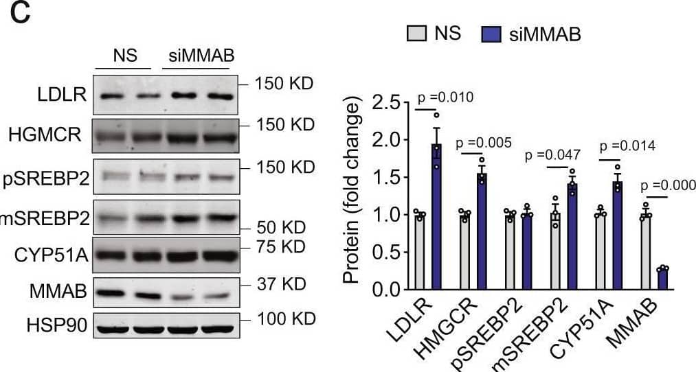


![Immunohistochemistry DDR1 Antibody [Unconjugated]](https://images.novusbio.com/images/antibody/DDR1_AF2396_Immunohistochemistry_7152.jpg)
![Simple Western DDR1 Antibody [Unconjugated]](https://images.novusbio.com/images/antibody/DDR1_AF2396_Immunohistochemistry_17635.jpg)
![Western Blot DDR1 Antibody [Unconjugated]](https://images.novusbio.com/images/antibody/DDR1_AF2396_Western_Blot_16723.jpg)
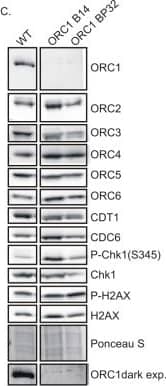


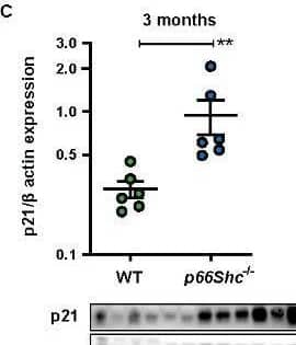
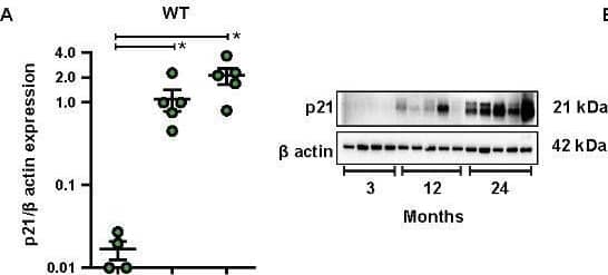
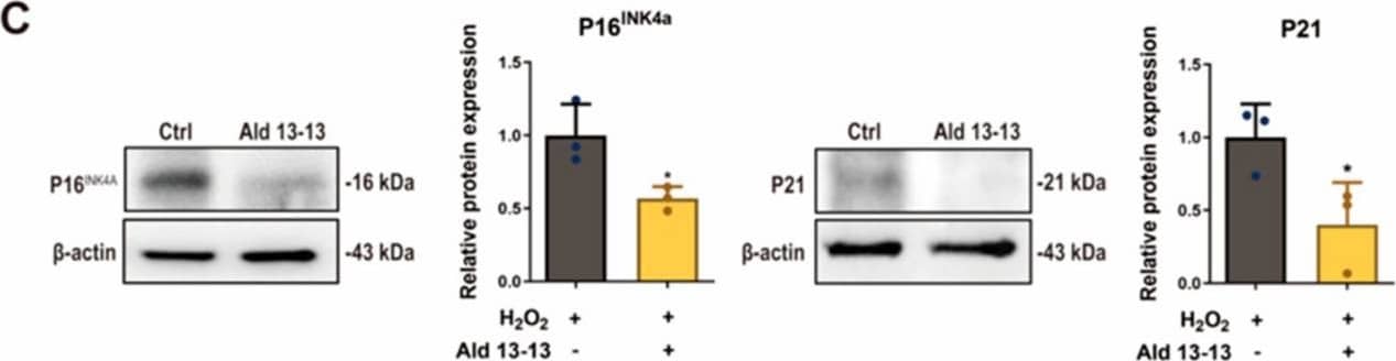


![Western Blot: Goat anti-Rabbit IgG (H+L) Secondary Antibody [HRP] [NB7160] - Western blot showing vemurafenib treatment in BRAFV600E CRC cells inhibits fission mediator DRP1 with no significant effect on fusion proteins (Mfn1 & 2) using MFN-1 antibody (NBP1-51841) and corresponding secondary antibody, goat anti-rabbit IgG-HRP (NB7160). Image collected and cropped by CiteAb from the following publication (https://pubmed.ncbi.nlm.nih.gov/33738242).](https://images.novusbio.com/images/Goat-anti-Rabbit-IgG-H+L-Secondary-Antibody-HRP-Western-Blot-NB7160-img0001.jpg "Western Blot: Goat anti-Rabbit IgG (H+L) Secondary Antibody [HRP] [NB7160] - Western blot showing vemurafenib treatment in BRAFV600E CRC cells inhibits fission mediator DRP1 with no significant effect on fusion proteins (Mfn1 & 2) using MFN-1 antibody (NBP1-51841) and corresponding secondary antibody, goat anti-rabbit IgG-HRP (NB7160). Image collected and cropped by CiteAb from the following publication (https://pubmed.ncbi.nlm.nih.gov/33738242).")
 followed by 30 min incubation with Goat anti Rabbit HRP conjugated secondary antibodies (Catalog # HAF008) at 1:20 dilution + DAB chromogen (brown). The tissue was counterstained with Hematoxylin (blue). Control was done by omitting primary antibody.")
![Flow Cytometry: Rabbit IgG Isotype Control [NBP2-24891] - An intracellular stain was performed on Raji cells with Adiponectin antibody NB100-65810 (blue) and a matched isotype control NBP2-24893 (orange). Cells were fixed with 4% PFA and then permeablized with 0.1% saponin. Cells were incubated in an antibody dilution of 1 ug/mL for 30 minutes at room temperature, followed by Dylight488-conjugated anti-rabbit secondary antibody. Image using the Azide Free form of this antibody.](https://images.novusbio.com/images/Rabbit--Mouse-IgG-Isotype-Control-Flow-Cytometry-NBP2-24891-img0006.jpg "Flow Cytometry: Rabbit IgG Isotype Control [NBP2-24891] - An intracellular stain was performed on Raji cells with Adiponectin antibody NB100-65810 (blue) and a matched isotype control NBP2-24893 (orange). Cells were fixed with 4% PFA and then permeablized with 0.1% saponin. Cells were incubated in an antibody dilution of 1 ug/mL for 30 minutes at room temperature, followed by Dylight488-conjugated anti-rabbit secondary antibody. Image using the Azide Free form of this antibody.")


