
6057-NG), Recombinant Human R-Spondin 1 (
4645-RS), and Recombinant Human Wnt-3a (
5036-WN), along with the other reagents listed in the intestinal organoid culture medium recipe in the
human intestinal organoid culture protocol. (A) Human intestinal organoids were stained using a Rat Anti-Human/Mouse/Rat Vimentin Monoclonal Antibody (
MAB2105; green) and a Goat Anti-Human/Mouse Desmin Antigen Affinity-purified Polyclonal Antibody (
AF3844; red) to visualize myofibroblast cells and counterstained with DAPI (
5748; blue). (B) Human intestinal organoids were stained using a Goat Anti-Human/Mouse E-Cadherin Antigen Affinity-purified Polyclonal Antibody (
AF748; green) and a Mouse Anti-Human MUC2 Monoclonal Antibody (Novus Biologicals, Catalog # NBP2-44431; red) and counterstained with DAPI (
5748; blue)." class="big_lightbox">

6057-NG), Recombinant Human R-Spondin 1 (
4645-RS), and Recombinant Human Wnt-3a (
5036-WN), along with the other reagents listed in the intestinal organoid culture medium recipe in the
human intestinal organoid culture protocol. (A) Human intestinal organoids were stained using a Rat Anti-Human/Mouse/Rat Vimentin Monoclonal Antibody (
MAB2105; green) and a Goat Anti-Human/Mouse Desmin Antigen Affinity-purified Polyclonal Antibody (
AF3844; red) to visualize myofibroblast cells and counterstained with DAPI (
5748; blue). (B) Human intestinal organoids were stained using a Goat Anti-Human/Mouse E-Cadherin Antigen Affinity-purified Polyclonal Antibody (
AF748; green) and a Mouse Anti-Human MUC2 Monoclonal Antibody (Novus Biologicals, Catalog # NBP2-44431; red) and counterstained with DAPI (
5748; blue)." class="big_thumb" />
6057-NG), Recombinant Human R-Spondin 1 (
4645-RS), and Recombinant Human Wnt-3a (
5036-WN), along with the other reagents listed in the intestinal organoid culture medium recipe in the
human intestinal organoid culture protocol. (A) Organoids were fixed and stained with a Mouse Anti-Human MUC2 Monoclonal Antibody (Novus Biologicals; Catalog # NBP2-44431; green) to visualize intestinal goblet cells and counterstained with a Goat Anti-Human/Mouse E-Cadherin Antigen Affinity-purified Polyclonal Antibody (
AF748; red) and DAPI (
5748; blue). The image shown was taken at 10x magnification. (B) Organoids were fixed and stained with a Mouse Anti-Human Chromogranin A Monoclonal Antibody (
MAB90981; green) to visualize enteroendocrine cells and counterstained with a Goat Anti-Human/Mouse E-Cadherin Antigen Affinity-purified Polyclonal Antibody (
AF748; red) and DAPI (
5748; blue). The image shown was taken at 20x magnification." class="big_lightbox">

6057-NG), Recombinant Human R-Spondin 1 (
4645-RS), and Recombinant Human Wnt-3a (
5036-WN), along with the other reagents listed in the intestinal organoid culture medium recipe in the
human intestinal organoid culture protocol. (A) Organoids were fixed and stained with a Mouse Anti-Human MUC2 Monoclonal Antibody (Novus Biologicals; Catalog # NBP2-44431; green) to visualize intestinal goblet cells and counterstained with a Goat Anti-Human/Mouse E-Cadherin Antigen Affinity-purified Polyclonal Antibody (
AF748; red) and DAPI (
5748; blue). The image shown was taken at 10x magnification. (B) Organoids were fixed and stained with a Mouse Anti-Human Chromogranin A Monoclonal Antibody (
MAB90981; green) to visualize enteroendocrine cells and counterstained with a Goat Anti-Human/Mouse E-Cadherin Antigen Affinity-purified Polyclonal Antibody (
AF748; red) and DAPI (
5748; blue). The image shown was taken at 20x magnification." class="big_thumb" />
1030-FN) to support cell attachment and spreading, the ITS and N-2 Plus Media Supplements (
AR013 and
AR003), and a panel of growth factors for effective oligodendrocyte differentiation, including Recombinant Human FGF-basic, Recombinant Human EGF (Catalog # 236-EG), and Recombinant Human PDGF-AA (
221-AA). Oligodendrocytes were detected using a Mouse Anti-Human/Mouse/Rat/Chicken Oligodendrocyte Marker O4 Monoclonal Antibody (
MAB1326). The cells were stained with the NorthernLights™-557 Affinity-purified Goat Anti-Mouse IgM Secondary Antibody (
NL019; red). The nuclei were counterstained with DAPI (
5748;blue)." class="big_lightbox">

1030-FN) to support cell attachment and spreading, the ITS and N-2 Plus Media Supplements (
AR013 and
AR003), and a panel of growth factors for effective oligodendrocyte differentiation, including Recombinant Human FGF-basic, Recombinant Human EGF (Catalog # 236-EG), and Recombinant Human PDGF-AA (
221-AA). Oligodendrocytes were detected using a Mouse Anti-Human/Mouse/Rat/Chicken Oligodendrocyte Marker O4 Monoclonal Antibody (
MAB1326). The cells were stained with the NorthernLights™-557 Affinity-purified Goat Anti-Mouse IgM Secondary Antibody (
NL019; red). The nuclei were counterstained with DAPI (
5748;blue)." class="big_thumb" />
 7754-BH) inhibits recombinant mouse IL-4 induced proliferation in the HT-2 mouse T cell line. The ED50 for this effect is 0.04-0.2 ng/mL." alt="Recombinant human TGF-beta 1 (7754-BH) inhibits recombinant mouse IL-4 induced proliferation in the HT-2 mouse T cell line. The ED50 for this effect is 0.04-0.2 ng/mL." class="big_thumb" />
 7754-BH) Recombinant Human TGF‑ beta 1 as measured by its ability to inhibit the IL-4-dependent proliferation of HT‑2 mouse T cell. (orange, green, respectively)." alt="Equivalent bioactivity of CHO-derived (Catalog # 240-B) and HEK293-derived (7754-BH) Recombinant Human TGF‑ beta 1 as measured by its ability to inhibit the IL-4-dependent proliferation of HT‑2 mouse T cell. (orange, green, respectively)." class="big_thumb" />


![Detection of LC3B in mouse ES cell lysates using Rabbit anti- [Catalog # NB100-2220]. LC3 detected at a molecular weight of approximately 17 kDa. Atg5-/- ES cells from Dr. Noboru Mizushima [Mizushima, N. et al. J. Cell Biol. 152 (2001)] Photo courtesy of Dr. Beth Levine, UT SW Medical Center.](http://images.novusbio.com/fullsize/LC3B-Antibody-Western-Blot-NB100-2220-img0047.jpg)


![<b>Biological Strategies Validation. </b>Detection of LC3I and LC3II in mouse cochlea cell line SV-K1 using Rabbit anti- [Catalog # NB100-2220]. Cells were treated with CQ (1 uM), and As2O3 (1 uM) for 24 hrs. Image from verified customer review.](http://images.novusbio.com/fullsize/LC3B-Antibody-Western-Blot-NB100-2220-img0056.jpg)


![Staining of rat brain tissue section. Analysis using Rabbit anti-LC3B Antibody [Catalog # NB100-2220]. Image from verified customer review.](http://images.novusbio.com/fullsize/LC3B-Antibody-Immunohistochemistry-NB100-2220-img0037.jpg)

![FFPE (Formalin-Fixed Paraffin-Embedded) tissue section of mouse brain using 1:200 dilution of Rabbit anti-LC3B antibody [Catalog # NB100-2220]. The specific signal of LC3 was detected using HRP-conjugated secondary antibody with DAB (3, 3 -diaminobenzidine) reagent, and nuclei of cells were counterstained using hematoxylin. This LC3B antibody generated a low to moderate levels of cytoplasmic staining in the glial cells. The neurons depicted a moderate to strong staining for LC3 in their cytoplasm.](http://images.novusbio.com/fullsize/LC3B-Antibody-Immunohistochemistry-Paraffin-NB100-2220-img0048.jpg)
![Analysis using the Biotin conjugate of Rabbit anti-LC3B Antibody [Catalog # NB100-2220]. Staining of brain, cerebral cortex, neurons with cell processes.](http://images.novusbio.com/fullsize/LC3B-Antibody-Immunohistochemistry-NB100-2220-img0053.jpg)


![Western Blot: LC3B Antibody - BSA Free [NB100-2220] - P53 mediates topotecan-induced autophagy through the activation of sestrin 2 & AMPK in colon cancer cells with wild-type p53.A. HCT116 & LS-174T cells were treated with various concentrations of TPT for 24 h. The levels of P53, sestrin2 & p-AMPK were analysed by immunoblotting. B. HCT116 cells were transfected with p53 or sestrin 2 siRNAs for 24 h, treated with or without 1 µg/mL TPT for an additional 24 h, & the indicated proteins were then analysed by immunoblotting. C. HCT116 cells were transfected with LKB1 or CaMKK beta siRNAs, treated with or without 1 µg/mL TPT for an additional 24 h, & the indicated proteins were detected by immunoblotting. Image collected & cropped by CiteAb from the following publication (//pubmed.ncbi.nlm.nih.gov/23024792), licensed under a CC-BY license. Not internally tested by Novus Biologicals.](http://images.novusbio.com/fullsize/nb100-2220_rabbit-polyclonal-lc3b-antibody-310202415334948.jpg)
![Western Blot: LC3B Antibody - BSA Free [NB100-2220] - Specific inhibitor SP600125 or siRNA directed JNK blocked ceramide-induced autophagy & up-regulation of LC3 expression. (A) SUNE1 cells were treated with 20 μM ceramide for 24 h in the absence or presence of SP600125 or JNK1/2 siRNA. Lysates were analyzed by immunoblotting. (B) Autophagosome formation was visualized using YFP-LC3 expressing & observed under a confocal microscope. Representative immunofluorescence pictures are shown at the original magnification × 1000. (C) The expression of LC3 mRNA was examined by RT-PCR analysis. Image collected & cropped by CiteAb from the following publication (//pubmed.ncbi.nlm.nih.gov/21943220), licensed under a CC-BY license. Not internally tested by Novus Biologicals.](http://images.novusbio.com/fullsize/nb100-2220_rabbit-polyclonal-lc3b-antibody-31020241535643.jpg)
![Western Blot: LC3B Antibody - BSA Free [NB100-2220] - Glutamine deprivation induces IL-8 secretion.(a) RT–PCR analysis of LC3, IL-8, CHOP & GADD34 in U2OS cells subjected to glutamine deprivation (−Q) or exposed to TPG (1 μM). (b) Immunoblot analysis of ER stress- & autophagy-related proteins in U2OS cells subjected to glutamine deprivation or TPG treatment. Samples are run in triplicate. (c) Cytokine array analysis of conditioned media from U2OS & A549 cells grown in the presence (+Q) or absence (−Q) of glutamine. (d) IL-8 enzyme-linked immunosorbent assay of conditioned media from U2OS & A549 cells grown in the presence (+Q) or absence (−Q) of glutamine. Error bars in all figures represent s.d. of three biological replicates. Image collected & cropped by CiteAb from the following publication (//www.nature.com/articles/ncomms5900), licensed under a CC-BY license. Not internally tested by Novus Biologicals.](http://images.novusbio.com/fullsize/nb100-2220_rabbit-polyclonal-lc3b-antibody-310202415395934.jpg)
![Western Blot: LC3B Antibody - BSA Free [NB100-2220] - NDs API improves ATO-based therapy in vivo. HepG2 tumor-bearing nude mice administered intravenously (i.v.) w/ NS (200 μL), NDs (5 mg kg−1), ATO (0.75 mg kg−1), ATO (1.5 mg kg−1), NDs–0.75 mg kg−1 ATO mixture,/NDs–1.5 mg kg−1 ATO mixture daily for 5 consecutive days every week, w/ 13-week cycles c Immunoblots of autophagy-related proteins LC3-II, p62 (left); semi-quantified analysis (n = 3) in tumor tissues from mice after treatment (right). GAPDH used as loading control. Normalized band densities shown below each band. Image collected & cropped by CiteAb from following publication (//pubmed.ncbi.nlm.nih.gov/30341298), licensed under a CC-BY license. Not internally tested by Novus Biologicals.](http://images.novusbio.com/fullsize/nb100-2220_rabbit-polyclonal-lc3b-antibody-31020241539454.jpg)
![Immunocytochemistry/ Immunofluorescence: LC3B Antibody - BSA Free [NB100-2220] - NDs API improves the ATO-based therapy in vivo. HepG2 tumor-bearing nude mice administered intravenously (i.v.) w/ NS (200 μL), NDs (5 mg kg−1), ATO (0.75 mg kg−1), ATO (1.5 mg kg−1), NDs–0.75 mg kg−1 ATO mixture,/NDs–1.5 mg kg−1 ATO mixture daily for 5 consecutive days every week, w/ 13-week cycles. g Immunostaining of LC3 & ATG13 in tumor tissues of NDs–1.5 mg kg−1 ATO-treated mice. Schematic showing (left) & imaging (right) indicate that LC3/Atg13 puncta in periphery & around the blood vessels inside the tumors. Scale bars: 20 μm. Error bars s.d. Image collected & cropped by CiteAb from the following publication (//pubmed.ncbi.nlm.nih.gov/30341298), licensed under a CC-BY license. Not internally tested by Novus Biologicals.](http://images.novusbio.com/fullsize/nb100-2220_rabbit-polyclonal-lc3b-antibody-310202415382444.jpg)
![Immunohistochemistry: LC3B Antibody - BSA Free [NB100-2220] - LC3, CRF, & HIF-1 alpha expression. (a) for each antibody the spatial distribution of immunohistochemical staining was the same in cases with spontaneous & induced labor. (b) The analysis of data from optical density values of Western blotting bands shows no differences of LC3-II & HIF-1 alpha expression between groups. On the contrary CRF is higher in spontaneous labor. LC3-II, HIF-1 alpha , & CRF are normalized onto alpha -tubulin. (c) No correlation between LC3-II & CRF expression was found. SP: spontaneous labor; PG: induction with prostaglandin; PO: induction with prostaglandin & oxytocin; OX: induction with oxytocin. Image collected & cropped by CiteAb from the following publication (//pubmed.ncbi.nlm.nih.gov/23956998), licensed under a CC-BY license. Not internally tested by Novus Biologicals.](http://images.novusbio.com/fullsize/nb100-2220_rabbit-polyclonal-lc3b-antibody-310202415392554.jpg)
![Immunocytochemistry/ Immunofluorescence: LC3B Antibody - BSA Free [NB100-2220] - The time-dependent effects of melatonin on the LC3-II & Beclin 1 expression in U87MG (a, b) & A172 (c, d) cells. The cells were treated with 1 mM melatonin for 4, 12, 24 or 36 h. PBS was used as a negative control, & rapamycin (Rap 200 nM) served as a positive control. The levels of LC3-II & Beclin-1 expression were determined by a Western blot analysis. The protein bands for each regimen were quantified by densitometry, & their differences are presented in the graph. The values represent the mean ± S.E.M. for three separate determinations. Statistical significance * at P 10 puncta per cell from three independent experiments is depicted. Bars represent mean±s.d. from three independent experiments (>50 cells per experiment). The statistical significance (P value) was determined by a two-tailed, paired Student’s t-test. *P<0.05. (c) Immunoblot analysis of U2OS cells subjected to glutamine deprivation with or without 400 nM BafA1. Cells were pretreated with BafA1 for 1 h before & during exposure to glutamine-deficient medium. Autophagic activity was monitored by detection of p62 & LC3-II proteins. (d) U2OS mCherry-GFP-LC3 cells were cultured in the presence (+Q) or absence (−Q) of glutamine for 18 h. Red vesicles denote autolysosomes, whereas yellow vesicles represent autophagosomes. Bars indicate numbers of yellow vesicles (autophagosomes) or red vesicles (autolysosomes) per cell±s.d. (e) Images of U2OS mCherry-GFP-LC3 cells cultured for 18 h in the presence (+Q) or absence (−Q) of glutamine. Scale bar, 10 μm. (f) Phagophore formation in mCherry-ULK1. Scale bar, 10 μm. (g) mCherry-ATG5-expressing U2OS cells after 24 h in the presence (+Q) or absence (−Q) of glutamine. Scale bar, 10 μm. Image collected & cropped by CiteAb from the following publication (//www.nature.com/articles/ncomms5900), licensed under a CC-BY license. Not internally tested by Novus Biologicals.](http://images.novusbio.com/fullsize/nb100-2220_rabbit-polyclonal-lc3b-antibody-31020241554193.jpg)
![Western Blot: LC3B Antibody - BSA Free [NB100-2220] - Glucocorticoids promote BCG survival in macrophages by inhibiting autophagy. (a) RAW264.7 cells were pretreated with dexamethasone (DEX) (1 μM) or vehicle control ethanol (mock) for 6 hr & then challenged with M. bovis BCG (MOI 5) or treated with the vehicle control DMSO or rapamycin (4 μM). LC3 level was detected with Western blot analysis. & GAPDH was used as the loading control. Full-length blots are presented in Supplementary Fig. 12. Data are representative of three independent experiments with similar results. (b,c) RAW264.7 cells were treated as described in (a), & viable bacilli were determined by CFU assays at 6 hpi (b). Survival rate was calculated compared with that of mock-treated cells (c). Data are shown as the mean ± SEM of three independent experiments. *p < 0.05. Image collected & cropped by CiteAb from the following publication (//www.nature.com/articles/s41598-017-01174-9), licensed under a CC-BY license. Not internally tested by Novus Biologicals.](http://images.novusbio.com/fullsize/nb100-2220_rabbit-polyclonal-lc3b-antibody-310202415532059.jpg)
![Western Blot: LC3B Antibody - BSA Free [NB100-2220] - CysC regulates AMPK activity during the induction of autophagy. (a) Immunoblotting analysis of autophagy regulators. N2a cells expressing SOD1 treated with or without CysC (1 μM) for 6 h. The lysates analyzed by immunoblotting using antibodies for phosphorylated AMPK (pAMPK), phosphorylated mTOR (pmTOR), phosphorylated PKCδ (pPKCδ), Myc & beta -actin. (b) Inactivation of AMPK by mutant SOD1 expression. Each relative pAMPK level normalized by beta -actin in (a) is quantified. *P<0.05, **P<0.01 versus mock. (c) Activation of AMPK by CysC treatment. Relative levels of pAMPK for CysC-treated samples normalized by that of PBS-treated control, which is shown as the broken line, in (a) quantified. *P<0.05 versus PBS-treated controls. (d) CysC induced autophagy through the AMPK activation. N2a cells treated with CysC (1 μM), CC (5 μM) or AICA-riboside (AICAR, 5 mM) for 12 h. The lysates analyzed by immunoblotting using antibodies against pAMPK, LC3 & Tubulin (left panel). Quantification of immunoblots was plotted (right panel). (e) The effect of pAMPK activation on neuroprotection by CysC. N2a cells expressing G85R SOD1 mutant treated with CysC (0.2 μM), CC (5 μM) or AICAR (5 mM). Cell viability was measured by the MTS assay. Data expressed as means±S.E.M. from 3 independent experiments. Each experiment was performed in triplicate. *P<0.01 compared to non-treated control, #P<0.01 compared to CysC-treated one. (f) Immunoblotting analysis of pAMPK in SOD1 transgenic mouse spinal cords. The spinal cord lysates from the transgenic mice at indicated ages analyzed for the levels of pAMPK, SOD1 & beta -actin (upper panel). Expression levels of pAMPK normalized by beta -actin (lower panel). All data expressed as means±S.E.M. from 3 independent experiments Image collected & cropped by CiteAb from the following publication (//www.nature.com/articles/cddis2014459), licensed under a CC-BY license. Not internally tested by Novus Biologicals.](http://images.novusbio.com/fullsize/nb100-2220_rabbit-polyclonal-lc3b-antibody-310202415535189.jpg)
![Immunocytochemistry/ Immunofluorescence: LC3B Antibody - BSA Free [NB100-2220] - Vps34 inhibition blocks autophagy initiation & causes accumulation of ubiquitin-positive & p62-positive structures. a Representative confocal images of cortical neurons treated with vehicle, Bafilomycin A1 (BafA1) at 50 nM, VPS34IN1 at 3 µM or cotreated for 3 h. Arrows highlight LC3 & p62 structures. Right panel, bar graphs denote average object intensity, per cell (mean ± SEM, N = 49-60 cells, from three independent experiments). Scale bar, 10 µm. ***p < 0.001 in one-way ANOVA, Holm–Sidak’s multiple comparisons test. b Representative confocal images of cortical neurons treated as in a & immunostained for LAMP-1, LC3, & p62. Airyscan insets highlight position of LC3 & p62 structures relative to LAMP-1-positive membranes. Scale bar, 10 µm. c Representative confocal images of cultured cortical neurons treated with vehicle or VPS34IN1 at 3 µM for 24 h. Arrows highlight p62 & ubiquitin colocalization. Scale bar, 10 µm Image collected & cropped by CiteAb from the following publication (//www.nature.com/articles/s41467-017-02533-w), licensed under a CC-BY license. Not internally tested by Novus Biologicals.](http://images.novusbio.com/fullsize/nb100-2220_rabbit-polyclonal-lc3b-antibody-310202415535128.jpg)
![Immunocytochemistry/ Immunofluorescence: LC3B Antibody - BSA Free [NB100-2220] - ORP1L & VAP-A form cholesterol-dependent ER-AV contact sites(a) MelJuSo cells expressing GFP or GFP-tagged ORP1L mutants were fixed & stained for LC3 & Calnexin. Y477 & D478 in the FFAT motif were mutated to alanines (A) in the ORP1L ydaa mutant. Scale bar, 10 μm. Right: co-immunoprecipitation for ORP1L (mutants) with VAP-A. GFP-ORP1L mutants or GFP were isolated from lysates of HEK293T cells co-overexpressing HA-VAPA using GFP-Trap beads. Western blot filters were probed for isolated GFP-tagged proteins, the associated HA-VAP-A & the input HA-VAP-A, as indicated. (b) Cryo-immuno-EM on HeLa cells expressing HA-LC3 & GFP-ORP1L delta ORD, as detected by HA10 nm & GFP15 nm gold antibodies. Insets show ORP1L labelling in the membrane contact site between ER & autophagosome. The membranes of the ER are depicted in the bottom inset. Scale bar, 50 nm. (c) Three-colour super-resolution image of an autophagosomal vesicle labelled by LC3 (green), ORP1L (blue) & the ER protein VAP-A (red). Scale bar, 500 nm (d) MelJuSo cells cultured either in lipid depleted serum or control medium were fixed & stained for LC3 & ER marker Calnexin. Scale bar, 10 μm. Right panel: Manders coefficient for LC3 localization to the ER was calculated on at least 10 cells over three independent experiments. Bars indicate mean+s.d. Student's t-test statistical analysis (****P<0.0001). Image collected & cropped by CiteAb from the following publication (//pubmed.ncbi.nlm.nih.gov/27283760), licensed under a CC-BY license. Not internally tested by Novus Biologicals.](http://images.novusbio.com/fullsize/nb100-2220_rabbit-polyclonal-lc3b-antibody-31020241554525.jpg)
![Immunohistochemistry: LC3B Antibody - BSA Free [NB100-2220] - OSI induces autophagy through upregulation of MCT1 in CRC cells.a Immunoblotting analysis of MCT1 expression in CRC cells treated with or without 5 μM OSI for 24 h. b Immunoblotting analysis of MCT1 & phosphorylated AMPK in tumor xenografts obtained from vehicle- or OSI-treated mice. (Each protein of interest from each group was electrophoretically transferred onto a PVDF membrane, incubated with indicated primary & secondary antibodies, & developed as a digital image.) c Immunohistochemical analysis of MCT1 expression in tumor xenografts. Scale bar, 50 μm. d Relative intensity of MCT1 staining in (c). e CRC cells were transfected with siScramble or siMCT1 for 24 h, followed by treatment with or without 5 μM OSI for another 24 h. The protein levels of LC3, phosphorylated AMPK, phosphorylated LKB1 & MCT1 were analyzed by immunoblotting. f CRC cells were treated as in (e), the endogenous LC3 puncta were analyzed by immunofluorescence. Scale bar, 10 μm. g The number of LC3 puncta in (f). h CRC cells were transfected with empty vector or Flag-MCT1 plasmid for 48 h, the protein levels of MCT1 & phosphorylated AMPK were analyzed by immunoblotting. i CRC cells were treated as in (h), the endogenous LC3 puncta were analyzed by immunofluorescence. Scale bar, 10 μm. j The number of LC3 puncta in (i). k Immunoblotting analysis of LC3, MCT1 & phosphorylated AMPK levels in CRC cells co-transfected with Flag-MCT1 & DN-AMPK plasmids for 48 h. l CRC cells were treated as in (k), the endogenous LC3 puncta were analyzed by immunofluorescence. Scale bar, 10 μm. m The number of LC3 puncta per cell in (l). Data are presented as mean SEM, Student’s t-test, & are representative of three independent experiments. *P < 0.05; ***P < 0.001 Image collected & cropped by CiteAb from the following publication (//pubmed.ncbi.nlm.nih.gov/31409796), licensed under a CC-BY license. Not internally tested by Novus Biologicals.](http://images.novusbio.com/fullsize/nb100-2220_rabbit-polyclonal-lc3b-antibody-310202415532016.jpg)
![Western Blot: LC3B Antibody - BSA Free [NB100-2220] - Abrogation of autophagy by siRNAs targeting autophagy-related genes in gastric cancer cells induced tumor-intrinsic PD-L1 expression. a Inhibition of autophagy by knockdown of ATG5 in AGS & NCI-n87 gastric cancer cells induced the expression of PD-L1 in the presence & absence of INF-gamma (200 U/mL) as shown by flow cytometry analysis at 48 h post-transfection. b The induction of PD-L1 was confirmed by Western blots at 72 h post-transfection. The knockdown efficacies of ATG5 & ATG7 siRNA were verified. The conversion of LC3B-I to LC3B-II was reduced. Results were averaged & blots were representative of 4 independent experiments. The ratio of PD-L1 MFI minus isotype control was shown as mean ± S.D. relative to Ctrl from 4 independent experiments, *p < 0.05 Image collected & cropped by CiteAb from the following publication (//pubmed.ncbi.nlm.nih.gov/30925913), licensed under a CC-BY license. Not internally tested by Novus Biologicals.](http://images.novusbio.com/fullsize/nb100-2220_rabbit-polyclonal-lc3b-antibody-31020241682127.jpg)
![Western Blot: LC3B Antibody - BSA Free [NB100-2220] - OSI induces autophagy through upregulation of MCT1 in CRC cells.a Immunoblotting analysis of MCT1 expression in CRC cells treated with or without 5 μM OSI for 24 h. b Immunoblotting analysis of MCT1 & phosphorylated AMPK in tumor xenografts obtained from vehicle- or OSI-treated mice. (Each protein of interest from each group was electrophoretically transferred onto a PVDF membrane, incubated with indicated primary & secondary antibodies, & developed as a digital image.) c Immunohistochemical analysis of MCT1 expression in tumor xenografts. Scale bar, 50 μm. d Relative intensity of MCT1 staining in (c). e CRC cells were transfected with siScramble or siMCT1 for 24 h, followed by treatment with or without 5 μM OSI for another 24 h. The protein levels of LC3, phosphorylated AMPK, phosphorylated LKB1 & MCT1 were analyzed by immunoblotting. f CRC cells were treated as in (e), the endogenous LC3 puncta were analyzed by immunofluorescence. Scale bar, 10 μm. g The number of LC3 puncta in (f). h CRC cells were transfected with empty vector or Flag-MCT1 plasmid for 48 h, the protein levels of MCT1 & phosphorylated AMPK were analyzed by immunoblotting. i CRC cells were treated as in (h), the endogenous LC3 puncta were analyzed by immunofluorescence. Scale bar, 10 μm. j The number of LC3 puncta in (i). k Immunoblotting analysis of LC3, MCT1 & phosphorylated AMPK levels in CRC cells co-transfected with Flag-MCT1 & DN-AMPK plasmids for 48 h. l CRC cells were treated as in (k), the endogenous LC3 puncta were analyzed by immunofluorescence. Scale bar, 10 μm. m The number of LC3 puncta per cell in (l). Data are presented as mean SEM, Student’s t-test, & are representative of three independent experiments. *P < 0.05; ***P < 0.001 Image collected & cropped by CiteAb from the following publication (//pubmed.ncbi.nlm.nih.gov/31409796), licensed under a CC-BY license. Not internally tested by Novus Biologicals.](http://images.novusbio.com/fullsize/nb100-2220_rabbit-polyclonal-lc3b-antibody-310202415541982.jpg)
![Immunocytochemistry/ Immunofluorescence: LC3B Antibody - BSA Free [NB100-2220] - Screening of potent NAPIs in HepG2. a Immunoblots for autophagy-related proteins LC3-II, p62 (left); semi-quantified analysis (n = 3) in various nanoparticles treated cells (right). CQ & Rapamycin (Rapa) were used as positive controls for autophagy inhibition & autophagy activation, respectively. Glyceraldehyde 3-phosphate dehydrogenase (GAPDH) was used as the loading control. Normalized band densities were shown below each band. b Fluorescence images of mCherry-GFP-LC3 cells after incubation with CQ or NDs for 48 h (autophagosomes: mCherry+/GFP+ yellow puncta; autolysosomes: mCherry+/GFP) & quantification of the number of LC3 puncta per cell in cells (10 cells per group). Scale bar: 10 μm. c Immunoblots for autophagy-related proteins LC3-II, p62 (left); semi-quantified analysis (n = 3) in CQ, NDs, or CQ–NDs-treated cells (right). &P < 0.05, significantly different from NDs. GAPDH was used as the loading control. Normalized band densities were shown below each band. d Left: Cell viability after incubation with ATO or various NAPIs–ATO mixture for 48 h (n = 3). ##P < 0.01 by t-test, significantly different from ATO. Right: Cell viability after 48 h NDs–ATO treatment with RNAi of autophagy proteins ATG5 & ATG7 (n = 3). e, f Immunoblots for autophagy-related protein LC3-II & autolysosomal process-related protein NUPR1, SNAP25, VAMP8 in NDs-treated cells. g Immunoblots for autolysosomal process-related protein NUPR1 after NDs treatment with RNAi of autophagy proteins ATG5 & ATG7. GAPDH was used as the loading control. Error bars are s.d. Image collected & cropped by CiteAb from the following publication (//pubmed.ncbi.nlm.nih.gov/30341298), licensed under a CC-BY license. Not internally tested by Novus Biologicals.](http://images.novusbio.com/fullsize/nb100-2220_rabbit-polyclonal-lc3b-antibody-310202415532088.jpg)
![Western Blot: LC3B Antibody - BSA Free [NB100-2220] - Screening of potent NAPIs in HepG2. a Immunoblots for autophagy-related proteins LC3-II, p62 (left); semi-quantified analysis (n = 3) in various nanoparticles treated cells (right). CQ & Rapamycin (Rapa) were used as positive controls for autophagy inhibition & autophagy activation, respectively. Glyceraldehyde 3-phosphate dehydrogenase (GAPDH) was used as the loading control. Normalized band densities were shown below each band. b Fluorescence images of mCherry-GFP-LC3 cells after incubation with CQ or NDs for 48 h (autophagosomes: mCherry+/GFP+ yellow puncta; autolysosomes: mCherry+/GFP) & quantification of the number of LC3 puncta per cell in cells (10 cells per group). Scale bar: 10 μm. c Immunoblots for autophagy-related proteins LC3-II, p62 (left); semi-quantified analysis (n = 3) in CQ, NDs, or CQ–NDs-treated cells (right). &P < 0.05, significantly different from NDs. GAPDH was used as the loading control. Normalized band densities were shown below each band. d Left: Cell viability after incubation with ATO or various NAPIs–ATO mixture for 48 h (n = 3). ##P < 0.01 by t-test, significantly different from ATO. Right: Cell viability after 48 h NDs–ATO treatment with RNAi of autophagy proteins ATG5 & ATG7 (n = 3). e, f Immunoblots for autophagy-related protein LC3-II & autolysosomal process-related protein NUPR1, SNAP25, VAMP8 in NDs-treated cells. g Immunoblots for autolysosomal process-related protein NUPR1 after NDs treatment with RNAi of autophagy proteins ATG5 & ATG7. GAPDH was used as the loading control. Error bars are s.d. Image collected & cropped by CiteAb from the following publication (//pubmed.ncbi.nlm.nih.gov/30341298), licensed under a CC-BY license. Not internally tested by Novus Biologicals.](http://images.novusbio.com/fullsize/nb100-2220_rabbit-polyclonal-lc3b-antibody-310202415523913.jpg)
![Western Blot: LC3B Antibody - BSA Free [NB100-2220] - OSI induces autophagy in CRC cells in vitro & in vivo.a Immunoblotting analysis of LC3, Atg5, & p62/SQSTM1 expression in CRC cells treated with indicated concentrations of OSI for 24 h. b The formation of endogenous LC3 puncta in cells treated with DMSO or 5 μM OSI for 24 h. c Total number of endogenous LC3 puncta per cell in (b). d, e LC3 expression in xenograft tissues was examined by IHC. Representative images were provided as indicated in (d) & relative intensity of LC3 staining was quantified in (e). f Immunoblotting analysis of LC3 & p62/SQSTM1 expression in tumor xenografts (Each protein of interest from each group was electrophoretically transferred onto a PVDF membrane, incubated with indicated primary & secondary antibodies, & developed as a digital image.) g Relative intensity of LC3 in (f). h Co-immunoprecipitation analysis of the interaction between Beclin 1 & Bcl-2 in CRC cells treated with or without 5 μM OSI for 24 h. i Immunoblotting analysis of LC3 expression in CRC cells treated with or without 5 μM OSI in the presence or absence of 5 mM 3-MA for 24 h. j CRC cells were treated as in (i), the LC3 puncta were analyzed by immunofluorescence. Scale bar, 10 μm. k, l Immunoblotting analysis of LC3 expression in CRC cells transfected with siScramble, siATG5 (k), or siBECN1 (l) for 24 h, followed by treatment with or without 5 μM OSI for another 24 h. m CRC cells were treated as in (k, l). The LC3 puncta were analyzed by immunofluorescence. Scale bar, 10 μm. Data are presented as mean SEM, Student’s t-test, & are representative of 3 independent experiments. **P < 0.01; ***P < 0.001 Image collected & cropped by CiteAb from the following publication (//pubmed.ncbi.nlm.nih.gov/31409796), licensed under a CC-BY license. Not internally tested by Novus Biologicals.](http://images.novusbio.com/fullsize/nb100-2220_rabbit-polyclonal-lc3b-antibody-310202415523911.jpg)
![Immunohistochemistry: LC3B Antibody - BSA Free [NB100-2220] - OSI induces autophagy in CRC cells in vitro & in vivo.a Immunoblotting analysis of LC3, Atg5, & p62/SQSTM1 expression in CRC cells treated with indicated concentrations of OSI for 24 h. b The formation of endogenous LC3 puncta in cells treated with DMSO or 5 μM OSI for 24 h. c Total number of endogenous LC3 puncta per cell in (b). d, e LC3 expression in xenograft tissues was examined by IHC. Representative images were provided as indicated in (d) & relative intensity of LC3 staining was quantified in (e). f Immunoblotting analysis of LC3 & p62/SQSTM1 expression in tumor xenografts (Each protein of interest from each group was electrophoretically transferred onto a PVDF membrane, incubated with indicated primary & secondary antibodies, & developed as a digital image.) g Relative intensity of LC3 in (f). h Co-immunoprecipitation analysis of the interaction between Beclin 1 & Bcl-2 in CRC cells treated with or without 5 μM OSI for 24 h. i Immunoblotting analysis of LC3 expression in CRC cells treated with or without 5 μM OSI in the presence or absence of 5 mM 3-MA for 24 h. j CRC cells were treated as in (i), the LC3 puncta were analyzed by immunofluorescence. Scale bar, 10 μm. k, l Immunoblotting analysis of LC3 expression in CRC cells transfected with siScramble, siATG5 (k), or siBECN1 (l) for 24 h, followed by treatment with or without 5 μM OSI for another 24 h. m CRC cells were treated as in (k, l). The LC3 puncta were analyzed by immunofluorescence. Scale bar, 10 μm. Data are presented as mean SEM, Student’s t-test, & are representative of 3 independent experiments. **P < 0.01; ***P < 0.001 Image collected & cropped by CiteAb from the following publication (//pubmed.ncbi.nlm.nih.gov/31409796), licensed under a CC-BY license. Not internally tested by Novus Biologicals.](http://images.novusbio.com/fullsize/nb100-2220_rabbit-polyclonal-lc3b-antibody-31020241553713.jpg)
![Western Blot: LC3B Antibody - BSA Free [NB100-2220] - Growth retardation & shortened lifespan of the mice with deletion of an autophagy-essential gene Atg7 in hematopoietic system. (A) Three genotypes for wild-type, heterozygote, & homozygote for Atg 7 deletion in hematopoietic system with representative images of the mice. The images were taken at age of 10 weeks. (B) PCR Genotyping analysis of the offsprings from Atg7f/f mice crossing Vav-iCre mice to screen Atg7f/f;Vav-iCre mice. The sequences for the primers used in PCR are given in the method section, & their PCR amplified bands representing specific genotypes were indicated in the agarose gel electrophoresis films. (C) Examination of Atg7 expression in wild-type & the Atg7-deleted mice. Upper panel, quantitative PCR analysis of Atg7 transcription normalized to Gapdh transcript in different organs; lower panel, western blotting analysis of autophagy-essential protein ATG7 & lipidation of LC3 in different organs. GAPDH used as a loading control. (D) Growth comparison between wild-type & Atg7-deleted mice. Wild-type mice progressively gain weight before age of 60 weeks (left panel), but Atg7-deleted mice cease weight gain at about age of 6 weeks (right panel). (E) Measurement of lifespan of wild-type & Atg7-deleted mice. (F) Immunohistological examination of heart, liver, lung & thymus from 10 weeks old wild-type & Atg7-deleted mice by HE staining. Image collected & cropped by CiteAb from the following publication (//pubmed.ncbi.nlm.nih.gov/31327762), licensed under a CC-BY license. Not internally tested by Novus Biologicals.](http://images.novusbio.com/fullsize/nb100-2220_rabbit-polyclonal-lc3b-antibody-310202415535185.jpg)
![Western Blot: LC3B Antibody - BSA Free [NB100-2220] - Overexpression of wild-type MAGEA6, but not mutant MAGEA6, suppresses autophagy in PDAC cell lines.(A) Immunoblot analysis of autophagy signaling in HPDE-iKRAS cells expressing GFP (Vec) & MAGEA6 variants. (B) Immunofluorescence staining of LC3B puncta in the transduced HPDE-iKRAS cells. Representative photos (left) & statistical analysis (mean ± standard deviation of counted cells, N=~100 per cohort) are shown. *p=0.002; two-tailed unpaired t-test. (C) Immunoblot analysis of autophagy substrate SQSTM1/p62 in the transduced HPDE-iKRAS cells treated with BafA1 for the indicated time points., Immunoblot analysis of (D) autophagy signaling & (E) SQSTM1/p62 accumulation in wild-type MAGEA6 expressing & (F) autophagy signaling in MAGEA6H305fs* expressing HPDE-iKRAS cells under nutrient-deficient conditions. (G) Immunoblot analysis of autophagy signaling in wild-type MAGEA6 expressing cells under prolonged nutrient-deficient conditions.Immunoblot analysis of autophagy signaling of transduced AsPC-1 & MIA PaCa-2.Immunoblot analysis of the accumulation of autophagy substrate SQSTM/p62 in the transduced HPDE-iKRAS cells under BafA1 for the indicated time points.Immunoblot analysis of autophagy activity in transduced HPDE-iKRAS cells under nutrient-depleted conditions as indicated. Image collected & cropped by CiteAb from the following publication (//pubmed.ncbi.nlm.nih.gov/32270762), licensed under a CC-BY license. Not internally tested by Novus Biologicals.](http://images.novusbio.com/fullsize/nb100-2220_rabbit-polyclonal-lc3b-antibody-310202415535165.jpg)
![Western Blot: LC3B Antibody - BSA Free [NB100-2220] - Inhibition of LRRK2 alters the autophagy/lysosomal pathway in H4 neuroglioma cells. A) LC3-II levels increase upon LRRK2-in1 inhibitor treatment (1 μM, overnight treatment; quantification from 3 independent replicates, the plot shows mean & SD, * indicates significance (p < 0.05)). B) Dose dependent increase in LC3-II upon overnight treatment with LRRK2-in1. C) MTT assay showing no alteration of cell viability upon over night treatment with LRRK2-in1 from 1 to 5 μM. A small toxic effect appeared with the higher dose (10 μM). The plot shows mean & SD, * indicates significance (p < 0.05). D) LRRK2 knockdown cells display reduced response to LRRK2-in1. LRRK2 protein levels are decreased in shRNA stable line compared to wild type cells (right panel), & knockdown of LRRK2 reduces response to 1 μM LRRK2-in1 treatment compared to wild type or scrambled shRNA cells (left panel). E) Western blot analysis of H4 cells treated with DMSO & LRRK2-in1 (5 μM, 2.5 hours treatment) in the presence & absence of 40 nM bafilomycin added at the same time as the inhibitor. Quantification of three replicates is shown in the right hand panel, the plot shows mean & SD, * indicates significance (p < 0.05). F) LRRK2-in1 increases LC3-II levels independent of mTORC1 activity. P70S6K & phoshoThr389-P70S6K; S6 & phosphoSer235/236-S6 levels are shown in control, starvation & amino-acid stimulated conditions. LRRK2-in1 (1 μM over night) treatment does not alter phosphorylation of P70S6K & S6 in conditions that show increased levels of LC3-II. G) Neutral red staining. The plot shows mean & SEM. * indicates significance (p < 0.05). Image collected & cropped by CiteAb from the following publication (//linkinghub.elsevier.com/retrieve/pii/S0167488913002954), licensed under a CC-BY license. Not internally tested by Novus Biologicals.](http://images.novusbio.com/fullsize/nb100-2220_rabbit-polyclonal-lc3b-antibody-31020241553728.jpg)
![Immunocytochemistry/ Immunofluorescence: LC3B Antibody - BSA Free [NB100-2220] - SQSTM1 recruited ARHGAP5-AS1 for autophagic degradation.a ARHGAP5-AS1 expression in resistant cells after transfecting siNC or SQSTM1 siRNAs was assessed using qRT-PCR. **p < 0.01. b ARHGAP5-AS1 expression in sensitive cells after SQSTM1 overexpression was detected by qRT-PCR. c The interaction of ARHGAP5-AS1 with SQSTM1 in SGC-R cells was analyzed by RIP assay followed with qRT-PCR. IgG was served as the negative control. d RNA pull down assay was performed to verify the binding of SQSTM1 to ARHGAP5-AS1 in SGC7901 or SGC-R cells. beta -actin was served as negative control. NC: no probe. The interaction of ARHGAP5-AS1 with SQSTM1 in SGC-R cells before & after EBSS treatment (4 h) were analyzed using RIP assay (e) & RNA pull down assay (f). g The interaction of various SQSTM1 constructs with ARHGAP5-AS1 were analyzed using RIP assay. h The interaction of various ARHGAP5-AS1 fragments with SQSTM1 was analyzed by RNA pull down assay. i Half-life of ARHGAP5-AS1 in SGC7901 & SGC-R cells with different SQSTM1 expression status were determined by qRT-PCR. The colocalization of ARHGAP5-AS with SQSTM1 (j) & LC3B (k) in SGC-R cells treated as indicated were analyzed by combined FISH & IFC assay (original magnification, ×100). Scale bar: 100 μm (j) & 10 μm (k) Image collected & cropped by CiteAb from the following publication (//pubmed.ncbi.nlm.nih.gov/31097692), licensed under a CC-BY license. Not internally tested by Novus Biologicals.](http://images.novusbio.com/fullsize/nb100-2220_rabbit-polyclonal-lc3b-antibody-31020241553716.jpg)
![Western Blot: LC3B Antibody - BSA Free [NB100-2220] - Effects of autophagy & caspase inhibitors on survival of neurons after glucose reperfusion (GR).After 3 h GD, neuro2a cells were replaced by 20-Methyladenine (3MA); Bafilomycin A1 (BA1) & z-VAD-FMK (z-VAD) were added at final concentrations of 5 mM, 5 nM & 20 uM, respectively. (A) Cell viability was assessed using the MTT assay after treatment of autophagy & pan-caspase inhibitors. MTT mitochondrial reduction was shown as a relative percentage of the MTT values at the indicated time point (3 h GD/48 h GR) from three independent experiments ± SD (*p<0.05, **p<0.001). (B) Changes in protein levels by inhibitors of autophagy or caspase activation were analyzed by immunoblot. Proteins were blotted with anti-LC3, p62, cleaved-caspase 3 & PARP antibodies. Tubulin was used as loading control. (C) Quantification of immunoblot data in (B). The amount of each protein was normalized against the amount of tubulin. Data represent the mean ± SD (*p<0.05, n = 3). (D) Cells transiently expressing GFP-LC3 (green) were treated with 3MA, BA1 or z-VAD for 48 h of GR after 3h GD. Nuclei were stained with DAPI (blue). Scale bar represents 5 µm. Quantification shown on the right graph represents mean GFP-LC3 puncta per cell (n = 10 cells per condition) from three independent experiments ± SD. The asterisks (*) indicate significant differences in the values.*p<0.05, **p<0.001. Image collected & cropped by CiteAb from the following publication (//dx.plos.org/10.1371/journal.pone.0076466), licensed under a CC-BY license. Not internally tested by Novus Biologicals.](http://images.novusbio.com/fullsize/nb100-2220_rabbit-polyclonal-lc3b-antibody-31020241553717.jpg)
![Immunocytochemistry/ Immunofluorescence: LC3B Antibody - BSA Free [NB100-2220] - Exposure of microglia to cART resulted in blockade of autophagosome–lysosome fusion. (A) rPMs were seeded into a 12-well plate followed by tandem fluorescent-tagged MAP1LC3B plasmid. Next, cells were exposed to cART (5 µM each of TDF, FTC, & DTG) for an additional 24 h & observed by confocal imaging. The results showed that cART exposure significantly increased the formation of autophagosomes (yellow puncta). (B) Representative bar graph showing the number of autophagosome (yellow puncta) per cell. (C) Representative bar graph showing the number of autolysosome (red puncta) per cell. (D) rPMs were seeded into 12-well plates followed with cART exposure for 24 h. Cells were then double immunostained with MAP1LC3B & LAMP2 antibody & observed by immunofluorescent microscopy. (E,F) Representative bar graphs showing cART-mediated decreased LAMP2 puncta & decreased colocalization of MAP1LC3B & LAMP2. BAF—autophagosome fusion inhibitor, & rapamycin (RAP—autophagy inducer) were used as controls for autophagy flux. Data is from three independent experiments & is expressed as means ± SEM & were analyzed using one-way ANOVA. *, p < 0.05 vs. control. Image collected & cropped by CiteAb from the following publication (//pubmed.ncbi.nlm.nih.gov/31569373), licensed under a CC-BY license. Not internally tested by Novus Biologicals.](http://images.novusbio.com/fullsize/nb100-2220_rabbit-polyclonal-lc3b-antibody-31020241683936.jpg)
![Western Blot: LC3B Antibody - BSA Free [NB100-2220] - The cytoprotection of in vitro sIPC is diminished by autophagy inhibitors in RPTC cells. RPTC cells were subjected to: (1) control; (2) CCCP-R; (3) sIPC + CCCP-R in the absence or presence of chloroquine (20 μM) & 3-methyladenine (10 mM). Both inhibitors were used for 1-h pretreatment & during 2-h recovery from prolonged CCCP treatment. Cells were collected for morphological & immunoblot analyses. (a) Representative images of phase contrast & fluorescence microscopy showing cellular & nuclear morphology of apoptosis. Scale bar: 200 μm. (b) Quantification of cell apoptosis. Data are expressed as mean ± SD. *, P < 0.05, significantly different from the control group; #, P < 0.05, significantly different from CCCP-R group. (c) Analysis of apoptosis inhibitory efficiency by sIPC. Data are expressed as mean ± SD. *, P < 0.05, significantly different from the group without inhibitors. (d & e) Immunoblots of LC3B & cleaved CASP3. ACTB was used as a loading control. The molecular mass marker lanes were labelled as kDa. For densitometric analysis of cleaved CASP3, after normalization with ACTB, the protein signals of the control were arbitrarily set as 1, & the signals of other conditions were normalized to the control to calculate fold changes. Data are expressed as mean ± SD. *, P < 0.05, significantly different from the control group; #, P < 0.05, significantly different from CCCP-R group. Image collected & cropped by CiteAb from the following publication (//pubmed.ncbi.nlm.nih.gov/31066324), licensed under a CC-BY license. Not internally tested by Novus Biologicals.](http://images.novusbio.com/fullsize/nb100-2220_rabbit-polyclonal-lc3b-antibody-310202415541971.jpg)
![Immunocytochemistry/ Immunofluorescence: LC3B Antibody - BSA Free [NB100-2220] - ORP1L & VAP-A form cholesterol-dependent ER-AV contact sites(a) MelJuSo cells expressing GFP or GFP-tagged ORP1L mutants were fixed & stained for LC3 & Calnexin. Y477 & D478 in the FFAT motif were mutated to alanines (A) in the ORP1L ydaa mutant. Scale bar, 10 μm. Right: co-immunoprecipitation for ORP1L (mutants) with VAP-A. GFP-ORP1L mutants or GFP were isolated from lysates of HEK293T cells co-overexpressing HA-VAPA using GFP-Trap beads. Western blot filters were probed for isolated GFP-tagged proteins, the associated HA-VAP-A & the input HA-VAP-A, as indicated. (b) Cryo-immuno-EM on HeLa cells expressing HA-LC3 & GFP-ORP1L delta ORD, as detected by HA10 nm & GFP15 nm gold antibodies. Insets show ORP1L labelling in the membrane contact site between ER & autophagosome. The membranes of the ER are depicted in the bottom inset. Scale bar, 50 nm. (c) Three-colour super-resolution image of an autophagosomal vesicle labelled by LC3 (green), ORP1L (blue) & the ER protein VAP-A (red). Scale bar, 500 nm (d) MelJuSo cells cultured either in lipid depleted serum or control medium were fixed & stained for LC3 & ER marker Calnexin. Scale bar, 10 μm. Right panel: Manders coefficient for LC3 localization to the ER was calculated on at least 10 cells over three independent experiments. Bars indicate mean+s.d. Student's t-test statistical analysis (****P<0.0001). Image collected & cropped by CiteAb from the following publication (//pubmed.ncbi.nlm.nih.gov/27283760), licensed under a CC-BY license. Not internally tested by Novus Biologicals.](http://images.novusbio.com/fullsize/nb100-2220_rabbit-polyclonal-lc3b-antibody-310202415541949.jpg)
![Western Blot: LC3B Antibody - BSA Free [NB100-2220] - Inhibition of LRRK2 alters the autophagy/lysosomal pathway in H4 neuroglioma cells. A) LC3-II levels increase upon LRRK2-in1 inhibitor treatment (1 μM, overnight treatment; quantification from 3 independent replicates, the plot shows mean & SD, * indicates significance (p < 0.05)). B) Dose dependent increase in LC3-II upon overnight treatment with LRRK2-in1. C) MTT assay showing no alteration of cell viability upon over night treatment with LRRK2-in1 from 1 to 5 μM. A small toxic effect appeared with the higher dose (10 μM). The plot shows mean & SD, * indicates significance (p < 0.05). D) LRRK2 knockdown cells display reduced response to LRRK2-in1. LRRK2 protein levels are decreased in shRNA stable line compared to wild type cells (right panel), & knockdown of LRRK2 reduces response to 1 μM LRRK2-in1 treatment compared to wild type or scrambled shRNA cells (left panel). E) Western blot analysis of H4 cells treated with DMSO & LRRK2-in1 (5 μM, 2.5 hours treatment) in the presence & absence of 40 nM bafilomycin added at the same time as the inhibitor. Quantification of three replicates is shown in the right hand panel, the plot shows mean & SD, * indicates significance (p < 0.05). F) LRRK2-in1 increases LC3-II levels independent of mTORC1 activity. P70S6K & phoshoThr389-P70S6K; S6 & phosphoSer235/236-S6 levels are shown in control, starvation & amino-acid stimulated conditions. LRRK2-in1 (1 μM over night) treatment does not alter phosphorylation of P70S6K & S6 in conditions that show increased levels of LC3-II. G) Neutral red staining. The plot shows mean & SEM. * indicates significance (p < 0.05). Image collected & cropped by CiteAb from the following publication (//linkinghub.elsevier.com/retrieve/pii/S0167488913002954), licensed under a CC-BY license. Not internally tested by Novus Biologicals.](http://images.novusbio.com/fullsize/nb100-2220_rabbit-polyclonal-lc3b-antibody-3102024165723.jpg)
![Immunocytochemistry/ Immunofluorescence: LC3B Antibody - BSA Free [NB100-2220] - TFEB overexpression in macrophages induces the autophagy markers LC3 & p62 & restores their co-localization in atherosclerotic aortic roots.(a,b) Representative immunofluorescence images of atherosclerotic aortic roots (2 months' western diet) from control & mφTFEB-TG mice (ApoE-null background) stained with antibodies against TFEB (a), TFEB & MOMA-2 (b; scale bar, 50 μm). (c) Quantification of the average TFEB intensity & co-localization with nuclear marker DAPI (n=4-5 mice per group). (d) Representative immunofluorescence images of atherosclerotic aortic roots from control & mφTFEB-TG mice stained with p62 & LC3 (scale bar, 50 μm). (e) Quantification of the p62 & LC3 average intensity from control & mφTFEB-TG-stained roots (n=13–14 mice per group). (f) Representative pseudocolour image of these p62/LC3 images (green represents co-localization) & graph depicting the increased p62/LC3 correlation seen in a representative mφTFEB-TG as compared to a control lesion (scale bar, 50 μm). (g) Quantification of the p62/LC3 co-localization from control & mφTFEB-TG-stained roots shown (n=13–14 mice per group). (h,i) FACS analysis of aortic macrophages isolated from atherosclerotic aortas of Control or mφTFEB-TG mice (western diet-fed ApoE-KO background, n=3–4 pooled aortas) & stained for either (h) p62 & LC3, or (i) Lamp2 & LC3 antibodies (per cent of macrophages expressing each marker is shown below plots). For all graphs, data are presented as mean±s.e.m. *P<0.05, ***P<0.001, two-tailed unpaired t-test. Image collected & cropped by CiteAb from the following publication (//www.nature.com/articles/ncomms15750), licensed under a CC-BY license. Not internally tested by Novus Biologicals.](http://images.novusbio.com/fullsize/nb100-2220_rabbit-polyclonal-lc3b-antibody-31020241553207.jpg)
![Immunohistochemistry: LC3B Antibody - BSA Free [NB100-2220] - Keap1 co-localizes with SQSTM1-immunopositive sarcoplasmic aggregates in AVM muscle. a-c. Only focal & minimal co-localization of LC3 (a) & Keap1 immunofluorescence (b) is seen in a representative toxic AVM sample (colchicine-treated subject #31); merged panel is shown in (c) (LC3, red; Keap1, green). d-f. In the same specimen, there is extensive co-localization of SQSTM1 (d) & Keap1 immunofluorescence (e); merged panel is shown in (f) (SQSTM1, red; Keap1, green). g-i. Similar to toxic AVM specimens, essentially no co-localization of LC3 (g) & Keap1 immunofluorescence (h) is seen in a representative IBM muscle biopsy (subject #46); merged panel is shown in (i) (LC3, red; Keap1, green). j-l. In the same specimen, there is extensive co-localization of SQSTM1 (j) & Keap1 immunofluorescence (k); merged panel is shown in (l) (SQSTM1, red; Keap1, green). Scale bar, 25 μm Image collected & cropped by CiteAb from the following publication (//actaneurocomms.biomedcentral.com/articles/10.1186/s40478-016-0384-6), licensed under a CC-BY license. Not internally tested by Novus Biologicals.](http://images.novusbio.com/fullsize/nb100-2220_rabbit-polyclonal-lc3b-antibody-310202415535125.jpg)
![Western Blot: LC3B Antibody - BSA Free [NB100-2220] - Glucose deprivation (GD) induces neuronal cell death.Time course of cell viability & cell death after GD was estimated by MTT & LDH assays, respectively. Bars depict rates of MTT reduction (A) & LDH release (B) time dependent manner after GD. MTT data represent relative percentages compared with controls (*p<0.05, **p<0.001, n = 3). LDH release from neuro2a cells was calculated as fold increase from the LDH release in controls (*p<0.01, **p<0.001, n = 3). (C) Levels of LC3 & p62 expression, cleaved-caspase 3 & PARP cleavage were determined at indicated times after GD by immunoblot analysis. Tubulin was used as loading control. (D) Quantitative analyses of the immunoblots are shown in (C). The amount of each protein was normalized against the amount of tubulin. Data represent the mean ± SD for each condition (*p<0.05, **p<0.001, n = 3). Image collected & cropped by CiteAb from the following publication (//dx.plos.org/10.1371/journal.pone.0076466), licensed under a CC-BY license. Not internally tested by Novus Biologicals.](http://images.novusbio.com/fullsize/nb100-2220_rabbit-polyclonal-lc3b-antibody-31020241552390.jpg)
![Immunohistochemistry: LC3B Antibody - BSA Free [NB100-2220] - OSI induces autophagy through upregulation of MCT1 in CRC cells.a Immunoblotting analysis of MCT1 expression in CRC cells treated with or without 5 μM OSI for 24 h. b Immunoblotting analysis of MCT1 & phosphorylated AMPK in tumor xenografts obtained from vehicle- or OSI-treated mice. (Each protein of interest from each group was electrophoretically transferred onto a PVDF membrane, incubated with indicated primary & secondary antibodies, & developed as a digital image.) c Immunohistochemical analysis of MCT1 expression in tumor xenografts. Scale bar, 50 μm. d Relative intensity of MCT1 staining in (c). e CRC cells were transfected with siScramble or siMCT1 for 24 h, followed by treatment with or without 5 μM OSI for another 24 h. The protein levels of LC3, phosphorylated AMPK, phosphorylated LKB1 & MCT1 were analyzed by immunoblotting. f CRC cells were treated as in (e), the endogenous LC3 puncta were analyzed by immunofluorescence. Scale bar, 10 μm. g The number of LC3 puncta in (f). h CRC cells were transfected with empty vector or Flag-MCT1 plasmid for 48 h, the protein levels of MCT1 & phosphorylated AMPK were analyzed by immunoblotting. i CRC cells were treated as in (h), the endogenous LC3 puncta were analyzed by immunofluorescence. Scale bar, 10 μm. j The number of LC3 puncta in (i). k Immunoblotting analysis of LC3, MCT1 & phosphorylated AMPK levels in CRC cells co-transfected with Flag-MCT1 & DN-AMPK plasmids for 48 h. l CRC cells were treated as in (k), the endogenous LC3 puncta were analyzed by immunofluorescence. Scale bar, 10 μm. m The number of LC3 puncta per cell in (l). Data are presented as mean SEM, Student’s t-test, & are representative of three independent experiments. *P < 0.05; ***P < 0.001 Image collected & cropped by CiteAb from the following publication (//pubmed.ncbi.nlm.nih.gov/31409796), licensed under a CC-BY license. Not internally tested by Novus Biologicals.](http://images.novusbio.com/fullsize/nb100-2220_rabbit-polyclonal-lc3b-antibody-310202415525226.jpg)
![Immunohistochemistry: LC3B Antibody - BSA Free [NB100-2220] - OSI induces autophagy in CRC cells in vitro & in vivo.a Immunoblotting analysis of LC3, Atg5, & p62/SQSTM1 expression in CRC cells treated with indicated concentrations of OSI for 24 h. b The formation of endogenous LC3 puncta in cells treated with DMSO or 5 μM OSI for 24 h. c Total number of endogenous LC3 puncta per cell in (b). d, e LC3 expression in xenograft tissues was examined by IHC. Representative images were provided as indicated in (d) & relative intensity of LC3 staining was quantified in (e). f Immunoblotting analysis of LC3 & p62/SQSTM1 expression in tumor xenografts (Each protein of interest from each group was electrophoretically transferred onto a PVDF membrane, incubated with indicated primary & secondary antibodies, & developed as a digital image.) g Relative intensity of LC3 in (f). h Co-immunoprecipitation analysis of the interaction between Beclin 1 & Bcl-2 in CRC cells treated with or without 5 μM OSI for 24 h. i Immunoblotting analysis of LC3 expression in CRC cells treated with or without 5 μM OSI in the presence or absence of 5 mM 3-MA for 24 h. j CRC cells were treated as in (i), the LC3 puncta were analyzed by immunofluorescence. Scale bar, 10 μm. k, l Immunoblotting analysis of LC3 expression in CRC cells transfected with siScramble, siATG5 (k), or siBECN1 (l) for 24 h, followed by treatment with or without 5 μM OSI for another 24 h. m CRC cells were treated as in (k, l). The LC3 puncta were analyzed by immunofluorescence. Scale bar, 10 μm. Data are presented as mean SEM, Student’s t-test, & are representative of 3 independent experiments. **P < 0.01; ***P < 0.001 Image collected & cropped by CiteAb from the following publication (//pubmed.ncbi.nlm.nih.gov/31409796), licensed under a CC-BY license. Not internally tested by Novus Biologicals.](http://images.novusbio.com/fullsize/nb100-2220_rabbit-polyclonal-lc3b-antibody-310202415535133.jpg)
![Immunocytochemistry/ Immunofluorescence: LC3B Antibody - BSA Free [NB100-2220] - Measurement of dysregulated autophagy by fluorescent microscopyOVCAR3 cells were treated with the indicated drugs for 24 hours & then fixed & immunostained for p62 (red) & LC3 (green), with a DAPI costain (blue). Confocal z-stacks were flattened for image analysis to capture all punctate area. A. Doses were: chloroquine (10 μM), nelfinavir (10 μM), rapamycin (10 nM), dasatinib (50 nM), & all were combined for Necramed. B. Similar immunostaining as in (A) Punctae were quantified for size & number per cell by ImageJ with at least 40 cells per condition. LC3 channel was analyzed for autophagosomes, & the p62 channel for sequestosomes. Doses were same as in (A), but with chloroquine & nelfinavir reduced to 5 μM. *p < 0.05, ***p < 0.001 by t-test. All error bars are s.e.m. C. OVCAR3 cells with a virally integrated mCherry-GFP-LC3 construct were studied by live microscopy, for the number of hours indicated following Necramed (doses as in (A)) or control treatment. Image collected & cropped by CiteAb from the following publication (//pubmed.ncbi.nlm.nih.gov/26418751), licensed under a CC-BY license. Not internally tested by Novus Biologicals.](http://images.novusbio.com/fullsize/nb100-2220_rabbit-polyclonal-lc3b-antibody-3102024165734.jpg)
![Western Blot: LC3B Antibody - BSA Free [NB100-2220] - Rop-DPRL combined with CR inhibits tumor growth. (A) Fluorescent imaging of major organs & tumor. Tumor-bearing mice were injected with cy5-labeled liposome or unlabelled liposome. The tumor & major organs from control or drug-treated mice were excised at 12 h or 24 h. (B) Fluorescent image of tumor sections. Tumor from control or Rop-DPRL treated mice were excised at 12 h. Tumor frozen sections were observed under confocal microscope. Scale bar = 20 µm. (C) Immunofluorescence staining of tumor sections for the autophagosome marker LC3, P62 & LAMP1. (D) Image of representative tumors. (E) Change in tumor volume. (F) Tumor weight. (G) TUNEL staining of tumor sections. Nuclei were stained with Hoechst. Scale bar = 20µm. (H) Body weight changes of mice under different treatments. CR: Calorie restriction. Arrows: Enlarged lysosomes. Data are presented as the mean ± SEM, ** p < 0.01. Image collected & cropped by CiteAb from the following publication (//pubmed.ncbi.nlm.nih.gov/32308756), licensed under a CC-BY license. Not internally tested by Novus Biologicals.](http://images.novusbio.com/fullsize/nb100-2220_rabbit-polyclonal-lc3b-antibody-31020241555242.jpg)
![Western Blot: LC3B Antibody - BSA Free [NB100-2220] - SsD & SsA inhibit autophagy in HeLa cells. a Structures of SsA, SsC, & SsD. b SsD (15 μM) & SsA (30 μM), but not SsC, significantly increased LC3-II yellow puncta formation but did not significantly affect LC3-II red-only puncta in RFP-GFP-LC3-expressing HeLa cells. Scale bar = 10 μm. Quantification of LC3 yellow puncta/red puncta (%) is presented as the mean ± S.E., n = ~80 cells from 3 independent experiments. c Treatment of HeLa cells with SsD for 6 h induced the accumulation of both LC3-II & SQSTM1 in a dose-dependent manner. d SsD (15 μM) induced the accumulation of both LC3-II & SQSTM1 in HeLa cells in a time-dependent manner. e SsA (30 μM) induced the accumulation of both LC3-II & SQSTM1 in HeLa cells in a time-dependent manner. f Treatment with different concentrations of SsC for 6 h failed to induce the accumulation of either LC3-II or SQSTM1 in HeLa cells. g SsD (15 μM) reversibly inhibited autophagy Image collected & cropped by CiteAb from the following publication (//pubmed.ncbi.nlm.nih.gov/30820356), licensed under a CC-BY license. Not internally tested by Novus Biologicals.](http://images.novusbio.com/fullsize/nb100-2220_rabbit-polyclonal-lc3b-antibody-31020241553736.jpg)
![Immunocytochemistry/ Immunofluorescence: LC3B Antibody - BSA Free [NB100-2220] - PERK pathway-independent induction of prosurvival autophagy in KG1 LSC-like cells upon G9a inhibition. a, LC3-I/II & p62 expression, analyzed by western blotting, after cell treatment with BIX-01294 (15 μmol/L) in the absence or presence of bafilomycin A1 (BafA1) for 24 h. b, Confocal microscopy images of GFP-LC3-transfected cells after treatment with BIX-01294 (5 μmol/L). c, Apoptotic fractions, measured using flow cytometry, after cell treatment with BIX-01294 (15 μmol/L) in the absence or presence of the autophagy inhibitor 3-MA (5 mmol/L) or BafA1 (2 nmol/L) for 48 h. d, Protein expression, analyzed by western blotting with the indicated antibodies, after treatment of PERK siRNA-transfected & non-transfected cells with BIX-01294 (15 μmol/L) for 48 h. e, Protein expression, analyzed by western blotting with the indicated antibodies, after cell treatment with BIX-01294 (10 μmol/L) in the absence or presence of GSK2606414 (20 μmol/L) for 48 h. f, Protein expression, analyzed by western blotting with the indicated antibodies, after treatment of NRF2 siRNA-transfected & non-transfected cells with BIX-01294 (10 μmol/L) for 48 h. g, Levels of apoptosis, evaluated by flow cytometry, after cell treatment for 48 h with BIX-01294 (10 μmol/L) in the absence or presence of the PERK inhibitor GSK2606414 (10 μmol/L) or the autophagy inhibitor 3-MA (5 mmol/L), or BafA1 (2 nmol/L). h, Levels of apoptosis, evaluated by flow cytometry, after treatment of PERK siRNA- or control siRNA-transfected cells with BIX-01294 (10 μmol/L) in the absence or presence of 3-MA (5 mmol/L) or BafA1 (2 nmol/L) Image collected & cropped by CiteAb from the following publication (//pubmed.ncbi.nlm.nih.gov/32293500), licensed under a CC-BY license. Not internally tested by Novus Biologicals.](http://images.novusbio.com/fullsize/nb100-2220_rabbit-polyclonal-lc3b-antibody-31020241682153.jpg)
![Immunohistochemistry: LC3B Antibody - BSA Free [NB100-2220] - OSI induces autophagy in CRC cells in vitro & in vivo.a Immunoblotting analysis of LC3, Atg5, & p62/SQSTM1 expression in CRC cells treated with indicated concentrations of OSI for 24 h. b The formation of endogenous LC3 puncta in cells treated with DMSO or 5 μM OSI for 24 h. c Total number of endogenous LC3 puncta per cell in (b). d, e LC3 expression in xenograft tissues was examined by IHC. Representative images were provided as indicated in (d) & relative intensity of LC3 staining was quantified in (e). f Immunoblotting analysis of LC3 & p62/SQSTM1 expression in tumor xenografts (Each protein of interest from each group was electrophoretically transferred onto a PVDF membrane, incubated with indicated primary & secondary antibodies, & developed as a digital image.) g Relative intensity of LC3 in (f). h Co-immunoprecipitation analysis of the interaction between Beclin 1 & Bcl-2 in CRC cells treated with or without 5 μM OSI for 24 h. i Immunoblotting analysis of LC3 expression in CRC cells treated with or without 5 μM OSI in the presence or absence of 5 mM 3-MA for 24 h. j CRC cells were treated as in (i), the LC3 puncta were analyzed by immunofluorescence. Scale bar, 10 μm. k, l Immunoblotting analysis of LC3 expression in CRC cells transfected with siScramble, siATG5 (k), or siBECN1 (l) for 24 h, followed by treatment with or without 5 μM OSI for another 24 h. m CRC cells were treated as in (k, l). The LC3 puncta were analyzed by immunofluorescence. Scale bar, 10 μm. Data are presented as mean SEM, Student’s t-test, & are representative of 3 independent experiments. **P < 0.01; ***P < 0.001 Image collected & cropped by CiteAb from the following publication (//pubmed.ncbi.nlm.nih.gov/31409796), licensed under a CC-BY license. Not internally tested by Novus Biologicals.](http://images.novusbio.com/fullsize/nb100-2220_rabbit-polyclonal-lc3b-antibody-31020241555224.jpg)
![Western Blot: LC3B Antibody - BSA Free [NB100-2220] - Metabolic profiling & biochemical assay. (A) Relative abundance of the substrates in the glycolytic pathway & TCA cycle in ORP compared to OCL by targeted metabolic profiling. When compared with OCL heart, ORP hearts have significantly lower glucose-6-phosphate & fructose-6-phosphate (both are glycolytic metabolites), & significantly higher a-ketoglutarate, fumarate, malate, & citrate (all are TCA cycle metabolites). *P < 0.05 compared with OCL. See Table S6 for numerical data. (B) A schematic diagram summarizing the changes in metabolism by rapamycin in old heart. (C) Western blots of autophagic markers show no significant change of LC3 II/I, p62, or beclin-1 in cardiac aging. However, OCR has significantly lower p62 than that in OCL. #P < 0.05 compared with OCL. (D) Both CR & RP significantly reduce the age-dependent increase in protein carbonyls (nmol mL−1). #P < 0.05 compared with OCL. (E). Both CR & RP significantly reduce the age-dependent increase in protein ubiquitination.*P < 0.05 compared with YCL & #P < 0.05 compared with OCL. n = 3–8. G6P: glucose 6-phosphate; G1P: glucose 1-phosphate; F6P: fructose 6-phosphate; F1P: fructose 1-phosphate; F16BP: fructose 1,6-bisphosphate; F26BP: fructose 2,6-biphosphate; G3P: glyceraldehyde 3-phosphate; DHAP:dihydroxyacetone phosphate; 2(3)-PGA: 2- or 3-phosphoglycerate; & PEP: phosphoenolpyruvate. Isomers of same molecular weight, that is, G6P versus G1P, F6P versus F1P, & F16BP versus F26BP, were not distinguishable by the LC-MS/MS-based metabolic profiling method. Image collected & cropped by CiteAb from the following publication (//pubmed.ncbi.nlm.nih.gov/24612461), licensed under a CC-BY license. Not internally tested by Novus Biologicals.](http://images.novusbio.com/fullsize/nb100-2220_rabbit-polyclonal-lc3b-antibody-310202415535191.jpg)
![Western Blot: LC3B Antibody - BSA Free [NB100-2220] - iNOS level is regulated by autophagy during LPS-stimulated condition. (A) Raw 264.7 cells were treated with LPS (200 ng/mL) for 12 h first, followed by removing the LPS & added CQ (30 µM), SAR (1 µM), Torin (1 µM), Rap (1 µM) for another 12 h. CTRL is blank control group with DMSO treatment. (B) Western blot analysis of iNOS in Raw 264.7 cells under LPS treatment. (C) Western blot analysis of p62 in Raw 264.7 cells under LPS treatment. (D) Raw 264.7 cells were treated LPS (200 ng/mL) for 12 h, then removed LPS added CHX (1 µg/mL) & autophagy related drugs for another 12 h. (E) Level of iNOS in Raw 264.7 cell after CHX treatment. (B–E) Values were analyzed from three individual experiments by GraphPad Prism, each experiment conducted three times. * p < 0.05, ** p < 0.01, *** p < 0.001. Error bars (mean ± SEM). One-way ANOVA with Turkeys as post hoc tests. Image collected & cropped by CiteAb from the following publication (//pubmed.ncbi.nlm.nih.gov/31618870), licensed under a CC-BY license. Not internally tested by Novus Biologicals.](http://images.novusbio.com/fullsize/nb100-2220_rabbit-polyclonal-lc3b-antibody-31020241682195.jpg)
![Immunocytochemistry/ Immunofluorescence: LC3B Antibody - BSA Free [NB100-2220] - Autophagic pathway regulates inflammasome activation in blood-derived monocytes stimulated with M. leprae. (A–C) Monocytes were purified from healthy donors’ peripheral blood mononuclear cells by 2-h adherence & stimulated with 10 µg/mL M. leprae (ML) in the presence or absence of autophagy regulators for 18 h. Autophagy was triggered with 200 ng/mL rapamycin (RP), & inhibited with 100 µM chloroquine (CQ, autophagic flux blocker) & 10 µM 3-methyladenine (3-MA). (A) Monocytes stimulated with M. leprae, RP, CQ, & 3-MA were fixed & stained with anti-NLRP3 (red), anti-LC3B (green) antibodies & DAPI (blue). Monocytes stimulated with M. leprae increase the number of LC3-II-decorated autophagosomes (puncta) per cell, which is reforced by RP addition, & reverted by 3-MA. Autophagy blocking with CQ & 3-MA was able to increase NLRP3 specks numbers per cell in relation to the well stimulated with M. leprae & RP. Immunofluorescence images were quantified & bars represent the mean values of the number of LC3 puncta & NLRP3 specks per cell ± SEM (n = 3). *(in relation to NS) or #(indicated by the dashes) p < 0.05. Scale bar: 10 µm. (B) Purified mRNAs from monocyte cultures stimulated with M. leprae, the autophagy blocker 3-MA, & RP, as a control of autophagy activation, were analyzed by RT-qPCR for LC3B & IL1B. Bars represent the mean of the fold change values in relation to the non-stimulated controls of three independent experiments with similar results. (C) The IL-1 beta , IL-6, & TNF levels in the monocyte cultures supernatants were assessed by ELISA. Bars represent the fold change values in relation to the non-stimulated controls ± SEM (n = 3). Image collected & cropped by CiteAb from the following publication (//pubmed.ncbi.nlm.nih.gov/29915584), licensed under a CC-BY license. Not internally tested by Novus Biologicals.](http://images.novusbio.com/fullsize/nb100-2220_rabbit-polyclonal-lc3b-antibody-31020241554517.jpg)
![Immunocytochemistry/ Immunofluorescence: LC3B Antibody - BSA Free [NB100-2220] - Melatonin induced LC3 aggregation & conversion. a GFP-LC3 aggregation & conversion in U87MG cells stably expressing an exogenous fusion protein, green fluorescent protein (GFP) & microtubule-associated light chain 3 (LC3) (U87-GFP-LC3), after treatment with DMSO (control), rapamycin (RAP 200 nM), melatonin (MEL 1 mM) or agomelatine (AGO 2 μM) for 24 h (×40); b U87MG cells & c A172 cells treated with DMSO (control), rapamycin (RAP 200 nM), melatonin (MEL 1 mM), agomelatine (AGO 2 μM) & luzindole (LUZ 5 μM/L) for 24 h were fixed & analyzed for endogenous LC3 dots by immunofluorescence using anti-LC3 antibodies (green), DAPI (blue) staining shows the nuclei (×40). d Stable U87-GFP-LC3 cell lines & GFP in vector control. U87MG cells stably expressing an exogenous fusion protein, green fluorescent protein (GFP) & microtubule-associated light chain 3 (LC3) (U87-GFP-LC3) (×40) Image collected & cropped by CiteAb from the following publication (//pubmed.ncbi.nlm.nih.gov/31870319), licensed under a CC-BY license. Not internally tested by Novus Biologicals.](http://images.novusbio.com/fullsize/nb100-2220_rabbit-polyclonal-lc3b-antibody-31020241612627.jpg)
![Immunocytochemistry/ Immunofluorescence: LC3B Antibody - BSA Free [NB100-2220] - Autophagy induction by atorvastatin in T24 human bladder cancer cells. (A) Immunocytochemistry for the co-localization of LC3 puncta (green) & Lysotracker (red, demarcation for the lysosome) after atorvastatin treatments (30 μM) for 24 h in T24 cells. Rapamycin treated T24 cells were the positive control of autophagy induction. 4′,6-diamidino-2-phenylindole (DAPI) was used for nucleus staining. Scale bar = 100 μm; (B) High magnification view of immunocytochemistry for the co-localization LC3 puncta (green) & Lysotracker after atorvastatin treatments (30 μM) for 24 h in T24 cells at the single-cell level. Scale bar = 50 μm; (C) Western blot analysis of autophagosome formation markers p62/SQSTM1, LC3-I & LC3-II in untreated (control) & atorvastatin (30 μM) treated T24 cells. Image collected & cropped by CiteAb from the following publication (//www.mdpi.com/1422-0067/15/5/8106), licensed under a CC-BY license. Not internally tested by Novus Biologicals.](http://images.novusbio.com/fullsize/nb100-2220_rabbit-polyclonal-lc3b-antibody-310202415541929.jpg)
![Immunocytochemistry/ Immunofluorescence: LC3B Antibody - BSA Free [NB100-2220] - Increased LC3B-positive puncta correlated positively with vascular remodeling during AngII-induced hypertension. (A) Western blot analysis of LC3B-II expression in aortas from sham or AngII-infused mice at different time points after operation. **P < 0.01 vs. the corresponding sham group, Student's t-test. n = 10 mice/group. (B) Representative immunofluorescence staining of LC3B (green) & alpha -SMA (red) in thoracic aorta section. Nuclei were stained with DAPI. Scale bars, 20 µm. (C) Quantification of LC3B puncta. Five random fields (2,500 µm2/filed) were measured in one section. Statistical significance was determined by one-way ANOVA. **P < 0.01 vs. sham, n = 12 sections from six mice per group. (D-G) Structural parameters of mouse aorta from sham & hypertensive mice. WD, wall diameter; LD, lumen diameter; CSA, cross-sectional area. *P < 0.05, **P < 0.01 vs. the corresponding sham group, Student's t-test. n = 10 mice/group. (H) Correlation between changes in LC3B puncta & CSA values. Image collected & cropped by CiteAb from the following publication (//pubmed.ncbi.nlm.nih.gov/32226533), licensed under a CC-BY license. Not internally tested by Novus Biologicals.](http://images.novusbio.com/fullsize/nb100-2220_rabbit-polyclonal-lc3b-antibody-31020241555241.jpg)
![Immunohistochemistry: LC3B Antibody - BSA Free [NB100-2220] - Representative images of the variation of LC3 staining associated with cavities in SDH activity & mitochondrial COX-1 staining in PAD gastrocnemius muscle sections. First column shows succinate dehydrogenase (SDH) activity (dark fibers are type I, intermediate fibers are type IIa, & light fibers are type IIa/x), second column shows mitochondrial COX-1 protein (green), third column shows LC3 (red) & fourth column shows COX-1 & LC3 merged. Examples of normal fibers with no LC3 staining or COX-1 cavities (rowA), punctate LC3 staining where a COX-1 cavitiy is forming (row B), elevated LC3 staining with low COX-1 staining (rowC), LC3 accumulation in areas lacking COX-1 staining (row D), & LC3 plaque in the center of an SDH/COX1 cavity (COX-1; rowE). Arrows within the same row point to areas of LC3 accumulation in same fiber. Scale bar = 100 µM Image collected & cropped by CiteAb from the following publication (//pubmed.ncbi.nlm.nih.gov/27687713), licensed under a CC-BY license. Not internally tested by Novus Biologicals.](http://images.novusbio.com/fullsize/nb100-2220_rabbit-polyclonal-lc3b-antibody-3102024168218.jpg)
![Immunocytochemistry/ Immunofluorescence: LC3B Antibody - BSA Free [NB100-2220] - Vps34 inhibition blocks autophagy initiation & causes accumulation of ubiquitin-positive & p62-positive structures. a Representative confocal images of cortical neurons treated with vehicle, Bafilomycin A1 (BafA1) at 50 nM, VPS34IN1 at 3 µM or cotreated for 3 h. Arrows highlight LC3 & p62 structures. Right panel, bar graphs denote average object intensity, per cell (mean ± SEM, N = 49-60 cells, from three independent experiments). Scale bar, 10 µm. ***p < 0.001 in one-way ANOVA, Holm–Sidak’s multiple comparisons test. b Representative confocal images of cortical neurons treated as in a & immunostained for LAMP-1, LC3, & p62. Airyscan insets highlight position of LC3 & p62 structures relative to LAMP-1-positive membranes. Scale bar, 10 µm. c Representative confocal images of cultured cortical neurons treated with vehicle or VPS34IN1 at 3 µM for 24 h. Arrows highlight p62 & ubiquitin colocalization. Scale bar, 10 µm Image collected & cropped by CiteAb from the following publication (//www.nature.com/articles/s41467-017-02533-w), licensed under a CC-BY license. Not internally tested by Novus Biologicals.](http://images.novusbio.com/fullsize/nb100-2220_rabbit-polyclonal-lc3b-antibody-3102024155521.jpg)
![Western Blot: LC3B Antibody - BSA Free [NB100-2220] - Mechanistic analysis of autophagic inhibition in ATO-treated HepG2. a The viability of HepG2 & NB4 cells after incubation with ATO at various concentrations for 48 h (n = 3; error bars are s.d.). Upper: Confocal images of NB4 or HepG2 cells after being stained with calcein AM. Scale bars: 10 μm. b The viability of HepG2 & NB4 cells after incubation with CQ, ATO, or CQ–ATO mixture for 48 h (n = 3). ##P < 0.01 by t-test, significantly different from ATO. ns not significant. c Immunoblots of autophagy-related proteins LC3-II, p62 (left); semi-quantified analysis (n = 3) in NDs, ATO, or NDs–ATO mixture-treated cells (right). GAPDH was used as the loading control. Normalized band densities were shown below each band. d Cell viability & Caspase-3 activity after incubation with ATO or NDs–ATO mixture for 48 h (n = 3). ##P < 0.01 by t-test, significantly different from ATO. e Immunostaining of LC3 in NDs, ATO, or NDs–ATO mixture-treated cells (left). Scale bar: 20 μm. TEM images of ATO or NDs–ATO mixture-treated cells (right). Scale bars: 1 μm. Typical structures of autophagosomes induced by ATO & autolysosome containing NDs induced by NDs–ATO mixture are indicated with green or blue arrows, respectively (magnification). Vacuoles inside cells are indicated with red arrows (magnification). Scale bars: 500 nm. Error bars are s.d. Image collected & cropped by CiteAb from the following publication (//pubmed.ncbi.nlm.nih.gov/30341298), licensed under a CC-BY license. Not internally tested by Novus Biologicals.](http://images.novusbio.com/fullsize/nb100-2220_rabbit-polyclonal-lc3b-antibody-310202415525276.jpg)
![Immunocytochemistry/ Immunofluorescence: LC3B Antibody - BSA Free [NB100-2220] - Rop-DPRL impairs autophagy. (A) Fluorescent images of GFP-LC3-HeLa cells treated with 1.5 mM ropivacaine for 0-24 h, or starvation for 0-24 h, & stained with 75 nM LysoTracker Red. Scale bar = 5 µm. (B) Western blot & (C) statistical results of P62, immature & mature cathepsin D (CathD), LC3I, LC3II, & beta -actin. HeLa cells were treated with ropivacaine or starvation for 0-24 h. (D) Western blot & (E) statistical results of P62, LC3I, LC3II, & beta -actin. HeLa cells were treated with PBS, CQ (50 µM), ropivacaine, or CQ+Rop for 24 h. (F) Fluorescent images of GFP-LC3-HeLa cells treated with empty liposome or Rop-DPRL for 24 h & stained with 75 nM LysoTracker Red. Scale bar = 5 μm. (G,I, K & M) Western blot & (H,J,L & N) statistical results for P62, LC3I, LC3II & beta -actin. Cells were treated with empty liposome, CQ, Rop-DPRL, starvation or indicated combination for 24 h. Data are presented as the mean ± SEM, *p < 0.05, ** p < 0.01, *** p < 0.001, ns = no significant difference, Rop: ropivacaine, starv: starvation. Image collected & cropped by CiteAb from the following publication (//pubmed.ncbi.nlm.nih.gov/32308756), licensed under a CC-BY license. Not internally tested by Novus Biologicals.](http://images.novusbio.com/fullsize/nb100-2220_rabbit-polyclonal-lc3b-antibody-31020241682123.jpg)
![Western Blot: LC3B Antibody - BSA Free [NB100-2220] - PERK pathway-independent induction of prosurvival autophagy in KG1 LSC-like cells upon G9a inhibition. a, LC3-I/II & p62 expression, analyzed by western blotting, after cell treatment with BIX-01294 (15 μmol/L) in the absence or presence of bafilomycin A1 (BafA1) for 24 h. b, Confocal microscopy images of GFP-LC3-transfected cells after treatment with BIX-01294 (5 μmol/L). c, Apoptotic fractions, measured using flow cytometry, after cell treatment with BIX-01294 (15 μmol/L) in the absence or presence of the autophagy inhibitor 3-MA (5 mmol/L) or BafA1 (2 nmol/L) for 48 h. d, Protein expression, analyzed by western blotting with the indicated antibodies, after treatment of PERK siRNA-transfected & non-transfected cells with BIX-01294 (15 μmol/L) for 48 h. e, Protein expression, analyzed by western blotting with the indicated antibodies, after cell treatment with BIX-01294 (10 μmol/L) in the absence or presence of GSK2606414 (20 μmol/L) for 48 h. f, Protein expression, analyzed by western blotting with the indicated antibodies, after treatment of NRF2 siRNA-transfected & non-transfected cells with BIX-01294 (10 μmol/L) for 48 h. g, Levels of apoptosis, evaluated by flow cytometry, after cell treatment for 48 h with BIX-01294 (10 μmol/L) in the absence or presence of the PERK inhibitor GSK2606414 (10 μmol/L) or the autophagy inhibitor 3-MA (5 mmol/L), or BafA1 (2 nmol/L). h, Levels of apoptosis, evaluated by flow cytometry, after treatment of PERK siRNA- or control siRNA-transfected cells with BIX-01294 (10 μmol/L) in the absence or presence of 3-MA (5 mmol/L) or BafA1 (2 nmol/L) Image collected & cropped by CiteAb from the following publication (//pubmed.ncbi.nlm.nih.gov/32293500), licensed under a CC-BY license. Not internally tested by Novus Biologicals.](http://images.novusbio.com/fullsize/nb100-2220_rabbit-polyclonal-lc3b-antibody-310202415525287.jpg)
![Western Blot: LC3B Antibody - BSA Free [NB100-2220] - TLR4 mediates Hsp70/Hsp90-induced catabolic response in myotubes. a Hsp70 & Hsp90-induced catabolic response in myotubes is dependent on TLR4. C2C12 myoblasts were transfected with siRNA as indicated. After differentiation, myotubes were treated with rHsp70 & rHsp90 & analyzed for catabolic response as described above. b LLC-released EVs induce muscle catabolism in C2C12 myotubes through TLR4. C2C12 myotubes with either TLR2 or TLR4 knockdown were treated with EVs isolated from LCM (AchE 6 mU) & analyzed for catabolic response. c CD9-positive EVs activate TLR4 in reporter cells. EVs isolated from LCM were subjected to immunoprecipitation (IP) using an antibody against CD9 with pre-immune IgG as control. Resulting pellet & supernatant were used to treat TLR4 reporter cell line HEK-Blue hTLR4 for 24 h, & compared with original EVs. TLR4 activation was measured as SEAP activity. Data (n = 3) were analyzed by analysis of variance. * denotes a difference (P < 0.05) Image collected & cropped by CiteAb from the following publication (//www.nature.com/articles/s41467-017-00726-x), licensed under a CC-BY license. Not internally tested by Novus Biologicals.](http://images.novusbio.com/fullsize/nb100-2220_rabbit-polyclonal-lc3b-antibody-310202415533814.jpg)
![Immunocytochemistry/ Immunofluorescence: LC3B Antibody - BSA Free [NB100-2220] - cIAP2 & XIAP regulate autophagosome fusion, but not cIAP1.a MEFs expressing mCherry-GFP-LC3b were transfected with siRNA against either cIAP1, cIAP2 or XIAP. Cells were analysed with the microscope & the number of mCherry+, & GFP + puncta/cell were calculated & the ratio of GFP + /mCherry + puncta is indicated. Shown are the means & the error bars represent the SEM of at least three independent experiments. Westerns show efficient knockdown of cIAP1 & XIAP expression. cIAP2 siRNA efficiency was determined by real time PCR as shown in the graph below the westerns. b Wild type & cIAP2−/− XIAP−/− dermal fibroblasts expressing mCherry-GFP-LC3b were treated analysed on the microscope & mCherry+, & GFP + puncta/cell were calculated. The ratio of GFP+/mCherry + puncta is indicated. c Wild type & cIAP2−/− XIAP−/− dermal fibroblasts were left in complete media (CM) or starved for 2 h in EBSS. Cells were lysed & proteins analysed by western blot for XIAP, LC3, & Actin. cIAP2−/− was confirmed by PCR due to lack of effective antibodies for mouse cIAP2 (see supplemental Fig. 2). d Immunofluoresence showing accumulation of LC3 in starved cIAP2−/− XIAP−/− dermal fibroblasts. Wild type & cIAP2−/− XIAP−/− dermal fibroblasts were incubated in complete media or starved in EBSS for 2 h, then fixed & stained for LC3 (green channel) & LAMP2 (red channel). Nuclei are stained blue with Hoechst. Shown in upper panels are overviews. Lower panels show zoomed in regions indicated in the upper panels Image collected & cropped by CiteAb from the following publication (//pubmed.ncbi.nlm.nih.gov/29743550), licensed under a CC-BY license. Not internally tested by Novus Biologicals.](http://images.novusbio.com/fullsize/nb100-2220_rabbit-polyclonal-lc3b-antibody-310202415541921.jpg)
![Western Blot: LC3B Antibody - BSA Free [NB100-2220] - Enhanced mTORC2 signaling is sufficient to protect MSC from calcification. Flag-tagged rictor or green fluorescent protein (GFP) as a control were introduced into MSC by lentiviral transfer. Cells were cultured for 21 days under control conditions, with osteoblast induction medium (OM) & vehicle, or OM & 20 nM Rapa as indicated. (A) Western blot analyses of rictor & flag confirming overexpression of rictor due to gene transfer, downstream targets of the two mTOR complexes (mTORC1: pp70-S6KSer389; mTORC2: pAKTSer473), inhibition of autophagy (LC3B II, p62), cellular senescence (p16INK4a), & apoptosis-related proteins (Bcl-2: negative regulator of apoptosis; cleaved caspase 3 (cl casp 3): executioner caspase). Representative western blots from six independent experiments are shown. For densitometric quantification, band intensities were normalized to GAPDH as a loading control. Control (no OM) was set to 1. (B) Lactate dehydrogenase (LDH) activity in cell culture supernatants normalized to total protein concentrations was determined to quantify cell death. Control was set to 1. (C) Alkaline phosphatase (ALP) activity normalized to total protein concentrations was measured after 7 days of incubation. (D) Calcium deposition was quantified with the ortho-cresolphthalein method & normalized to total protein concentrations. Bars represent mean + SEM, *P < 0.05, **P < 0.01, ***P < 0.001. Numbers of independent experiments are indicated at the bottom of each graph. (E) Alizarin red S staining of calcium deposits. Representative experiment out of six, phase contrast microscopy, original magnification x200, scale bar = 100 µm. Image collected & cropped by CiteAb from the following publication (//pubmed.ncbi.nlm.nih.gov/31882658), licensed under a CC-BY license. Not internally tested by Novus Biologicals.](http://images.novusbio.com/fullsize/nb100-2220_rabbit-polyclonal-lc3b-antibody-310202415523919.jpg)
![Immunocytochemistry/ Immunofluorescence: LC3B Antibody - BSA Free [NB100-2220] - Autophagy induction by atorvastatin in T24 human bladder cancer cells. (A) Immunocytochemistry for the co-localization of LC3 puncta (green) & Lysotracker (red, demarcation for the lysosome) after atorvastatin treatments (30 μM) for 24 h in T24 cells. Rapamycin treated T24 cells were the positive control of autophagy induction. 4′,6-diamidino-2-phenylindole (DAPI) was used for nucleus staining. Scale bar = 100 μm; (B) High magnification view of immunocytochemistry for the co-localization LC3 puncta (green) & Lysotracker after atorvastatin treatments (30 μM) for 24 h in T24 cells at the single-cell level. Scale bar = 50 μm; (C) Western blot analysis of autophagosome formation markers p62/SQSTM1, LC3-I & LC3-II in untreated (control) & atorvastatin (30 μM) treated T24 cells. Image collected & cropped by CiteAb from the following publication (//www.mdpi.com/1422-0067/15/5/8106), licensed under a CC-BY license. Not internally tested by Novus Biologicals.](http://images.novusbio.com/fullsize/nb100-2220_rabbit-polyclonal-lc3b-antibody-310202415532040.jpg)
![Western Blot: LC3B Antibody - BSA Free [NB100-2220] - OSI induces autophagy by activating LKB1/AMPK signaling in CRC cells.a Immunoblotting analysis of AMPK, phosphorylated AMPK (Thr172) & LC3 levels in CRC cells treated with indicated concentration of OSI for 24 h. b Immunohistochemistry analysis of AMPK phosphorylation levels in xenograft tissues. Scale bar, 50 μm. c Relative intensity of phosphorylated AMPK staining in (b). d CRC cells were transfected with siScramble or siAMPK for 24 h & followed by treatment with or without 5 μM for another 24 h. LC3 & phosphorylated AMPK (Thr172) levels were detected by immunoblotting. e CRC cells were transfected with empty vector or DN-AMPK plasmid for 24 h, followed by treatment with or without 5 μM OSI for another 24 h. LC3 & AMPK phosphorylation levels were determined by immunoblotting. f CRC cells were treated as in (d), the endogenous LC3 puncta in CRC cells were assessed by immunofluorescence. Scale bar, 10 μm. g The number of LC3 puncta per cell in (f). h CRC cells were treated as in (e), the endogenous LC3 puncta in CRC cells were assessed by immunofluorescence, Scale bar, 10 μm. i The number of LC3 puncta per cell in (h). j Immunoblotting analysis of phosphorylated LKB1 & phosphorylated CaMKK beta levels in CRC cells treated with or without 5 μM OSI. Data are presented as mean SEM, Student’s t-test, & are representative of three independent experiments. ***P < 0.001 Image collected & cropped by CiteAb from the following publication (//pubmed.ncbi.nlm.nih.gov/31409796), licensed under a CC-BY license. Not internally tested by Novus Biologicals.](http://images.novusbio.com/fullsize/nb100-2220_rabbit-polyclonal-lc3b-antibody-310202415532067.jpg)
![Western Blot: LC3B Antibody - BSA Free [NB100-2220] - Cathepsin inhibitors promote neuronal survival after glucose reperfusion (GR).After 3 h GD, neuro2a cells were replaced by 20; pepstatin A (PA, 10 uM), leupeptin (Leu, 10 uM) or both (PA+Leu). (A) Cell viability was assessed using the MTT assay. MTT mitochondrial reduction was shown as a relative percentage of the MTT values at the indicated time point (3 h GD/48 h GR) from three independent experiments ± SD (*p<0.05, **p<0.001). (B) Changes of protein level by cathepsin inhibitors were analyzed by immunoblot analysis. Proteins from cell lysates were fractionated on SDS-PAGE; blots were probed with anti-LC3, anti-p62, anti-cleaved caspase-3, & anti-PARP antibodies. Tubulin was used as loading control. (C) The graphs represent relative band density of each protein shown in (B). The amount of each protein was normalized against the amount of tubulin. Data represent the mean ± SD (*p<0.05, n = 3). Image collected & cropped by CiteAb from the following publication (//dx.plos.org/10.1371/journal.pone.0076466), licensed under a CC-BY license. Not internally tested by Novus Biologicals.](http://images.novusbio.com/fullsize/nb100-2220_rabbit-polyclonal-lc3b-antibody-31020241553712.jpg)


![Simple Western: HIF-1 alpha Antibody (H1alpha67) [NB100-105] - Effect of hypoxia on human airway epithelial cells (A549).A549 cells were subjected to hypoxia for 6, 24, and 48 hours. Cell lysates were collected and analyzed for the expression levels of various hypoxic markers by qPCR (n = 4). (A-C) The expression levels of HIF-1 alpha (A), GLUT-1 (B), and VEGFA (C) were measured. (D-G) Additionally, whole-lung lysate was used for Western blots to determine the protein expression of HIF-1 alpha, VEGFA, and FIH-1. After analyzing the data, we used the unpaired t test with Welch's correction to determine its statistical significance. *P < 0.05, ***P < 0.001, ****P < 0.0001. Image collected and cropped by CiteAb from the following publication (//pubmed.ncbi.nlm.nih.gov/37737265 ), licensed under a CC-BY license. Not internally tested by Novus Biologicals.](http://images.novusbio.com/fullsize/nb100-105_mouse-monoclonal-hif-1-alpha-antibody-h1alpha67-30820241044555.jpg)

![Western Blot: HIF-1 alpha Antibody (H1alpha67) [NB100-105] - Inhibition of tumor growth in vivo by various treatments. (a) Macroscopic view of mouse tumors at the end of the study. (b) Tumor growth curves from day 0 to 40, with tumor sizes measured every 5 days, in various treatment groups as indicated. (c) Wet weight of tumors measured at the end of the study. (d) Protein expression of HIF-1 alpha examined by western blot in PC-3 xenografts exposed to various treatments. Data were presented as mean ± SD (n = 6). *p < 0.05 versus control group; #p < 0.05 versus si-HIF-1 alpha or DDP group. The original blots are presented in Supplementary Figure 6. Image collected & cropped by CiteAb from the following publication (//www.nature.com/articles/s41598-017-07973-4), licensed under a CC-BY license. Not internally tested by Novus Biologicals.](http://images.novusbio.com/fullsize/nb100-105_mouse-monoclonal-hif-1-alpha-antibody-h1alpha67-310202415284537.jpg)
![Western Blot: HIF-1 alpha Antibody (H1alpha67) [NB100-105] - AAM inhibits hypoxia-induced VM formation involving HIF-1 alpha & MMP2. The HCT-116 (A) & LoVo (B) cells were treated with different concentrations of AAM under hypoxia & normoxia; the protein expression of HIF-1 alpha & MMP2 was detected by western blotting. (C, D) The mRNA expression of HIF-1 alpha & MMP2 was measured by RT-PCR (*P < 0.05, **P < 0.01, ***P < 0.001). Image collected & cropped by CiteAb from the following publication (//pubmed.ncbi.nlm.nih.gov/32499699), licensed under a CC-BY license. Not internally tested by Novus Biologicals.](http://images.novusbio.com/fullsize/nb100-105_mouse-monoclonal-hif-1-alpha-antibody-h1alpha67-310202415371973.jpg)
![Immunocytochemistry/ Immunofluorescence: HIF-1 alpha Antibody (H1alpha67) [NB100-105] - Muscle repair following eccentric contraction–induced injury is concomitant with dynamic alterations of HIF2A & HIF1A expression in SCs.(A–C) Representative images of EDL myofibers from injured muscles at various time points (n >50 myofibers from 3 mice/group/time point) & stained for Pax7, DAPI, & EdU (A), HIF2A (B), or HIF1A (C). Scale bars: 20 μm. Arrowheads indicate SCs. (D) Number of Pax7+ SCs per myofiber at various time points. (E) Percentage of EdU+ SCs at various time points. (F) Percentage of HIF2A+ SCs at various time points. (G) Percentage of HIF1A+ SCs at various time points. Data represent the mean ± SEM. Image collected & cropped by CiteAb from the following publication (//www.jci.org/articles/view/96208), licensed under a CC-BY license. Not internally tested by Novus Biologicals.](http://images.novusbio.com/fullsize/nb100-105_mouse-monoclonal-hif-1-alpha-antibody-h1alpha67-310202415371945.jpg)
![Western Blot: HIF-1 alpha Antibody (H1alpha67) [NB100-105] - mTORC2 not mTORC1 regulates HIF‐2 alpha , GOT1 in PDAC under prolonged hypoxia. (A & B) HIF‐2 alpha mRNA & protein levels determined by qRT‐PCR (mRNA) & Western blot (protein) after treatment of Panc‐1 & Capan‐2 cells with mTORC1 inhibitor rapamycin for 48 hrs at 3% or 1% O2. beta ‐Actin was used as loading control. (C & D) Panc‐1 & Capan‐2 cells were treated with mTORC1/mTORC2 inhibitor PP242 & cultured for 48 hrs at 20%, 3% or 1% O2, & HIF‐2 alpha , GOT1 mRNA & protein levels were determined by qRT‐PCR (mRNA) & Western blot (protein). beta ‐Actin was used as loading control. Data are presented as mean ± S.D. from three independent experiments. *P < 0.05, **P < 0.01. Image collected & cropped by CiteAb from the following publication (//pubmed.ncbi.nlm.nih.gov/28544376), licensed under a CC-BY license. Not internally tested by Novus Biologicals.](http://images.novusbio.com/fullsize/nb100-105_mouse-monoclonal-hif-1-alpha-antibody-h1alpha67-310202415371960.jpg)
![Western Blot: HIF-1 alpha Antibody (H1alpha67) [NB100-105] - mTORC2 not mTORC1 regulates HIF‐2 alpha , GOT1 in PDAC under prolonged hypoxia. (A & B) HIF‐2 alpha mRNA & protein levels determined by qRT‐PCR (mRNA) & Western blot (protein) after treatment of Panc‐1 & Capan‐2 cells with mTORC1 inhibitor rapamycin for 48 hrs at 3% or 1% O2. beta ‐Actin was used as loading control. (C & D) Panc‐1 & Capan‐2 cells were treated with mTORC1/mTORC2 inhibitor PP242 & cultured for 48 hrs at 20%, 3% or 1% O2, & HIF‐2 alpha , GOT1 mRNA & protein levels were determined by qRT‐PCR (mRNA) & Western blot (protein). beta ‐Actin was used as loading control. Data are presented as mean ± S.D. from three independent experiments. *P < 0.05, **P < 0.01. Image collected & cropped by CiteAb from the following publication (//pubmed.ncbi.nlm.nih.gov/28544376), licensed under a CC-BY license. Not internally tested by Novus Biologicals.](http://images.novusbio.com/fullsize/nb100-105_mouse-monoclonal-hif-1-alpha-antibody-h1alpha67-31020241539593.jpg)
![Western Blot: HIF-1 alpha Antibody (H1alpha67) [NB100-105] - Murine tumor cells express connexin 43 (Cx43) & hypoxic-induced factor-1 alpha (HIF-1 alpha ). The expression of Cx43 & HIF-1 alpha was measured by Western blot analysis. beta -actin expression served as loading controls & total protein. Inserted values indicated relative protein expression in comparison with beta -actin. Image collected & cropped by CiteAb from the following publication (//www.mdpi.com/1422-0067/16/1/439), licensed under a CC-BY license. Not internally tested by Novus Biologicals.](http://images.novusbio.com/fullsize/nb100-105_mouse-monoclonal-hif-1-alpha-antibody-h1alpha67-310202415371957.jpg)
![Immunohistochemistry: HIF-1 alpha Antibody (H1alpha67) [NB100-105] - Representative microscopy images of staining for hypoxia markers in prostate tissues (MO, 400×). A) HIF-1 alpha - notice the granular cytoplasmic immunoreactivity of the malignant epithelial cells. In this case, more than 50% of the glands stained. B) LOX - strong & diffuse nuclear immunoreactivity of the epithelial cells. C) CAIX - note a focal apical cytoplasmic immunoreactivity in epithelial cells. D) VEGFR2 - moderate nuclear & weak cytoplasmic expression of the epithelial cells Image collected & cropped by CiteAb from the following publication (//pubmed.ncbi.nlm.nih.gov/28143503), licensed under a CC-BY license. Not internally tested by Novus Biologicals.](http://images.novusbio.com/fullsize/nb100-105_mouse-monoclonal-hif-1-alpha-antibody-h1alpha67-310202415395927.jpg)
![Western Blot: HIF-1 alpha Antibody (H1alpha67) [NB100-105] - Effect of renal medullary transfection of PHD2 plasmids on the levels of PHD2 & HIF-1 alpha in the renal medulla. (A) PHD2 mRNA levels. (B) Representative ECL gel documents of Western blot analyses depicting the protein levels of HIF-1 alpha . (C) Summarized intensities of the HIF-1 alpha blots (normalized to LS). *P < 0.05 versus others (n = 7–8). LS = low salt, HS = high salt, Ctrl = control vectors, PHD2 = PHD2 expression vectors, CoCl2 = sample from cells treated with CoCl2 as positive control. Renal medullary tissues were obtained at the end of blood pressure recording after 3 week high salt challenge. Image collected & cropped by CiteAb from the following publication (//pubmed.ncbi.nlm.nih.gov/22686466), licensed under a CC-BY license. Not internally tested by Novus Biologicals.](http://images.novusbio.com/fullsize/nb100-105_mouse-monoclonal-hif-1-alpha-antibody-h1alpha67-310202415345343.jpg)
![Immunohistochemistry: HIF-1 alpha Antibody (H1alpha67) [NB100-105] - Immunohistochemical analysis of STC1 expression in esophageal cancer. (A) Normal squamous epithelium was negative for STC1 expression. (B) Normal gastric glands showed positive staining for STC1, which was used as a positive control. (C) Positive staining for STC1 in esophageal squamous cell carcinoma (ESCC). (D) Negative staining for STC1 in ESCC. (E) Positive staining for HIF-1 alpha in ESCC. (F) Positive staining for p53 in ESCC. Magnification, ×100. Image collected & cropped by CiteAb from the following publication (//www.spandidos-publications.com/10.3892/or.2011.1607), licensed under a CC-BY license. Not internally tested by Novus Biologicals.](http://images.novusbio.com/fullsize/nb100-105_mouse-monoclonal-hif-1-alpha-antibody-h1alpha67-310202415334940.jpg)
![Immunohistochemistry-Paraffin: HIF-1 alpha Antibody (H1alpha67) [NB100-105] - HIF-1alpha & CAIX expression in PDAC & adjacent normal pancreatic tissue. Adjacent normal exocrine pancreas with weak nuclear & cytoplasmic HIF-1alpha staining (a). Adjacent normal pancreatic tissue with weak membranous CAIX staining in the ducts & absent staining in exocrine pancreatic tissue (b). PDAC with weak (intensity 1) nuclear & cytoplasmic HIF-1alpha staining (c). PDAC with strong (intensity 3) nuclear & moderate (intensity 2) cytoplasmic HIF-1alpha staining (d). PDAC with weak to moderate (intensity 1–2) membranous CAIX staining (e). PDAC with strong (intensity 3) membranous CAIX staining (f) Image collected & cropped by CiteAb from the following publication (//pubmed.ncbi.nlm.nih.gov/29973215), licensed under a CC-BY license. Not internally tested by Novus Biologicals.](http://images.novusbio.com/fullsize/nb100-105_mouse-monoclonal-hif-1-alpha-antibody-h1alpha67-31020241535668.jpg)
![Immunohistochemistry: HIF-1 alpha Antibody (H1alpha67) [NB100-105] - HIF-1 alpha staining within injured muscle area (representative figure).The animal was ventilated with 100% O2. 450 minutes after trauma, the Musculus gastrocnemius of the traumatized right hind limb was harvested & section of the muscle specimen was analyzed for HIF-1 alpha expression within the injured area. No significant staining for HIF-1 alpha of the muscle cells. Scale bar: 150 µm. Inlet: Positive staining for HIF-1 alpha expression by myeloid cells (brown), invading to the traumatized muscle. Scale bar: 10 µm. Image collected & cropped by CiteAb from the following publication (//dx.plos.org/10.1371/journal.pone.0111151), licensed under a CC-BY license. Not internally tested by Novus Biologicals.](http://images.novusbio.com/fullsize/nb100-105_mouse-monoclonal-hif-1-alpha-antibody-h1alpha67-31020241535620.jpg)
![Immunocytochemistry/ Immunofluorescence: HIF-1 alpha Antibody (H1alpha67) [NB100-105] - HIF1 alpha is activated in VSMCs during Ang II-induced vascular remodeling.WT mice were infused with saline or 1000 ng/kg/min Ang II for 28 days. a Immunofluorescence analysis of representative cross-sections of mice aortas for HIF1 alpha (red) & alpha -SMA (green), nuclei was stained with DAPI. VSMCs were isolated form WT mice & treated with 1 μM Ang II for 24 h. bHif1a mRNA was measured by qPCR analysis. c HIF1 alpha protein was detected by western blot. **P < 0.01, ***P < 0.001, n = 3 per group, statistical significance was determined by the unpaired t-test Image collected & cropped by CiteAb from the following publication (//pubmed.ncbi.nlm.nih.gov/31320613), licensed under a CC-BY license. Not internally tested by Novus Biologicals.](http://images.novusbio.com/fullsize/nb100-105_mouse-monoclonal-hif-1-alpha-antibody-h1alpha67-31020241534367.jpg)
![Western Blot: HIF-1 alpha Antibody (H1alpha67) [NB100-105] - Hypoxia is implicated in the adaptive response after short-term bevacizumab treatment. Expression of HIF-1 alpha in pulmonary tumor nodules of the four groups. (A) A representative western blot is shown. beta -actin was used as a loading control. (B) While most tumors showed little expression of HIF-1 alpha protein in PBS & cisplatin groups, mice that received bevacizumab & bevacizumab + cisplatin therapy showed a markedly increased level of HIF-1 alpha expression.. *P < 0.05, **P < 0.01. Image collected & cropped by CiteAb from the following publication (//pubmed.ncbi.nlm.nih.gov/22357313), licensed under a CC-BY license. Not internally tested by Novus Biologicals.](http://images.novusbio.com/fullsize/nb100-105_mouse-monoclonal-hif-1-alpha-antibody-h1alpha67-310202415395944.jpg)
![Immunohistochemistry-Paraffin: HIF-1 alpha Antibody (H1alpha67) [NB100-105] - HIF-1alpha & CAIX expression in PDAC & adjacent normal pancreatic tissue. Adjacent normal exocrine pancreas with weak nuclear & cytoplasmic HIF-1alpha staining (a). Adjacent normal pancreatic tissue with weak membranous CAIX staining in the ducts & absent staining in exocrine pancreatic tissue (b). PDAC with weak (intensity 1) nuclear & cytoplasmic HIF-1alpha staining (c). PDAC with strong (intensity 3) nuclear & moderate (intensity 2) cytoplasmic HIF-1alpha staining (d). PDAC with weak to moderate (intensity 1–2) membranous CAIX staining (e). PDAC with strong (intensity 3) membranous CAIX staining (f) Image collected & cropped by CiteAb from the following publication (//pubmed.ncbi.nlm.nih.gov/29973215), licensed under a CC-BY license. Not internally tested by Novus Biologicals.](http://images.novusbio.com/fullsize/nb100-105_mouse-monoclonal-hif-1-alpha-antibody-h1alpha67-310202415395930.jpg)
![Immunohistochemistry: HIF-1 alpha Antibody (H1alpha67) [NB100-105] - DC101, evofosfamide, & low dose doxorubicin multimodal treatment of KP sarcomasA. Tumor volume of KP sarcomas. Groups were treated by intraperitoneal injection of control IgG 20 mg/kg 3 times per week, DC101 20 mg/kg 3 times per week, evofosfamide (Evo) 50 mg/kg 5 times per week, and/or low dose doxorubicin (Dox) 1 mg/kg 3 times per week. Photos B. & graphs C. of PCNA immunohistochemistry for proliferation, TUNEL immunofluorescence for apoptosis, CD31 immunohistochemistry for microvessel density, CD31 & TUNEL immunofluorescence for endothelial cell (EC)-specific apoptosis, & nuclear HIF-1 alpha immunohistochemistry for HIF-1 alpha activity. Bars represent standard deviation. **p < 0.05 compared to all other groups. Image collected & cropped by CiteAb from the following publication (//www.oncotarget.com/lookup/doi/10.18632/oncotarget.10212), licensed under a CC-BY license. Not internally tested by Novus Biologicals.](http://images.novusbio.com/fullsize/nb100-105_mouse-monoclonal-hif-1-alpha-antibody-h1alpha67-31020241535697.jpg)
![Western Blot: HIF-1 alpha Antibody (H1alpha67) [NB100-105] - Overexpression of HIF alpha proteins increases IL-1 alpha transcription in A549 & HeLa cells but not in WI-38 fibroblasts.(A) Western blot using anti-HIF-1 alpha or anti-HIF-2 alpha antibodies of nuclear fractions obtained from HEK-T293 cells transfected with the annotated vectors. WI-38 (B), A549 (C), or HeLa (D) cells were transfected with either control vector or with HIF-1 alpha (P402A/P564A) & HIF-2 alpha (P405A/P531A), & 24 h later were analyzed for IL-1 alpha & VEGF RQ of mRNA levels by real-time PCR. Graphs represent mean ± SEM of four independent experiments for each cell type. P values of HIF alpha -transfected cells vs. control (Ct) transfected are annotated in the graphs. Image collected & cropped by CiteAb from the following publication (//pubmed.ncbi.nlm.nih.gov/23049530), licensed under a CC-BY license. Not internally tested by Novus Biologicals.](http://images.novusbio.com/fullsize/nb100-105_mouse-monoclonal-hif-1-alpha-antibody-h1alpha67-31020241538246.jpg)
![Immunohistochemistry-Paraffin: HIF-1 alpha Antibody (H1alpha67) [NB100-105] - Effects of F2 on tumor-related biomarkers in vivoImmunohistochemical analysis of tumor-related proteins (HIF-1 alpha , VEGF, MMP-2, CD31 & KI67) in tumor sections from U87 xenograft mice treated with F2 (mg/kg i.g.). Red arrows indicate the related protein detected in the tissue samples. Values are means ± SEM of three experiments. * P<0.05 compared with DDW group. Image collected & cropped by CiteAb from the following publication (//www.oncotarget.com/lookup/doi/10.18632/oncotarget.19654), licensed under a CC-BY license. Not internally tested by Novus Biologicals.](http://images.novusbio.com/fullsize/nb100-105_mouse-monoclonal-hif-1-alpha-antibody-h1alpha67-310202415382424.jpg)
![Immunohistochemistry: HIF-1 alpha Antibody (H1alpha67) [NB100-105] - Immunohistochemical labeling of normal brain & glioma tissueRepresentative images of normal temporal lobe, anaplastic astrocytoma grade II & grade III (AAII & AAIII), & three GBMs with low, medium, & high labeling of (A) MAOB, (B) HiF-1 alpha , & (C) GFAP are shown, along with (D) Sp1 & e) Sp3. The antibodies were detected with DAB & the nuclei were labeled with hematoxylin. Each image in a column was taken from a single slide, where each tissue slice was treated identically & imaged using the same microscope setting & with no electronic image manipulation. Arrows indicate nuclear HiF-1 alpha . Image collected & cropped by CiteAb from the following publication (//www.oncotarget.com/lookup/doi/10.18632/oncotarget.6582), licensed under a CC-BY license. Not internally tested by Novus Biologicals.](http://images.novusbio.com/fullsize/nb100-105_mouse-monoclonal-hif-1-alpha-antibody-h1alpha67-310202415284524.jpg)
![Western Blot: HIF-1 alpha Antibody (H1alpha67) [NB100-105] - Expression of PACAP, VIP, VIPRs, HIF-1 alpha , HIF-2 alpha , & EGFR in glioblastoma multiforme (GBM). (A) Representative immunoblot of PACAP & VIP precursor peptides & PAC1R, VPAC1R, & VPAC2R expression on frozen glioblastoma sample. (B) Representative immunoblot & photomicrographs of signals detected by antibodies direct against HIF-1 alpha , HIF-2 alpha , & EGFR in a frozen glioblastoma sample. Image collected & cropped by CiteAb from the following publication (//journal.frontiersin.org/Article/10.3389/fphar.2016.00139/abstract), licensed under a CC-BY license. Not internally tested by Novus Biologicals.](http://images.novusbio.com/fullsize/nb100-105_mouse-monoclonal-hif-1-alpha-antibody-h1alpha67-31020241529190.jpg)
![Immunocytochemistry/ Immunofluorescence: HIF-1 alpha Antibody (H1alpha67) [NB100-105] - Regulation of cartilage degradation by HIF-1 alpha . (A) Amount of glycosaminoglycans released into the medium determined by the dimethylmethylene blue assay during 3 days culture of Hif1afl/fl (Cntl) & Col2a1-CreERT2;Hif1afl/fl (KO) femoral heads treated with or without 10 ng/mL IL-1 beta under the hypoxic condition (3% O2). Tamoxifen induction was performed three days before sacrifice. Bars show the mean ± SD of three samples per group. *P < 0.05. (B) Immunofluorescence of Mmp13, Hif2a, & Hif1a in the Cntl & KO femoral head cartilage after 3-day culture with or without 10 ng/mL IL-1 beta . Scale bars, 50 µm. Image collected & cropped by CiteAb from the following publication (//pubmed.ncbi.nlm.nih.gov/32214220), licensed under a CC-BY license. Not internally tested by Novus Biologicals.](http://images.novusbio.com/fullsize/nb100-105_mouse-monoclonal-hif-1-alpha-antibody-h1alpha67-310202415395911.jpg)
![Western Blot: HIF-1 alpha Antibody (H1alpha67) [NB100-105] - HIF1 alpha /PFKFB3 is upregulated in beta -cells of HIP rats & humans with type 2 diabetes. Representative immunofluorescence images of islets from a WT & HIP rats & b non-diabetic (ND) & T2D patients stained for PFKFB3 (red), insulin (green), & nuclei (blue). c Frequency of PFKFB3 positive beta -cells in HIP versus WT rats (left panel) & T2D versus ND patients (right panel). d Representative Western blot of PFKFB3 & HIF1 alpha levels in nuclear-enriched- & whole cell extracts from non-diabetic (ND) & T2D donor islets. Data are presented as mean ± SEM, n = 3 independent biological samples for each group. Statistical significance was analyzed by Student t-test (*p < 0.05, ***p < 0.001) Image collected & cropped by CiteAb from the following publication (//pubmed.ncbi.nlm.nih.gov/31213603), licensed under a CC-BY license. Not internally tested by Novus Biologicals.](http://images.novusbio.com/fullsize/nb100-105_mouse-monoclonal-hif-1-alpha-antibody-h1alpha67-310202415291982.jpg)
![Western Blot: HIF-1 alpha Antibody (H1alpha67) [NB100-105] - HIF-1 alpha & HIF-2 alpha expression is differentially modulated by Hsp90 inhibition in CCRCC cells. UMRC2 & 786-O cells were treated for 20 h with the Hsp90 inhibitors 17-AAG (1 μM), EC154 (100 nM), the HDAC inhibitor LBH589 (100 nM), or DMSO vehicle control. Nuclear protein was analyzed for HIF-1 alpha & HIF-2 alpha & cytosolic protein was analyzed for RACK1 by SDS-PAGE & Western blot. Topoisomerase II & GAPDH were used as nuclear & cytosolic loading controls, respectively. Bands corresponding to HIF-1 alpha & HIF-2 alpha Western blots were quantified, normalized to TOPOII, & are expressed as a percentage of control samples. Image collected & cropped by CiteAb from the following publication (//pubmed.ncbi.nlm.nih.gov/22172030), licensed under a CC-BY license. Not internally tested by Novus Biologicals.](http://images.novusbio.com/fullsize/nb100-105_mouse-monoclonal-hif-1-alpha-antibody-h1alpha67-31020241517526.jpg)
![Immunocytochemistry/ Immunofluorescence: HIF-1 alpha Antibody (H1alpha67) [NB100-105] - Comparison of MAOB expression, HiF-1 alpha expression, & peroxide generation in response to a MAOB substrate in normal human astrocytes & glioma cellsHiF-1 alpha (i, red), MAOB (ii, green), & superimposed HiF-1 alpha /MAOB signals with blue DAPI labeling (iii) of NHAs (A) & GBM cells (B). (C) Relative expression of HiF-1 alpha (white bars) & MAOB (black bars) in NHAs & in three GBM low-passage cultures, n = 6, mean ± SD. (D) MAOB inhibited (gray bars), endogenous (white bars) & 4-FBA-stimulated (black bars) reactive oxygen species (hydrogen peroxide) generation, shown as DCF per cell, in NHA & the three GBM cultures, n = 16, mean ± SD. Image collected & cropped by CiteAb from the following publication (//www.oncotarget.com/lookup/doi/10.18632/oncotarget.6582), licensed under a CC-BY license. Not internally tested by Novus Biologicals.](http://images.novusbio.com/fullsize/nb100-105_mouse-monoclonal-hif-1-alpha-antibody-h1alpha67-310202415392560.jpg)
![Western Blot: HIF-1 alpha Antibody (H1alpha67) [NB100-105] - HIF1 alpha is activated in VSMCs during Ang II-induced vascular remodeling.WT mice were infused with saline or 1000 ng/kg/min Ang II for 28 days. a Immunofluorescence analysis of representative cross-sections of mice aortas for HIF1 alpha (red) & alpha -SMA (green), nuclei was stained with DAPI. VSMCs were isolated form WT mice & treated with 1 μM Ang II for 24 h. bHif1a mRNA was measured by qPCR analysis. c HIF1 alpha protein was detected by western blot. **P < 0.01, ***P < 0.001, n = 3 per group, statistical significance was determined by the unpaired t-test Image collected & cropped by CiteAb from the following publication (//pubmed.ncbi.nlm.nih.gov/31320613), licensed under a CC-BY license. Not internally tested by Novus Biologicals.](http://images.novusbio.com/fullsize/nb100-105_mouse-monoclonal-hif-1-alpha-antibody-h1alpha67-310202415304218.jpg)
![Western Blot: HIF-1 alpha Antibody (H1alpha67) [NB100-105] - Effects of one- & five-dosage dry needling (1D, 5D) on iNOS, HIF-1 alpha , COX-2, & VEGF expressions in a needling-treated muscle. (a) Representative Western blot images. The quantification of the protein levels for (b) iNOS, (c) HIF-1 alpha , (d) COX-2, & (e) VEGF. Values are expressed as mean ± SD. *Indicates the significant difference (P < 0.05) between the sham groups (s1D & s5D). #Represents the significant difference (P < 0.05) between the 1D & 5D groups. DN: dry needling. Image collected & cropped by CiteAb from the following publication (//pubmed.ncbi.nlm.nih.gov/23346198), licensed under a CC-BY license. Not internally tested by Novus Biologicals.](http://images.novusbio.com/fullsize/nb100-105_mouse-monoclonal-hif-1-alpha-antibody-h1alpha67-310202415175262.jpg)
![Western Blot: HIF-1 alpha Antibody (H1alpha67) [NB100-105] - Effect of ENMD-1198 on tumor vascularization & tumor cell proliferation. A) Immunohistochemical analysis of tumor vascularization (CD31-positive vessels). Densitometric analysis of images from CD31-stained tissue sections from all tumors showed that vessel area was significantly reduced in ENMD-1198-treated tumors (*P < 0.01). B) ENMD-1198 significantly decreased the number of proliferating (BrdUrd-positive) tumor cells in tissue sections (*P < 0.01). For both vessel area & cell proliferation, representative images are illustrated. Results shown are the mean ± SEM. C) Western blot analysis for HIF-1 alpha expression in HCC tumors showed a substantial reduction of HIF-1 alpha in ENMD-1198 treated tumors. Image collected & cropped by CiteAb from the following publication (//pubmed.ncbi.nlm.nih.gov/18651980), licensed under a CC-BY license. Not internally tested by Novus Biologicals.](http://images.novusbio.com/fullsize/nb100-105_mouse-monoclonal-hif-1-alpha-antibody-h1alpha67-310202415345342.jpg)
![Immunohistochemistry-Paraffin: HIF-1 alpha Antibody (H1alpha67) [NB100-105] - Enhanced expression of glycolytic genes in livers of DEN-Wwox delta Hep mice.a mRNA expression levels of HIF1 alpha glycolytic target genes in DEN-treated control & Wwox delta Hep (cKO) mice at the age of 1 a, 3 b, 6 c & 10 d months (n = 3 for each group). e Immunohistochemical staining of HIF1 alpha , PKM2, & GLUT1 in 6 & 10 months of DEN-treated Wwox delta Hep & control liver tissues. Images were taken at × 40 magnification (Bar = 100μm). f ChIP experiment with HIF1 alpha antibody on DEN-treated control & Wwox delta Hep (cKO) mice followed by qRT-PCR analysis of HIF1 alpha target genes. * P value < 0.05, ** P value < 0.01, *** P value < 0.001. Error bars indicate ± SEM Image collected & cropped by CiteAb from the following publication (//www.nature.com/articles/s41419-018-0510-4), licensed under a CC-BY license. Not internally tested by Novus Biologicals.](http://images.novusbio.com/fullsize/nb100-105_mouse-monoclonal-hif-1-alpha-antibody-h1alpha67-310202415175253.jpg)
![Immunohistochemistry-Paraffin: HIF-1 alpha Antibody (H1alpha67) [NB100-105] - HIF-1alpha & CAIX expression in PDAC & adjacent normal pancreatic tissue. Adjacent normal exocrine pancreas with weak nuclear & cytoplasmic HIF-1alpha staining (a). Adjacent normal pancreatic tissue with weak membranous CAIX staining in the ducts & absent staining in exocrine pancreatic tissue (b). PDAC with weak (intensity 1) nuclear & cytoplasmic HIF-1alpha staining (c). PDAC with strong (intensity 3) nuclear & moderate (intensity 2) cytoplasmic HIF-1alpha staining (d). PDAC with weak to moderate (intensity 1–2) membranous CAIX staining (e). PDAC with strong (intensity 3) membranous CAIX staining (f) Image collected & cropped by CiteAb from the following publication (//pubmed.ncbi.nlm.nih.gov/29973215), licensed under a CC-BY license. Not internally tested by Novus Biologicals.](http://images.novusbio.com/fullsize/nb100-105_mouse-monoclonal-hif-1-alpha-antibody-h1alpha67-310202415363727.jpg)
![Chromatin Immunoprecipitation: HIF-1 alpha Antibody (H1alpha67) [NB100-105] - Involvement of CBP in BNIP3 expression regulated by HIF-1 alpha & FOXO3 under hypoxia. (A) UCB-hMSCs were incubated with hypoxia condition for 24 h. Co-immunoprecipitation of HIF-1 alpha & FOXO3 with IgG & CBP were shown in left panel. IgG was used as a negative control. The total protein expressions of HIF-1 alpha , FOXO3, CBP & beta -actin in lysate were shown in right panel. n = 3. (B) CBP (20 μM) was pretreated to UCB-hMSCs, & cells were incubated with hypoxia for 24 h. The BNIP3 mRNA expression level was analyzed by qPCR. n = 6. (C) BNIP3 & beta -actin protein expressions were analyzed by western blot. Data represent mean ± S.E. n = 4. (D, E) Sample DNA was immuno-precipitated with RNA polymerase, IgG, HIF-1 alpha & FOXO3 specific antibodies. CHIP (top panel) & lysate (bottom panel) samples were amplified with the primers of GAPDH & BNIP3 promoters. Quantitative CHIP data was analyzed by qPCR, & shown in the right panel. n = 4. Western blot data were normalized by beta -actin, & qPCR data were normalized by ACTB mRNA expression level. Quantitative data are presented as a mean ± S.E.M. All blot images are representative. *p < 0.05 versus control, #p < 0.05 versus hypoxia. Image collected & cropped by CiteAb from the following publication (//linkinghub.elsevier.com/retrieve/pii/S2213231717303804), licensed under a CC-BY license. Not internally tested by Novus Biologicals.](http://images.novusbio.com/fullsize/nb100-105_mouse-monoclonal-hif-1-alpha-antibody-h1alpha67-310202415535186.jpg)
![Western Blot: HIF-1 alpha Antibody (H1alpha67) [NB100-105] - Lack of HIF-P4H-1 increases the amount of p53. (a) Western blot analysis of p53 in wt & Hif-p4h-1−/− MEFs cultured in 21% or 1% O2 for 24 h. (b,c) Western blot analysis of p53 in Hif-p4h-1 & scrambled (Scrm) siRNA transfected HEK293 cells (b) & wt MEFs treated with 50 µM FG4497 for 24 h (c). (d) qPCR analysis of p53 mRNA in wt & Hif-p4h-1−/− MEFs cultured in 21% or 1% O2 for 24 h. (e) Analysis of p53 protein turnover rate. Hif-p4h-1−/− & wt MEFs were treated with 200 μg/ml of cycloheximide for the indicated time points & whole cell lysates were blotted for p53. (f) Western blot analysis of MDM2 in wt & Hif-p4h-1−/− MEFs cultured in 21% or 1% O2 for 24 h. (g) Western blot analysis of p53 & MDM2 in wt MEFs treated with 10 μM nutlin-3a for 24 h with or without overexpression of human HIF-P4H-1 (OE). (h,i) Western blot analysis of HIF1 alpha in wt & Hif-p4h-1−/− MEFs cultured in 21% or 1% O2 for 24 h (h) & in scrambled & Hif-p4h-1 siRNA transfected HEK293 cells (i). (j) Western blot analysis of ubiquitination of p53 in Hif-p4h-1 & scrambled siRNA transfected HEK293 cells. The cells were transfected with HA-ubiquitin & endogenous p53 was immunoprecipitated followed by Western blotting with anti-HA & anti-P53 antibodies. (k) Western blot analysis of p53 in Hif-p4h-1−/− & wt MEFs were treated with or without 10 μM MG132 for 5 h. Data are presented as representative Western blots & as mean ± s.d., n = at least 3 individual MEF isolates or experiments. *P < 0.05, **P < 0.01 & ***P < 0.001, two-tailed Student’s t-test. Unprocessed original scans of blots are shown in Supplementary Fig. 5. Image collected & cropped by CiteAb from the following publication (//www.nature.com/articles/s41598-017-17376-0), licensed under a CC-BY license. Not internally tested by Novus Biologicals.](http://images.novusbio.com/fullsize/nb100-105_mouse-monoclonal-hif-1-alpha-antibody-h1alpha67-31020241554198.jpg)
![Western Blot: HIF-1 alpha Antibody (H1alpha67) [NB100-105] - HIF-1 alpha -mediated repression of ENT1 expression in Caki-1 cells under hypoxia. Comparison of HIF-1 alpha & HIF-2 alpha protein expression in human matched renal normal-tumor samples (n=11) (A) & 3 RCC cell lines (786-O, 769-P, Caki-1) under normoxia or hypoxia for 6 h (B). (C) Schematic diagram of two hypoxia response elements (HRE) in the promoter of ENT1. (D) CHIP-qPCR analysis of HIF-1 alpha at the promoter of ENT1 in Caki-1 cells, exposed under normoxia or hypoxia for 48 h. Two different HIF-1 alpha antibodies (610958, BD Biosciences; NB100-105, Novus) were used to pull down HIF-1 alpha . The protein expression level of HIF-1 alpha (E) & mRNA expression level of SLC29A1 (F) in Caki-1 under normoxia or hypoxia & HIF-1 alpha knockout-Caki-1 cells. (G) The expression level of ARNT1 (HIF-1 beta ) was detected in Caki-1, transfected with ARNT1 specific siRNAs or NC. Student's t test (two-tailed) was used. (H, I) The expression level of ENT1 in Caki-1, transfected with ARNT1 specific siRNAs or NC, under normoxia or hypoxia. Oneway-ANOVA analysis was used. The results are expressed as mean ± SEM (n=3). ***P<0.001. Image collected & cropped by CiteAb from the following publication (//pubmed.ncbi.nlm.nih.gov/32206108), licensed under a CC-BY license. Not internally tested by Novus Biologicals.](http://images.novusbio.com/fullsize/nb100-105_mouse-monoclonal-hif-1-alpha-antibody-h1alpha67-310202415541955.jpg)
![Western Blot: HIF-1 alpha Antibody (H1alpha67) [NB100-105] - L. donovani infection induces HIF-1 alpha expression in CD11chi splenic DCs in an IRF-5 dependent manner.Mice were infected with 2x10⁷ amastigotes intravenously. (A) Real-time PCR analysis of HIF-1 alpha mRNA expression in CD11c+ cells purified from C57BL/6 mice at various time points after infection. (B) Immunoblot analysis of HIF-1 alpha expression in CD11c+ cells from C57BL/6 mice (upper panel) & densitometric analysis normalized to beta -actin expression & expressed as fold increase to results obtained with naïve mice (lower panel). (C) Real-time PCR analysis of HIF-1 alpha expression in sorted CD11c+ cells from Irf-5flox/floxCre- & Irf-5flox/floxCMV-Cre+. (D) Immunoblot analysis of Hif-1 alpha expression in CD11c+ cells population of Irf5flox/floxCre- (left upper panel) & Irf-5flox/floxCMV-Cre+ (right upper panel), & densitometric analysis normalized to beta -actin expression & expressed as fold increase to results obtained with naïve mice (lower panels). (E) Real-time PCR analysis of Hif-1 alpha expression in CD11c- splenocytes from Irf-5flox/floxCre- & Irf-5flox/floxCMV-Cre+. (F) Real-time PCR analysis of HIF-1 alpha mRNA expression in BMDC from Irf-5flox/floxCMV-Cre+ & Cre- mice. All data represent mean ± SEM combined from 3 independent experiments. Image collected & cropped by CiteAb from the following publication (//dx.plos.org/10.1371/journal.ppat.1004938), licensed under a CC-BY license. Not internally tested by Novus Biologicals.](http://images.novusbio.com/fullsize/nb100-105_mouse-monoclonal-hif-1-alpha-antibody-h1alpha67-31020241553389.jpg)
![Western Blot: HIF-1 alpha Antibody (H1alpha67) [NB100-105] - Optical redox ratio (ORR) demonstrates reduced glycolytic phenotype following 24 hours of YC-1 treatment. ORR of A549 & A549RR cells before, 12 & 24 hours following 50 μM YC-1 treatment to inhibit HIF-1 (a,b). Immunoblotting for HIF-1 alpha (c) & PDK-1 (d) before & 24 hours following YC-1 treatment to verify in of HIF-1 alpha . Representative immunoblots were horizontally cropped at the molecular weight indicated (e). Uncropped exposures can be seen in Supplemental Fig. 1. Experiments were performed in triplicate across 3 independent experiments. *p < 0.05 compared to A549 0 hr control, †p < 0.05 compared to A549RR 0hrs. Scale bar in images represents 50 μm. ME – Main effect of cell type & time; however, no significant interactions were found between the two main effects. Image collected & cropped by CiteAb from the following publication (//www.nature.com/articles/s41598-018-27262-y), licensed under a CC-BY license. Not internally tested by Novus Biologicals.](http://images.novusbio.com/fullsize/nb100-105_mouse-monoclonal-hif-1-alpha-antibody-h1alpha67-310202415532011.jpg)
![Western Blot: HIF-1 alpha Antibody (H1alpha67) [NB100-105] - A beta facilitates HIF1 alpha synthesis & autophagy inhibition via mTOR activation. (A) SK-N-MC cells exposed to A beta (5 μM) for 0–48 h. HIF1 alpha & beta -actin expression analyzed by WB. n = 3. (B) Cells pretreated w/ NAC (1 mM) for 30 min prior to A beta treatment for 24 h. HIF1 alpha & beta -actin expression analyzed by WB. n = 3. (C,E) Cells incubated w/ rapamycin (10 nM) for 30 min prior to A beta treatment for 24 h. Phosphorylation of 4EBP1 (Thr 37/46) & 4EBP1, phosphorylation of p70S6K1 (Thr 389), HIF1 alpha & beta -actin analyzed by WB. n = 6. (D) Protein samples immunoprecipitated by eukaryotic translation initiation factor 4E (eIF4E) antibody-conjugated protein A/G agarose beads. Samples blotted w/ 4EBP1 & eIF4E-specific antibodies. n = 3. (F) Cells exposed to PF4708671 (10 μM) for 30 min prior to A beta treatment for 24 h. HIF1 alpha & beta -actin expression detected by WB. n = 6. (G) Cells exposed to cycloheximide (4 μM) for 30 min prior to A beta treatment for 24 h. HIF1 alpha & beta -actin expressions detected by WB. n = 6. (H) Cells pretreated w/ rapamycin (10 nM) for 30 min, incubated w/ A beta for 24 h & analyzed by WB w/ LC3, p62 & beta -actin specific antibodies. n = 3–6. (I) LC3 puncta visualized by confocal microscopy. Presented results merged images. Green & red fluorescents indicate LC3 & PI respectively. Scale bars, 50 μm (magnification × 600). (J) Cells pretreated w/ trehalose (10 μM) for 30 min prior to A beta treatment for 24 h. Cytotoxicity measured by MTT assay at an absorbance of 545 nm using a microplate reader. Data present the mean ± SE. n = 6. (K) Cell viability measured by trypan blue exclusion assay. Data presented as a mean ± SE. n = 6. Each blot image presented as representative image. *p < 0.05 vs. control, #p < 0.05 vs. A beta treatment. Image collected & cropped by CiteAb from the following publication (//pubmed.ncbi.nlm.nih.gov/28790888), licensed under a CC-BY license. Not internally tested by Novus Biologicals.](http://images.novusbio.com/fullsize/nb100-105_mouse-monoclonal-hif-1-alpha-antibody-h1alpha67-310202415532019.jpg)
![Western Blot: HIF-1 alpha Antibody (H1alpha67) [NB100-105] - FDFT1 inhibits glycolysis through suppressing AKT-mTOR-HIF1 alpha pathway in CRC.aFDFT1 overexpression reduced glucose uptake in CT26 cells. bFDFT1 overexpression decreased lactate production via glycolysis in CT26 cells. c ECAR reduced when FDFT1 overexpressed in CT26 cells. d OCR increased when FDFT1 overexpressed in CT26 cells. eFDFT1 knockdown increased glucose uptake in CT26 cells. fFDFT1 knockdown increased lactate production via glycolysis in CT26 cells. g ECAR increased when FDFT1 knocked down in CT26 cells. h OCR decreased when FDFT1 knocked down in CT26 cells. i, mFDFT1 overexpression inhibited protein & mRNA expression of mTOR-targeted glycolytic enzymes, including GLUT1, HK2, PGK1, GPI, & LDHA, in CT26 cells. j, mFDFT1 knockdown increased protein & mRNA expression of mTOR-targeted glycolytic enzymes, including GLUT1, HK2, PGK1, GPI, & LDHA, in CT26 cells. k, nFDFT1 overexpression decreased protein & mRNA expression of AKT, mTOR, & HIF1 alpha . l, nFDFT1 knockdown increased protein & mRNA expression of AKT, mTOR, & HIF1 alpha . o Photograph of dissected tumors (the first line: normal diet, second line: FMD + glucose, third line: FMD, n = 5). p The tumor volumes measured every 2 days after 13th day. The FMD + glucose group can reverse tumor growth inhibition induced by FMD (n = 5; ns: P = 0.1838; P = 0.0001). q The protein level of FDFT1 & mTOR in dissected tumor samples from normal diet group, FMD group & FMD + glucose group measured by western blotting. r The glucose level in these three groups. Error bars, mean ± SD, data are from three independent experiments. Two-sided t tests. *P < 0.05, **P < 0.01, ***P < 0.001, compared w/ control group (or normal diet group). #P < 0.05, ##P < 0.01. Image collected & cropped by CiteAb from following publication (//pubmed.ncbi.nlm.nih.gov/32313017), licensed under a CC-BY license. Not internally tested by Novus Biologicals.](http://images.novusbio.com/fullsize/nb100-105_mouse-monoclonal-hif-1-alpha-antibody-h1alpha67-310202415532069.jpg)
![Western Blot: HIF-1 alpha Antibody (H1alpha67) [NB100-105] - L. donovani infection induces HIF-1 alpha expression in CD11chi splenic DCs in an IRF-5 dependent manner.Mice were infected with 2x10⁷ amastigotes intravenously. (A) Real-time PCR analysis of HIF-1 alpha mRNA expression in CD11c+ cells purified from C57BL/6 mice at various time points after infection. (B) Immunoblot analysis of HIF-1 alpha expression in CD11c+ cells from C57BL/6 mice (upper panel) & densitometric analysis normalized to beta -actin expression & expressed as fold increase to results obtained with naïve mice (lower panel). (C) Real-time PCR analysis of HIF-1 alpha expression in sorted CD11c+ cells from Irf-5flox/floxCre- & Irf-5flox/floxCMV-Cre+. (D) Immunoblot analysis of Hif-1 alpha expression in CD11c+ cells population of Irf5flox/floxCre- (left upper panel) & Irf-5flox/floxCMV-Cre+ (right upper panel), & densitometric analysis normalized to beta -actin expression & expressed as fold increase to results obtained with naïve mice (lower panels). (E) Real-time PCR analysis of Hif-1 alpha expression in CD11c- splenocytes from Irf-5flox/floxCre- & Irf-5flox/floxCMV-Cre+. (F) Real-time PCR analysis of HIF-1 alpha mRNA expression in BMDC from Irf-5flox/floxCMV-Cre+ & Cre- mice. All data represent mean ± SEM combined from 3 independent experiments. Image collected & cropped by CiteAb from the following publication (//dx.plos.org/10.1371/journal.ppat.1004938), licensed under a CC-BY license. Not internally tested by Novus Biologicals.](http://images.novusbio.com/fullsize/nb100-105_mouse-monoclonal-hif-1-alpha-antibody-h1alpha67-310202415535145.jpg)
![Western Blot: HIF-1 alpha Antibody (H1alpha67) [NB100-105] - WNT11 is induced by hypoxia or hypoxic mimetics in different cell types.(A) Increased Wnt11 mRNA in EMSC adipocytes (Day 12) after hypoxia-mimetic treatments. EMSC adipocytes were treated with CoCl2 (0.1 mM), DFO (0.1 mM) or DMOG (0.1 mM) for 24 hrs. Values were normalized to Tbp mRNA & are expressed relative to control (n = 3). (B,C) Increased Wnt11 mRNA by hypoxia in EMSC preadipocytes & adipocytes (Day 0–12 after differentiation) (B), & C2C12 myoblast & myocyte (Day 0 & 8 after differentiation) (C). Wnt11 mRNA was assessed by quantitative PCR in cells exposed to air (21% O2) or hypoxia (1% O2) for 24 hrs. (n = 4). Values were normalized to Tbp mRNA & are expressed relative to 21% O2 samples (left panel). (D) Immunoblot analyses of HeLa cells under normal air or hypoxia for 24 hrs. (E,F) Induction of Wnt11 by increasing concentrations of DMOG in MDA-MB-231 cells (E) & 4T1 cells (F). (G) EMSCs treated with 0.1 mM DMOG for the indicated times. Wnt11 & Vegf mRNA expression was measured by qPCR & normalized to Tbp mRNA (n = 4). (H) WNT11 protein levels after DMOG treatment normalized to alpha -Tubulin (upper panel; n = 4). Representative immunoblots of EMSCs treated with 0.1 mM DMOG for the indicated times (Lower panel). (I) Protein expression in MDA-MB-231 cells treated with 0.1 mM DMOG. (J) Induction of Wnt11 promoter activity by hypoxia or hypoxia mimetics. pGL3-Wnt11 promoter plasmid was transfected into C2C12 cells. Cells were incubated with DMOG (left panel, n = 4) or under 21% O2 or 1% O2 (right panel, n = 8) for 24 hrs. For panels (A–C,G,H,J), values are mean ± s.e.m. *p < 0.05, **p < 0.01. For panels of immunoblotting, laminin, alpha -tubulin, & ERK were used as loading controls, WNT11 normalized to alpha -Tubulin was shown. Image collected & cropped by CiteAb from the following publication (//www.nature.com/articles/srep21520), licensed under a CC-BY license. Not internally tested by Novus Biologicals.](http://images.novusbio.com/fullsize/nb100-105_mouse-monoclonal-hif-1-alpha-antibody-h1alpha67-31020241612666.jpg)
![Western Blot: HIF-1 alpha Antibody (H1alpha67) [NB100-105] - Ccl7 is a HIF1 alpha direct target gene.a qPCR analysis of Ccl7 mRNA expression in Hif1afl/fl & Hif1a delta SMC VSMCs treated with vehicle, CoCl2, normoxia, or hypoxia (2% O2) for 6, 12, & 24 h. *P < 0.05, **P < 0.01, ***P < 0.001, n = 6 (independent experiments) per group. VSMCs isolated from Hif1afl/fl mice & Hif1a delta SMC mice were infected with oxygen-stable HIF1 alpha -expressing lentivirus, & then treated with Ang II for 24 h, bCcl7 mRNA was measured by qPCR & c CCL7 protein was detected by ELISA. d qPCR analysis of Ccl7 mRNA expression in vehicle or Ang II-treated Hif2afl/fl & Hif2a delta SMC VSMCs. e Schematic diagram of the mouse Ccl7 promoter illustrating the HREs in the regulatory region; the upstream regions were numbered in relation to the transcription initiation site. f Luciferase-reporter constructs under the control of the mouse Ccl7 promoter. HEK293T human embryonic kidney cells transiently transfected with the luciferase construct, & cotransfected with empty vector or HIF1a expression plasmids. Standard dual-luciferase assays were performed. EV, empty vector. **P < 0.01, n = 3. g, h ChIP assays of vehicle or Ang II-treated wild-type VSMCs using HIF1 alpha or HIF2 alpha antibodies. Data were normalized to input. *P < 0.05, **P < 0.01, n = 6 per group. i, j ChIP assays of vehicle or Ang II-treated Hif1afl/fl & Hif1a delta SMC VSMCs using HIF1 alpha antibody. Data were normalized to input. *P < 0.05, **P < 0.01, n = 6 per group. Statistical significance was determined by one-way ANOVA test followed by the unpaired t-test Image collected & cropped by CiteAb from the following publication (//pubmed.ncbi.nlm.nih.gov/31320613), licensed under a CC-BY license. Not internally tested by Novus Biologicals.](http://images.novusbio.com/fullsize/nb100-105_mouse-monoclonal-hif-1-alpha-antibody-h1alpha67-31020241554534.jpg)
![Immunohistochemistry: HIF-1 alpha Antibody (H1alpha67) [NB100-105] - Hypoxic/necrotic core in GemOE tumors enhances Ac-HMGB1 secretion(A–C) Adjacent sections from a GemOE tumor IHC stained for geminin (A), c-Abl (B) & HMGB1 (C). (D) The level of circulating HMGB1 measured using specific ELISA assay performed on serum isolated from samples collected 7 weeks after mice were injected in mammary fat pads with naïve HME cells (n = 10, no tumors developed) or GemOE cells (n = 30, tumor-bearing, p = 0.00042). Two different sets (E, G, I & K) & (F, H, J & L) of adjacent sections from GemOE orthotopic mammary tumors stained with H & E (E & F), or IHC stained for geminin (G & H), HMGB1 (I & J) as well as HIF-1 alpha (K) or hypoxyprobe (L). N denotes necrosis within these tumors that are shown adjacent to the hypoxic cells as indicated by high HIF-1 alpha or hypoxyprobe staining. These cells are also expressing cytoplasmic HMGB1. (M) The levels of HMGB1 detected using specific ELISA assay released from iGem9, iG197, iG240 or iG257 cells grown under normoxic (N) or hypoxic (H) conditions. Experiments were done in triplicates 3 different times, **represents p < 0.001. (N) The level of acetylated HMGB1 passively diffused from naïve HME, iG197, iG240 or iG257 cells after repeated freeze & thaw cycles. Image collected & cropped by CiteAb from the following publication (//pubmed.ncbi.nlm.nih.gov/26989079), licensed under a CC-BY license. Not internally tested by Novus Biologicals.](http://images.novusbio.com/fullsize/nb100-105_mouse-monoclonal-hif-1-alpha-antibody-h1alpha67-310202415535166.jpg)
![Western Blot: HIF-1 alpha Antibody (H1alpha67) [NB100-105] - A beta induces the expression of cell cycle regulatory proteins. (A) SK-N-MC cells were exposed to A beta (5 μM) for 24 h. The mRNA expression levels of CDK5, P35 & P39 were analyzed by real-time PCR. The mRNA expression level was normalized by beta -actin mRNA expression level. Data represent the mean ± SE. n = 4. (B)hif1 alpha specific- & non-targeting (NT) siRNA were transfected to the cells for 24 h prior to A beta treatment. Cyclin D1, CDK4, cyclin E, CDK2, HIF1 alpha & beta -actin was detected by western blot. n = 3. (C–F) Cells were pretreated with trehalose (10 μM), rapamycin (10 nM), PF4708671 (10 μM) & cycloheximide (4 μM) for 30 min prior to A beta treatment for 24 h. Cyclin D1, CDK4, cyclin E, CDK2 & beta -actin were detected by western blot. n = 3–6. (G) Mouse hippocampal neurons were transfected with hif1 alpha specific- & NT siRNAs for 24 h prior to A beta treatment for 24 h. Samples were blotted with Cyclin D1, CDK4, cyclin E, CDK2 & beta -actin specific antibodies. n = 3–6. (H) Mouse hippocampal neurons were pretreated with trehalose (10 μM) for 30 min & incubated with A beta for 24 h. cyclin D1, CDK4, cyclin E, CDK2, HIF1 alpha & beta -actin were analyzed by western blot. n = 3–6. Data are presented as a mean ± SE. *p < 0.05 vs. control, #p < 0.05 vs. A beta treatment. Image collected & cropped by CiteAb from the following publication (//pubmed.ncbi.nlm.nih.gov/28790888), licensed under a CC-BY license. Not internally tested by Novus Biologicals.](http://images.novusbio.com/fullsize/nb100-105_mouse-monoclonal-hif-1-alpha-antibody-h1alpha67-31020241612678.jpg)
![Western Blot: HIF-1 alpha Antibody (H1alpha67) [NB100-105] - Hypoxia induces expression of WNT11 through VHL.(A,B) Higher basal levels of WNT11 protein in Vhl-deleted cells (lenti-Cre infected Vhlf/f). EMSCs isolated from Vhlf/f mouse were infected with lentivirus carrying either GFP gene (for control) or Cre recombinase (for knockout). Non-infected cells were also used as a control. Immunoblot analysis of control or Vhl KO EMSCs treated with 0.1 mM DMOG (A), & EMSCs exposed to air (21% O2) or hypoxia (1% O2) for 24 hrs (B). Laminin, alpha -tubulin, & lamin A/C were used as loading controls, WNT11 normalized to alpha -Tubulin was shown. (C,D) Inactivation of the Vhl gene results in increased Wnt11 mRNA. Wnt11 & Vegf mRNA levels in liver (C) or duodenum (D) were measured by qPCR in Liver-VhlcKO or duodenum-VhlcKO & control mice (n = 5 per group). Values normalized to Tbp mRNA are expressed relative to tissues from control mice. For panels (C,D), values are mean ± s.e.m. *p < 0.05, **p < 0.01. Image collected & cropped by CiteAb from the following publication (//www.nature.com/articles/srep21520), licensed under a CC-BY license. Not internally tested by Novus Biologicals.](http://images.novusbio.com/fullsize/nb100-105_mouse-monoclonal-hif-1-alpha-antibody-h1alpha67-3102024155525.jpg)
![Immunocytochemistry/ Immunofluorescence: HIF-1 alpha Antibody (H1alpha67) [NB100-105] - QSCs are hypoxic in the niche & express HIF2A, but not HIF1A.(A) Timeline of in vivo pimonidazole labeling in SC-INTACT mice & representative confocal images of uninjured/resting EDL myofibers (n >50 myofibers from n = 3 mice) showing that nmGFP+ QSCs were pimonidazole+. Scale bars: 50 μm & 10 μm (insets). Inset images show that pimonidazole signals were relatively enriched in the cytoplasm of QSCs. Arrowheads indicate a QSC; asterisks indicate a myonucleus. (B) Percentage of pimonidazole+ QSCs. (C) Timeline of in vivo CCI-103F labeling in C57BL/6 mice & representative images of uninjured/resting EDL myofibers (n >50 myofibers from 3 mice) showing that nmGFP+ QSCs were CCI-103F+. Arrowheads indicate a QSC; asterisks indicate a myonucleus. Scale bar: 20 μm. (D) Percentage of CCI-103F+ QSCs. (E & F) Representative images of uninjured/resting EDL myofibers from C57BL/6 mice (n >50 myofibers from 6 mice/group) showing that most QSCs were HIF2A+, but HIF1A–. Scale bars: 10 μm. (G) Percentage of HIF1A+ & HIF2A+ QSCs. Data represent the mean ± SEM. Image collected & cropped by CiteAb from the following publication (//www.jci.org/articles/view/96208), licensed under a CC-BY license. Not internally tested by Novus Biologicals.](http://images.novusbio.com/fullsize/nb100-105_mouse-monoclonal-hif-1-alpha-antibody-h1alpha67-310202415525228.jpg)
![Western Blot: HIF-1 alpha Antibody (H1alpha67) [NB100-105] - FDFT1 inhibits glycolysis through suppressing AKT-mTOR-HIF1 alpha pathway in CRC.aFDFT1 overexpression reduced glucose uptake in CT26 cells. bFDFT1 overexpression decreased lactate production via glycolysis in CT26 cells. c ECAR reduced when FDFT1 overexpressed in CT26 cells. d OCR increased when FDFT1 overexpressed in CT26 cells. eFDFT1 knockdown increased glucose uptake in CT26 cells. fFDFT1 knockdown increased lactate production via glycolysis in CT26 cells. g ECAR increased when FDFT1 knocked down in CT26 cells. h OCR decreased when FDFT1 knocked down in CT26 cells. i, mFDFT1 overexpression inhibited protein & mRNA expression of mTOR-targeted glycolytic enzymes, including GLUT1, HK2, PGK1, GPI, & LDHA, in CT26 cells. j, mFDFT1 knockdown increased protein & mRNA expression of mTOR-targeted glycolytic enzymes, including GLUT1, HK2, PGK1, GPI, & LDHA, in CT26 cells. k, nFDFT1 overexpression decreased protein & mRNA expression of AKT, mTOR, & HIF1 alpha . l, nFDFT1 knockdown increased protein & mRNA expression of AKT, mTOR, & HIF1 alpha . o Photograph of dissected tumors (the first line: normal diet, second line: FMD + glucose, third line: FMD, n = 5). p The tumor volumes measured every 2 days after 13th day. The FMD + glucose group can reverse tumor growth inhibition induced by FMD (n = 5; ns: P = 0.1838; P = 0.0001). q The protein level of FDFT1 & mTOR in dissected tumor samples from normal diet group, FMD group & FMD + glucose group measured by western blotting. r The glucose level in these three groups. Error bars, mean ± SD, data are from three independent experiments. Two-sided t tests. *P < 0.05, **P < 0.01, ***P < 0.001, compared w/ control group (or normal diet group). #P < 0.05, ##P < 0.01. Image collected & cropped by CiteAb from following publication (//pubmed.ncbi.nlm.nih.gov/32313017), licensed under a CC-BY license. Not internally tested by Novus Biologicals.](http://images.novusbio.com/fullsize/nb100-105_mouse-monoclonal-hif-1-alpha-antibody-h1alpha67-31020241612625.jpg)
![Western Blot: HIF-1 alpha Antibody (H1alpha67) [NB100-105] - Role of HIF-1 alpha , FOXO3 in hypoxia-induced BNIP3 expression. (A) UCB-hMSCs were incubated with various durations of hypoxia (0–48 h). The protein expressions of HIF-1 alpha & beta -actin were detected by western blot. n = 4. (B) UCB-hMSCs were pretreated with NAC (5 mM) for 30 min prior to hypoxia incubation for 24 h. The protein expressions of HIF-1 alpha , lamin A/C & beta -tubulin in non-nuclear & nuclear fractionized cell samples were assessed by using western blot. n = 3. (C) UCB-hMSCs were immuno-stained with HIF-1 alpha & PI (magnification ×600). Scale bars, 37.5 µm. (D) HIF1A siRNA or NT siRNA was transfected to cells prior to hypoxia treatment for 24 h. The mRNA expression of BNIP3 was analyzed by qPCR. n = 6. (E) The protein expressions of BNIP3 & HIF-1 alpha were detected by western blot. n = 4. (F) NAC (5 mM) was pretreated to UCB-hMSCs prior to hypoxia treatment for 24 h. FOXO3, lamin A/C & beta -tubulin proteins expressions were assessed by western blot. n = 3. (G) FOXO3 siRNA transfected to UCB-hMSCs prior to hypoxia treatment for 24 h. The FOXO3 mRNA expression was measured by qPCR. n = 6. (H) BNIP3, FOXO3 & beta -actin expressions were detected by western blot. n = 3. Western blot data were normalized by beta -actin, & qPCR data were normalized by ACTB mRNA expression level. Lamin A/C & beta -tubulin were used as nuclear & non-nuclear protein controls, respectively. Quantitative data are presented as a mean ± S.E.M. All blots & confocal images are representative. *p < 0.05 versus control, #p < 0.05 versus hypoxia. Image collected & cropped by CiteAb from the following publication (//linkinghub.elsevier.com/retrieve/pii/S2213231717303804), licensed under a CC-BY license. Not internally tested by Novus Biologicals.](http://images.novusbio.com/fullsize/nb100-105_mouse-monoclonal-hif-1-alpha-antibody-h1alpha67-310202415532090.jpg)
![Western Blot: HIF-1 alpha Antibody (H1alpha67) [NB100-105] - ROS overproduction mediated apoptosis induced by combined treatment of DDP & HIF-1 alpha in PC-3 xenografts & cell culture. (a) ROS monitored by DHE (red) & nuclei by DAPI (blue) staining in PCa xenografts (scale bars, 50 µm). (b) MDA formation of PCa xenografts was examined after various treatments (c,d). PC-3 cells were treated with DDP, si-HIF-1 alpha plasmid, or both, in the presence or absence of NAC (5 mM) or DHLA (0.25 mM) for 24 h. Total lysates (c) & culture media (d) were used to detect cellular H2O2 level. (e,f) Western analysis for HIF-1 alpha as well as cleaved caspase-3 & PARP in PC-3 cells following various treatments. Data were presented as mean ± SD of three independent experiments. *p < 0.05 versus control group; #p < 0.05 versus si-HIF-1 alpha or DDP group; $p < 0.05 versus DDP/si-HIF-1 alpha group. The original blots are presented in Supplementary Figure 8. Image collected & cropped by CiteAb from the following publication (//www.nature.com/articles/s41598-017-07973-4), licensed under a CC-BY license. Not internally tested by Novus Biologicals.](http://images.novusbio.com/fullsize/nb100-105_mouse-monoclonal-hif-1-alpha-antibody-h1alpha67-31020241552521.jpg)
![Western Blot: HIF-1 alpha Antibody (H1alpha67) [NB100-105] - miR-128 downmodulation regulates MAFG increase in hypoxic condition both in vitro & in vivo. C2C12 cells exposed to hypoxia (2% O2) for 4 h, & miR-128 levels (a) determined by RT-qPCR on total RNAs after normalization w/ the small RNU6. Data are expressed as relative to values obtained in untreated control which set equal to 1. Each column in the panels represents the mean ± SD of 3 independent experiments. ∗p < 0.05. Western blotting analysis (b) of HIF-1 alpha , MAFG, & HMOX-1 performed on total extracts from the same cells, as described in Materials & Methods. Tubulin used as a loading control. mRNA levels of HMOX-1 & x-CT genes (c) determined by RT-qPCR on total RNAs, & relative changes calculated by comparing treated cells versus control, after normalization w/ c-ABL. Data reported as relative to values obtained in normoxia, which set equal to 1. Each column in the panel represents the mean ± SD of 3 independent experiments. ∗p < 0.05. Representative laser Doppler analysis (d) of blood flow before & 4 hours after (post 4 h) hindlimb ischemia procedure (n = 3-4 mice/group). Representative Western blot analysis & densitometric analysis (e) of MAFG & HMOX-1 protein levels performed on hindlimb muscle lysates from littermates after sham procedure (sham) or not-ischemic limb (NIL) & ischemic limb (IL) 4 h after femoral artieriectomy. Tubulin protein levels used as loading controls (∗p < 0.05 versus sham; n = 3-4 hindlimb/group). miR-128 levels (f) determined by RT-qPCR on total RNAs after normalization w/ the small RNU6. The values obtained in not-ischemic limb (NIL) set equal to 1. The data are expressed as the mean ± standard error & are representative of 3 independent experiments. ∗p < 0.05. Image collected & cropped by CiteAb from the following publication (//pubmed.ncbi.nlm.nih.gov/29138682), licensed under a CC-BY license. Not internally tested by Novus Biologicals.](http://images.novusbio.com/fullsize/nb100-105_mouse-monoclonal-hif-1-alpha-antibody-h1alpha67-310202415525236.jpg)
![Western Blot: HIF-1 alpha Antibody (H1alpha67) [NB100-105] - Effect of ENMD-1198 on HIF-1 alpha & VEGF expression in HCC cells. To determine effects of ENMD-1198 on nuclear HIF-1 alpha expression, HUH-7 cells were incubated for 16 h ± ENMD-1198 & subsequently exposed to hypoxia (1% O2, 4 h). A) Western blot analysis of whole protein showed that ENMD-1198 effectively blunted hypoxic induction of HIF-1 alpha protein. B) In addition, treatment with ENMD-1198 down-regulated constitutive VEGF-A mRNA levels in HCC cells (HepG2), as measured by real-time PCR (* P < 0.01) (n = 3/group). VEGF-A mRNA expression is normalized to beta -actin. C) ELISA analysis for VEGF in culture supernatants (HepG2). Hypoxia (20 h, 1% O2) markedly increased VEGF protein. ENMD-1198 treatment lowered VEGF secretion under hypoxic conditions (*P < 0.05). D) Western blot analysis for VEGF. Cells were incubated under either non-hypoxic, or hypoxic conditions ± ENMD-1198. Treatment with ENMD-1198 slightly lowered the hypoxic induction of VEGF, as determined by densitometry. Bars: SEM. Image collected & cropped by CiteAb from the following publication (//pubmed.ncbi.nlm.nih.gov/18651980), licensed under a CC-BY license. Not internally tested by Novus Biologicals.](http://images.novusbio.com/fullsize/nb100-105_mouse-monoclonal-hif-1-alpha-antibody-h1alpha67-310202415532060.jpg)
![Western Blot: HIF-1 alpha Antibody (H1alpha67) [NB100-105] - Upregulation of HIF-1 alpha in human PCa. (a) HIF-1 alpha protein was detected by western blot in nonmalignant (RWPE-1 & BPH1) & PCa cell lines (PC-3, DU145, LNCaP, & 22RV1) as indicated. (b–d) Total RNA extracted from RWPE-1, BPH1, PC-3, DU145, LNCaP, & 22RV1 cells was subjected to qRT-PCR for HIF-1 alpha (b), VEGF (c) & GLUT4 (d). (e) The HIF-1 alpha promoter-driven reporter (firefly luciferase) & a control vector (Renilla luciferase) were co-transfected into RWPE-1, BPH1, PC-3, DU145, LNCaP, & 22RV1 cells for measurement of luciferase activity. HIF-1 alpha promoter activity was calculated as a ratio of firefly to Renilla activity. (f) Human normal & malignant tissue (Gleason score 9) sections were probed with HIF-1 alpha antibody (scale bars, 100 µm). (g) Protein expression of HIF-1 alpha , VEGF, & GLUT4 were examined with western blot, in PC-3, DU145, & LNCaP cells after various treatments as indicated. Data are expressed as mean ± SD of seven independent experiments. $p < 0.05 versus RWPE-1 or BPH1 cells or normal tissue. *p < 0.05 versus control group. #p < 0.05 versus si-HIF-1 alpha or DDP group. Original blots are shown in Supplementary Figure 5. C: Ctrl; D: DDP; S: si-HIF-1 alpha ; D/S: DDP/si-HIF-1 alpha . Image collected & cropped by CiteAb from the following publication (//www.nature.com/articles/s41598-017-07973-4), licensed under a CC-BY license. Not internally tested by Novus Biologicals.](http://images.novusbio.com/fullsize/nb100-105_mouse-monoclonal-hif-1-alpha-antibody-h1alpha67-31020241612610.jpg)
![Western Blot: HIF-1 alpha Antibody (H1alpha67) [NB100-105] - Cx43-overexpressing cells regulate the expression of HIF-1 alpha , vascular endothelial growth factor (VEGF), & the proliferation of endothelial cells. (a) The expression of Cx43 & HIF-1 alpha was measured in Cx43-overexpressing cells by Western blot analysis. beta -actin expression served as loading controls for & total protein. Inserted values indicated relative proteins expression in comparison with beta -actin; (b) Cx43-overexpressing cells were cotransfected with pCLNCX-6× HRELuc & pTCYLacZ plasmids. At 6 h post-transfection, their luciferase activities were determined & normalized with beta -gal activity. Data shown were the mean ± SD (n = 4); (c) The conditioned medium of Cx43-overexpressing cells was measured by ELISA. Data shown were the mean ± SD (n = 4); & (d) The conditioned medium of Cx43-overexpressing cells reduced the proliferation of endothelial cells. The HEMC-1 cells treated with conditioned medium of Cx43-overexpressing cells were examined the proliferation activity. Cell viability was assessed by the WST-1 assay. Data shown were the mean ± SD (n = 4). * p < 0.05; *** p < 0.001. Image collected & cropped by CiteAb from the following publication (//www.mdpi.com/1422-0067/16/1/439), licensed under a CC-BY license. Not internally tested by Novus Biologicals.](http://images.novusbio.com/fullsize/nb100-105_mouse-monoclonal-hif-1-alpha-antibody-h1alpha67-31020241554522.jpg)
![Western Blot: HIF-1 alpha Antibody (H1alpha67) [NB100-105] - Prolonged hypoxia increases glutamine metabolism in PDAC cells, & HIF‐2 alpha can promote non‐canonical glutamine metabolism in chronic hypoxic conditions. (A) Time course of glutamine consumption at 1%, 3% or 20% O2, each time data point is an average of triplicate experiments. (B) Panc‐1 & Capan‐2 were incubated for 48 hrs at 1%, 3% or 20% O2, GLS1, GOT1 & GOT2 protein were measured by Western blot. beta ‐Actin was used as loading control. (C) Panc‐1 & Capan‐2 were incubated for 48 hrs at 1%, 3% or 20% O2, GLS1, GOT1 & GOT2 mRNA were measured by qRT‐PCR. Data are presented as mean ± S.D. from three independent experiments. (D & F) Si‐HIF‐2 alpha ‐transfected Panc‐1 & Capan‐2 cultured at 1% or 3%O2 for 48 hrs. The level of HIF‐2 alpha & glutamine metabolism enzymes mRNA & protein were determined by qRT‐PCR (mRNA) & Western blot (protein), & beta ‐actin was used as loading control. Data are presented as mean ± S.D. from three independent experiments. *P < 0.05, **P < 0.01. Image collected & cropped by CiteAb from the following publication (//pubmed.ncbi.nlm.nih.gov/28544376), licensed under a CC-BY license. Not internally tested by Novus Biologicals.](http://images.novusbio.com/fullsize/nb100-105_mouse-monoclonal-hif-1-alpha-antibody-h1alpha67-310202415523943.jpg)
![Western Blot: HIF-1 alpha Antibody (H1alpha67) [NB100-105] - HIF1 alpha & PFKFB3 are protective against hIAPP mediated beta -cell death. a Representative immunoblotting (left) & quantification (right) of the whole cell extract (WCE) from asynchronous INS 832/13 cells treated with either HIF1 alpha inhibitor KC7F2 (10 μM) or PFKFB3 siRNA (75 nM) for 56 h versus control. Cells were transduced with hIAPP adenovirus (75 MOI) or LacZ adenovirus as control (75 MOI) for last 32 h of culture. DMSO or CTRL siRNA, respectively were added to LacZ or hIAPP to equal DMSO or PFKFB3 siRNA used in experimental groups. GAPDH was used as a loading control. Data are presented as mean ± SEM, n = 4, *p < 0.05, **p < 0.01. b Quantification of DNA content distribution after FACS analysis of INS 832/13 cells treated as described in (a). Data are the mean ± SEM, n = 4, *p < 0.05, **p < 0.01. c The frequency of TUNEL positive INS 832/13 cells treated as described in (a). Data are the mean ± SEM, n = 4 in (a) & (b), & n = 3 independent experiments in (c). Statistical significance was analyzed by one-way ANOVA test with Tukey’s post-test (*p < 0.05, **p < 0.01). See Supplementary Fig. 10 for additional supportive data Image collected & cropped by CiteAb from the following publication (//pubmed.ncbi.nlm.nih.gov/31213603), licensed under a CC-BY license. Not internally tested by Novus Biologicals.](http://images.novusbio.com/fullsize/nb100-105_mouse-monoclonal-hif-1-alpha-antibody-h1alpha67-310202415523937.jpg)
![Immunohistochemistry: HIF-1 alpha Antibody (H1alpha67) [NB100-105] - Immunohistochemical & Western blot analysis of HIF-1 alpha & VEGF expression in OVA-challenged mice.(A & B) Immunostaining for HIF-1 alpha & VEGF in nasal mucosa following the last challenge. Positive HIF-1 alpha signals are brown & are predominantly nuclear & VEGF is predominantly cytoplasmic (magnification 400×, scale bar = 20 µm). (C & D) Representative Western blot analysis showing HIF-1 alpha & VEGF (with beta -actin as a loading control) expression in the nasal mucosa 24 h after the last challenge. All densitometric analyses are presented as the relative ratio of each molecule to beta -actin & the ratio in negative control mice was set to 100. Values represent means±SEM (n = 6 mice). *Significantly different from negative control, p<0.05; #significantly different from positive control, p<0.05. Image collected & cropped by CiteAb from the following publication (//dx.plos.org/10.1371/journal.pone.0048618), licensed under a CC-BY license. Not internally tested by Novus Biologicals.](http://images.novusbio.com/fullsize/nb100-105_mouse-monoclonal-hif-1-alpha-antibody-h1alpha67-31020241612694.jpg)
![Immunohistochemistry: HIF-1 alpha Antibody (H1alpha67) [NB100-105] - DC101, evofosfamide, & HIF-1 alpha shRNA treatment of HT1080 sarcomasA. Western blot of HT1080 cells stably transduced with HIF-1 alpha shRNA (sh.HIF-1 alpha ) or scrambled control shRNA (sh.Scr) & grown in 21% oxygen or 1% oxygen. beta -actin serves as loading control. B. Growth of HT1080 cells stably transduced with sh.HIF-1 alpha or sh.Scr in athymic nude mice. Groups were treated with control IgG 20 mg/kg 3 times per week, DC101 20 mg/kg 3 times per week, and/or evofosfamide (Evo) 50 mg/kg 5 times per week. C. Photos of H&E staining, cleaved caspase 3 IF, cleaved caspase 3 & CD31 IF, CD31 IHC, pimonidazole IHC, & HIF-1 alpha IHC. D. Graphs of microvessel density, pimondizale area, & nuclear HIF-1a expression. Bars represent standard deviation. **p < 0.05 compared to all other groups. Image collected & cropped by CiteAb from the following publication (//www.oncotarget.com/lookup/doi/10.18632/oncotarget.10212), licensed under a CC-BY license. Not internally tested by Novus Biologicals.](http://images.novusbio.com/fullsize/nb100-105_mouse-monoclonal-hif-1-alpha-antibody-h1alpha67-310202415523933.jpg)
![Western Blot: HIF-1 alpha Antibody (H1alpha67) [NB100-105] - WNT11 is induced by hypoxia or hypoxic mimetics in different cell types.(A) Increased Wnt11 mRNA in EMSC adipocytes (Day 12) after hypoxia-mimetic treatments. EMSC adipocytes were treated with CoCl2 (0.1 mM), DFO (0.1 mM) or DMOG (0.1 mM) for 24 hrs. Values were normalized to Tbp mRNA & are expressed relative to control (n = 3). (B,C) Increased Wnt11 mRNA by hypoxia in EMSC preadipocytes & adipocytes (Day 0–12 after differentiation) (B), & C2C12 myoblast & myocyte (Day 0 & 8 after differentiation) (C). Wnt11 mRNA was assessed by quantitative PCR in cells exposed to air (21% O2) or hypoxia (1% O2) for 24 hrs. (n = 4). Values were normalized to Tbp mRNA & are expressed relative to 21% O2 samples (left panel). (D) Immunoblot analyses of HeLa cells under normal air or hypoxia for 24 hrs. (E,F) Induction of Wnt11 by increasing concentrations of DMOG in MDA-MB-231 cells (E) & 4T1 cells (F). (G) EMSCs treated with 0.1 mM DMOG for the indicated times. Wnt11 & Vegf mRNA expression was measured by qPCR & normalized to Tbp mRNA (n = 4). (H) WNT11 protein levels after DMOG treatment normalized to alpha -Tubulin (upper panel; n = 4). Representative immunoblots of EMSCs treated with 0.1 mM DMOG for the indicated times (Lower panel). (I) Protein expression in MDA-MB-231 cells treated with 0.1 mM DMOG. (J) Induction of Wnt11 promoter activity by hypoxia or hypoxia mimetics. pGL3-Wnt11 promoter plasmid was transfected into C2C12 cells. Cells were incubated with DMOG (left panel, n = 4) or under 21% O2 or 1% O2 (right panel, n = 8) for 24 hrs. For panels (A–C,G,H,J), values are mean ± s.e.m. *p < 0.05, **p < 0.01. For panels of immunoblotting, laminin, alpha -tubulin, & ERK were used as loading controls, WNT11 normalized to alpha -Tubulin was shown. Image collected & cropped by CiteAb from the following publication (//www.nature.com/articles/srep21520), licensed under a CC-BY license. Not internally tested by Novus Biologicals.](http://images.novusbio.com/fullsize/nb100-105_mouse-monoclonal-hif-1-alpha-antibody-h1alpha67-310202415535163.jpg)
![Western Blot: HIF-1 alpha Antibody (H1alpha67) [NB100-105] - CAPE inhibits MCT-induced HIF-1 alpha expression & PDGF-BB production in rats. (A) Rats were treated with PBS or CAPE (5 or 10 mg/kg) from days 14–28 after MCT treatment 14 days (60 mg/kg). Preparation of lung tissues was analyzed by Western blot to determine the levels of HIF-1 alpha protein. Glyceraldehyde-3-phosphate dehydrogenase (GAPDH)was used as a loading control. (B) Serum levels of PDGF-BB in rat was assayed by an ELISA kit. (C) Representative images of immunohistochemical staining for HIF-1 alpha in lung sections from the rats. Scale bars, 50 μm. (D) Immunofluorescence staining of alpha-smooth muscle actin ( alpha -SMA) & HIF-1 alpha in lung sections from rats. Scale bars, 50 μm. Data in (A,B) are expressed as mean ± SEM of three independent experiments. *** p < 0.01, as compared with the PBS group. ### p < 0.01, as compared with the rats exposed to MCT alone. Results are representative of three rats per experimental group. Image collected & cropped by CiteAb from the following publication (//pubmed.ncbi.nlm.nih.gov/30909527), licensed under a CC-BY license. Not internally tested by Novus Biologicals.](http://images.novusbio.com/fullsize/nb100-105_mouse-monoclonal-hif-1-alpha-antibody-h1alpha67-310202415541970.jpg)
![Western Blot: HIF-1 alpha Antibody (H1alpha67) [NB100-105] - mTOR expression is inversely correlated with FDFT1 expression.aFDFT1 overexpression inhibited the protein level of mTOR, whereas FDFT1 knockdown increased the protein level of mTOR in CT26 & SW620 cells. bFDFT1 expression & mTOR expression were negatively correlated in CRC patient samples. Scale bar: 200 µm. c The mTOR silencing efficiency of the siRNA in CT26 & SW620 cells was validated by western blotting. d The effect of mTOR silencing on FDFT1 expression level in CT26 was evaluated by western blotting. e The protein level of mTOR when FDFT1 knockdown combined with or without mTOR inhibitor in CT26. f The protein level of pS6k, S6k, pS6, & S6 under the effect of fasting & FDFT1 overexpression in CRC cells. g, h CCK8 proliferation assays showed that the silencing of mTOR decreased the proliferation of CT26 & SW620 cells. k, n The silencing of mTOR reduced glucose uptake & lactate production in CT26 & SW620 cells. i, j, l, o The silencing of mTOR decreased the ECAR & increased the OCR in CT26 & SW620 cells. m The silencing of mTOR decreased the expression of AKT, HIF1 alpha , & proteins encoded by relevant glycolytic genes, such as GLUT1, HK2, LDHA, GPI, PGK1, in CT26 & SW620 cells. p–t Based on TCGA data set analysis, mTOR expression was positively correlated with AKT1, HIF1 alpha , GLUT1, HK2, & LDHA expression. Error bars, mean ± SD, the data are from three independent experiments. Two-sided t tests. *P < 0.05, **P < 0.01, ***P < 0.001, compared with the control group. Image collected & cropped by CiteAb from the following publication (//pubmed.ncbi.nlm.nih.gov/32313017), licensed under a CC-BY license. Not internally tested by Novus Biologicals.](http://images.novusbio.com/fullsize/nb100-105_mouse-monoclonal-hif-1-alpha-antibody-h1alpha67-310202415532043.jpg)
![Chromatin Immunoprecipitation: HIF-1 alpha Antibody (H1alpha67) [NB100-105] - HIF-1 alpha -mediated repression of ENT1 expression in Caki-1 cells under hypoxia. Comparison of HIF-1 alpha & HIF-2 alpha protein expression in human matched renal normal-tumor samples (n=11) (A) & 3 RCC cell lines (786-O, 769-P, Caki-1) under normoxia or hypoxia for 6 h (B). (C) Schematic diagram of two hypoxia response elements (HRE) in the promoter of ENT1. (D) CHIP-qPCR analysis of HIF-1 alpha at the promoter of ENT1 in Caki-1 cells, exposed under normoxia or hypoxia for 48 h. Two different HIF-1 alpha antibodies (610958, BD Biosciences; NB100-105, Novus) were used to pull down HIF-1 alpha . The protein expression level of HIF-1 alpha (E) & mRNA expression level of SLC29A1 (F) in Caki-1 under normoxia or hypoxia & HIF-1 alpha knockout-Caki-1 cells. (G) The expression level of ARNT1 (HIF-1 beta ) was detected in Caki-1, transfected with ARNT1 specific siRNAs or NC. Student's t test (two-tailed) was used. (H, I) The expression level of ENT1 in Caki-1, transfected with ARNT1 specific siRNAs or NC, under normoxia or hypoxia. Oneway-ANOVA analysis was used. The results are expressed as mean ± SEM (n=3). ***P<0.001. Image collected & cropped by CiteAb from the following publication (//pubmed.ncbi.nlm.nih.gov/32206108), licensed under a CC-BY license. Not internally tested by Novus Biologicals.](http://images.novusbio.com/fullsize/nb100-105_mouse-monoclonal-hif-1-alpha-antibody-h1alpha67-310202415535192.jpg)
![Western Blot: HIF-1 alpha Antibody (H1alpha67) [NB100-105] - Upregulation of HIF-1 alpha in human PCa. (a) HIF-1 alpha protein was detected by western blot in nonmalignant (RWPE-1 & BPH1) & PCa cell lines (PC-3, DU145, LNCaP, & 22RV1) as indicated. (b–d) Total RNA extracted from RWPE-1, BPH1, PC-3, DU145, LNCaP, & 22RV1 cells was subjected to qRT-PCR for HIF-1 alpha (b), VEGF (c) & GLUT4 (d). (e) The HIF-1 alpha promoter-driven reporter (firefly luciferase) & a control vector (Renilla luciferase) were co-transfected into RWPE-1, BPH1, PC-3, DU145, LNCaP, & 22RV1 cells for measurement of luciferase activity. HIF-1 alpha promoter activity was calculated as a ratio of firefly to Renilla activity. (f) Human normal & malignant tissue (Gleason score 9) sections were probed with HIF-1 alpha antibody (scale bars, 100 µm). (g) Protein expression of HIF-1 alpha , VEGF, & GLUT4 were examined with western blot, in PC-3, DU145, & LNCaP cells after various treatments as indicated. Data are expressed as mean ± SD of seven independent experiments. $p < 0.05 versus RWPE-1 or BPH1 cells or normal tissue. *p < 0.05 versus control group. #p < 0.05 versus si-HIF-1 alpha or DDP group. Original blots are shown in Supplementary Figure 5. C: Ctrl; D: DDP; S: si-HIF-1 alpha ; D/S: DDP/si-HIF-1 alpha . Image collected & cropped by CiteAb from the following publication (//www.nature.com/articles/s41598-017-07973-4), licensed under a CC-BY license. Not internally tested by Novus Biologicals.](http://images.novusbio.com/fullsize/nb100-105_mouse-monoclonal-hif-1-alpha-antibody-h1alpha67-310202415525216.jpg)
![Western Blot: HIF-1 alpha Antibody (H1alpha67) [NB100-105] - Upregulation of HIF-1 alpha in the spleen restricts CD8+ T cell expansion.(A-B) Mice were infected with 2x10⁷ amastigotes intravenously & euthanized at various time points after infection. HIF-1 alpha expression in splenocytes was assessed by real-time PCR (A) & immunoblot analysis (B). (C-F) 2x10⁴ OT-I CD8+ T cells were adoptively transferred into recipient mice a day prior to infection with ovalbumin-transgenic (PINK) L. donovani amastigotes. (C) Graphs represent the average number of OT-I CD8+ T cells found in the spleen from Hif1aflox/flox & Hif1a+/-mice at d7 & d14 p.i.. (D) Percentage of OT-I CD8+ T cells at d7 & d14 p.i.. (E) Percentage of gated OT-I CD8+ expressing CD62Llo/int & negative for CD127. (F) Percentage of OT-I CD8+ T cells that were CD44+ CD127+CD62Lhi. All data represent mean ± SEM of one of 3 independent experiments, n = 5. * denotes p<0.05 & ** denotes p<0.01. Image collected & cropped by CiteAb from the following publication (//dx.plos.org/10.1371/journal.ppat.1004938), licensed under a CC-BY license. Not internally tested by Novus Biologicals.](http://images.novusbio.com/fullsize/nb100-105_mouse-monoclonal-hif-1-alpha-antibody-h1alpha67-310202415532057.jpg)
![Western Blot: HIF-1 alpha Antibody (H1alpha67) [NB100-105] - Ccl7 is a HIF1 alpha direct target gene.a qPCR analysis of Ccl7 mRNA expression in Hif1afl/fl & Hif1a delta SMC VSMCs treated with vehicle, CoCl2, normoxia, or hypoxia (2% O2) for 6, 12, & 24 h. *P < 0.05, **P < 0.01, ***P < 0.001, n = 6 (independent experiments) per group. VSMCs isolated from Hif1afl/fl mice & Hif1a delta SMC mice were infected with oxygen-stable HIF1 alpha -expressing lentivirus, & then treated with Ang II for 24 h, bCcl7 mRNA was measured by qPCR & c CCL7 protein was detected by ELISA. d qPCR analysis of Ccl7 mRNA expression in vehicle or Ang II-treated Hif2afl/fl & Hif2a delta SMC VSMCs. e Schematic diagram of the mouse Ccl7 promoter illustrating the HREs in the regulatory region; the upstream regions were numbered in relation to the transcription initiation site. f Luciferase-reporter constructs under the control of the mouse Ccl7 promoter. HEK293T human embryonic kidney cells transiently transfected with the luciferase construct, & cotransfected with empty vector or HIF1a expression plasmids. Standard dual-luciferase assays were performed. EV, empty vector. **P < 0.01, n = 3. g, h ChIP assays of vehicle or Ang II-treated wild-type VSMCs using HIF1 alpha or HIF2 alpha antibodies. Data were normalized to input. *P < 0.05, **P < 0.01, n = 6 per group. i, j ChIP assays of vehicle or Ang II-treated Hif1afl/fl & Hif1a delta SMC VSMCs using HIF1 alpha antibody. Data were normalized to input. *P < 0.05, **P < 0.01, n = 6 per group. Statistical significance was determined by one-way ANOVA test followed by the unpaired t-test Image collected & cropped by CiteAb from the following publication (//pubmed.ncbi.nlm.nih.gov/31320613), licensed under a CC-BY license. Not internally tested by Novus Biologicals.](http://images.novusbio.com/fullsize/nb100-105_mouse-monoclonal-hif-1-alpha-antibody-h1alpha67-310202415541951.jpg)
![Western Blot: HIF-1 alpha Antibody (H1alpha67) [NB100-105] - WNT11 is induced by hypoxia or hypoxic mimetics in different cell types.(A) Increased Wnt11 mRNA in EMSC adipocytes (Day 12) after hypoxia-mimetic treatments. EMSC adipocytes were treated with CoCl2 (0.1 mM), DFO (0.1 mM) or DMOG (0.1 mM) for 24 hrs. Values were normalized to Tbp mRNA & are expressed relative to control (n = 3). (B,C) Increased Wnt11 mRNA by hypoxia in EMSC preadipocytes & adipocytes (Day 0–12 after differentiation) (B), & C2C12 myoblast & myocyte (Day 0 & 8 after differentiation) (C). Wnt11 mRNA was assessed by quantitative PCR in cells exposed to air (21% O2) or hypoxia (1% O2) for 24 hrs. (n = 4). Values were normalized to Tbp mRNA & are expressed relative to 21% O2 samples (left panel). (D) Immunoblot analyses of HeLa cells under normal air or hypoxia for 24 hrs. (E,F) Induction of Wnt11 by increasing concentrations of DMOG in MDA-MB-231 cells (E) & 4T1 cells (F). (G) EMSCs treated with 0.1 mM DMOG for the indicated times. Wnt11 & Vegf mRNA expression was measured by qPCR & normalized to Tbp mRNA (n = 4). (H) WNT11 protein levels after DMOG treatment normalized to alpha -Tubulin (upper panel; n = 4). Representative immunoblots of EMSCs treated with 0.1 mM DMOG for the indicated times (Lower panel). (I) Protein expression in MDA-MB-231 cells treated with 0.1 mM DMOG. (J) Induction of Wnt11 promoter activity by hypoxia or hypoxia mimetics. pGL3-Wnt11 promoter plasmid was transfected into C2C12 cells. Cells were incubated with DMOG (left panel, n = 4) or under 21% O2 or 1% O2 (right panel, n = 8) for 24 hrs. For panels (A–C,G,H,J), values are mean ± s.e.m. *p < 0.05, **p < 0.01. For panels of immunoblotting, laminin, alpha -tubulin, & ERK were used as loading controls, WNT11 normalized to alpha -Tubulin was shown. Image collected & cropped by CiteAb from the following publication (//www.nature.com/articles/srep21520), licensed under a CC-BY license. Not internally tested by Novus Biologicals.](http://images.novusbio.com/fullsize/nb100-105_mouse-monoclonal-hif-1-alpha-antibody-h1alpha67-310202415525295.jpg)
![Immunocytochemistry/ Immunofluorescence: HIF-1 alpha Antibody (H1alpha67) [NB100-105] - CAPE inhibits MCT-induced HIF-1 alpha expression & PDGF-BB production in rats. (A) Rats were treated with PBS or CAPE (5 or 10 mg/kg) from days 14–28 after MCT treatment 14 days (60 mg/kg). Preparation of lung tissues was analyzed by Western blot to determine the levels of HIF-1 alpha protein. Glyceraldehyde-3-phosphate dehydrogenase (GAPDH)was used as a loading control. (B) Serum levels of PDGF-BB in rat was assayed by an ELISA kit. (C) Representative images of immunohistochemical staining for HIF-1 alpha in lung sections from the rats. Scale bars, 50 μm. (D) Immunofluorescence staining of alpha-smooth muscle actin ( alpha -SMA) & HIF-1 alpha in lung sections from rats. Scale bars, 50 μm. Data in (A,B) are expressed as mean ± SEM of three independent experiments. *** p < 0.01, as compared with the PBS group. ### p < 0.01, as compared with the rats exposed to MCT alone. Results are representative of three rats per experimental group. Image collected & cropped by CiteAb from the following publication (//pubmed.ncbi.nlm.nih.gov/30909527), licensed under a CC-BY license. Not internally tested by Novus Biologicals.](http://images.novusbio.com/fullsize/nb100-105_mouse-monoclonal-hif-1-alpha-antibody-h1alpha67-3102024165710.jpg)
![Western Blot: HIF-1 alpha Antibody (H1alpha67) [NB100-105] - Role of HIF-1 alpha , FOXO3 in hypoxia-induced BNIP3 expression. (A) UCB-hMSCs were incubated with various durations of hypoxia (0–48 h). The protein expressions of HIF-1 alpha & beta -actin were detected by western blot. n = 4. (B) UCB-hMSCs were pretreated with NAC (5 mM) for 30 min prior to hypoxia incubation for 24 h. The protein expressions of HIF-1 alpha , lamin A/C & beta -tubulin in non-nuclear & nuclear fractionized cell samples were assessed by using western blot. n = 3. (C) UCB-hMSCs were immuno-stained with HIF-1 alpha & PI (magnification ×600). Scale bars, 37.5 µm. (D) HIF1A siRNA or NT siRNA was transfected to cells prior to hypoxia treatment for 24 h. The mRNA expression of BNIP3 was analyzed by qPCR. n = 6. (E) The protein expressions of BNIP3 & HIF-1 alpha were detected by western blot. n = 4. (F) NAC (5 mM) was pretreated to UCB-hMSCs prior to hypoxia treatment for 24 h. FOXO3, lamin A/C & beta -tubulin proteins expressions were assessed by western blot. n = 3. (G) FOXO3 siRNA transfected to UCB-hMSCs prior to hypoxia treatment for 24 h. The FOXO3 mRNA expression was measured by qPCR. n = 6. (H) BNIP3, FOXO3 & beta -actin expressions were detected by western blot. n = 3. Western blot data were normalized by beta -actin, & qPCR data were normalized by ACTB mRNA expression level. Lamin A/C & beta -tubulin were used as nuclear & non-nuclear protein controls, respectively. Quantitative data are presented as a mean ± S.E.M. All blots & confocal images are representative. *p < 0.05 versus control, #p < 0.05 versus hypoxia. Image collected & cropped by CiteAb from the following publication (//linkinghub.elsevier.com/retrieve/pii/S2213231717303804), licensed under a CC-BY license. Not internally tested by Novus Biologicals.](http://images.novusbio.com/fullsize/nb100-105_mouse-monoclonal-hif-1-alpha-antibody-h1alpha67-310202415535122.jpg)
![Western Blot: HIF-1 alpha Antibody (H1alpha67) [NB100-105] - Cx43-knockdown cells regulate the expression of HIF-1 alpha , c-Src, VEGF & the proliferation of endothelial cells. (a) The expression of Cx43, c-Src, & HIF-1 alpha was measured in Cx43-knocknown cells by Western blot analysis. beta -actin expression served as loading controls for & total protein. Inserted values indicated relative protein expression in comparison with beta -actin; (b) Cx43-knockdown cells were cotransfected with pCLNCX-6× HRELuc & pTCYLacZ plasmids. Then, cells were treated with 100 ng/μL PP2 or DMSO for 1 h. At 6 h post-transfection, their luciferase activities were determined & normalized with beta -gal activity. Data shown were the mean ± SD (n = 4); (c) The conditioned medium of Cx43-knockdown cells was measured by ELISA. Data shown were the mean ± SD (n = 4); (d) The conditioned medium of Cx43-knockdown cells reduced the proliferation of endothelial cells. The HEMC-1 cells treated with conditioned medium of Cx43-knockdown cells were examined for proliferation activity. Cell viability was assessed by the WST-1 assay. Data shown were the mean ± SD (n = 4). * p < 0.05; ** p < 0.01; *** p < 0.001. Image collected & cropped by CiteAb from the following publication (//www.mdpi.com/1422-0067/16/1/439), licensed under a CC-BY license. Not internally tested by Novus Biologicals.](http://images.novusbio.com/fullsize/nb100-105_mouse-monoclonal-hif-1-alpha-antibody-h1alpha67-310202415535175.jpg)
![Western Blot: HIF-1 alpha Antibody (H1alpha67) [NB100-105] - Involvement of CBP in BNIP3 expression regulated by HIF-1 alpha & FOXO3 under hypoxia. (A) UCB-hMSCs were incubated with hypoxia condition for 24 h. Co-immunoprecipitation of HIF-1 alpha & FOXO3 with IgG & CBP were shown in left panel. IgG was used as a negative control. The total protein expressions of HIF-1 alpha , FOXO3, CBP & beta -actin in lysate were shown in right panel. n = 3. (B) CBP (20 μM) was pretreated to UCB-hMSCs, & cells were incubated with hypoxia for 24 h. The BNIP3 mRNA expression level was analyzed by qPCR. n = 6. (C) BNIP3 & beta -actin protein expressions were analyzed by western blot. Data represent mean ± S.E. n = 4. (D, E) Sample DNA was immuno-precipitated with RNA polymerase, IgG, HIF-1 alpha & FOXO3 specific antibodies. CHIP (top panel) & lysate (bottom panel) samples were amplified with the primers of GAPDH & BNIP3 promoters. Quantitative CHIP data was analyzed by qPCR, & shown in the right panel. n = 4. Western blot data were normalized by beta -actin, & qPCR data were normalized by ACTB mRNA expression level. Quantitative data are presented as a mean ± S.E.M. All blot images are representative. *p < 0.05 versus control, #p < 0.05 versus hypoxia. Image collected & cropped by CiteAb from the following publication (//linkinghub.elsevier.com/retrieve/pii/S2213231717303804), licensed under a CC-BY license. Not internally tested by Novus Biologicals.](http://images.novusbio.com/fullsize/nb100-105_mouse-monoclonal-hif-1-alpha-antibody-h1alpha67-3102024155459.jpg)
![Immunohistochemistry: HIF-1 alpha Antibody (H1alpha67) [NB100-105] - Precision quantitative image analysis workflow for the characterization of functional ITH.(a) Representative areas with positive immunohistochemical (IHC) staining for HIF-1 alpha , HIF-2 alpha , phospho-mTOR S2448, phospho-S6RP S235/236 & Ki-67. Only nuclear HIF-1 alpha & HIF-2 alpha expression was included in the analysis. Scale bar, 100 μm. (b) Overview of the quantitative IHC analysis process using a representative image of a whole-slide scanned pT1M1 tumour after phospho-mTOR S2448 staining. The image was overlaid with a virtual grid consisting of 1 mm2 squares that defines the tumour regions to be analysed. Non-cancerous tissue was subtracted manually (dark grey) & a positive pixel count was performed on the corrected area. Positivity per square was expressed as percentage of positive pixels. If a square contained non-tumorous tissue, the positivity was normalized to the corrected square area. Computed values were depicted as a heat map. Coloured squares represent tumour, whereas open squares indicate completely non-tumorous areas. Squares defined as peripheral tumour zone are marked by a black line. Scale bar, 1 cm. Image collected & cropped by CiteAb from the following publication (//pubmed.ncbi.nlm.nih.gov/27291893), licensed under a CC-BY license. Not internally tested by Novus Biologicals.](http://images.novusbio.com/fullsize/nb100-105_mouse-monoclonal-hif-1-alpha-antibody-h1alpha67-3102024168213.jpg)
![Western Blot: HIF-1 alpha Antibody (H1alpha67) [NB100-105] - ROS overproduction mediated apoptosis induced by combined treatment of DDP & HIF-1 alpha in PC-3 xenografts & cell culture. (a) ROS monitored by DHE (red) & nuclei by DAPI (blue) staining in PCa xenografts (scale bars, 50 µm). (b) MDA formation of PCa xenografts was examined after various treatments (c,d). PC-3 cells were treated with DDP, si-HIF-1 alpha plasmid, or both, in the presence or absence of NAC (5 mM) or DHLA (0.25 mM) for 24 h. Total lysates (c) & culture media (d) were used to detect cellular H2O2 level. (e,f) Western analysis for HIF-1 alpha as well as cleaved caspase-3 & PARP in PC-3 cells following various treatments. Data were presented as mean ± SD of three independent experiments. *p < 0.05 versus control group; #p < 0.05 versus si-HIF-1 alpha or DDP group; $p < 0.05 versus DDP/si-HIF-1 alpha group. The original blots are presented in Supplementary Figure 8. Image collected & cropped by CiteAb from the following publication (//www.nature.com/articles/s41598-017-07973-4), licensed under a CC-BY license. Not internally tested by Novus Biologicals.](http://images.novusbio.com/fullsize/nb100-105_mouse-monoclonal-hif-1-alpha-antibody-h1alpha67-3102024165739.jpg)
![Immunocytochemistry/ Immunofluorescence: HIF-1 alpha Antibody (H1alpha67) [NB100-105] - Regulation of OA development by HIF-1 alpha . Plain radiographs of the entire bodies at 8 weeks of age (A), & knee joints 8 weeks after OA surgery (B) of Cntl & cKO littermates. Scale bars, 10 mm & 1 mm, respectively. (C) Safranin O & TUNEL staining’s of Cntl & KO knee joints at 8 weeks after OA surgery (n = 9 & 11, respectively). Tamoxifen induction was performed at 7 weeks. Inset boxes in top images indicate the regions of high magnification Safranin O & TUNEL images. Inset boxes in middle images indicate the regions of immunofluorescence in (G). White dotted lines in TUNEL images indicate the cartilage area. Scale bars, 400 µm & 100 µm, respectively. (D) OARSI (Osteoarthritis Research Society International) score of OA development. Lines show the mean ± SD. *P < 0.05. (E) Rate of TUNEL-positive cells in the cartilage area. Bars show the mean ± SD. *P < 0.05. (F) mRNA levels of Hif1a in articular cartilage tissues of Cntl & KO. Bars show the mean ± SD. *P < 0.05. (G) Immunofluorescence of Hif1a, Mmp13, Hif2a, I kappa B alpha , Ser 32 & 36 dual phosphorylated I kappa B alpha , Rela, & C1qtnf3. Scale bars, 20 µm. The percentage of positive cells in the immunofluorescence is shown below. *P < 0.05. Image collected & cropped by CiteAb from the following publication (//pubmed.ncbi.nlm.nih.gov/32214220), licensed under a CC-BY license. Not internally tested by Novus Biologicals.](http://images.novusbio.com/fullsize/nb100-105_mouse-monoclonal-hif-1-alpha-antibody-h1alpha67-3102024155454.jpg)
![Western Blot: HIF-1 alpha Antibody (H1alpha67) [NB100-105] - A beta facilitates HIF1 alpha synthesis & autophagy inhibition via mTOR activation. (A) SK-N-MC cells exposed to A beta (5 μM) for 0–48 h. HIF1 alpha & beta -actin expression analyzed by WB. n = 3. (B) Cells pretreated w/ NAC (1 mM) for 30 min prior to A beta treatment for 24 h. HIF1 alpha & beta -actin expression analyzed by WB. n = 3. (C,E) Cells incubated w/ rapamycin (10 nM) for 30 min prior to A beta treatment for 24 h. Phosphorylation of 4EBP1 (Thr 37/46) & 4EBP1, phosphorylation of p70S6K1 (Thr 389), HIF1 alpha & beta -actin analyzed by WB. n = 6. (D) Protein samples immunoprecipitated by eukaryotic translation initiation factor 4E (eIF4E) antibody-conjugated protein A/G agarose beads. Samples blotted w/ 4EBP1 & eIF4E-specific antibodies. n = 3. (F) Cells exposed to PF4708671 (10 μM) for 30 min prior to A beta treatment for 24 h. HIF1 alpha & beta -actin expression detected by WB. n = 6. (G) Cells exposed to cycloheximide (4 μM) for 30 min prior to A beta treatment for 24 h. HIF1 alpha & beta -actin expressions detected by WB. n = 6. (H) Cells pretreated w/ rapamycin (10 nM) for 30 min, incubated w/ A beta for 24 h & analyzed by WB w/ LC3, p62 & beta -actin specific antibodies. n = 3–6. (I) LC3 puncta visualized by confocal microscopy. Presented results merged images. Green & red fluorescents indicate LC3 & PI respectively. Scale bars, 50 μm (magnification × 600). (J) Cells pretreated w/ trehalose (10 μM) for 30 min prior to A beta treatment for 24 h. Cytotoxicity measured by MTT assay at an absorbance of 545 nm using a microplate reader. Data present the mean ± SE. n = 6. (K) Cell viability measured by trypan blue exclusion assay. Data presented as a mean ± SE. n = 6. Each blot image presented as representative image. *p < 0.05 vs. control, #p < 0.05 vs. A beta treatment. Image collected & cropped by CiteAb from the following publication (//pubmed.ncbi.nlm.nih.gov/28790888), licensed under a CC-BY license. Not internally tested by Novus Biologicals.](http://images.novusbio.com/fullsize/nb100-105_mouse-monoclonal-hif-1-alpha-antibody-h1alpha67-31020241683940.jpg)
![Western Blot: HIF-1 alpha Antibody (H1alpha67) [NB100-105] - TSA reduces HIF-1 alpha & VEGF expression & up-regulates the expression of PEDF.Real-time PCR (A) & Western blot assays (B) were performed on RPE cells treated with 0–0.5 μM TSA & 150 μM CoCl2. (A) Cells were treated with 0–0.5 μM TSA for 14 h & then co-treated with 150 μM CoCl2 for 6 h for the analysis of gene expression by real-time PCR. Changes in HIF-1 alpha mRNA levels were not statistically significant. CoCl2 causes a fourfold enhancement of VEGF mRNA expression; but at 0.5 μM TSA, the mRNA level of VEGF reduces to less than half of that in cells treated with CoCl2 only. TSA induces a statistically significant increase in the mRNA level of PEDF. (B) Cells were treated with 0–0.5 μM TSA for 18 h & then co-treated with 150 μM CoCl2 for 6 h for Western blot analysis. TSA reduces the CoCl2-induced HIF-1 alpha & VEGF protein levels by 4.3-fold & 5.7-fold, respectively, & up-regulates PEDF protein level by threefold. (C) Densitometry data for Western blot of HIF-1 alpha , VEGF & PEDF. (*: t test p<0.05; **: t test p<0.01). Image collected & cropped by CiteAb from the following publication (//dx.plos.org/10.1371/journal.pone.0120587), licensed under a CC-BY license. Not internally tested by Novus Biologicals.](http://images.novusbio.com/fullsize/nb100-105_mouse-monoclonal-hif-1-alpha-antibody-h1alpha67-31020241555218.jpg)
![Western Blot: HIF-1 alpha Antibody (H1alpha67) [NB100-105] - A beta facilitates HIF1 alpha synthesis & autophagy inhibition via mTOR activation. (A) SK-N-MC cells exposed to A beta (5 μM) for 0–48 h. HIF1 alpha & beta -actin expression analyzed by WB. n = 3. (B) Cells pretreated w/ NAC (1 mM) for 30 min prior to A beta treatment for 24 h. HIF1 alpha & beta -actin expression analyzed by WB. n = 3. (C,E) Cells incubated w/ rapamycin (10 nM) for 30 min prior to A beta treatment for 24 h. Phosphorylation of 4EBP1 (Thr 37/46) & 4EBP1, phosphorylation of p70S6K1 (Thr 389), HIF1 alpha & beta -actin analyzed by WB. n = 6. (D) Protein samples immunoprecipitated by eukaryotic translation initiation factor 4E (eIF4E) antibody-conjugated protein A/G agarose beads. Samples blotted w/ 4EBP1 & eIF4E-specific antibodies. n = 3. (F) Cells exposed to PF4708671 (10 μM) for 30 min prior to A beta treatment for 24 h. HIF1 alpha & beta -actin expression detected by WB. n = 6. (G) Cells exposed to cycloheximide (4 μM) for 30 min prior to A beta treatment for 24 h. HIF1 alpha & beta -actin expressions detected by WB. n = 6. (H) Cells pretreated w/ rapamycin (10 nM) for 30 min, incubated w/ A beta for 24 h & analyzed by WB w/ LC3, p62 & beta -actin specific antibodies. n = 3–6. (I) LC3 puncta visualized by confocal microscopy. Presented results merged images. Green & red fluorescents indicate LC3 & PI respectively. Scale bars, 50 μm (magnification × 600). (J) Cells pretreated w/ trehalose (10 μM) for 30 min prior to A beta treatment for 24 h. Cytotoxicity measured by MTT assay at an absorbance of 545 nm using a microplate reader. Data present the mean ± SE. n = 6. (K) Cell viability measured by trypan blue exclusion assay. Data presented as a mean ± SE. n = 6. Each blot image presented as representative image. *p < 0.05 vs. control, #p < 0.05 vs. A beta treatment. Image collected & cropped by CiteAb from the following publication (//pubmed.ncbi.nlm.nih.gov/28790888), licensed under a CC-BY license. Not internally tested by Novus Biologicals.](http://images.novusbio.com/fullsize/nb100-105_mouse-monoclonal-hif-1-alpha-antibody-h1alpha67-310202415541924.jpg)
![Western Blot: HIF-1 alpha Antibody (H1alpha67) [NB100-105] - Role of HIF-1 alpha , FOXO3 in hypoxia-induced BNIP3 expression. (A) UCB-hMSCs were incubated with various durations of hypoxia (0–48 h). The protein expressions of HIF-1 alpha & beta -actin were detected by western blot. n = 4. (B) UCB-hMSCs were pretreated with NAC (5 mM) for 30 min prior to hypoxia incubation for 24 h. The protein expressions of HIF-1 alpha , lamin A/C & beta -tubulin in non-nuclear & nuclear fractionized cell samples were assessed by using western blot. n = 3. (C) UCB-hMSCs were immuno-stained with HIF-1 alpha & PI (magnification ×600). Scale bars, 37.5 µm. (D) HIF1A siRNA or NT siRNA was transfected to cells prior to hypoxia treatment for 24 h. The mRNA expression of BNIP3 was analyzed by qPCR. n = 6. (E) The protein expressions of BNIP3 & HIF-1 alpha were detected by western blot. n = 4. (F) NAC (5 mM) was pretreated to UCB-hMSCs prior to hypoxia treatment for 24 h. FOXO3, lamin A/C & beta -tubulin proteins expressions were assessed by western blot. n = 3. (G) FOXO3 siRNA transfected to UCB-hMSCs prior to hypoxia treatment for 24 h. The FOXO3 mRNA expression was measured by qPCR. n = 6. (H) BNIP3, FOXO3 & beta -actin expressions were detected by western blot. n = 3. Western blot data were normalized by beta -actin, & qPCR data were normalized by ACTB mRNA expression level. Lamin A/C & beta -tubulin were used as nuclear & non-nuclear protein controls, respectively. Quantitative data are presented as a mean ± S.E.M. All blots & confocal images are representative. *p < 0.05 versus control, #p < 0.05 versus hypoxia. Image collected & cropped by CiteAb from the following publication (//linkinghub.elsevier.com/retrieve/pii/S2213231717303804), licensed under a CC-BY license. Not internally tested by Novus Biologicals.](http://images.novusbio.com/fullsize/nb100-105_mouse-monoclonal-hif-1-alpha-antibody-h1alpha67-3102024155458.jpg)
![Western Blot: HIF-1 alpha Antibody (H1alpha67) [NB100-105] - HIF1 alpha protein is suppressed by ED71 but not by 1,25(OH)2D3.(A) Western analysis of Raw264.7 cells cultured in hypoxic conditions with or without 10−7 M of ED71 or 1,25(OH)2D3 (1,25D). (B) Hif1 alpha mRNA levels in Raw264.7 cells cultured in hypoxic conditions were analyzed by realtime PCR in the presence or absence of 10−7 M ED71 or 1,25(OH)2D3. Data represent mean Hif1 alpha expression relative to that of Actb ± SD (n = 5). (C) Levels of VDR transcripts in Raw264.7 cells transfected with shRNA targeting the VDR (shVDR) or control shRNA (Control) were determined by realtime PCR. Data represent mean VDR expression relative to that of Actb ± SD (n = 5). (D) Western analysis of control (shControl) or VDR-suppressed (shVDR#1 or shVDR#2) Raw264.7 transformants cultured in hypoxic conditions with ED71 or 1,25(OH)2D3 (1,25D), both at 10−7 M. (E) M-CSF-dependent Ctsk Cre/Hifflox/flox cells were cultured in normoxic conditions to suppress HIF1 alpha in the presence of M-CSF (50 ng/ml) plus RANKL (25 ng/ml) with either ED71 or 1,25(OH)2D3 (1,25D) both at 10−7M for 4 days. Expression of Ctsk & NFATc1 was then assessed by realtime PCR. Data represent mean Ctsk or NFATc1 expression relative to that of Actb ± SD (n = 5). *P<0.05; **P<0.01; ***P<0.001. Image collected & cropped by CiteAb from the following publication (//dx.plos.org/10.1371/journal.pone.0111845), licensed under a CC-BY license. Not internally tested by Novus Biologicals.](http://images.novusbio.com/fullsize/nb100-105_mouse-monoclonal-hif-1-alpha-antibody-h1alpha67-31020241682192.jpg)
![Western Blot: HIF-1 alpha Antibody (H1alpha67) [NB100-105] - Lack of HIF-P4H-1 increases the amount of p53. (a) Western blot analysis of p53 in wt & Hif-p4h-1−/− MEFs cultured in 21% or 1% O2 for 24 h. (b,c) Western blot analysis of p53 in Hif-p4h-1 & scrambled (Scrm) siRNA transfected HEK293 cells (b) & wt MEFs treated with 50 µM FG4497 for 24 h (c). (d) qPCR analysis of p53 mRNA in wt & Hif-p4h-1−/− MEFs cultured in 21% or 1% O2 for 24 h. (e) Analysis of p53 protein turnover rate. Hif-p4h-1−/− & wt MEFs were treated with 200 μg/ml of cycloheximide for the indicated time points & whole cell lysates were blotted for p53. (f) Western blot analysis of MDM2 in wt & Hif-p4h-1−/− MEFs cultured in 21% or 1% O2 for 24 h. (g) Western blot analysis of p53 & MDM2 in wt MEFs treated with 10 μM nutlin-3a for 24 h with or without overexpression of human HIF-P4H-1 (OE). (h,i) Western blot analysis of HIF1 alpha in wt & Hif-p4h-1−/− MEFs cultured in 21% or 1% O2 for 24 h (h) & in scrambled & Hif-p4h-1 siRNA transfected HEK293 cells (i). (j) Western blot analysis of ubiquitination of p53 in Hif-p4h-1 & scrambled siRNA transfected HEK293 cells. The cells were transfected with HA-ubiquitin & endogenous p53 was immunoprecipitated followed by Western blotting with anti-HA & anti-P53 antibodies. (k) Western blot analysis of p53 in Hif-p4h-1−/− & wt MEFs were treated with or without 10 μM MG132 for 5 h. Data are presented as representative Western blots & as mean ± s.d., n = at least 3 individual MEF isolates or experiments. *P < 0.05, **P < 0.01 & ***P < 0.001, two-tailed Student’s t-test. Unprocessed original scans of blots are shown in Supplementary Fig. 5. Image collected & cropped by CiteAb from the following publication (//www.nature.com/articles/s41598-017-17376-0), licensed under a CC-BY license. Not internally tested by Novus Biologicals.](http://images.novusbio.com/fullsize/nb100-105_mouse-monoclonal-hif-1-alpha-antibody-h1alpha67-310202415541934.jpg)
![Western Blot: HIF-1 alpha Antibody (H1alpha67) [NB100-105] - NDRG1 was regulated by HIF‐1/2 alpha . (A‐E) The protein expression of the shown genes was detected by Western blot, respectively; beta ‐actin was used as the loading control. (A) RCC4 & 786‐O cells (B) were infected with VHL vector (VHL) or empty vector (EV). (C) Caki‐1 cells were transfected with VHL shRNAs (shVHL) or non‐specific control (NC). (D) RCC4 cells were infected with HIF‐1 alpha siRNAs (siHIF‐1 alpha ) or non‐specific control (NC). (E) 786‐O cells were transfected with HIF‐2 alpha shRNAs (shHIF‐2 alpha ) or non‐specific control (NC). (F) Representative images of HIF‐1 alpha , HIF‐2 alpha & NDRG1 protein expression from the IHC staining of human clear cell renal cell cancer specimens. Patient I: The protein levels of HIF‐1 alpha , HIF‐2 alpha & NDRG1 were low. Patient II: The protein levels of HIF‐1 alpha , HIF‐2 alpha & NDRG1 were medium. Patient III: The protein levels of HIF‐1 alpha , HIF‐2 alpha & NDRG1 were high (scale bar, 50 μm). (G & H) The protein level of NDRG1 is significantly correlated with HIF‐1 alpha (G) & HIF‐2 alpha (H). The protein levels were evaluated according to IHC scores, representing very low (score 0‐2), low (score 3‐4), high (score 5‐8) & strong (score 9‐12). The subjects were divided into four groups according to the IHC scores of HIFs in the tumours. (I) The protein level of NDRG1 is significantly correlated with the average of HIF‐1 alpha & HIF‐2 alpha in ccRCC tissues. The protein levels are evaluated according to IHC scores, representing very low (score < 3), low (score < 5), high (score < 8) & strong (score 8‐12). The subjects were divided into four groups according to the IHC scores of the average of HIF‐1 alpha & HIF‐2 alpha . Significance p‐values & r values according to Spearman's rank correlation Image collected & cropped by CiteAb from the following publication (//pubmed.ncbi.nlm.nih.gov/32537867), licensed under a CC-BY license. Not internally tested by Novus Biologicals.](http://images.novusbio.com/fullsize/nb100-105_mouse-monoclonal-hif-1-alpha-antibody-h1alpha67-31020241682138.jpg)
![Western Blot: HIF-1 alpha Antibody (H1alpha67) [NB100-105] - WNT11 is induced by hypoxia or hypoxic mimetics in different cell types.(A) Increased Wnt11 mRNA in EMSC adipocytes (Day 12) after hypoxia-mimetic treatments. EMSC adipocytes were treated with CoCl2 (0.1 mM), DFO (0.1 mM) or DMOG (0.1 mM) for 24 hrs. Values were normalized to Tbp mRNA & are expressed relative to control (n = 3). (B,C) Increased Wnt11 mRNA by hypoxia in EMSC preadipocytes & adipocytes (Day 0–12 after differentiation) (B), & C2C12 myoblast & myocyte (Day 0 & 8 after differentiation) (C). Wnt11 mRNA was assessed by quantitative PCR in cells exposed to air (21% O2) or hypoxia (1% O2) for 24 hrs. (n = 4). Values were normalized to Tbp mRNA & are expressed relative to 21% O2 samples (left panel). (D) Immunoblot analyses of HeLa cells under normal air or hypoxia for 24 hrs. (E,F) Induction of Wnt11 by increasing concentrations of DMOG in MDA-MB-231 cells (E) & 4T1 cells (F). (G) EMSCs treated with 0.1 mM DMOG for the indicated times. Wnt11 & Vegf mRNA expression was measured by qPCR & normalized to Tbp mRNA (n = 4). (H) WNT11 protein levels after DMOG treatment normalized to alpha -Tubulin (upper panel; n = 4). Representative immunoblots of EMSCs treated with 0.1 mM DMOG for the indicated times (Lower panel). (I) Protein expression in MDA-MB-231 cells treated with 0.1 mM DMOG. (J) Induction of Wnt11 promoter activity by hypoxia or hypoxia mimetics. pGL3-Wnt11 promoter plasmid was transfected into C2C12 cells. Cells were incubated with DMOG (left panel, n = 4) or under 21% O2 or 1% O2 (right panel, n = 8) for 24 hrs. For panels (A–C,G,H,J), values are mean ± s.e.m. *p < 0.05, **p < 0.01. For panels of immunoblotting, laminin, alpha -tubulin, & ERK were used as loading controls, WNT11 normalized to alpha -Tubulin was shown. Image collected & cropped by CiteAb from the following publication (//www.nature.com/articles/srep21520), licensed under a CC-BY license. Not internally tested by Novus Biologicals.](http://images.novusbio.com/fullsize/nb100-105_mouse-monoclonal-hif-1-alpha-antibody-h1alpha67-310202415532037.jpg)


 6057-NG), Recombinant Human R-Spondin 1 (
 6057-NG), Recombinant Human R-Spondin 1 (
 1030-FN) to support cell attachment and spreading, the ITS and N-2 Plus Media Supplements (


![<b>Genetic Strategies Validation. </b>Reduction of HIF-2alpha levels leads to protection in UV-triggered apoptosis, but not for apoptosis caused by glucose and serum starvation in 786-O cells. Parental 786-O or those either stably expressing wild-type VHLp19 or stably infected with a control vector (pSuperRetro) or a pool of two HIF-2alpha shRNAs vectors [21] were grown to confluence and lysed. Cell alpha-tubulin.](http://images.novusbio.com/fullsize/HIF-2-alpha-EPAS1-Antibody-Knockdown-Validated-NB100-122-img0033.jpg)

![Western Blot: Rabbit Polyclonal HIF-2 alpha/EPAS1 Antibody [NB100-122] - Analysis of HIF-2 alpha/EPAS1 antibody on mouse liver nuclear extracts. Image from a verified customer review.](http://images.novusbio.com/fullsize/antibody/nb100-122_rabbit-polyclonal-hif-2-alpha-epas1-antibody-western-blot-222024141448..jpg)
![Immunocytochemistry/ Immunofluorescence: HIF-2 alpha/EPAS1 Antibody - BSA Free [NB100-122] - Muscle repair following eccentric contraction–induced injury is concomitant with dynamic alterations of HIF2A & HIF1A expression in SCs.(A–C) Representative images of EDL myofibers from injured muscles at various time points (n >50 myofibers from 3 mice/group/time point) & stained for Pax7, DAPI, & EdU (A), HIF2A (B), or HIF1A (C). Scale bars: 20 μm. Arrowheads indicate SCs. (D) Number of Pax7+ SCs per myofiber at various time points. (E) Percentage of EdU+ SCs at various time points. (F) Percentage of HIF2A+ SCs at various time points. (G) Percentage of HIF1A+ SCs at various time points. Data represent the mean ± SEM. Image collected & cropped by CiteAb from the following publication (//www.jci.org/articles/view/96208), licensed under a CC-BY license. Not internally tested by Novus Biologicals.](http://images.novusbio.com/fullsize/nb100-122_rabbit-polyclonal-hif-2-alpha-epas1-antibody-31020241535648.jpg)
![Immunocytochemistry/ Immunofluorescence: HIF-2 alpha/EPAS1 Antibody - BSA Free [NB100-122] - Genetic ablation of HIF2A in QSCs leads to transient activation, proliferation, & differentiation of SCs.(B) Representative images of myofibers from SC-HIF2AKO mice & control littermates (n >50 myofibers from 5 mice/group; 10 dpr). Immunofluorescence of Pax7 (red), HIF2A (green), MyoD (purple), & DAPI (blue) staining revealed HIF2A–MyoD+ & HIF2A+MyoD– SCs (arrowheads) in SC-HIF2AKO & control mice, respectively. Scale bar: 10 μm. Image collected & cropped by CiteAb from following publication (//www.jci.org/articles/view/96208), licensed under a CC-BY license. Not internally tested by Novus Biologicals.](http://images.novusbio.com/fullsize/nb100-122_rabbit-polyclonal-hif-2-alpha-epas1-antibody-3102024153569.jpg)
![Immunocytochemistry/ Immunofluorescence: HIF-2 alpha/EPAS1 Antibody - BSA Free [NB100-122] - EpEX & EpCAM activate a STAT3-HIF2 alpha signal for EpEX/EpCAM-mediated iPSC formation.(A) iPSCs were infected with two different EpCAM shRNAs (two clones, #1 & #2). The protein expressions of EpCAM & HIF2 alpha were detected by Western blotting. (B) MEFs were stimulated by EpEX (1 μg/mL) at the indicated times. Nuclear-translocation was detected with a specific antibody against HIF2 alpha (n = 3). (C) Immunofluorescence staining was performed to detect subcellular localization of HIF2 alpha . Nuclei were stained with DAPI. Scale bar: 10 μm. (D) MEFs were treated with STAT3 inhibitor (WP1066, 10 μM), & then stimulated with EpEX for 30 min. The nuclear-translocation of HIF2 alpha was detected by Western blotting with anti-HIF2 alpha antibody (n = 3). (E) iPSC morphology was observed at day 20 after induction. Reprogramming of Oct4-GFP MEFs was induced by transfection of OSKM, OE + EpEX, & KE + EpEX with or without STAT3 inhibitor WP1066, or HIF2 alpha shRNA (n = 3). Scale bar: 50 μm. Image collected & cropped by CiteAb from the following publication (//www.nature.com/articles/srep41852), licensed under a CC-BY license. Not internally tested by Novus Biologicals.](http://images.novusbio.com/fullsize/nb100-122_rabbit-polyclonal-hif-2-alpha-epas1-antibody-310202415532070.jpg)
![Gel Super Shift Assays: HIF-2 alpha/EPAS1 Antibody - BSA Free [NB100-122] - Manipulation of DNA methylation at HRE sequences alters HIF binding. (A) The Hypoxia Response Element (HRE) contains a CpG site that can be methylated which prevents HIF1 & HIF2 binding in vitro. EMSA of in vitro–translated HIF1 & HIF2 binding to 32P-labelled unmethylated & methylated HRE probes. Competition with 250X molar excess of unlabelled/unmethylated HRE probe (lanes 3, 7,12, 16) or unlabelled control HRE-free probe (lanes 4, 8, 13, 17). HIF1 & HIF2 complexes bound to unmethylated HRE or methylated HRE supershifted with anti-HIF1 alpha (lanes 9 & 18) & anti-HIF2 alpha (lanes 5 & 14). (B) RCC4-VHL cells were grown in normoxia (NT) or treated with 25 µM decitabine (5aza) or grown in hypoxia (1%) for 48 hrs & with the addition of 25 µM decitabine (1% + 5aza). QPCR was performed using primers specific to VEGF, uPAR, TGF alpha , GUS, U1AsnRNP1. Samples were normalised to relative expression of the housekeeping gene, ACTIN. Image collected & cropped by CiteAb from the following publication (//www.nature.com/articles/s41598-018-21524-5), licensed under a CC-BY license. Not internally tested by Novus Biologicals.](http://images.novusbio.com/fullsize/nb100-122_rabbit-polyclonal-hif-2-alpha-epas1-antibody-310202415532046.jpg)
![Immunocytochemistry/ Immunofluorescence: HIF-2 alpha/EPAS1 Antibody - BSA Free [NB100-122] - HIF-1 alpha /2 alpha expression in myeloid-specific KO mice targeting the HIF pathway. (A) Images of the colon of wild-type (WT), myeloid-specific Hif-1a KO (hMRP8 Hif-1a KO) or von Hippel Lindau (Vhl) KO (hMRP8 Vhl KO) mice, immunostained for MRP8 (green) & the DNA-binding regions of Hif-1a mRNA (red). Mice were fed with 5% DSS for 4 days prior to immunostaining analyses. Note that there were no MRP8-positive cells that were positive for Hif-1a mRNA in hMRP8 Hif-1a KO (middle column) mice, but we observed many cells that were double positive for MRP8 & Hif-1a mRNA in hMRP8 Vhl KO mice (right column). (B) Images of the colon of hMRP8 Vhl KO mice fed with 5% DSS as in A, immunostained for MRP8 (green) & HIF-1 alpha (red, upper row) or HIF-2 alpha (red, bottom row). DAPI-stained nuclei are shown in blue. White boxes in A & B indicate the regions magnified in the lower or right images, respectively. Yellow arrowheads in A & B indicate cells positive for both markers. Scale bars: 100 μm. Image collected & cropped by CiteAb from the following publication (//pubmed.ncbi.nlm.nih.gov/29967068), licensed under a CC-BY license. Not internally tested by Novus Biologicals.](http://images.novusbio.com/fullsize/nb100-122_rabbit-polyclonal-hif-2-alpha-epas1-antibody-31020241612668.jpg)
![Immunocytochemistry/ Immunofluorescence: HIF-2 alpha/EPAS1 Antibody - BSA Free [NB100-122] - HIF2 alpha stabilization results in exocrine cell atrophy & expansion of duct-like tubular structures. (A) Body weight (left panel), pancreas weight (middle panel) & body/pancreas weight ratio (right panel) in Pdx1-Cre;HIF2dPA & control mice at 2 & 8 weeks of age. Data are presented as mean ± SD. (B) HIF2 alpha accumulation in Pdx1-Cre;HIF2dPA analyzed by Western blot with anti-HA antibody. Two independent two-week-old control & mutant mice are shown. beta -actin protein was used for loading control. Full-length blots are presented in Supplementary Fig. 2. (D) Immunofluorescence analysis of HIF2 alpha in two-week-old control pancreata. Endogenous HIF2 alpha expression is observed in islets (marked by an white asterisk) but not in exocrine tissue. (D) Robust HIF2 alpha accumulation in the pancreas of two-week-old Pdx1-Cre;HIF2dPA mice. Hematoxylin/Eosin-stained pancreatic sections from P0 (E,F), two- (I,J) & eight-week-old (M,N) Pdx1-Cre;HIF2dPA & control mice. Inset in N shows an area with adipose tissue in Pdx1-Cre;HIF2dPA pancreata. Immunofluorescence of amylase & KRT19 shows no differences between Pdx1-Cre;HIF2dPA & control mice at P0 (G,H). Duct-like tubular structures & loss of amylase immunoreactivity in two- (K,L) & eight-week-old (O,P) Pdx1-Cre;HIF2dPA mice compared to control mice. Note areas with normal acini in 8-week-old Pdx1-Cre;HIF2dPA mice (white asterisk in O). Insets in (H,L & P) show higher magnification pictures. DAPI staining is shown in blue in (C,D,G,H,K,L,O & P). Scale bars = 50 µm for (C,D); 100 µm for (E–P). ***P 50 myofibers from n = 3 mice) showing that nmGFP+ QSCs were pimonidazole+. Scale bars: 50 μm & 10 μm (insets). Inset images show that pimonidazole signals were relatively enriched in the cytoplasm of QSCs. Arrowheads indicate a QSC; asterisks indicate a myonucleus. (B) Percentage of pimonidazole+ QSCs. (C) Timeline of in vivo CCI-103F labeling in C57BL/6 mice & representative images of uninjured/resting EDL myofibers (n >50 myofibers from 3 mice) showing that nmGFP+ QSCs were CCI-103F+. Arrowheads indicate a QSC; asterisks indicate a myonucleus. Scale bar: 20 μm. (D) Percentage of CCI-103F+ QSCs. (E & F) Representative images of uninjured/resting EDL myofibers from C57BL/6 mice (n >50 myofibers from 6 mice/group) showing that most QSCs were HIF2A+, but HIF1A–. Scale bars: 10 μm. (G) Percentage of HIF1A+ & HIF2A+ QSCs. Data represent the mean ± SEM. Image collected & cropped by CiteAb from the following publication (//www.jci.org/articles/view/96208), licensed under a CC-BY license. Not internally tested by Novus Biologicals.](http://images.novusbio.com/fullsize/nb100-122_rabbit-polyclonal-hif-2-alpha-epas1-antibody-310202415541931.jpg)
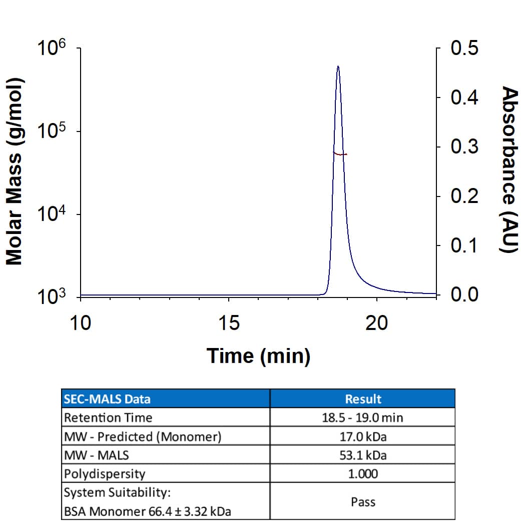


![<b>Genetic Strategies Validation. </b>Depletion of PNUTS and NPAT induces a global DDR.(A) Induction of telomere dysfunction–induced foci (TIF) upon candidate knockdown in HeLa cells from Fig 5. Depletion of TRF2 was used as a positive control. 53BP1 was detected by indirect IF and telomeres by FISH with a Cy3-[CCCTAA]3 probe. (B) Frequency distribution of the number of TIFs per cell from (A). n indicates the number of cells analyzed for each condition and red lines mark the mean.](http://images.novusbio.com/fullsize/nb100-304_rabbit-polyclonal-53bp1-antibody-271220231261934.jpg)


![Western Blot: 53BP1 Antibody - BSA Free [NB100-304] - Depletion of TPX2 causes defects in 53BP1 ionizing radiation-induced foci formation.(A) The protein level of 53BP1 is not affected by miRNA-mediated depletion of TPX2. Levels of actin used as loading controls. Image collected & cropped by CiteAb from the following publication (//dx.plos.org/10.1371/journal.pone.0110994), licensed under a CC-BY license. Not internally tested by Novus Biologicals.](http://images.novusbio.com/fullsize/nb100-304_rabbit-polyclonal-53bp1-antibody-310202415334914.jpg)
![Immunocytochemistry/ Immunofluorescence: 53BP1 Antibody - BSA Free [NB100-304] - Nuclear intensity of key BER proteins.A) Quantification of mean nuclear fluorescence intensity is shown for PARP1 (**** P < 0.0001, MDA-231 to MDA-157, HCC1806, & MDA-468), PAR (**** P < 0.0001, MDA-468 to MDA-157, MDA-231, & HCC1806), & p53 (**** P < 0.0001, MDA-157 to MDA-231, MDA-157 to MDA-468, MDA-157 to HCC1806, MDA-231 to MDA-468, MDA-231 to HCC1806, & MDA-468 to HCC1806). B) Representative fluorescent images used for quantification in A for PARP1, PAR, & p53 for all cell lines tested. Scale bar = 20 μm. Image collected & cropped by CiteAb from the following publication (//pubmed.ncbi.nlm.nih.gov/31596905), licensed under a CC-BY license. Not internally tested by Novus Biologicals.](http://images.novusbio.com/fullsize/nb100-304_rabbit-polyclonal-53bp1-antibody-31020241535658.jpg)
![Immunohistochemistry: 53BP1 Antibody - BSA Free [NB100-304] - Representative immunohistochemistry staining for 53BP1 expression in pancreatic adenocarcinoma. (A) is high intensity of 53BP1 expression & (B) is low intensity of 53BP1 expression. Image collected & cropped by CiteAb from the following publication (//pubmed.ncbi.nlm.nih.gov/23530749), licensed under a CC-BY license. Not internally tested by Novus Biologicals.](http://images.novusbio.com/fullsize/nb100-304_rabbit-polyclonal-53bp1-antibody-31020241535636.jpg)
![Immunocytochemistry/ Immunofluorescence: 53BP1 Antibody - BSA Free [NB100-304] - H2A.J accumulates in the aging human epidermis.(a) H2A.J & Ki-67 co-localization in sections of human skin of the indicated ages by immunofluorescent staining. H2A.J showed mutually exclusive staining with the Ki-67 proliferation marker. (b) H2A.J & 53BP1 foci increase in aging human epidermis. Arrows indicate positively-stained nuclei. A quantification of positive cells for three biological replicates is shown below the immunofluorescence images, & the one-sided Mann–Whitney U-test was used to determine the statistical significance (*P<0.05, **P<0.01). Error bars represent mean±s.e. Image collected & cropped by CiteAb from the following publication (//www.nature.com/articles/ncomms14995), licensed under a CC-BY license. Not internally tested by Novus Biologicals.](http://images.novusbio.com/fullsize/nb100-304_rabbit-polyclonal-53bp1-antibody-31020241539714.jpg)
![Western Blot: 53BP1 Antibody - BSA Free [NB100-304] - 53BP1 cofractionates with lamins A/C in HDF. (A) HDF were treated with IR (5 Gy, 1 h) & fractionated by sequential treatments of detergent (CSK/T) & DNase I. Extracts were then processed for immunoblotting using the indicated antibodies. WCE, whole cell extract. P, pellet; S, supernatant. (B) HDF were treated with IR (2 Gy; 1 h) & extracted in situ with CSK/T followed by DNase I digestion before being processed for immunofluorescence using lamin A/C & 53BP1 antibodies. Scale bar, 10 μm. Image collected & cropped by CiteAb from the following publication (//pubmed.ncbi.nlm.nih.gov/25645366), licensed under a CC-BY license. Not internally tested by Novus Biologicals.](http://images.novusbio.com/fullsize/nb100-304_rabbit-polyclonal-53bp1-antibody-31020241539728.jpg)
![Immunocytochemistry/ Immunofluorescence: 53BP1 Antibody - BSA Free [NB100-304] - Depletion of Ajuba results in activation of the DNA damage response. (A) Staining for p53BP1 foci on cells fixed 72 h after transfection with siRNA #3, & siGFP as a negative control. Quantification of the number of p53BP1 foci on three independent siRNA experiments shown in (B). (C, D) Western blots for induction of Chk1 phosphorylation & induction of p53 phosphorylation, showed for the three target sites, 72 h after siRNA transfection. Image collected & cropped by CiteAb from the following publication (//pubmed.ncbi.nlm.nih.gov/23755068), licensed under a CC-BY license. Not internally tested by Novus Biologicals.](http://images.novusbio.com/fullsize/nb100-304_rabbit-polyclonal-53bp1-antibody-310202415291948.jpg)
![Immunocytochemistry/ Immunofluorescence: 53BP1 Antibody - BSA Free [NB100-304] - Depletion of PNUTS & NPAT induces a global DDR.(A) Induction of telomere dysfunction–induced foci (TIF) upon candidate knockdown in HeLa cells from Fig 5. Depletion of TRF2 was used as a positive control. 53BP1 was detected by indirect IF & telomeres by FISH with a Cy3-[CCCTAA]3 probe. (B) Frequency distribution of the number of TIFs per cell from (A). n indicates the number of cells analyzed for each condition & red lines mark the mean. Image collected & cropped by CiteAb from the following publication (//pubmed.ncbi.nlm.nih.gov/30456372), licensed under a CC-BY license. Not internally tested by Novus Biologicals.](http://images.novusbio.com/fullsize/nb100-304_rabbit-polyclonal-53bp1-antibody-310202415371914.jpg)
![Western Blot: 53BP1 Antibody - BSA Free [NB100-304] - HPV+ HNSCCs have decreased expression of NHEJ & HR proteins including DNA-Pk & BRCA2Cells were treated with mock or 4 Gy radiation, harvested & lysed at 15 minutes post-treatment, & analyzed by western blot for relative expression of indicated proteins. beta -actin was used as a loading control. Shown is a representative blot from 2 independent experiments. Image collected & cropped by CiteAb from the following publication (//pubmed.ncbi.nlm.nih.gov/26336991), licensed under a CC-BY license. Not internally tested by Novus Biologicals.](http://images.novusbio.com/fullsize/nb100-304_rabbit-polyclonal-53bp1-antibody-310202415384176.jpg)
![Immunocytochemistry/ Immunofluorescence: 53BP1 Antibody - BSA Free [NB100-304] - Spontaneous DNA breakage in G1-phase cells: GamGFP shows large spontaneous G1 53BP1 foci to contain multi-break clusters.The large spontaneous 53BP1 foci in undamaged cells, which occur solely in G1 (Harrigan et al., 2011;Lukas et al., 2011a), contain multiple DSBs that are marked by GamGFP. The GamGFP-53BP1 co-localization is more apparent in the absence of Ku. Data are from three (Ku80-proficient) or four (Ku80-defective) independent experiments of >25 cells each with Z-stacked (optically sectioned) nuclei.DOI://dx.doi.org/10.7554/eLife.01222.017 Image collected & cropped by CiteAb from the following publication (//elifesciences.org/articles/01222), licensed under a CC-BY license. Not internally tested by Novus Biologicals.](http://images.novusbio.com/fullsize/nb100-304_rabbit-polyclonal-53bp1-antibody-310202415382416.jpg)
![Immunocytochemistry/ Immunofluorescence: 53BP1 Antibody - BSA Free [NB100-304] - 53BP1 cofractionates with lamins A/C in HDF. (A) HDF were treated with IR (5 Gy, 1 h) & fractionated by sequential treatments of detergent (CSK/T) & DNase I. Extracts were then processed for immunoblotting using the indicated antibodies. WCE, whole cell extract. P, pellet; S, supernatant. (B) HDF were treated with IR (2 Gy; 1 h) & extracted in situ with CSK/T followed by DNase I digestion before being processed for immunofluorescence using lamin A/C & 53BP1 antibodies. Scale bar, 10 μm. Image collected & cropped by CiteAb from the following publication (//pubmed.ncbi.nlm.nih.gov/25645366), licensed under a CC-BY license. Not internally tested by Novus Biologicals.](http://images.novusbio.com/fullsize/nb100-304_rabbit-polyclonal-53bp1-antibody-310202415384168.jpg)
![Western Blot: 53BP1 Antibody - BSA Free [NB100-304] - 53BP1 focus formation. (A) Bars indicate the mean ± standard error of 53BP1 focus-forming cells in three independent control & three independent Asciz−/− primary MEF cultures under basal conditions (open bars), in response to 24-h aphidicolin treatment (black bars) & following 30 min recovery after ionizing radiation (grey bars). More detailed quantifications of foci/cell are shown in Supp. Fig. S2. (B) Subcelllular distribution of 53BP1 in cell fractionation assays. M601 is used as a mitochondrial marker, & tubulin as a cytosolic marker. Image collected & cropped by CiteAb from the following publication (//linkinghub.elsevier.com/retrieve/pii/S1568786417301349), licensed under a CC-BY license. Not internally tested by Novus Biologicals.](http://images.novusbio.com/fullsize/nb100-304_rabbit-polyclonal-53bp1-antibody-310202415395939.jpg)
![Immunocytochemistry/ Immunofluorescence: 53BP1 Antibody - BSA Free [NB100-304] - hsRAD51-II3A expressing cells accumulate 53BP1 foci after treatment with HU. Representative images depicting 53BP1 foci (green) in PCNA (red) positive cells after the indicated treatments. DNA is stained in blue. Scale bar = 10 μm. Dot plot depicts quantitation of 53BP1 foci after indicated treatments. Data is combined data consisting of at least 100 nuclei from two independent experiments. Red line represents the mean. Values are presented as mean ± SD. Statistical significance was determined using the Mann–Whitney U test. Source data are provided as a source data file Image collected & cropped by CiteAb from the following publication (//pubmed.ncbi.nlm.nih.gov/31562309), licensed under a CC-BY license. Not internally tested by Novus Biologicals.](http://images.novusbio.com/fullsize/nb100-304_rabbit-polyclonal-53bp1-antibody-310202415382411.jpg)
![Immunocytochemistry/ Immunofluorescence: 53BP1 Antibody - BSA Free [NB100-304] - Persistent DNA damage exists in ALT cells. (A) IF-FISH showing DNA damage response (53BP1) in ALT (VA13, U2OS, SAOS2, SKLU-1), telomerase positive (MCF7, A549), & human normal fibroblast cells (MRC5, BJ). Scale bar: 10 μm. (B) Quantification of (A). Data represent the mean ± SEM, n = 3. More than 100 cells were counted for each experiment. (C) FACS analysis of apoptosis in cells listed in (A). (D) Quantification of (C). Data represent the mean ± SEM, n = 3. (E) Proliferation of p53-positive (VA13, U2OS) & p53-defictive (SAOS2, SKLU-1) ALT cells. (F) Western blot showing activation of JNK (phosphorylation) & accumulation of c-myc in ALT cells. Human normal BJ cells were used as a control Image collected & cropped by CiteAb from the following publication (//pubmed.ncbi.nlm.nih.gov/31115790), licensed under a CC-BY license. Not internally tested by Novus Biologicals.](http://images.novusbio.com/fullsize/nb100-304_rabbit-polyclonal-53bp1-antibody-31020241539594.jpg)
![Immunocytochemistry/ Immunofluorescence: 53BP1 Antibody - BSA Free [NB100-304] - LNCaP cells in culture were treated for 24 hrs with DMSO or ENZ, then irradiated with 0 or 1 Gy.Cell were fixed & immunostained for 53BP1 0.5, 1.5 & 24 hrs after irradiation. Immunofluorescent images were obtained by confocal microscopy. White bar = 20 microns. A) DMSO alone, B) ENZ alone, C) 1 Gy, D) ENZ + 1 Gy. White bar = 20 microns. Image collected & cropped by CiteAb from the following publication (//pubmed.ncbi.nlm.nih.gov/30933998), licensed under a CC-BY license. Not internally tested by Novus Biologicals.](http://images.novusbio.com/fullsize/nb100-304_rabbit-polyclonal-53bp1-antibody-310202415293335.jpg)
![Immunocytochemistry/ Immunofluorescence: 53BP1 Antibody - BSA Free [NB100-304] - IMR90 cells depleted for Ajuba undergo a DNA damage response. (A,C,D) Western blots for Ajuba, Cyclin A, Rb, Chk1 phosphorylation, & PARP cleavage on lysates prepared from IMR90 cells 72 h after transfection with siRNA #3. The loading controls with GAPDH for each blot are shown. (B) Staining for p53BP1 in Ajuba-depleted cells (siRNA#3), with siGFP as a control. Image collected & cropped by CiteAb from the following publication (//pubmed.ncbi.nlm.nih.gov/23755068), licensed under a CC-BY license. Not internally tested by Novus Biologicals.](http://images.novusbio.com/fullsize/nb100-304_rabbit-polyclonal-53bp1-antibody-310202415371983.jpg)
![Western Blot: 53BP1 Antibody - BSA Free [NB100-304] - DYNLL1 is required for 53BP1-dependent p53 responses to Nutlin-3. a Immunoblot analysis of the MCF-7 cell lines used in (a) with anti-53BP1 or anti-RPA32 antibodies prior to N3 treatment. b Quantification of n = 3 independent experiments represented in (a), each performed in triplicate. Mean ± SD. c Indicated parental or stably complemented 53BP1-/- MCF-7 cell lines were incubated in the presence (11 days) or absence (7 days) of Nutlin-3 (4 µM) Image collected & cropped by CiteAb from the following publication (//pubmed.ncbi.nlm.nih.gov/30559443), licensed under a CC-BY license. Not internally tested by Novus Biologicals.](http://images.novusbio.com/fullsize/nb100-304_rabbit-polyclonal-53bp1-antibody-310202415394519.jpg)
![Immunocytochemistry/ Immunofluorescence: 53BP1 Antibody - BSA Free [NB100-304] - RNA:DNA hybrids promote 53BP1 foci formation.(a) Immunofluorescence using the anti-flag & anti-53BP1 antibody in cells expressing Flag-tagged hybrid-binding (HB) domain or flag-tagged active RNaseH1. 53BP1 foci were quantified in cells expressing HB-domain or RNaseH1. Column bars represent the mean of n number of cells (described on each column), from three independent experiments. Error bars represent SEM. *P ≤ 0.05; **P < 0.01, ***P < 0.001 using the Kruskal-Wallis test followed by Dunn’s post hoc test. (b) Higher zoom representative images show HB-domain & 53BP1 co-localization, specifically in the euchromatin area. Image collected & cropped by CiteAb from the following publication (//pubmed.ncbi.nlm.nih.gov/30345028), licensed under a CC-BY license. Not internally tested by Novus Biologicals.](http://images.novusbio.com/fullsize/nb100-304_rabbit-polyclonal-53bp1-antibody-31020241535612.jpg)
![Immunocytochemistry/ Immunofluorescence: 53BP1 Antibody - BSA Free [NB100-304] - RNA:DNA hybrids promote 53BP1 foci formation.(a) Immunofluorescence using the anti-flag & anti-53BP1 antibody in cells expressing Flag-tagged hybrid-binding (HB) domain or flag-tagged active RNaseH1. 53BP1 foci were quantified in cells expressing HB-domain or RNaseH1. Column bars represent the mean of n number of cells (described on each column), from three independent experiments. Error bars represent SEM. *P ≤ 0.05; **P < 0.01, ***P < 0.001 using the Kruskal-Wallis test followed by Dunn’s post hoc test. (b) Higher zoom representative images show HB-domain & 53BP1 co-localization, specifically in the euchromatin area. Image collected & cropped by CiteAb from the following publication (//pubmed.ncbi.nlm.nih.gov/30345028), licensed under a CC-BY license. Not internally tested by Novus Biologicals.](http://images.novusbio.com/fullsize/nb100-304_rabbit-polyclonal-53bp1-antibody-310202415284547.jpg)
![Immunocytochemistry/ Immunofluorescence: 53BP1 Antibody - BSA Free [NB100-304] - General chromatin effects of A∆rod & A∆rod∆hbs. (a) DNA staining (DAPI) in U2OS cells expressing A∆rod. Chromatin staining tended to be stronger on or around lamin A∆rod structures. (b) pRb is a known binding partner of lamin A, & so, it was co-stained as a positive control, revealing strong co-localization with the A∆ structures. (c) No co-localization between A∆rod structures & 53BP1 in HeLa cells indicates that chromatin is not damaged by the structures. (d) Similarly, gamma H2AX did not accumulate on A∆ structures. Image collected & cropped by CiteAb from the following publication (//www.mdpi.com/2073-4409/6/2/9), licensed under a CC-BY license. Not internally tested by Novus Biologicals.](http://images.novusbio.com/fullsize/nb100-304_rabbit-polyclonal-53bp1-antibody-310202415384125.jpg)
![Immunocytochemistry/ Immunofluorescence: 53BP1 Antibody - BSA Free [NB100-304] - UFL1 regulates the ATM signaling. a U2OS cells expressing UFL1 tet-on shRNA1 were irradiated with 2 Gy IR. Thirty minutes later, cells were lysed & blotted with indicated antibodies. b Representative picture of gamma H2AX, 53BP1, & BRCA1 foci in control (Dox-) & UFL1 knockdown (Dox+) U2OS cells 1 h after 0.5 Gy treatment. Scale bars, 10 µm. c–e Quantification of intensities of gamma H2AX, 53BP1, & BRCA1 foci in control (Dox-) & UFL1 knockdown (Dox+) U2OS cells. Data presented as mean ± SD of n = 50 cells. **p < 0.01 by Student’s t-test. Source data are provided as a Source Data file Image collected & cropped by CiteAb from the following publication (//pubmed.ncbi.nlm.nih.gov/30886146), licensed under a CC-BY license. Not internally tested by Novus Biologicals.](http://images.novusbio.com/fullsize/nb100-304_rabbit-polyclonal-53bp1-antibody-310202415394518.jpg)
![Immunocytochemistry/ Immunofluorescence: 53BP1 Antibody - BSA Free [NB100-304] - The DNA damage response at telomeres uncapped through TRF2 depletion does not require BRCA1 or CtIPImmortalized Brca1F/− MEFs were infected with retroviruses expressing the indicated shRNAs and/or Cre recombinase, followed by selection with puromycin for 72 h. Cell extracts were prepared 48 h later & analysed by Western blotting as indicated. SMC1 & tubulin were used as loading controls. *non-specific band.Cells treated as in (A) were fixed 48 h after selection & stained with an anti-53BP1 antibody (green). Telomeres were visualized with a Cy3-conjugated (CCCTAA)3-PNA probe (red). Yellow arrowheads point to 53BP1 foci that co-localize with telomeres.Quantification of TIFs in cells treated as in (B). A minimum of 200 nuclei were scored for each sample. Error bars represent SD of two independent experiments. P-values were calculated using an unpaired two-tailed t-test. *P ≤ 0.05; NS, P > 0.05. Image collected & cropped by CiteAb from the following publication (//pubmed.ncbi.nlm.nih.gov/25582120), licensed under a CC-BY license. Not internally tested by Novus Biologicals.](http://images.novusbio.com/fullsize/nb100-304_rabbit-polyclonal-53bp1-antibody-310202415525242.jpg)
![Western Blot: 53BP1 Antibody - BSA Free [NB100-304] - Hypoxia causes DNA damage in patient-derived colonosphere cultures in vitro(a) Two independent patient-derived colonospheres were cultured in normoxia (21%) or hypoxia (0.1%) for 24 hours. Immunofluorescence was then used to asses gamma H2AX (green) in the nuclei DAPI (blue). (b) Bar graphs showing the quantification of gamma H2AX observed in (a) (n=3), shown as percentage of the mean gamma H2AX intensity at normoxia. (c) Representative confocal pictures of the comet assay of colonospheres cultured as in (a). (d) Bar graphs showing the quantification of DNA damage observed with the comet assay (Extend tail moment) (n=73). (e) Colonospheres were cultured as in (a) & gamma H2AX was assessed by FACS. The plots show normoxic gamma H2AX levels in red & hypoxic gamma H2AX levels in blue in two independent cell lines. (f) Human colonospheres were exposed to hypoxia for the indicated periods of time. Cell lysates were then analyzed by Western blotting for the indicated markers (cropped). Image collected & cropped by CiteAb from the following publication (//www.oncotarget.com/lookup/doi/10.18632/oncotarget.21145), licensed under a CC-BY license. Not internally tested by Novus Biologicals.](http://images.novusbio.com/fullsize/nb100-304_rabbit-polyclonal-53bp1-antibody-31020241683928.jpg)
![Immunocytochemistry/ Immunofluorescence: 53BP1 Antibody - BSA Free [NB100-304] - TRF1HP1 alpha results in reduced TIFs induced by mutant hTR expression. Cells stably expressing TRF1HP1 alpha (WT or mutant variants of HP1 alpha ) were infected with lentivirus containing WT hTR, or mutant hTR (47A or TSQ1) on day 0, selected for stable expression after 48 h, & analyzed on day 5. a Fluorescence microscopy images of representative cells expressing mutant hTR 47A stained with telomeric (Tel-Cy3) peptide nucleic acid (PNA) probes (magenta in merged image) via fluorescent in situ hybridization (FISH), antibody against DNA-damage repair protein marker 53BP1 (green in merged image), & counterstained with DAPI. Zoom-in images (the last row) correspond to yellow-squared regions of the row above. b % TIF per telomere of each nucleus is quantified; n = 94–159 nuclei combining data of three independent experiments. ****p < 0.0001; n.s. (no significance). a, b Scale bar: 10 µm. c Upon TSQ1 expression, TRF1HP1 alpha results in fewer TIFs compared to Vonly, TRF1 or HP1 alpha controls. TRFHP1 alpha ~11.4% shows decreased TIFs compared to Vonly: ~19.7% ***p = 0.0008; TRF1: ~20.4% ***p = 0.0005; HP1 alpha : ~17.9% *p = 0.0137 (n = 30–38 nuclei). b, c Significance is assessed by one-way ANOVA & Dunnett’s multiple comparison test with 95% confidence level. d Fluorescence images of control cells overexpressing WT hTR (n = 27–36 nuclei). Same color scheme as a. TIFs quantification in the presence of e WT hTR or f Vonly (n = 28–36 nuclei) show minimal baseline DNA damage at telomeres. b, c; e, f Error bars represent s.e.m. Image collected & cropped by CiteAb from the following publication (//pubmed.ncbi.nlm.nih.gov/30181605), licensed under a CC-BY license. Not internally tested by Novus Biologicals.](http://images.novusbio.com/fullsize/nb100-304_rabbit-polyclonal-53bp1-antibody-31020241612690.jpg)
![Immunocytochemistry/ Immunofluorescence: 53BP1 Antibody - BSA Free [NB100-304] - Absence of Roma Leads to Activation of the DNA Damage Response & Genomic Instability(A) Representative immunofluorescence analysis of RPA2-pT21, gamma H2AX, & 53BP1 foci in tissue sections from WT & Roma−/− glands at 10dL. The white arrow indicates nucleolar gamma H2AX. Scale bars, 10 μm. The higher magnification panels on the right have been chosen to highlight the foci & are not zoomed images of the left panels.(B) Immunoblot analysis of cell-cycle checkpoint & DNA damage response in extracts from WT & Roma−/− glands at 10dL. Extracts from U2OS cells treated with 5mM HU were used as a damage control.(C) PCR analysis of p21Cip1 & MDM2 & quantitative real-time PCR analysis of p21Cip1 in extracts from 10dL WT & Roma−/− glands (p = 0.0007, Student’s t test).(D) Representative spectral karyotypes (top) & metaphase spreads (bottom panel) prepared from MECs isolated from WT & Roma−/− glands after a full natural wean.See also Figure S2. Image collected & cropped by CiteAb from the following publication (//pubmed.ncbi.nlm.nih.gov/27149840), licensed under a CC-BY license. Not internally tested by Novus Biologicals.](http://images.novusbio.com/fullsize/nb100-304_rabbit-polyclonal-53bp1-antibody-31020241553711.jpg)
![Immunocytochemistry/ Immunofluorescence: 53BP1 Antibody - BSA Free [NB100-304] - A-T post-mitotic neurons are defective in DDR. In a, D30 post-mitotic neurons (D30) were tested by western blot with antibodies specific for the various ATM phosphosubstrates & cleaved PARP at various times after treatment with 5 Gy IR. beta -Actin was used as a loading control. DNA SSBs (b) & DSBs (c) were analyzed in neurons (D30) by alkaline & neutral comet assay, respectively, at two different times after treatment. 20 min after H2O2 or IR treatment was considered the time point with the maximum DNA damage. In b & c, representative photos of comets from untreated cells & at different time points following treatment. The ratio between treated & untreated tail moments of at least 50–70 cells per experimental point is shown in the graphs (values in %). One representative experiment out of three is shown. In d & e, formation & resolution of IR-induced nuclear foci. Neurons (D30) were irradiated with 0.5 Gy, collected at the indicated times & labeled for gamma -H2AX (d) & 53BP1 (e). For each treatment, the number of foci was scored from 100 cell nuclei per duplicate preparations & from three independent experiments (mean±S.D.) (d & e, bottom) For each time point, the difference between Ctrl & A-T was statistically significant (#P<0.01) (analysis performed by the Student's t-test) Image collected & cropped by CiteAb from the following publication (//pubmed.ncbi.nlm.nih.gov/25032865), licensed under a CC-BY license. Not internally tested by Novus Biologicals.](http://images.novusbio.com/fullsize/nb100-304_rabbit-polyclonal-53bp1-antibody-31020241682117.jpg)
![Immunocytochemistry/ Immunofluorescence: 53BP1 Antibody - BSA Free [NB100-304] - Exposure to radiation induces persistent genomic instability.(a) Time course of micronucleus frequency in binucleated cells exposed to 1Gy X-rays or Fe ions (average of two independent irradiations, error bars represent SEM). Paired Student's t-test, *p<0.05, **p<0.01. (b) Time course of gamma H2AX- 53BP1 positive foci per cell distribution in a population of 50 cells per condition following exposure to 1Gy of the indicated radiation. One of two experiments is shown. Chi-Square test NI vs. X-rays or Fe ions. day 7 & day 14 p<0.001. Day 21 p>0.05. (c) Immunofluorescence staining of gamma H2AX-53BP1 positive foci 7 days following exposure to 1Gy of the indicated radiation type. Scale bar is 10 µm. (d) Immunofluorescence staining for BrdU & 53BP1 7 days following exposure to 1Gy Fe ions. Scale bar is 10 µm. The graph represents the average of scoring in 5 different fields a total of 70 cells. Image collected & cropped by CiteAb from the following publication (//dx.plos.org/10.1371/journal.pone.0108234), licensed under a CC-BY license. Not internally tested by Novus Biologicals.](http://images.novusbio.com/fullsize/nb100-304_rabbit-polyclonal-53bp1-antibody-310202415532010.jpg)
![Immunocytochemistry/ Immunofluorescence: 53BP1 Antibody - BSA Free [NB100-304] - SPOC1 is recruited to I-SceI-induced DSBs where it colocalizes with 53BP1. (A) In situ immunofluorescence studies with U2OS19 ptight13 GFP-LacR cells with a stably integrated I-SceI cleavage site flanked by lac operator repeats reveals localization of the GFP-lac repressor protein (GFP-LacR) at the lac-operator DNA sequences in the nucleus before (−I-SceI) & 16 h after I-SceI-induced (+ I-SceI) DSB. In contrast, SPOC1 (white) & 53BP1 (red) are distributed throughout the nucleus in the absence of I-Scel, & partially colocalized in naturally occurring repair foci. After I-SceI cleavage to generate DSBs, SPOC1 & 53BP1 colocalize at distinct foci, including the cleaved DNA adjacent to DNA-bound GFP-LacR. Proteins were visualized by immunostaining & confocal microscopy. Scale bars = 10 μm. (B) Quantitative analysis of SPOC1 recruitment to the 53BP1 & GFP-LacR positive lacO array before & 16 h after I-SceI induction. (C) Monitoring of the kinetics of SPOC1 & 53BP1 recruitment to DSBs between 0 & 24 h post-I-SceI induction. (D) Quantifying ATM kinase inhibitor-mediated effects on recruitment of SPOC1 & 53BP1 to DSBs as evident 16 h after I-SceI-induced cleavage. (E) SPOC1 & 53BP1 colocalize at a large discrete endogenous repair focus as observed in some non-irradiated U2OS cells by immunostaining. Scale bar = 10µm. Image collected & cropped by CiteAb from the following publication (//academic.oup.com/nar/article-lookup/doi/10.1093/nar/gks868), licensed under a CC-BY license. Not internally tested by Novus Biologicals.](http://images.novusbio.com/fullsize/nb100-304_rabbit-polyclonal-53bp1-antibody-310202415525292.jpg)
![Western Blot: 53BP1 Antibody - BSA Free [NB100-304] - A-type lamin depletion inhibits DNA repair & reduces cellular fitness to DNA damage. (A) Wild-type (WT) or Y259X HDF treated w/ proteasome inhibitor MG-132 (30 μm). Cell extracts prepared & levels of 53BP1 assessed by immunoblotting. beta -tubulin a loading control. WCE, whole cell extract. The relative levels of 53BP1 expressed relative to levels of untreated WT HDF (1.00). In untreated Y259X cells, 53BP1 levels fell to 15% & restored to ∽50% following treatment w/ MG-132. (B) Cell extracts prepared from WT & Y259X HDF & assessed by immunoblotting w/ indicated antibodies. Shown is a representative set of blots from two independent experiments. (C) WT & Y259X HDF biochemically extracted into cytoplasmic & nuclear fractions before immunoblotting for levels of 53BP1. (D) HDF & Y259X cells treated w/ IR (3 Gy), fixed at the times indicated & stained w/ gamma -H2AX antibody. The number of gamma -H2AX foci at each time point quantified & data represents the mean ± SD from 3 independent experiments. (E) U2OS cells transfected w/ siCTRL or siLMNA siRNA & plated onto 60-mm dishes. The cells then exposed to varying doses of IR & fixed 12 days later in a solution containing crystal violet. The surviving fraction at each IR dose calculated based on the plating efficiency of untreated plates. Data represent the mean ± SD from 3 independent experiments. (F) HDF or Y259X cells treated w/ DMSO or KU55933 ATM inhibitor (10 μm) before exposure to indicated doses of IR then fixed, processed & stained w/ gamma -H2AX antibody. Residual gamma -H2AX foci enumerated & the data represent the mean ± SD from 3 independent experiments. (G) HDF or Y259X cells exposed to IR, briefly extracted w/ CSK buffer & processed for IF w/ gamma -H2AX & pKAP-1 antibodies. Scale bar, 10 μm. Image collected & cropped by CiteAb from the following publication (//pubmed.ncbi.nlm.nih.gov/25645366), licensed under a CC-BY license. Not internally tested by Novus Biologicals.](http://images.novusbio.com/fullsize/nb100-304_rabbit-polyclonal-53bp1-antibody-31020241612631.jpg)
![Immunocytochemistry/ Immunofluorescence: 53BP1 Antibody - BSA Free [NB100-304] - TRF2/Terc double null mutation induces DNA damage response resulting in terminal differentiation of metastatic tumorsHistopathology of primary SCC from K14Cre;TRF2+/+;Terc+/+ A. & K14Cre;TRF2f/f;Terc-/- C. mice is shown by H&E staining. Scale bar = 10 μm. Histopathology of metastatic SCC from K14Cre;TRF2+/+;Terc+/+ B. & K14Cre;TRF2f/f;Terc-/- D., E. mice is shown by H&E staining. Scale bar in (D) = 50 μm. Apoptotic cells in primary SCC from K14Cre;TRF2+/+;Terc+/+ F. & K14Cre;TRF2f/f;Terc-/- G. mice are shown by TUNEL assay. Scale bar = 10 μm. Proliferating cells were detected in K14Cre;TRF2+/+;Terc+/+ H. & K14Cre;TRF2f/f;Terc-/- I. primary SCC by PCNA immunohistochemistry & quantitated in J.. Error bars indicate SEM. Asterisk indicates statistically significant difference. Co-localization of 53BP1 (shown by immunofluorescence, AlexaFluor 488) at telomeres (shown by fluorescence in situ hybridization, Cy3) in histopathologic sections from K14Cre;TRF2+/+;Terc+/+ K. & K14Cre;TRF2f/f;Terc-/- L. primary SCC & quantitated in M. Error bars indicate SEM. Nuclei were counterstained with DAPI. Scale bar = 5 μm. N. Average telomere length ratios in CD34+ stem, Lgr6+ stem, & basal cells from K14Cre;TRF2+/+;Terc+/+ & K14Cre;TRF2f/f;Terc-/- primary SCC were determined by qPCR. Error bars represent SEM. O. Phospho-ATM, phospho-Chk2, & p53 expression in K14Cre;TRF2+/+;Terc+/+ & K14Cre;TRF2f/f;Terc-/- primary SCC is shown by western blot. beta -actin expression was used to control for equal loading of each lane. Representative blots are shown. Image collected & cropped by CiteAb from the following publication (//pubmed.ncbi.nlm.nih.gov/29113290), licensed under a CC-BY license. Not internally tested by Novus Biologicals.](http://images.novusbio.com/fullsize/nb100-304_rabbit-polyclonal-53bp1-antibody-310202415532050.jpg)
![Immunocytochemistry/ Immunofluorescence: 53BP1 Antibody - BSA Free [NB100-304] - Increased Neurogenesis in the Absence of INK4a/ARF Expression Is Independent of Apoptosis & Microglial Activation(A) Quantification of the number of apoptotic cells, as detected by TUNEL, in the DG 6 hr following exposure of WT, ink4a/arf-deficient (INK4a/ARF KO), & p53-deficient (p53 KO) mice to 6 Gy cranial IR. Indicated is the average number of apoptotic cells per DG section (n = 4 mice per group).(B) Quantification of CD68 expression in the DG 24 hr following exposure of mice to IR.(C) Representative confocal images showing CD68+ cells (in red) next to apoptotic TUNEL+ cells (in green) following exposure of WT mice to IR. Note the increase in CD68 signal & side by side localization of CD68+ & TUNEL+ cells following IR.(D–F) Proportion of neurons (NeuN, in green), young neurons (DCX, in green), & progenitor cells (Sox2, in green) showing persistent DNA damage foci (53BP1, in red) in the hippocampus of mice sacrificed 8 weeks post exposure to IR. Nuclei were stained with DAPI.(G) Representative confocal images of brain sections from irradiated WT mice displaying 53BP1 DNA damage foci with the indicated cell markers are shown. Images were acquired by a Leica microsystems TSC SP8 HyVolution confocal microscope & an HC PL APO CS2 40×/1.3 oil objective. The p values (∗p < 0.05; ∗∗∗p < 0.001) were obtained by performing a Student's t test. ND (no cells were detected). Image collected & cropped by CiteAb from the following publication (//linkinghub.elsevier.com/retrieve/pii/S2213671118301528), licensed under a CC-BY license. Not internally tested by Novus Biologicals.](http://images.novusbio.com/fullsize/nb100-304_rabbit-polyclonal-53bp1-antibody-31020241554539.jpg)
![Immunocytochemistry/ Immunofluorescence: 53BP1 Antibody - BSA Free [NB100-304] - GamGFP marks DSBs in mammalian cells & is inhibited by Ku.(A) GamGFP co-localizes with 53BP1 on laser-induced DNA breaks. (B) Ku inhibits recruitment of GamGFP to laser-induced damage, live cells. (C & D) Ku inhibits recruitment of GamGFP, fixed cells. Mean ± SEM of three experiments, n >25 cells each. (E) GamGFP forms IR-induced foci in Ku80-defective MEFs. (F) Zoomed image from E. (G) IR-induced foci containing Gam only, 53BP1 only or both Gam & 53BP1 (>2600 total foci counted in three independent experiments). Error bars, SEM. Scale bars = 5 μm.DOI://dx.doi.org/10.7554/eLife.01222.012GamGFP marks DSBs in mammalian cells.(A) Live analysis of GamGFP localization to laser-induced DNA damage. Hela cells producing GamGFP were laser damaged along the cell track indicated by the red line at 0 min (m) & images were taken at the indicated times as shown. (B) GamGFP co-localizes with gamma H2AX in fixed Hela cells.DOI://dx.doi.org/10.7554/eLife.01222.013Ku inhibits GamGFP recruitment at DSBs independently of non-homologous end joining.Cells lacking either Ku80 or LigIV are defective in non-homologous end joining (NHEJ), yet the presence of Ku still inhibits recruitment of GamGFP to laser-induced DSBs even in NHEJ-defective cells, & thus independently of the cell's ability to complete NHEJ. Whereas it could have been possible that reduced GamGFP recruitment in the presence of Ku was caused by reduced persistence of DSBs due to their repair by NHEJ, our data show instead that Ku inhibits recruitment independently of successful NHEJ & imply that Ku binding to DSEs itself is inhibitory.DOI://dx.doi.org/10.7554/eLife.01222.014 Image collected & cropped by CiteAb from the following publication (//elifesciences.org/articles/01222), licensed under a CC-BY license. Not internally tested by Novus Biologicals.](http://images.novusbio.com/fullsize/nb100-304_rabbit-polyclonal-53bp1-antibody-310202415525217.jpg)
![Immunocytochemistry/ Immunofluorescence: 53BP1 Antibody - BSA Free [NB100-304] - Loss of functional PRPF31 induce RNA:DNA hybrid dependent genomic instability but not in mice retinal neurons.(a andb) gamma H2AX & 53BP1 foci analysis in PRPF31 siRNA-transfected RPE-1 cells. (c & d) gamma H2AX & 53BP1 foci analysis in vasculo-stromal fraction derived primary cells fromPrpf31+/A216P mice (Prpf31-ki). (e) gamma H2AX & 53BP1 foci analysis in retina fromPrpf31+/A216P mice on postnatal day 20. All column bars represent the mean. For (a-d) “n”, mentioned on respective column, signify number of cells analyzed from two independent experiments. For (e) n=16 for each column & signify number of retinal sections analyzed; acquired from n=4 eyes. Error bars represent Standard error of Mean (SEM). *P≤0.05; **P<0.01, ***P<0.001 using Mann-Whitney test (a,b), Kruskal-Wallis test followed by Dunn’s post hoc test (c,d); & two tailed unpaired Student’s t-test (e). Image collected & cropped by CiteAb from the following publication (//pubmed.ncbi.nlm.nih.gov/30345028), licensed under a CC-BY license. Not internally tested by Novus Biologicals.](http://images.novusbio.com/fullsize/nb100-304_rabbit-polyclonal-53bp1-antibody-31020241553741.jpg)
![Western Blot: 53BP1 Antibody - BSA Free [NB100-304] - Lamin A/C-53BP1 interaction is regulated in a DNA damage-dependent manner. (A) Left; HDF were subjected to IR (10 Gy) & allowed to recover for 1 h. Association between A-type lamins & 53BP1 was assessed by immunoprecipitation of endogenous lamin A/C followed by immunoblotting with 53BP1 & lamin A/C antibodies. WCE, whole cell extract, IP: immunoprecipitates. WCE represents 5% input. Right; endogenous lamin A/C was immunoprecipitated & supernatants or pellets were analysed by immunoblotting for interaction with JMJD2A. (B) U2OS/GFP-lamin A cells were pretreated with caffeine (20 mm) for 1 h before exposure to IR (10 Gy, 1 h recovery). Cell extracts were then subjected to immunoprecipitation using GFP-Trap beads, & bound complexes were then analysed by immunoblotting using 53BP1 & GFP antibodies. WCE represents 1% input. (C) As in (C) except cells were pretreated with 10 μm ATMi for 1 h before IR. WCE represents 1% input. (D) U2OS/GFP-lamin A cells were subjected to laser micro-irradiation, fixed 1 h later & immunostained with gamma -H2AX antibody. Scale bar, 10 μm. (E) U2OS cells were transfected with siCTRL or siLMNA & subjected to laser micro-irradiation, fixed 15 min later & then processed for immunofluorescence with gamma -H2AX & 53BP1 antibodies. Scale bar, 10 μm. Image collected & cropped by CiteAb from the following publication (//pubmed.ncbi.nlm.nih.gov/25645366), licensed under a CC-BY license. Not internally tested by Novus Biologicals.](http://images.novusbio.com/fullsize/nb100-304_rabbit-polyclonal-53bp1-antibody-31020241553729.jpg)
![Immunocytochemistry/ Immunofluorescence: 53BP1 Antibody - BSA Free [NB100-304] - SPOC1 is recruited to I-SceI-induced DSBs where it colocalizes with 53BP1. (A) In situ immunofluorescence studies with U2OS19 ptight13 GFP-LacR cells with a stably integrated I-SceI cleavage site flanked by lac operator repeats reveals localization of the GFP-lac repressor protein (GFP-LacR) at the lac-operator DNA sequences in the nucleus before (−I-SceI) & 16 h after I-SceI-induced (+ I-SceI) DSB. In contrast, SPOC1 (white) & 53BP1 (red) are distributed throughout the nucleus in the absence of I-Scel, & partially colocalized in naturally occurring repair foci. After I-SceI cleavage to generate DSBs, SPOC1 & 53BP1 colocalize at distinct foci, including the cleaved DNA adjacent to DNA-bound GFP-LacR. Proteins were visualized by immunostaining & confocal microscopy. Scale bars = 10 μm. (B) Quantitative analysis of SPOC1 recruitment to the 53BP1 & GFP-LacR positive lacO array before & 16 h after I-SceI induction. (C) Monitoring of the kinetics of SPOC1 & 53BP1 recruitment to DSBs between 0 & 24 h post-I-SceI induction. (D) Quantifying ATM kinase inhibitor-mediated effects on recruitment of SPOC1 & 53BP1 to DSBs as evident 16 h after I-SceI-induced cleavage. (E) SPOC1 & 53BP1 colocalize at a large discrete endogenous repair focus as observed in some non-irradiated U2OS cells by immunostaining. Scale bar = 10µm. Image collected & cropped by CiteAb from the following publication (//academic.oup.com/nar/article-lookup/doi/10.1093/nar/gks868), licensed under a CC-BY license. Not internally tested by Novus Biologicals.](http://images.novusbio.com/fullsize/nb100-304_rabbit-polyclonal-53bp1-antibody-31020241612674.jpg)
![Immunocytochemistry/ Immunofluorescence: 53BP1 Antibody - BSA Free [NB100-304] - Comparative kinetics of 53BP1 & SPOC1 recruitment to radiomimetic drug NCS-induced DSBs. U2OS cells fixed before (mock) or at the indicated times post-treatment (+NCS), immunostained for SPOC1 (green) & 53BP1 (red) & analyzed by confocal microcopy. At the earliest observation period after NCS treatment (15 min) a large number of small 53BP1 stained DSB repair foci emerged, but were not detectably stained by SPOC1. Between 2 to 24 h post-NCS treatment the number of small 53BP1 stained foci gradually, then almost completely disappeared, concomitant with emerging larger repair foci. At around 8 h post-NCS treatment most of the large 53BP1-stained repair foci were also strongly stained by SPOC1, suggesting SPOC1 is recruited to & accumulates at DSBs in heterochromatin known for their slow repair kinetics. Scale bars = 10µm. Image collected & cropped by CiteAb from the following publication (//academic.oup.com/nar/article-lookup/doi/10.1093/nar/gks868), licensed under a CC-BY license. Not internally tested by Novus Biologicals.](http://images.novusbio.com/fullsize/nb100-304_rabbit-polyclonal-53bp1-antibody-310202415525239.jpg)
![Immunocytochemistry/ Immunofluorescence: 53BP1 Antibody - BSA Free [NB100-304] - RNA:DNA hybrid formation is central to ATM-53BP1 repair pathway.(a) Immunofluorescence using the anti-flag to quantify nuclear hybrid-binding (HB) domain foci signal in pre-permeabilized RPE cells & quantification of HB domain signal in cells treated with siATM, (b) Western blot showing ATM depletion. (c) Immunofluorescence & HB-domain foci quantification in cells treated with ATM inhibitor. (d) Quantitation of gamma H2AX foci in cells expressing HB domain or active RNAseH1, in presence or absence of H2O2 dependent oxidative stress. siC, non-targeted control siLuciferease RNA. Column bars represent the mean of n number of cells (described on each column) from three independent experiments. Error bars represent SEM. *P ≤ 0.05; **P < 0.01, ***P < 0.001 using Mann-Whitney test (a,c) & Kruskal-Wallis test followed by Dunn’s post hoc test (d). Image collected & cropped by CiteAb from the following publication (//pubmed.ncbi.nlm.nih.gov/30345028), licensed under a CC-BY license. Not internally tested by Novus Biologicals.](http://images.novusbio.com/fullsize/nb100-304_rabbit-polyclonal-53bp1-antibody-31020241683919.jpg)
![Immunocytochemistry/ Immunofluorescence: 53BP1 Antibody - BSA Free [NB100-304] - UMI acidic residues are required for RNF168-mediated site-specific ubiquitination & the DDR.a Sequence alignment analysis of RNF168 UMI domains across species. The evolutionary conserved E143/E144 are highlighted in red. b Deletion of residues 143-144 attenuates 53BP1 IRIF formation. U2OS RNF168 KO cells were transiently transfected with GFP-RNF168 or RNF168 delta 143-144. Cells were irradiated with 10 Gy, allowed to recover for 1 h, followed by immunofluorescence analysis with indicated antibodies. Repeated three times with similar results. c Deletion of 143-144 abolishes RNF168-mediated ubiquitination. Co-transfection of GFP-RNF168 or mutant with Flag-H2AX K13/K15 only vector in HEK293T cells followed by western blot analysis. Repeated three times with similar results. d RNF168 delta 143-144 is recruited to laser damage. U2OS RNF168 KO cells expressing GFP-RNF168 & RNF168 delta 143-144 were damaged using a 405 nm laser source & analyzed 15 min later by confocal microscopy. Dotted line indicates the laser path. Repeated three times independently with similar results. e–f U2OS cells were transfected with GFP-RNF168 wildtype & mutants. Proximity ligation assay (PLA) was performed using specific antibodies against GFP & gamma H2AX. Mean intensity of PLA signals in GFP-positive cells were measured & analyzed using ImageJ software. Data presented as mean ± SD. One-way ANOVA, Dunn’s multiple comparison test was used in statistical analysis, wildtype vs. R57D P = 0.1455, wildtype vs. delta 143-144 P = 0.1589, R57D vs. delta 143-144, P > 0.9999, ****P < 0.0001 with 95% confidence interval. n = 75 (wildtype), 48 (R57D), 74 ( delta 143-144), 53 (R57D/ delta 143-144) over 3 independent experiments. Source data are provided as Source Data file. Image collected & cropped by CiteAb from the following publication (//pubmed.ncbi.nlm.nih.gov/32424115), licensed under a CC-BY license. Not internally tested by Novus Biologicals.](http://images.novusbio.com/fullsize/nb100-304_rabbit-polyclonal-53bp1-antibody-3102024168215.jpg)
![Immunocytochemistry/ Immunofluorescence: 53BP1 Antibody - BSA Free [NB100-304] - TRF2/Terc double null mutant mice exhibit DNA damage response at short telomeres in epidermisA. Average telomere length ratios in CD34+ stem, Lgr6+ stem, & basal cells from K14Cre;TRF2+/+;Terc+/+, K14Cre;TRF2f/f;Terc+/+, K14Cre;TRF2+/+;Terc-/-, & K14Cre;TRF2f/f;Terc-/- epidermis were determined by qPCR. Error bars represent SEM. Co-localization of 53BP1 (shown by immunofluorescence, AlexaFluor 488) at telomeres (shown by fluorescence in situ hybridization, Cy3) in histopathologic sections from K14Cre;TRF2+/+;Terc+/+ B., K14Cre;TRF2f/f;Terc-/- C., K14Cre;TRF2+/+;Terc-/- D., & K14Cre;TRF2f/f;Terc+/+ E. epidermis is shown. Nuclei are counterstained with DAPI. Scale bar = 5 μm. Phospho-ATM expression in histopathologic sections from K14Cre;TRF2+/+;Terc+/+ F., K14Cre;TRF2f/f;Terc-/- G., K14Cre;TRF2+/+;Terc-/- H., & K14Cre;TRF2f/f;Terc+/+ I. epidermis. Phospho-Chk2 expression in histopathologic sections from K14Cre;TRF2+/+;Terc+/+ J., K14Cre;TRF2f/f;Terc-/- K., K14Cre;TRF2+/+;Terc-/- L., & K14Cre;TRF2f/f;Terc+/+ M. epidermis. p53 protein expression in histopathologic sections from K14Cre;TRF2+/+;Terc+/+ N., K14Cre;TRF2f/f;Terc-/- O., K14Cre;TRF2+/+;Terc-/- P., & K14Cre;TRF2f/f;Terc+/+ Q. epidermis. Representative sections are shown. Image collected & cropped by CiteAb from the following publication (//pubmed.ncbi.nlm.nih.gov/29113290), licensed under a CC-BY license. Not internally tested by Novus Biologicals.](http://images.novusbio.com/fullsize/nb100-304_rabbit-polyclonal-53bp1-antibody-31020241612626.jpg)
![Western Blot: 53BP1 Antibody - BSA Free [NB100-304] - Vismodegib increases radiation-induced gamma H2AX/53BP1 nuclear foci. BCC-1 or SCC-25 cells were treated for 24 h with 10 or 40 µM vismodegib or with DMSO as control followed by irradiation with 4 Gy. (A) Representative images of phosphorylated gamma histone-2AX ( gamma H2AX)/p53-binding protein 1 (53BP1) foci from non-irradiated & 4 Gy-irradiated DMSO control versus 40 µM vismodegib-treated cells are shown. Nuclei were counterstained with DAPI. Scale bar, 10 µm. (B) Quantification of persistent gamma H2AX/53BP1 foci in vismodegib treated & irradiated BCC-1 (left panel) & SCC-25 (right panel) cells. Bars represent means + SD from three independent experiments performed in duplicate. ** p < 0.01 vismodegib- versus DMSO-treated cells & #p < 0.05, ##p < 0.01 4 Gy versus non-irradiated cells (t-test). Gy, Gray; Vism., vismodegib. Image collected & cropped by CiteAb from the following publication (//pubmed.ncbi.nlm.nih.gov/30142876), licensed under a CC-BY license. Not internally tested by Novus Biologicals.](http://images.novusbio.com/fullsize/nb100-304_rabbit-polyclonal-53bp1-antibody-310202416577.jpg)
![Western Blot: 53BP1 Antibody - BSA Free [NB100-304] - The hypoxia-activated pro-drug Tirapazamine (TPZ) reduces stem-like Aldefluorbright population in vitro & in liver metastases(a) Human colonospheres cultured in hypoxia (0.1%) & normoxia (21%) for 24 hrs in absence or presence of TPZ for 4 hrs. Cells processed for IF analysis of gamma H2AX (green). DAPI (blue) used to visualize cell nuclei. (b) Bar graphs showing quantification of gamma H2AX foci observed in (a), shown as % of mean gamma H2AX intensity at normoxia (n=3). (c) Representative confocal pictures of comet assay of colonospheres cultured as in (a). (d) Bar graphs showing quantification of DNA damage observed w/ the comet assay (Extend tail moment) (n=77). (e) Experiment performed as in (a), but cells either treated or untreated for 16 hrs. Cells lysed & analyzed by WB for the indicated markers (cropped). (f) Colonospheres cultured in hypoxia & treated w/ TPZ for 16 hrs. The cells FACS sorted into Aldefluorbright & Aldefluordim populations & analyzed by WB for the indicated markers. (g) Human colonospheres cultured in hypoxia (0.1%) for 24 hrs in absence or presence of TPZ for 4 hrs & FACS sorted into Aldefluorbright & Aldefluordim populations. Bar graph the fold change Aldefluordim & Aldefluorbright cells upon TPZ treatment (n=3). (h) C26 tumor cells injected into liver parenchyma of immune-deficient mice. Following tumor initiation, tumor-bearing liver lobes subjected to a vascular clamping protocol [24] to induce hypoxia & treated w/ saline (control) or TPZ (treatment) for 10 days. At endpoint, livers excised & expression of ALDH1 examined by IHC. Magnification 20x, T=tumor. (i) Bar graphs showing quantification of ALDH1 (n=28) observed in (h) (n=14). * = significant (p>0.05). ns = not significant. Image collected & cropped by CiteAb from the following publication (//www.oncotarget.com/lookup/doi/10.18632/oncotarget.21145), licensed under a CC-BY license. Not internally tested by Novus Biologicals.](http://images.novusbio.com/fullsize/nb100-304_rabbit-polyclonal-53bp1-antibody-31020241612670.jpg)
![Immunocytochemistry/ Immunofluorescence: 53BP1 Antibody - BSA Free [NB100-304] - TRF1HP1 alpha results in reduced TIFs induced by mutant hTR expression. Cells stably expressing TRF1HP1 alpha (WT or mutant variants of HP1 alpha ) were infected with lentivirus containing WT hTR, or mutant hTR (47A or TSQ1) on day 0, selected for stable expression after 48 h, & analyzed on day 5. a Fluorescence microscopy images of representative cells expressing mutant hTR 47A stained with telomeric (Tel-Cy3) peptide nucleic acid (PNA) probes (magenta in merged image) via fluorescent in situ hybridization (FISH), antibody against DNA-damage repair protein marker 53BP1 (green in merged image), & counterstained with DAPI. Zoom-in images (the last row) correspond to yellow-squared regions of the row above. b % TIF per telomere of each nucleus is quantified; n = 94–159 nuclei combining data of three independent experiments. ****p < 0.0001; n.s. (no significance). a, b Scale bar: 10 µm. c Upon TSQ1 expression, TRF1HP1 alpha results in fewer TIFs compared to Vonly, TRF1 or HP1 alpha controls. TRFHP1 alpha ~11.4% shows decreased TIFs compared to Vonly: ~19.7% ***p = 0.0008; TRF1: ~20.4% ***p = 0.0005; HP1 alpha : ~17.9% *p = 0.0137 (n = 30–38 nuclei). b, c Significance is assessed by one-way ANOVA & Dunnett’s multiple comparison test with 95% confidence level. d Fluorescence images of control cells overexpressing WT hTR (n = 27–36 nuclei). Same color scheme as a. TIFs quantification in the presence of e WT hTR or f Vonly (n = 28–36 nuclei) show minimal baseline DNA damage at telomeres. b, c; e, f Error bars represent s.e.m. Image collected & cropped by CiteAb from the following publication (//pubmed.ncbi.nlm.nih.gov/30181605), licensed under a CC-BY license. Not internally tested by Novus Biologicals.](http://images.novusbio.com/fullsize/nb100-304_rabbit-polyclonal-53bp1-antibody-310202415525232.jpg)
![Immunocytochemistry/ Immunofluorescence: 53BP1 Antibody - BSA Free [NB100-304] - MRE11 poly(T)11/12 sequence variant has no apparent functional consequence on DNA damage detection & repair.(A) Electropherograms & sequencing traces of the MRE11 poly(T)11 tract in leukemia/lymphoma cells: Daudi (heterozygous insertion of one bp, poly(T)11/12); Jurkat (heterozygous deletion of one bp, poly(T)10/11); & Raji (wild-type, poly(T)11). (B) MRE11 expression in Daudi, Jurkat & Raji cells. qRT-PCR was used to calculate the ratio of MRE11 expression (amplification of exons 2 to 4) relative to the GAPDH control, independently of the presence of the aberrant transcript observed in Jurkat. PCR products were loaded on the gel at saturation of the reaction, being not quantitative. MRE11 protein levels were assessed by Western blot, & were considerably lower in Jurkat. (C) ATM-pS1981 levels were assessed by Western blot in Daudi, Jurkat & Raji after 1.5 Gy irradiation of cells & 1 h, 4 h or 24 h recovery. There was an increase of ATM phosphorylation 1 h after irradiation in Daudi & Raji cells that decreased at the later time points. Jurkat showed no ATM phosphorylation after exposure to irradiation. (D) DNA repair foci was observed by immunofluorescence after 1.5 Gy IR followed by 1 h & 4 h recovery. Irradiation resulted in the formation of both MRE11 & 53BP1 foci in Daudi & Raji cells, whereas Jurkat failed to form MRE11 foci. Nuclei were counterstained with DAPI. Merged figure presents co-localization of MRE11 & 53BP1 foci visualized in yellow. Image collected & cropped by CiteAb from the following publication (//pubmed.ncbi.nlm.nih.gov/21637783), licensed under a CC-BY license. Not internally tested by Novus Biologicals.](http://images.novusbio.com/fullsize/nb100-304_rabbit-polyclonal-53bp1-antibody-31020241552397.jpg)
![Immunocytochemistry/ Immunofluorescence: 53BP1 Antibody - BSA Free [NB100-304] - Cry2−/− cells accumulate damaged DNA.(A) Representative early passage (P3–4) primary wildtype (WT), Cry1−/−, Cry2−/−, & Cry1−/−;Cry2−/− adult ear fibroblasts stained with anti-53BP1 antibody (green) & DAPI (blue). Insets show enlarged view of indicated cells. (B) Quantitation of 53BP1-positive cells prepared as described in (A). Nuclei containing more than five 53BP1 puncta & negative for BrdU labeling were considered positive for DNA damage. Data represent the mean ± s.d. for at least 200 cells per genotype. (C) Chi-squared analysis of the distributions of Cry1 & Cry2 wildtype (black), heterozygous (gray), & homozygous knockout (red) genotypes establishes a significantly reduced survival of Cry2−/− mice. **p < 0.01 by chi-squared analysis with 2 degrees of freedom (χ2 = 10.39).DOI://dx.doi.org/10.7554/eLife.04883.020 Image collected & cropped by CiteAb from the following publication (//pubmed.ncbi.nlm.nih.gov/25756610), licensed under a CC-BY license. Not internally tested by Novus Biologicals.](http://images.novusbio.com/fullsize/nb100-304_rabbit-polyclonal-53bp1-antibody-310202415532034.jpg)
![Immunocytochemistry/ Immunofluorescence: 53BP1 Antibody - BSA Free [NB100-304] - Exposure to radiation induces persistent genomic instability.(a) Time course of micronucleus frequency in binucleated cells exposed to 1Gy X-rays or Fe ions (average of two independent irradiations, error bars represent SEM). Paired Student's t-test, *p<0.05, **p<0.01. (b) Time course of gamma H2AX- 53BP1 positive foci per cell distribution in a population of 50 cells per condition following exposure to 1Gy of the indicated radiation. One of two experiments is shown. Chi-Square test NI vs. X-rays or Fe ions. day 7 & day 14 p<0.001. Day 21 p>0.05. (c) Immunofluorescence staining of gamma H2AX-53BP1 positive foci 7 days following exposure to 1Gy of the indicated radiation type. Scale bar is 10 µm. (d) Immunofluorescence staining for BrdU & 53BP1 7 days following exposure to 1Gy Fe ions. Scale bar is 10 µm. The graph represents the average of scoring in 5 different fields a total of 70 cells. Image collected & cropped by CiteAb from the following publication (//dx.plos.org/10.1371/journal.pone.0108234), licensed under a CC-BY license. Not internally tested by Novus Biologicals.](http://images.novusbio.com/fullsize/nb100-304_rabbit-polyclonal-53bp1-antibody-31020241682145.jpg)
![Western Blot: 53BP1 Antibody - BSA Free [NB100-304] - Lamin A/C-53BP1 interaction is regulated in a DNA damage-dependent manner. (A) Left; HDF were subjected to IR (10 Gy) & allowed to recover for 1 h. Association between A-type lamins & 53BP1 was assessed by immunoprecipitation of endogenous lamin A/C followed by immunoblotting with 53BP1 & lamin A/C antibodies. WCE, whole cell extract, IP: immunoprecipitates. WCE represents 5% input. Right; endogenous lamin A/C was immunoprecipitated & supernatants or pellets were analysed by immunoblotting for interaction with JMJD2A. (B) U2OS/GFP-lamin A cells were pretreated with caffeine (20 mm) for 1 h before exposure to IR (10 Gy, 1 h recovery). Cell extracts were then subjected to immunoprecipitation using GFP-Trap beads, & bound complexes were then analysed by immunoblotting using 53BP1 & GFP antibodies. WCE represents 1% input. (C) As in (C) except cells were pretreated with 10 μm ATMi for 1 h before IR. WCE represents 1% input. (D) U2OS/GFP-lamin A cells were subjected to laser micro-irradiation, fixed 1 h later & immunostained with gamma -H2AX antibody. Scale bar, 10 μm. (E) U2OS cells were transfected with siCTRL or siLMNA & subjected to laser micro-irradiation, fixed 15 min later & then processed for immunofluorescence with gamma -H2AX & 53BP1 antibodies. Scale bar, 10 μm. Image collected & cropped by CiteAb from the following publication (//pubmed.ncbi.nlm.nih.gov/25645366), licensed under a CC-BY license. Not internally tested by Novus Biologicals.](http://images.novusbio.com/fullsize/nb100-304_rabbit-polyclonal-53bp1-antibody-310202415525286.jpg)
![Immunocytochemistry/ Immunofluorescence: 53BP1 Antibody - BSA Free [NB100-304] - Persistence of repair foci at suboptimal pHe. (A) Left panel shows immunofluorescence detection of gamma H2AFX & 53BP1 following 24 h recovery from bleomycin treatment. Control, sample before damage induction. Colocalization is shown in white; PCC, Pearson correlation coefficient score. Right panel corresponding to gamma H2AFX foci/cell at 24 h & 48 h following recovery. Only foci larger than 10 pixels were counted. Experiments were performed in triplicate. Values are mean ± SEM. *P < 0.05, ****P < 0.00005. (B) Immunoblots for the detection of gamma H2AFX & histone H4. Right panel shows gamma H2AFX/H4 ratios at indicated pHe values. C, control before damage induction. Quantitation of three independent immunoblots is shown on the right. Values are mean ± SEM. *P < 0.05. (C) Immunofluorescence detection of gamma H2AFX after 48‐h incubation in range of pHe values tested without BLM treatment. Right panel corresponding to gamma H2AFX foci/cell at 48 h of pHe incubation. Only foci larger than 10 pixels were counted. Experiments were performed in triplicate. Values are mean ± SEM. *P < 0.05. Image collected & cropped by CiteAb from the following publication (//pubmed.ncbi.nlm.nih.gov/29511618), licensed under a CC-BY license. Not internally tested by Novus Biologicals.](http://images.novusbio.com/fullsize/nb100-304_rabbit-polyclonal-53bp1-antibody-31020241682130.jpg)
![Western Blot: 53BP1 Antibody - BSA Free [NB100-304] - BETd blocks 53BP1 function in senescent cells.a Early passage (control) or late passage (replicative senescence) TIG-3 cells were treated with 10 nM ARV825 (+) or vehicle (−) for 4 days. These cells were then subjected to western blotting using the antibodies shown on the right after immunoprecipitation with the antibodies shown at the top of the panel (IP:). b–f Early passage (control) or late passage (replicative senescence) TIG-3 cells were transfected with previously validated two different siRNA oligos against 53BP1 or control siRNA oligo twice at 2 days intervals. These cells were then subjected to RT-qPCR analysis (b), western blotting analysis (c), the cell proliferation analysis (d), immunofluorescence staining using the antibodies shown on the left (e) or neutral comet assay (f). Representative photographs of the cells in the indicated culture conditions are shown & the histogram shown at the bottom of the panel indicates the relative cell number (d). The number of gamma H2AX foci, above threshold intensity per nucleus (n = 50) was quantified & shown at the bottom of the e. The average tail olive moments were shown at the bottom of the f. Each box indicates the median & 25% & 75% quantiles, & the whiskers represent the minimum & maximum observations (e, f). For all graphs, error bars indicate mean ± s.d. (n = 3 for b, n = 4 for d) & the representative data from three independent experiments were shown. Statistical significance was determined with two-tailed unpaired Student’s t-test (b), one-way ANOVA followed by Tukey multiple comparison test (d), one-way ANOVA followed by Holm-Sidak multiple comparison test (e, f). P values < 0.05 were considered significant. Source data are provided as a Source Data file. Image collected & cropped by CiteAb from the following publication (//pubmed.ncbi.nlm.nih.gov/32321921), licensed under a CC-BY license. Not internally tested by Novus Biologicals.](http://images.novusbio.com/fullsize/nb100-304_rabbit-polyclonal-53bp1-antibody-31020241553519.jpg)
![Immunocytochemistry/ Immunofluorescence: 53BP1 Antibody - BSA Free [NB100-304] - Bystander signaling evokes DNA damage foci formation in urothelial cancer & normal urothelial cells(A) Schematic representation of experimental conditions including non-irradiated, uniformly-irradiated & shielding protocols where 50% of the slide was exposed & 50% shielded with MCP alloy. Cells were fixed & processed for 53BP1-immunofluorescence, one hour following irradiation. Micrographs of urothelial cancer T24 cells labelled with anti-53BP1 (green) to label nuclear DNA double stand break foci & DAPI to counterstain nuclei are shown for each experimental condition. (B) Summary graph of mean foci/cell in T24 cells (N=3), measured from confocal z-stacks in Volocity software. There were significantly more foci in shielded cells 0.5mm from the shield than in non-irradiated controls, indicative of a bystander effect (p=0.019). (C) Summary data for HT1376 cancer cells (N=3) where there was no difference in the mean foci/cell between non-irradiated controls & shielded cells. (D) Summary data for normal urothelial HUC (N=3). Mean foci/cell was significantly greater in shielded cells in the 0-5mm (p=0.005) & 5-10mm (p=0.04) analysis regions from the shield. * denotes p<0.05, ** denotes p<0.01 Image collected & cropped by CiteAb from the following publication (//pubmed.ncbi.nlm.nih.gov/29228614), licensed under a CC-BY license. Not internally tested by Novus Biologicals.](http://images.novusbio.com/fullsize/nb100-304_rabbit-polyclonal-53bp1-antibody-310202415533843.jpg)
![Immunocytochemistry/ Immunofluorescence: 53BP1 Antibody - BSA Free [NB100-304] - Pot1a prevents DDR in HSCs. a, b Telomeric DDR in 8 week-old LSK cells upon Pot1a knockdown. a Immunocytochemical staining of TRF1 (green) & 53BP1 (red). Foci co-stained with TRF1 & 53BP1 were identified as TIFs. Nuclei were stained with TOTO3 (blue). Scale bar, 2 μm. b Frequency of TIFs per cell after 1 week of culture. Data are expressed as the mean ± SD (n = 100, *p < 0.01 by t-test). Representative data from three independent experiments are shown. c–f Telomeric DDR in donor-derived control-GFP or Pot1a overexpressing 8 week-old LSK cells 4 months post BMT. c Immunocytochemical staining of TRF1 (green) & 53BP1 (red). Nuclei were stained with TOTO3 (blue). Scale bar, 2 μm. d Frequency of TIFs per cell. Data are expressed as the mean ± SD (n = 120, *p < 0.01 by t-test). Representative data from 3 independent experiments are shown. e Immunocytochemical staining of TRF1 (green) & RPA32 (red). f Immunocytochemical staining of TRF1 (green) & pChk1 (red). Scale bar, 2 μm. g Flow cytometric analysis of Chk1 & pChk1 in donor-derived GFP+ LSKCD48–CD150+ cells after 5 months of 2nd BMT. Mean fluorescence intensity of Chk1 & pChk1 (left panels). Data are expressed as the mean ± SD (n = 3, *p < 0.01 by t-test). Representative FACS profiles of Chk1 & pChk1 in donor-derived GFP+ LSKCD48–CD150+ cells (right panels) Image collected & cropped by CiteAb from the following publication (//pubmed.ncbi.nlm.nih.gov/28986560), licensed under a CC-BY license. Not internally tested by Novus Biologicals.](http://images.novusbio.com/fullsize/nb100-304_rabbit-polyclonal-53bp1-antibody-310202415535123.jpg)
![Western Blot: 53BP1 Antibody - BSA Free [NB100-304] - Aphidicolin-induced ATM signaling in immortalized MEFs. (A) Cells (WT, MJI-53; KO, MJI-1) were treated for 24 h using 0 or 3 μM aphidicolin. (B) Cells (lane 1–3, WT, MJI-91; lanes 4–6, KO, MJI-186; lanes 7–9, WT, MJI-105; lanes 1–12, KO, MJI-84) were treated for 24 h using 0, 1 or 3 μM aphidicolin. Actin & 53BP1 serve as loading controls. DYNLL1 is used as a surrogate marker for loss of ASCIZ. (C) Quantification of western blot band intensities. An arbitrary unit of 100 represents the average band intensity for the respective phospho-protein in the 3 μM aphidicolin-treated wildtype samples on each membrane. Graphs indicate the mean ± standard error, n = 3. Additional loading controls for total KAP1, p53 & H2AX are shown in Supplementary Fig. S1A & B. Image collected & cropped by CiteAb from the following publication (//linkinghub.elsevier.com/retrieve/pii/S1568786417301349), licensed under a CC-BY license. Not internally tested by Novus Biologicals.](http://images.novusbio.com/fullsize/nb100-304_rabbit-polyclonal-53bp1-antibody-310202415532056.jpg)
![Immunocytochemistry/ Immunofluorescence: 53BP1 Antibody - BSA Free [NB100-304] - POT1 prevents DDR in human HSC. a Expression of POT1 in hCB CD34+ cells & LT-HSCs (Lin–CD34+CD38−CD45RA−CD90+CD49f+). b hCB-derived LT-HSCs were cultured with MTM-POT1. After 10 days of culture, cells were isolated & re-cultured in methylcellulose medium (200 cells per dish). The number of CFU-C & HPP-CFC (>1.0 mm, >2.0 mm) are shown. Data are expressed as the mean ± SD (n = 3, *p < 0.01 by t-test). Representative data from three independent experiments are shown. c, d hCB LT-HSCs were cultured for 10 days with control MTM protein or MTM-POT1. After 10 days of culture, LT-HSCs were re-isolated & number of TIF was examined. c Immunocytochemical staining of TRF1 (green), 53BP1 (red), & TOTO3 (blue). Scale bar, 2 μm (left). Frequencies of TIFs after 10 days of culture (right). Data are expressed as the mean ± SD (n = 100: control, n = 100: MTM-POT1, *p < 0.01 by t-test). Representative data from 2 independent experiments are shown. d Immunocytochemical staining of TRF1 (green), RPA32 (red), & TOTO3 (blue). Scale bar, 2 μm (left). Frequencies of TIFs after 10 days of culture (right). Data are expressed as the mean ± SD (n = 110–120: control, n = 110: MTM-POT1, *p < 0.01 by t-test). Representative data from two independent experiments are shown Image collected & cropped by CiteAb from the following publication (//pubmed.ncbi.nlm.nih.gov/28986560), licensed under a CC-BY license. Not internally tested by Novus Biologicals.](http://images.novusbio.com/fullsize/nb100-304_rabbit-polyclonal-53bp1-antibody-310202415535149.jpg)
![Immunocytochemistry/ Immunofluorescence: 53BP1 Antibody - BSA Free [NB100-304] - Telomere-initiated senescent cells retain active DDR foci for years after senescence establishment.a. DDR, in the form of ATM pS1981 foci co-localizing with 53BP1 & gamma H2AX foci, is detectable three years after senescence establishment. Scale bar, 10 µm. Below, bar graphs show the percentage of cells positive ± s.e.m. for the indicated DDR markers, in senescent (sen), early passage proliferating (prol) or telomerized proliferating (tel) skin fibroblasts from two independent centenarian donors (cen2 & cen3). Cells were considered positive if bearing more than 3 DDR foci (*** p-value <0.001). b. SA-beta -gal staining of the two batches, cen2 & cen3, is shown together with the percentage of BrdU-positive cells. Scale bar, 100 µm. Image collected & cropped by CiteAb from the following publication (//dx.plos.org/10.1371/journal.pone.0110969), licensed under a CC-BY license. Not internally tested by Novus Biologicals.](http://images.novusbio.com/fullsize/nb100-304_rabbit-polyclonal-53bp1-antibody-310202415533842.jpg)
![Immunocytochemistry/ Immunofluorescence: 53BP1 Antibody - BSA Free [NB100-304] - VPA at a safe dose can lead to the dysfunction of DNA repair function. (A) The images represent gamma H2AX & 53BP1 foci formation in U2OS cells treated with 0.5 or 1.0 mM VPA at 24 h post-IR treatment (left panel), & the percentage of cells with gamma H2AX foci or 53BP1 foci formation in each group was calculated (right panel, the cell with >10 foci was called positive & counted). DAPI was used for nuclear staining; (B) The relative DNA damage in primary-culture tumor cells at 120 min post-IR was analyzed by comet assay (upper), & gamma H2AX foci formation in primary-culture tumor cells at 24 h post-IR was presented,“+” & “−“ indicated whether VPA was added in the groups (lower left) & calculated (lower right); (C) The clonogenic survival assay was used to detect survival in the U2OS cells treated with the combination of 0.5 or 1.0 mM VPA with 10 μM ABT888 (PARPi). Each data point in the graphs was from three independent experiments (mean ± SD). p-Values were calculated by Student’s t-test (* p < 0.05). Image collected & cropped by CiteAb from the following publication (//pubmed.ncbi.nlm.nih.gov/28489060), licensed under a CC-BY license. Not internally tested by Novus Biologicals.](http://images.novusbio.com/fullsize/nb100-304_rabbit-polyclonal-53bp1-antibody-310202415525269.jpg)
![Western Blot: 53BP1 Antibody - BSA Free [NB100-304] - BETd blocks 53BP1 function in senescent cells.a Early passage (control) or late passage (replicative senescence) TIG-3 cells were treated with 10 nM ARV825 (+) or vehicle (−) for 4 days. These cells were then subjected to western blotting using the antibodies shown on the right after immunoprecipitation with the antibodies shown at the top of the panel (IP:). b–f Early passage (control) or late passage (replicative senescence) TIG-3 cells were transfected with previously validated two different siRNA oligos against 53BP1 or control siRNA oligo twice at 2 days intervals. These cells were then subjected to RT-qPCR analysis (b), western blotting analysis (c), the cell proliferation analysis (d), immunofluorescence staining using the antibodies shown on the left (e) or neutral comet assay (f). Representative photographs of the cells in the indicated culture conditions are shown & the histogram shown at the bottom of the panel indicates the relative cell number (d). The number of gamma H2AX foci, above threshold intensity per nucleus (n = 50) was quantified & shown at the bottom of the e. The average tail olive moments were shown at the bottom of the f. Each box indicates the median & 25% & 75% quantiles, & the whiskers represent the minimum & maximum observations (e, f). For all graphs, error bars indicate mean ± s.d. (n = 3 for b, n = 4 for d) & the representative data from three independent experiments were shown. Statistical significance was determined with two-tailed unpaired Student’s t-test (b), one-way ANOVA followed by Tukey multiple comparison test (d), one-way ANOVA followed by Holm-Sidak multiple comparison test (e, f). P values < 0.05 were considered significant. Source data are provided as a Source Data file. Image collected & cropped by CiteAb from the following publication (//pubmed.ncbi.nlm.nih.gov/32321921), licensed under a CC-BY license. Not internally tested by Novus Biologicals.](http://images.novusbio.com/fullsize/nb100-304_rabbit-polyclonal-53bp1-antibody-31020241553201.jpg)
![Western Blot: 53BP1 Antibody - BSA Free [NB100-304] - The susceptibility of human LECs to low-dose ionizing radiation. The human lens cell line FHL124 was exposed to low-dose IR up to 2.28 Gy. Exposed cells were then processed for both immunofluoresence microscopy (a) & immunoblotting (b) 1 h later. The signals obtained by immunoblotting were quantified & the mean from three independent experiments calculated & plotted (c) against IR dose. GAPDH was used as a loading control. Both gamma H2AX & RAD51 increased linearly with IR dose. Signals for other markers of DNA repair, 53BP1, MRE11 & TP53, remained unchanged as assessed by immunoblotting (b). By immunofluoresence microscopy (a), MRE11 & 53BP1 redistributed into nuclear foci, particularly at the 2.28 Gy level. TP53 remained uniformly distributed throughout the nuclear compartment, but excluded from nucleoli (a). As the levels of gamma H2AX & RAD51 increased after exposure to IR (c), so the number of nuclear foci also increased (a). Scale bar, 10 µm. Image collected & cropped by CiteAb from the following publication (//pubmed.ncbi.nlm.nih.gov/25924630), licensed under a CC-BY license. Not internally tested by Novus Biologicals.](http://images.novusbio.com/fullsize/nb100-304_rabbit-polyclonal-53bp1-antibody-31020241682143.jpg)
![Western Blot: 53BP1 Antibody - BSA Free [NB100-304] - A-type lamin depletion inhibits DNA repair & reduces cellular fitness to DNA damage. (A) Wild-type (WT) or Y259X HDF treated w/ proteasome inhibitor MG-132 (30 μm). Cell extracts prepared & levels of 53BP1 assessed by immunoblotting. beta -tubulin a loading control. WCE, whole cell extract. The relative levels of 53BP1 expressed relative to levels of untreated WT HDF (1.00). In untreated Y259X cells, 53BP1 levels fell to 15% & restored to ∽50% following treatment w/ MG-132. (B) Cell extracts prepared from WT & Y259X HDF & assessed by immunoblotting w/ indicated antibodies. Shown is a representative set of blots from two independent experiments. (C) WT & Y259X HDF biochemically extracted into cytoplasmic & nuclear fractions before immunoblotting for levels of 53BP1. (D) HDF & Y259X cells treated w/ IR (3 Gy), fixed at the times indicated & stained w/ gamma -H2AX antibody. The number of gamma -H2AX foci at each time point quantified & data represents the mean ± SD from 3 independent experiments. (E) U2OS cells transfected w/ siCTRL or siLMNA siRNA & plated onto 60-mm dishes. The cells then exposed to varying doses of IR & fixed 12 days later in a solution containing crystal violet. The surviving fraction at each IR dose calculated based on the plating efficiency of untreated plates. Data represent the mean ± SD from 3 independent experiments. (F) HDF or Y259X cells treated w/ DMSO or KU55933 ATM inhibitor (10 μm) before exposure to indicated doses of IR then fixed, processed & stained w/ gamma -H2AX antibody. Residual gamma -H2AX foci enumerated & the data represent the mean ± SD from 3 independent experiments. (G) HDF or Y259X cells exposed to IR, briefly extracted w/ CSK buffer & processed for IF w/ gamma -H2AX & pKAP-1 antibodies. Scale bar, 10 μm. Image collected & cropped by CiteAb from the following publication (//pubmed.ncbi.nlm.nih.gov/25645366), licensed under a CC-BY license. Not internally tested by Novus Biologicals.](http://images.novusbio.com/fullsize/nb100-304_rabbit-polyclonal-53bp1-antibody-310202415533835.jpg)
![Immunocytochemistry/ Immunofluorescence: 53BP1 Antibody - BSA Free [NB100-304] - UDM1 is required for RNF168-mediated 53BP1 recruitment.a Schematic illustration of RNF8 & RNF168 structural domain organization, RNF168 fragments, & chimeric proteins used as in b & c. FHA–forkhead-association domain; coiled-coil–coiled-coil domain; RING–ubiquitin E3 ligase RING domain; Rs-arginine anchor; LRM–LR motif; UMI–UIM-and MIU-related ubiquitin binding domain; MIU–motif interacting with ubiquitin. b–c Transient transfection of GFP-tagged RNF168 fragments & chimeric proteins in U2OS RNF168 KO cells. Cells were irradiated with 10 Gy & allowed to recover for 1 h followed by immunofluorescence analysis using 53BP1 antibody as indicated. Cells were counterstained with DAPI. Repeated at least two times independently with similar results. Source data are provided as Source Data file. Image collected & cropped by CiteAb from the following publication (//pubmed.ncbi.nlm.nih.gov/32424115), licensed under a CC-BY license. Not internally tested by Novus Biologicals.](http://images.novusbio.com/fullsize/nb100-304_rabbit-polyclonal-53bp1-antibody-310202415533817.jpg)
![Western Blot: 53BP1 Antibody - BSA Free [NB100-304] - Loss of ATM function in retina.(a) Retinal explants pretreated with ATM inhibitor (ATMi) or PI3K inhibitor (DDRi) are irradiated & sections analyzed for gamma H2AX. (b) ATM knockout adult mice retina analyzed for irradiation dependent gamma H2AXaccumulation. (c) Western blot analysis of ATM & 53BP1 in micro-dissected neuronal retina. LC, Coomassie-equivalent staining of gels used as loading control (representation of n=2 independent repeats). (d) Cartoon depiction of domains of ATM protein. (e) Neural retina of mice was microdissected & cDNA was prepared. PCR performed using indicated domain-specific primers onATM mRNA. Unlike FATC & TAN domain coding mRNA; the FAT-domain containing mRNA could be observed even in 5-month-old adult mice retina, indicating the presence of an alternatively spliced form of ATM in the retina. Image collected & cropped by CiteAb from the following publication (//pubmed.ncbi.nlm.nih.gov/30345028), licensed under a CC-BY license. Not internally tested by Novus Biologicals.](http://images.novusbio.com/fullsize/nb100-304_rabbit-polyclonal-53bp1-antibody-31020241552528.jpg)
![Immunocytochemistry/ Immunofluorescence: 53BP1 Antibody - BSA Free [NB100-304] - TIFs measured in leukocytes. (A) The percentage of cells with one or more TIFs in griffon vultures. The age & number of cells measured for each individual was as follows: 8.06 years: 77 cells, 10 years: 29 cells, 10.9 years: 153 cells, 12.3 years: 146 cells, 15.3 years: 125 cells, & 21.4 years: 173 cells. (B) The percentage of cells with one or more TIFs in American flamingos. The age & number of cells measured for each individual was as follows: 0.79 years: 3 cells, 1.75 years: 12 cells, 1.8 years: 34 cells, 6.75 years: 21 cells, 13.8 years: 11 cells, 16.9 years: 9 cells, 19.9 years: 7 cells, 19.9 years: 7 cells, 34.9 years: 20 cells, & 35.9 years: 6 cells. In the representative images, the nuclei are stained blue with DAPI, the telomeres are red, & the 53BP1 stain is in green. 53BP1 staining at the very edge of nuclei was not counted as foci. The top row of the representative images shows maximum projections which are the result of taking the maximum value of several different z-planes. The magnification image is displayed for a single plane rather than a maximum projection to show co-localization of the stains. A white arrow indicates a colocalization of 53BP1 & a telomere spot (a TIF). Image collected & cropped by CiteAb from the following publication (//pubmed.ncbi.nlm.nih.gov/31730540), licensed under a CC-BY license. Not internally tested by Novus Biologicals.](http://images.novusbio.com/fullsize/nb100-304_rabbit-polyclonal-53bp1-antibody-310202415525218.jpg)
![Immunocytochemistry/ Immunofluorescence: 53BP1 Antibody - BSA Free [NB100-304] - UDM1 is required for RNF168-mediated 53BP1 recruitment.a Schematic illustration of RNF8 & RNF168 structural domain organization, RNF168 fragments, & chimeric proteins used as in b & c. FHA–forkhead-association domain; coiled-coil–coiled-coil domain; RING–ubiquitin E3 ligase RING domain; Rs-arginine anchor; LRM–LR motif; UMI–UIM-and MIU-related ubiquitin binding domain; MIU–motif interacting with ubiquitin. b–c Transient transfection of GFP-tagged RNF168 fragments & chimeric proteins in U2OS RNF168 KO cells. Cells were irradiated with 10 Gy & allowed to recover for 1 h followed by immunofluorescence analysis using 53BP1 antibody as indicated. Cells were counterstained with DAPI. Repeated at least two times independently with similar results. Source data are provided as Source Data file. Image collected & cropped by CiteAb from the following publication (//pubmed.ncbi.nlm.nih.gov/32424115), licensed under a CC-BY license. Not internally tested by Novus Biologicals.](http://images.novusbio.com/fullsize/nb100-304_rabbit-polyclonal-53bp1-antibody-31020241683911.jpg)
![Immunocytochemistry/ Immunofluorescence: 53BP1 Antibody - BSA Free [NB100-304] - NRK1 deletion curbs PARP activity & exacerbates DNA damage. a NAD+ levels in the liver of NRK1 LKO mice upon LFD & HFD (n = 8 for all groups except NRK1 LKO on HFD, n = 10; 24 weeks of age). b NR (left), NMN (center) & NAM (right) levels in the liver of NRK1 LKO & control mice upon HFD (n = 6 mice per group). c mRNA level of genes involved in the NAD+ biosynthesis pathways (n = 8 mice for the control group, n = 10 mice for the NRK1 LKO group). d Levels of poly(ADP-ribose) protein modification, PARP1, SIRT1, NAMPT, & GAPDH in the liver of NRK1 LKO & control mice on HFD. e Representative staining (top) & quantification (bottom) of gamma H2AX (left) & 53BP1 (right) immunofluorescence on liver sections from NRK1 LKO & control mice on HFD (n = 4 mice per group, ×10 magnification, DAPI counterstaining, scale bars: 200 μm). Results shown are mean ± SEM, * & *** indicate statistical difference vs. control group at p < 0.05 & p < 0.001, respectively. The individual values & statistical tests used for each panel can be found in the Source Data file Image collected & cropped by CiteAb from the following publication (//pubmed.ncbi.nlm.nih.gov/31541116), licensed under a CC-BY license. Not internally tested by Novus Biologicals.](http://images.novusbio.com/fullsize/nb100-304_rabbit-polyclonal-53bp1-antibody-31020241612643.jpg)
![Immunocytochemistry/ Immunofluorescence: 53BP1 Antibody - BSA Free [NB100-304] - GamGFP marks DSBs in mammalian cells & is inhibited by Ku.(A) GamGFP co-localizes with 53BP1 on laser-induced DNA breaks. (B) Ku inhibits recruitment of GamGFP to laser-induced damage, live cells. (C & D) Ku inhibits recruitment of GamGFP, fixed cells. Mean ± SEM of three experiments, n >25 cells each. (E) GamGFP forms IR-induced foci in Ku80-defective MEFs. (F) Zoomed image from E. (G) IR-induced foci containing Gam only, 53BP1 only or both Gam & 53BP1 (>2600 total foci counted in three independent experiments). Error bars, SEM. Scale bars = 5 μm.DOI://dx.doi.org/10.7554/eLife.01222.012GamGFP marks DSBs in mammalian cells.(A) Live analysis of GamGFP localization to laser-induced DNA damage. Hela cells producing GamGFP were laser damaged along the cell track indicated by the red line at 0 min (m) & images were taken at the indicated times as shown. (B) GamGFP co-localizes with gamma H2AX in fixed Hela cells.DOI://dx.doi.org/10.7554/eLife.01222.013Ku inhibits GamGFP recruitment at DSBs independently of non-homologous end joining.Cells lacking either Ku80 or LigIV are defective in non-homologous end joining (NHEJ), yet the presence of Ku still inhibits recruitment of GamGFP to laser-induced DSBs even in NHEJ-defective cells, & thus independently of the cell's ability to complete NHEJ. Whereas it could have been possible that reduced GamGFP recruitment in the presence of Ku was caused by reduced persistence of DSBs due to their repair by NHEJ, our data show instead that Ku inhibits recruitment independently of successful NHEJ & imply that Ku binding to DSEs itself is inhibitory.DOI://dx.doi.org/10.7554/eLife.01222.014 Image collected & cropped by CiteAb from the following publication (//elifesciences.org/articles/01222), licensed under a CC-BY license. Not internally tested by Novus Biologicals.](http://images.novusbio.com/fullsize/nb100-304_rabbit-polyclonal-53bp1-antibody-310202415541936.jpg)
![Immunocytochemistry/ Immunofluorescence: 53BP1 Antibody - BSA Free [NB100-304] - DNA repair response to irradiation-induced DNA-breaks in adult mice retina.(a) Immunofluorescence performed using anti-gamma H2AX & 53BP1 antibodies in mice retina 1hr after exposure to 5 Gyrase of ionizing radiation. gamma H2AX appears in all cell types in response to DNA breaks. 53BP only observed in the ganglion cell layer (GC) & inner strata of inner nuclear layer (INL) (seeFigure 1a). Zoom in show foci formation in cells expressing 53BP1. (b) H2AX phosphorylation in all retinal cell types is euchromatin specific. (c) Kinetics of DNA repair in mice retina. Post-Irradiation, mice were sacrificed at indicated times & retinal sections were analysed for gamma H2AX & 53BP1. Left panel show all nuclear layers of retina;right panel show zoom in images to emphasize foci formation. Two mice were used for each condition, which were processed & stained together. Random image were taken using confocal microscope on each eye. Image collected & cropped by CiteAb from the following publication (//pubmed.ncbi.nlm.nih.gov/30345028), licensed under a CC-BY license. Not internally tested by Novus Biologicals.](http://images.novusbio.com/fullsize/nb100-304_rabbit-polyclonal-53bp1-antibody-310202415541979.jpg)
![Immunocytochemistry/ Immunofluorescence: 53BP1 Antibody - BSA Free [NB100-304] - Detection of 53BP1 in CTCs from MBC patients in the DETECT IV trial. (a) Selection of MBC patients with CTC-positivity (HER2-) & primary TNBC or primary HER2-, HR+ breast cancer for Eribulin monotherapy & blood sample collection. Blood samples were drawn before therapy (baseline visit), four, twelve, & 24 weeks after the start of Eribulin treatment (1st, 2nd, & 3rd treatment visit) as well as one year after treatment initiation or during premature termination due to disease progression (final visit). The numbers of patients recruited at the different visits are indicated by black columns, the numbers of CTC+ & CTC- patients among them by red & grey columns, respectively. Note that not all of the patients described in Table 1 entered the study during the baseline visit but during later visits which explains the differences in patient numbers. (b) Evaluation of 53BP1 signals in CTCs. Following EpCAM-based CTC enrichment & immunofluorescence microscopic imaging (Cellsearch®), CTCs (CK+, CD45-) were enumerated & 53BP1-staining intensity assessed per individual CTC as indicated by representative examples. A 53BP1 score per patient sample was calculated from the percentage of CTCs without & with light, moderate or strong staining. PBMC, peripheral blood mononuclear cell (CK-, CD45+). Image collected & cropped by CiteAb from the following publication (//pubmed.ncbi.nlm.nih.gov/32283863), licensed under a CC-BY license. Not internally tested by Novus Biologicals.](http://images.novusbio.com/fullsize/nb100-304_rabbit-polyclonal-53bp1-antibody-310202415533810.jpg)
![Immunocytochemistry/ Immunofluorescence: 53BP1 Antibody - BSA Free [NB100-304] - Treatment w/ exogenous POT1 protein protects LT-HSCs in culture. a Effect of MTM-POT1a on colony formation of HSCs. LT-HSCs (8 week-old) cultured w/ MTM-POT1a for 2 weeks. After culture, Lin− cells isolated & re-cultured in MethoCult™ GF M3434 medium (200 cells per dish). Data are expressed as the mean ± SD (n = 3, *p < 0.01 by Tukey’s test). Representative data from two independent experiments are shown. b, c LT-HSCs (8 week-old) cultured w/ MTM-POT1a or control MTM protein. b Number of total cells & LT-HSCs on day 4, 7, & 10 of culture. Data are expressed as the mean ± SD (n = 6, *p < 0.01 by t-test). c Percentage of Annexin V+PI+ apoptotic cells in LT-HSCs on day 4, 7, & 10 of culture. Data are expressed as the mean ± SD (n = 6, *p < 0.01 by t-test). d LT-HSCs (8 week-old) transduced w/ control shRNA or shPot1a-1. After 2 days of shRNA transduction, GFP+LT-HSCs re-sorted & cultured w/ MTM-POT1a or control MTM protein. After 1 week of culture, Annexin V assay performed. Percentage of Annexin V+PI+ apoptotic cells in GFP+LT-HSC fraction is shown. (n = 3, **p < 0.05 by Tukey’s test). e Immunocytochemical staining of TRF (green), 53BP1 (red), & TOTO3 (blue) in 8 week-old LT-HSCs cultured for 3 weeks (left). Frequencies of TIFs after 3 weeks of culture (right). Data are expressed as the mean ± SD (n = 80–100, *p < 0.01 by t-test). f Representative FACS profiles of 8 week-old LT-HSCs after 3 weeks of culture w/ control MTM protein or MTM-POT1a (left). Numbers of LSK cells & LT-HSCs after 3 week culture (starting from 2400 cells) are shown in right panels. Data are expressed as the mean ± SD (n = 3, *p < 0.01 by t-test). Representative data from five independent experiments are shown Image collected & cropped by CiteAb from the following publication (//pubmed.ncbi.nlm.nih.gov/28986560), licensed under a CC-BY license. Not internally tested by Novus Biologicals.](http://images.novusbio.com/fullsize/nb100-304_rabbit-polyclonal-53bp1-antibody-31020241682196.jpg)
![Immunocytochemistry/ Immunofluorescence: 53BP1 Antibody - BSA Free [NB100-304] - Induction of senescence in hepatocytes causes mitochondrial dysfunction & reduces fatty acid oxidation capacity.Hepatocytes isolated from wild-type C57Bl/6 mice acquire a senescent phenotype 6 days after 10 Gy X-ray irradiation: (a) representative images showing SA-beta -Gal in non-irradiated cells (NOIR) & 6 days after irradiation, Merge: light blue=DAPI, dark blue=SA-beta -Gal, scale bar, 20 μm; (b) frequencies of SA-beta -Gal-positive hepatocytes & (c) representative images showing 53BP1 staining in NOIR & 6 days after irradiation. Merge: blue=DAPI, red=53BP1, scale bar 10 μm; (d) average number of 53BP1 foci per cell are significantly increased 6 days after irradiation. (e) Representative images showing Nile red staining in NOIR & 6 days after IR. Merge: blue=DAPI, red=Nile red, scale bar scale bar 20 μm. (f) Average fluorescence intensity of Nile red staining in senescent or non-senescent hepatocytes. (g) Change in OCR in senescent or non-senescent hepatocytes after addition of the fatty acid palmitate as substrate; (h) change in OCR in senescent or non-senescent hepatocytes after inhibition of fatty acid oxidation by etomoxir. Data in b,g,h are mean±s.e.m. & in d,f are mean±s.d. with 3–4 animals per group. Significant differences (t-test) are indicated with *P≤0.05 & **P≤0.001. Image collected & cropped by CiteAb from the following publication (//pubmed.ncbi.nlm.nih.gov/28608850), licensed under a CC-BY license. Not internally tested by Novus Biologicals.](http://images.novusbio.com/fullsize/nb100-304_rabbit-polyclonal-53bp1-antibody-310202415535184.jpg)


![N/A TNF-alpha [Biotin]](http://images.novusbio.com/fullsize/elisa/DATA_TNFalpha_DY410_ELISA_2323.jpg "N/A TNF-alpha [Biotin]")
![N/A IL-6 [HRP]](http://images.novusbio.com/fullsize/elisa/DATA_IL6_M6000_ELISA_936.jpg "N/A IL-6 [HRP]")
![N/A IL-6 [HRP]](http://images.novusbio.com/fullsize/elisa/IL-6_M6000_ELISA_415.jpg)


![N/A VEGF [HRP]](http://images.novusbio.com/fullsize/elisa/DATA_VEGF_DVE00_ELISA_871.jpg "N/A VEGF [HRP]")
![N/A VEGF [HRP]](http://images.novusbio.com/fullsize/elisa/DATA_VEGF_DVE00_ELISA_872.jpg)
![N/A VEGF [HRP]](http://images.novusbio.com/fullsize/elisa/VEGF_DVE00_ELISA_208.jpg)


