| Jeong, H;Joshi, P;Hu, Y;Kim, J;Vu, AH;Rosenstein, JK;Wong, IY; Label-Free All-Electrical Tracking of Individual and Collective Cell Migration on a Megapixel CMOS Capacitance Sensor bioRxiv : the preprint server for biology 2026-06-17 [PMID: 42368019] (Human, Bioassay) |
Bioassay |
Human |
| Weidman, MP;Campbell, NB;Headings, C;Chung, S;Khan, M;Kandukuri, A;Lim, V;Olubowale, G;Kim, M;Devor, A;Zeldich, E;Thunemann, M; A pipeline for cell migration analysis in live-cell imaging data from human iPSC-derived forebrain assembloids bioRxiv : the preprint server for biology 2026-05-18 [PMID: 42239207] (Bioassay, Human) |
Bioassay |
Human |
| Grasso, D;Diaz, MA;Sobrado, L;Yzetta, V;Cucher, MA;Cerutti, ML;Papademetrio, DL;Alvarez, E;Garcia, MN; Small CD63?+?CD81+ extracellular vesicles induce autophagy via the FOXO3a pathway in pancreatic cancer cells during starvation Scientific reports 2026-05-15 [PMID: 42141110] (Bioassay, Human) |
Bioassay |
Human |
| Yang, L;Bhattacharya, A;Li, Y;Peterson, D;Zhang, Y; A degrader of HER2 and EGFR abolishes p95HER2 and shows robust antitumor efficacy in HER2-positive breast cancer Scientific reports 2026-04-20 [PMID: 42009845] (Bioassay, Human) |
Bioassay |
Human |
| Cohen, BM;Koh, E;Levental, KR;Levental, I;Sonntag, KC; Inherent Lipid Composition Abnormalities in Astrocytes Associated with Late-Onset Alzheimers Disease (LOAD) Cells 2026-03-19 [PMID: 41892340] (Immunocytochemistry, Human) |
Immunocytochemistry |
Human |
| Li, C;Li, X;Biemans, R;Zhang, M;Dubois, LJ; MonoHER selectively enhances the radiotherapy response in p53 wild-type breast cancer via stabilization of p53 Clinical and translational radiation oncology 2026-05-01 [PMID: 41884186] (Bioassay, Human) |
Bioassay |
Human |
| Demeter, F;Debreczeni, ML;Németh, Z;Kajdácsi, E;Doleschall, Z;Cervenak, L; Nature versus nurture: genetic background and media composition shape endothelial cell transcriptomes in vitro Scientific reports 2026-03-16 [PMID: 41840119] (Cell Culture, Human) |
Cell Culture |
Human |
| Gong, EY;Lee, HW;Oh, H;Lee, JH;Byun, SJ;Hwang, JW;Bae, SU;Hong, H;Do, YW; Nitroxoline Enhances Radiosensitivity in Non-Small Cell Lung Cancer by Suppressing STAT3-AKT-MTOR Survival Signaling International journal of molecular sciences 2026-03-09 [PMID: 41828719] (Bioassay, Human) |
Bioassay |
Human |
| Kim, J;Jeong, H;Falcó, C;Hruska, AM;Martinson, WD;Marzoratti, A;Araiza, M;Yang, H;Franck, C;Carrillo, JA;Guo, M;Wong, IY; Collective transitions from orbiting to matrix invasion in three-dimensional multicellular spheroids Nature Physics 2026-01-26 [PMID: 41728630] (Bioassay, Human) |
Bioassay |
Human |
| Wu, J;Chen, X;Zhang, J;Wettschurack, K;Robinson, M;Li, W;Zhao, Y;Yoo, YE;Deming, BA;Shu, Y;Abeyaratna, AD;Que, Z;Du, D;Tegtmeyer, M;Yuan, C;Skarnes, WC;Zhang, ZY;Rochet, JC;Wu, LJ;Yang, Y; Human microglia in brain assembloids display region-specific diversity and respond to hyperexcitable neurons carrying SCN2A mutation Science advances 2026-02-20 [PMID: 41706855] (Bioassay, Human) |
Bioassay |
Human |
| Yu, WD;Lee, S;Cho, HS;Kwon, O;Lim, JH;Jung, CR;Jang, B;Lee, KJ;Yoo, J;Kim, DS;Lee, H;Son, MY; Drug-induced gastrointestinal toxicity and barrier integrity: cytoskeleton-mediated impairment in a clinically relevant human intestinal epithelium model Experimental & molecular medicine 2026-02-12 [PMID: 41680471] (Bioassay, Human) |
Bioassay |
Human |
| Aguirre-Flores, ME;Tokach, LE;Cheang, E;Trahan, G;Webster, DA;Watson, AL;Carlson, DF;Deyle, DR; Development of a large porcine model of osteogenesis imperfecta type I Bone reports 2026-03-01 [PMID: 41626399] (Bioassay, pig) |
Bioassay |
pig |
| Qi, Y;Zhao, R;Zhang, X;Xia, H;Zhang, P;Wang, Q;Zhao, S;Wang, S;Zhao, H;Guo, X;Qiu, W;Li, B;Pan, Z;Qiu, J;Gao, Z;Wang, C;Lu, H;Li, G;Xue, H; Targeting hypoxic exosomal IGFBP2 overcomes CD47-mediated immune evasion in glioblastoma Cell death & disease 2026-01-31 [PMID: 41620401] (Bioassay, Human) |
Bioassay |
Human |
| Shen, C;Popova, LV;Chopyk, DM;Hartshorn, L;Li, Z;Shu, Y;Araujo, C;Priya, S;Thakur, V;Phay, JE;Miller, BS;Bedogni, B;Li, H;Dedhia, PH; Targeting matrix metalloproteinase-14 disrupts DNA repair and reduces viability in adrenocortical carcinoma bioRxiv : the preprint server for biology 2026-01-07 [PMID: 41542511] (Bioassay, Human) |
Bioassay |
Human |
| Fernández-Simón, E;Tejedera-Villafranca, A;Fernández-Garabay, X;Clark, J;Monceau, A;Villalobos, E;Cox, D;Azcón, JR;Fernández-Costa, JM;Manera, JD; Investigating the role of EGFR signalling in muscle dystrophies: implications for Duchenne muscular dystrophy Cell death & disease 2026-01-09 [PMID: 41513629] (Bioassay, Human) |
Bioassay |
Human |
| Mevizou, R;Naim, D;Cauchois, F;Naudin, C;Greaves, G;Espie, K;Felipe, B;Simon, V;Boublik, Y;Nguyen, J;Urbach, S;Roche, S;Sirvent, A; SLAP controls mTORC2 integrity via UBE3C-mediated non-degradative mLST8 ubiquitination to suppress colorectal tumorigenesis Cell death and differentiation 2025-12-15 [PMID: 41398047] (Bioassay, Human) |
Bioassay |
Human |
| Widener, AE;Manson, CT;Barra, JM;Spencer, CP;Grodman, AD;Ladd, AM;Russ, HA;Phelps, EA; Anti-CD3 microporous annealed particle hydrogel protects stem cell derived beta cells from autoreactive T cells bioRxiv : the preprint server for biology 2025-11-12 [PMID: 41292879] (Bioassay, Human) |
Bioassay |
Human |
| Osorio-Vasquez, V;Zhu, J;Lumibao, JC;Lande, K;Peck, KL;Stamp, MK;Okhovat, SR;Song, H;Ogawa, S;Kubota, C;Pantazopoulou, V;Dai, Y;Rock, A;Bottomley, C;Thomas, E;Hsu, J;Morales, AH;Fowler, A;McGriff, T;Evensen, KG;Larsen, S;Abdulla, M;Greer, P;Gibson, J;Downes, M;Evans, R;Lowy, AM;Whitcomb, DC;Zou, J;Molinolo, A;Oh, TG;White, R;Bellin, M;Tiriac, H;Engle, DD; Chronic pancreatitis patient-derived organoids reveal new paths to precision therapeutics bioRxiv : the preprint server for biology 2025-11-14 [PMID: 41279927] (Bioassay, Human) |
Bioassay |
Human |
| Bera, K;McNally, DL;Kirkpatrick, BE;Petrich, NR;Yavitt, FM;Coulombe, M;Quintero, M;Skillin, NP;Khang, A;McGrath, PS;Samuelson, LC;Lele, TP;Dempsey, PJ;Anseth, KS; Nuclei sense complex tissue shape and direct intestinal stem cell fate bioRxiv : the preprint server for biology 2025-10-24 [PMID: 41278926] (Bioassay, Human) |
Bioassay |
Human |
| Bhatia, TN;Ganta, S;Meselhe, M;Sojka, C;Martija, A;Nieland, L;Rufen-Blanchette, U;Sing, A;King, A;Hub, BO;Bhaduri, A;Hoang, K;Nduom, E;Read, RD;Olson, J;Sloan, SA; Region-specific human brain organoids reveal synaptic and cell state drivers of glioblastoma invasion bioRxiv : the preprint server for biology 2025-10-15 [PMID: 41278684] (Bioassay, Human) |
Bioassay |
Human |
| Ehnes, DD;Morishita, A;Phal, A;Jung, HY;Lim, YC;Foreman, Z;Cirulli, V;Mathieu, J;Ruohola-Baker, H; Human iPSC-derived salivary gland organoids model diabetic salivary gland dysfunction bioRxiv : the preprint server for biology 2025-10-03 [PMID: 41256700] (Bioassay, Human) |
Bioassay |
Human |
| Lu, J;Kim, H;Zhu, J;Martin, C;Zhang, J;Polavarapu, V;Lacko, LA;Ariyachet, C;Dattoli, AA;Liu, T;Chen, X;Xia, Q;Huang, X;Zhou, Q; Modeling in vivo induction of gastric insulin-secreting cells using transplanted human stomach organoids Stem cell reports 2025-11-06 [PMID: 41202817] (Bioassay, Human) |
Bioassay |
Human |
| Pabst, O;Schülke, C;Balasingham, I;Krauss, S;Glover, JC;Martinsen, ØG; Recording and modeling of pinched hysteresis loops, the fingerprint of a memristor, in neurons Scientific reports 2025-10-24 [PMID: 41136441] (Bioassay, Human) |
Bioassay |
Human |
| Tehrani, GA;Kubick, RN;Zarodniuk, M;Datta, M; Histone deacetylase inhibitors sensitize glioblastoma models to temozolomide and reprogram immunosuppressive myeloid cells Scientific reports 2025-10-21 [PMID: 41120507] (Bioassay, Human) |
Bioassay |
Human |
| Ahmed, S;Fine, JL;Chalmers, JJ;Kiriakopulos, K;Clarke, LA;Suárez, CC;Soleimanpour, K;Oliveros, W;Delfosse, K;Mokhtaridoost, M;Wong, CJ;Gingras, AC;Ciruna, B;Fabian, MR;Blencowe, BJ;Maass, PG; tRNA-overlapping long non-coding RNA loci repress codon-biased genes Cell reports 2025-10-09 [PMID: 41075247] (Flow Cytometry, Human) |
Flow Cytometry |
Human |
| McLean, IC;Gross, SM;Liby, TA;Dane, MA;Derrick, DS;Heiser, LM; Oncostatin M orchestrates collective epithelial migration via HIF1A activation bioRxiv : the preprint server for biology 2025-09-27 [PMID: 41040379] (Bioassay, Human) |
Bioassay |
Human |
| Nikeghbal, P;Zamanian, D;Burke, D;Steinkamp, MP; Organoid models established from primary tumors and patient-derived xenograft tumors reflect platinum sensitivity of ovarian cancer patients BMC cancer 2025-09-30 [PMID: 41029529] (Bioassay, Human) |
Bioassay |
Human |
| Cirigliano, SM;Singhania, R;Nicholson, J;Monga, I;Wan, Y;Haywood, C;Muley, A;Giacobetti, S;Fine, HA; Targeting Glioblastoma Cell State Plasticity for Enhanced Therapeutic Efficacy bioRxiv : the preprint server for biology 2025-09-15 [PMID: 41000723] (Bioassay, Human) |
Bioassay |
Human |
| Xie, X;Tong, W;Xie, Y;Jiang, H;Jiang, A;Huang, J;Liu, Z;Yu, J; Targeting the SYVN1-EGFR axis: a breakthrough strategy for TKI-resistant NSCLC Cell death & disease 2025-08-28 [PMID: 40877231] (Bioassay, Human) |
Bioassay |
Human |
| Zhang, Y;Wang, R;Li, Y;Zhu, W;Liu, Y;Zhang, Z;Qi, X;Chen, H;Zhou, Q;Xie, L; Hyperglycemia-Reduced Platelet-Derived Growth Factor-BB Expression Impairs Corneal Wound Healing in Diabetic Mice Investigative ophthalmology & visual science 2025-08-01 [PMID: 40856651] (Bioassay, Human) |
Bioassay |
Human |
| Brown, ME;Barra, JM;Pina, MR;Proia, J;Brusko, TM;Russ, HA; Human Stem Cell-Derived ?-cells Expressing An Optimized CD155 Reduce Cytotoxic Immune Cell Function for Application in Type 1 Diabetes bioRxiv : the preprint server for biology 2025-08-15 [PMID: 40832357] (Bioassay, Human) |
Bioassay |
Human |
| Huang, M;Chen, M;Yuan, G;Cui, Y;Shen, B;Liu, Z;Zhang, B;Chen, J;Chen, D;Qiu, S;Zhang, Y;Liu, L;Qin, L;Zhu, Y;Liu, J;Zhang, H;Wu, J;Yuan, Y;Sha, J; Establishment of human gastrulating stem cells with the capacity of stable differentiation into multiple gastrulating cell types Cell research 2025-08-06 [PMID: 40770543] (Bioassay, Human) |
Bioassay |
Human |
| Chesnokova, V;Zonis, S;Ainsworth, R;Apaydin, T;Valencia, CW;Greiner, EC;Barrett, R;Kettenbach, AN;Melmed, S; Local Growth Hormone Facilitates Aging of the Colon Epithelial Microenvironment Aging cell 2025-08-05 [PMID: 40761135] (Bioassay, Human) |
Bioassay |
Human |
| Seo, S;Park, MJ;Park, MG;Gwak, M;Kim, Y;Jang, J;Hong, N;Lee, BS;Kim, C;Jo, S;Shim, HB;Kim, HJ;Kim, MH;Yoo, SH;Yoon, S;Kim, S;Lee, JH;Choi, SH;Lee, SY;Yeon, GB;Park, SH;Kim, SH;Lee, H;Lee, JY;Kim, DS;Lee, BC;Park, JW;Kim, H; DHRS13 suppresses differentiation and mitophagy in glioma via retinoic acid and mitochondrial reactive oxygen species Nature communications 2025-07-30 [PMID: 40739132] (Bioassay, Human) |
Bioassay |
Human |
| Lu, Z;Yao, X;Jiang, J;Hou, G; ERCC6L-mediated stabilization of HIF-1? enhances glycolysis and stemness properties of lung adenocarcinoma cells Cell death & disease 2025-07-21 [PMID: 40691138] (Bioassay, Human) |
Bioassay |
Human |
| Kumari, P;Friedman, RZ;Curtis, SW;Pi, L;Paraiso, K;Visel, A;Rhea, L;Dunnwald, M;Patni, AP;Mar, D;Bomsztyk, K;Mathieu, J;Ruohola-Baker, H;Leslie-Clarkson, EJ;White, MA;Cohen, BA;Cornell, RA; Identification of functional non-coding variants associated with orofacial cleft Nature communications 2025-07-16 [PMID: 40670354] (Bioassay, Human) |
Bioassay |
Human |
| Ge, X;Singh, J;Li, W;Markham, CS;Ruiz, CF;Bhattacharyya, M;Liu, Y;Muzumdar, MD; Phosphoinositide 3-kinase regulates wild-type RAS signaling to confer resistance to KRAS inhibition bioRxiv : the preprint server for biology 2025-06-25 [PMID: 40666995] (Bioassay, Human) |
Bioassay |
Human |
| Papatheodorou, I;Blaková, G;Bosáková, V;Tomáiková, Z;Spearing, E;Klieber, R;Ostaov, P;tíchová, J;Dvon?ová, M;Mýtniková, A;Emmer, J;Bendí?ková, K;Tomá, T;rámek, V;Kolovos, P;Holubová, M;Helán, M;Vlková, M;Fri?, J;Hortová-Kohoutková, M; Redefining the role of IL-18 in post-surgical recovery and sepsis: a key mediator of inflammation resolution Journal of translational medicine 2025-07-02 [PMID: 40604830] (Bioassay, Human) |
Bioassay |
Human |
| Liu, Z;Wang, H;Zhang, C;Qi, H;Li, Z;Sun, Y;Wang, B;Shen, F;Hong, Z;Zhu, J;Li, X;Ding, Y;Li, K; The interplay between oxytocin receptor and YAP in regulating gastric cancer progression Oncogene 2025-07-02 [PMID: 40604321] (Bioassay, Human) |
Bioassay |
Human |
| Jang, HL;Kim, ST;Hong, JY;Jang, KT;Heo, J;Chon, HJ;Park, YS;Park, JO; L-Ascorbic acid preferentially kills KRAS mutant pancreatic cancer cells through DNA damage Scientific reports 2025-07-02 [PMID: 40595145] (Bioassay, Human) |
Bioassay |
Human |
| Liu, Q;Ma, H;Liao, J;Zhu, Z;Chen, H;Sun, D;Wang, L;Lu, L;Chen, X;Zhang, X;Wang, F; MiR-125a-5p in extracellular vesicles of neural stem cells acts as a crosstalk signal modulating neuroinflammatory microenvironment to alleviate cerebral ischemia-reperfusion injury Theranostics 2025-06-12 [PMID: 40585973] (Bioassay, Human) |
Bioassay |
Human |
| Min, PX;Feng, LL;Zhang, YX;Jiang, CC;Zhang, HZ;Chen, Y;Fukunaga, K;Liu, F;Zhang, YJ;Sasaki, T;Qian, X;Horimoto, K;Jiang, JD;Lu, YM;Han, F; Endothelial cells-derived SEMA3G suppresses glioblastoma stem cells by inducing c-Myc degradation Cell death and differentiation 2025-06-18 [PMID: 40533501] (Bioassay, Human) |
Bioassay |
Human |
| Pan, T;Lin, G;Li, X;VanHeyningen, D;Walker, JC;Kohli, S;Saravanan, A;Kondur, A;Jaklic, DC;Pantoja-Gutierrez, S;Vaid, S;Sturza, J;Inoki, K;Imamichi, T;Chang, W;Dang, LT; Delayed forebrain excitatory and inhibitory neurogenesis in STRADA-related megalencephaly via mTOR hyperactivity bioRxiv : the preprint server for biology 2025-05-14 [PMID: 40462897] (Bioassay, Human) |
Bioassay |
Human |
| Nonaka, T;Nakamura, A;Murata, D;Yoshizato, H;Kashimoto, S;Nagaishi, Y;Itoh, M;Zujur, D;Zhao, C;Inada, Y;Ikeya, M;Toguchida, J;Mawatari, M;Nakayama, K; Xenograft of bio-3D printed scaffold-free cartilage constructs derived from human iPSCs to regenerate articular cartilage in immunodeficient pigs Regenerative therapy 2025-06-01 [PMID: 40453698] (Bioassay, Human) |
Bioassay |
Human |
| Zhang, J;Gu, Y;Tong, L;Feng, B;Dong, S;Shao, Q;Chen, Y;Tu, H;Wang, Z;Wang, Y;Li, X;Yu, H;Lin, Z;Wang, X;Li, Z;Ai, Z;Xiang, Y;Jiang, Z;Jin, Z;Li, Z;Chen, Y;Shen, Z;Huang, C;Liu, J;Liu, J;Xu, P;Yu, Y;Xia, P;Liang, H;Huang, H;Chen, D; IGF2BP1 restricts the induction of human primordial germ cell fate in an m6A-dependent manner Cell stem cell 2025-05-21 [PMID: 40436019] (Bioassay, Human) |
Bioassay |
Human |
| Zhao, Y;Xiao, C;Li, S;Huang, A;Li, H;Dong, J;Qu, Q;Liu, X;Gao, B;Shao, N; CD71-Mediated Effects of Soluble Vasorin on Tumor Progression, Angiogenesis and Immunosuppression International journal of molecular sciences 2025-05-20 [PMID: 40430053] (Bioassay, Human) |
Bioassay |
Human |
| Perez-Stable, C;de Las Pozas, A;Wangpaichitr, M;Sha, W;Wang, H;Cai, R;Schally, AV; Growth Hormone-Releasing Hormone (GHRH) Antagonist Peptides Combined with PI3K Isoform Inhibitors Enhance Cell Death in Prostate Cancer Cancers 2025-05-13 [PMID: 40427140] (Bioassay, Human) |
Bioassay |
Human |
| Wang, Z;Yu, H;Gu, Z;Shi, X;Ma, J;Shao, Q;Yao, Y;Yao, S;Xu, Y;Gu, Y;Dai, J;Liu, Q;Shi, J;Qi, R;Jin, Y;Liu, Y;Shen, X;Huang, W;Liu, HJ;Jin, M;Liu, W;Brook, M;Chen, D; RNA-binding proteins DND1 and NANOS3 cooperatively suppress the entry of germ cell lineage Nature communications 2025-05-23 [PMID: 40410171] (Bioassay, Human) |
Bioassay |
Human |
| Saito, S;Kato, S;Arai, U;En, A;Tsunezumi, J;Mizushima, T;Tateishi, K;Adachi, N; HR eye & MMR eye: one-day assessment of DNA repair-defective tumors eligible for targeted therapy Nature communications 2025-05-12 [PMID: 40355434] (Bioassay, Human) |
Bioassay |
Human |
| Li, N;Yang, Z;Su, Y;Ma, W;Zhao, J;Wang, X;Wan, W;Xie, S;Li, H;Wang, M;Zhao, Y;Han, S;Li, T;Xiehe, S;Guo, J;Yue, L;Li, X;Wang, A;Jiang, F;Qing, S;Liu, X;Liu, J;Lei, A;Tang, Y; Establishing Bovine Embryonic Stem Cells and Dissecting Their Self-Renewal Mechanisms International journal of molecular sciences 2025-04-09 [PMID: 40331984] (Bioassay, Bovine) |
Bioassay |
Bovine |
| Navarro-Traxler, AJ;Ghisolfi, L;Lien, EC;Toker, A; The glycosyltransferase ALG3 is an AKT substrate that regulates protein N-glycosylation bioRxiv : the preprint server for biology 2025-04-03 [PMID: 40236010] (Bioassay, Human) |
Bioassay |
Human |
| Zhu, Y;Abedini, A;Rodriguez, GM;McCloskey, CW;Abou-Hamad, J;Salah, OS;Larocque, J;Tsoi, MF;Boerboom, D;Cook, D;Vanderhyden, B; Loss of LATS1 and LATS2 promotes ovarian tumor formation by enhancing AKT activity and PD-L1 expression Oncogene 2025-04-12 [PMID: 40221530] (Bioassay, Transgenic Mouse) |
Bioassay |
Transgenic Mouse |
| Wu, L;Weng, Z;Yang, X;Huang, Y;Lin, Y;Li, S;Fu, L;Yun, J; ARL8B regulates lysosomal function and predicts poor prognosis in hepatocellular carcinoma Scientific reports 2025-04-10 [PMID: 40210693] (Bioassay, Human) |
Bioassay |
Human |
| Li, TL;Blair, JD;Yoo, T;Grant, GA;Hockemeyer, D;Porter, BE;Bateup, HS; mTORC1 activation drives astrocyte reactivity in cortical tubers and brain organoid models of TSC bioRxiv : the preprint server for biology 2025-03-18 [PMID: 40093155] (Bioassay, Human) |
Bioassay |
Human |
| Hei Yu, KK;Abou-Mrad, Z;Törkenczy, K;Schulze, I;Gantchev, J;Baquer, G;Hopland, K;Bander, ED;Tosi, U;Brennan, C;Moss, NS;Hamard, PJ;Koche, R;Lareau, C;Agar, NYR;Merghoub, T;Tabar, V; A pathogenic subpopulation of human glioma associated macrophages linked to glioma progression bioRxiv : the preprint server for biology 2025-02-17 [PMID: 40027797] (Bioassay, Human) |
Bioassay |
Human |
| Zhu, D;Chen, Z;Guo, K;Xie, Q;Zou, Y;Mou, Q;Zhou, Z;Jin, G; Enhanced viability and functional maturity of iPSC-derived islet organoids by collagen-VI-enriched ECM scaffolds Cell stem cell 2025-02-15 [PMID: 39999846] (Bioassay, Human) |
Bioassay |
Human |
| Hasel, P;Cooper, ML;Marchildon, AE;Rufen-Blanchette, U;Kim, RD;Ma, TC;Groh, AMR;Hill, EJ;Lewis, EM;Januszewski, M;Light, SEW;Smith, CJ;Stratton, JA;Sloan, SA;Kang, UJ;Chao, MV;Liddelow, SA; Defining the molecular identity and morphology of glia limitans superficialis astrocytes in vertebrates Cell reports 2025-02-20 [PMID: 39982817] (Bioassay, Human) |
Bioassay |
Human |
| Bhattacharya, S;Tei, G;Singh, PNP;Malagola, E;Eskiocak, O;He, R;Kraiczy, J;Gu, W;Perlov, Y;Beyaz, S;Wang, TC;Zhou, Q;Shivdasani, RA; Niche-driven phenotypic plasticity and cis-regulatory dynamics of a revised model for intestinal secretory differentiation bioRxiv : the preprint server for biology 2025-02-08 [PMID: 39975049] (Bioassay, Human) |
Bioassay |
Human |
| Urbanska, M;Ge, Y;Winzi, M;Abuhattum, S;Ali, SS;Herbig, M;Kräter, M;Toepfner, N;Durgan, J;Florey, O;Dori, M;Calegari, F;Lolo, FN;del Pozo, MÁ;Taubenberger, A;Cannistraci, CV;Guck, J; De novo identification of universal cell mechanics gene signatures eLife 2025-02-17 [PMID: 39960760] (Bioassay, Human) |
Bioassay |
Human |
| Xiao, H;Yao, Z;Li, T;Fang, X;Xu, X;Hu, S;Yang, Y;Jin, C;Fei, Y;Liu, C;Du, Q; SERPINH1 secretion by cancer-associated fibroblasts promotes hepatocellular carcinoma malignancy through SENP3-mediated SP1/SQLE pathway International immunopharmacology 2025-02-12 [PMID: 39946769] (Bioassay, Human) |
Bioassay |
Human |
| Mima, A;Kimura, A;Ito, R;Hatano, Y;Tsujimoto, H;Mae, SI;Yamane, J;Fujibuchi, W;Uza, N;Toyoda, T;Seno, H;Osafune, K; Mechanistic elucidation of human pancreatic acinar development using single-cell transcriptome analysis on a human iPSC differentiation model Scientific reports 2025-02-07 [PMID: 39920294] (Bioassay, Human) |
Bioassay |
Human |
| Arthurs, AL;Dietrich, B;Knöfler, M;Lushington, CJ;Thomas, PQ;Adikusuma, F;Williamson, JM;Babikha, S;Damhuis, T;Jankovic-Karasoulos, T;Smith, MD;Pringle, KG;Roberts, CT; Genetically edited human placental organoids cast new light on the role of ACE2 Cell death & disease 2025-02-07 [PMID: 39920116] (Bioassay, Human) |
Bioassay |
Human |
| Goswami, Y;Baghel, A;Sharma, G;Sharma, PK;Biswas, S;Yadav, R;Garg, PK;Shalimar, ;Tandon, R; Liver Organoids From Hepatocytes of Healthy Humans and Non-alcoholic Fatty Liver Disease (NAFLD) Patients Display Multilineage Architecture and can be Used to Develop an In Vitro Model of Steatohepatitis Journal of clinical and experimental hepatology 2024-12-03 [PMID: 39872219] (Bioassay, Human) |
Bioassay |
Human |
| Liao, Y;Chen, J;Yao, H;Zheng, T;Tu, J;Chen, W;Guo, Z;Zou, Y;Wen, L;Xie, X; Single-cell profiling of SLC family transporters: uncovering the role of SLC7A1 in osteosarcoma Journal of translational medicine 2025-01-22 [PMID: 39844299] (Bioassay, Human) |
Bioassay |
Human |
| Lee, SM;Smela, MP;Surani, MA; The role of KLF4 in human primordial germ cell development Open biology 2025-01-01 [PMID: 39837498] (Bioassay, Human) |
Bioassay |
Human |
| Sojka, C;Wang, HV;Bhatia, TN;Li, Y;Chopra, P;Sing, A;Voss, A;King, A;Wang, F;Joseph, K;Ravi, VM;Olson, J;Hoang, K;Nduom, E;Corces, VG;Yao, B;Sloan, SA; Mapping the developmental trajectory of human astrocytes reveals divergence in glioblastoma Nature cell biology 2025-01-08 [PMID: 39779941] (Bioassay, Human) |
Bioassay |
Human |
| Ruffilli, C;Röth, S;Zelcer, N;Moreau, K; Orthogonal validation of PROTAC mediated degradation of the integral membrane proteins EGFR and c-MET Scientific reports 2025-01-02 [PMID: 39748066] (Bioassay, Human) |
Bioassay |
Human |
| Tang, M;Dirks, K;Kim, SY;Qiu, Z;Gao, Y;Sun, D;Peruggia, G;Sallavanti, J;Li, W; Inhibition of thioredoxin reductase 1 sensitizes glucose-starved glioblastoma cells to disulfidptosis Cell death and differentiation 2024-12-23 [PMID: 39715824] (Bioassay, Human) |
Bioassay |
Human |
| Stefanski, KM;Huang, H;Luu, DD;Hutchison, JM;Saksena, N;Fisch, AJ;Hasaka, TP;Bauer, JA;Kenworthy, AK;Van Horn, WD;Sanders, CR; Small-Molecule Modulators of Lipid Raft Stability and Protein-Raft Partitioning bioRxiv : the preprint server for biology 2024-12-11 [PMID: 39713458] (Bioassay, Human) |
Bioassay |
Human |
| Hammond, T;Choi, JB;Membreño, MW;Demeter, J;Ng, R;Bhattacharya, D;Nguyen, TN;Hartmann, GG;Bossard, C;Skotheim, JM;Jackson, PK;Pasca, A;Rubin, SM;Sage, J; THE FAM53C/DYRK1A axis regulates the G1/S transition of the cell cycle bioRxiv : the preprint server for biology 2024-12-11 [PMID: 39713326] (Bioassay, Human) |
Bioassay |
Human |
| Harel, K;Czamanski-Cohen, J;Cohen, M;Lane, RD;Dines, M;Caspi, O;Weihs, KL; Differences in Emotional Awareness Moderate Cytokine-Symptom Associations Among Breast Cancer Survivors Brain, behavior, and immunity 2024-12-12 [PMID: 39674559] (Bioassay, Human) |
Bioassay |
Human |
| Zhao, C;Inada, Y;Motoike, S;Kamiya, D;Hino, K;Ikeya, M; BMP-9 mediates fibroproliferation in fibrodysplasia ossificans progressiva through TGF-? signaling EMBO molecular medicine 2024-12-03 [PMID: 39627568] (Bioassay, Human) |
Bioassay |
Human |
| Shan, H;Chen, M;Zhao, S;Wei, X;Zheng, M;Li, Y;Lin, Q;Jiang, Z;Chen, Z;Fei, C;Li, Z;Chen, Z;Chen, X; Acoustic virtual 3D scaffold for direct-interacting tumor organoid-immune cell coculture systems Science advances 2024-11-22 [PMID: 39576870] (Bioassay, Human) |
Bioassay |
Human |
| Ounadjela, JR;Zhang, K;Kobayashi-Kirschvink, KJ;Jin, K;J C Russell, A;Lackner, AI;Callahan, C;Viggiani, F;Dey, KK;Jagadeesh, K;Maxian, T;Prandstetter, AM;Nadaf, N;Gong, Q;Raichur, R;Zvezdov, ML;Hui, M;Simpson, M;Liu, X;Min, W;Knöfler, M;Chen, F;Haider, S;Shu, J; Spatial multiomic landscape of the human placenta at molecular resolution Nature medicine 2024-11-20 [PMID: 39567716] (Bioassay, Human) |
Bioassay |
Human |
| He, C;Chen, Y;Zhang, X;Feng, H;Rao, Y;Ji, T;Wang, W; Down-regulation of ESRP2 inhibits breast cancer cell proliferation via inhibiting cyclinD1 Scientific reports 2024-11-18 [PMID: 39557898] (Bioassay, Human) |
Bioassay |
Human |
| Popova, LV;Garfinkle, EAR;Chopyk, DM;Navarro, JB;Rivaldi, A;Shu, Y;Lomonosova, E;Phay, JE;Miller, BS;Sattuwar, S;Mullen, M;Mardis, ER;Miller, KE;Dedhia, PH; Single Nuclei Sequencing Reveals Intratumoral Cellular Heterogeneity and Replication Stress in Adrenocortical Carcinoma bioRxiv : the preprint server for biology 2024-10-28 [PMID: 39554059] (Bioassay, Human) |
Bioassay |
Human |
| Jun, I;Choi, YJ;Kim, BR;Lee, HK;Seo, KY;Kim, TI; Activation of the mTOR pathway enhances PPAR?/SREBP-mediated lipid synthesis in human meibomian gland epithelial cells Scientific reports 2024-11-15 [PMID: 39548144] (Bioassay, Human) |
Bioassay |
Human |
| Sánchez-Castillo, A;Savelkouls, KG;Baldini, A;Hounjet, J;Sonveaux, P;Verstraete, P;De Keersmaecker, K;Dewaele, B;Björkblom, B;Melin, B;Wu, WY;Sjöberg, RL;Rouschop, KMA;Broen, MPG;Vooijs, M;Kampen, KR; Sertraline/chloroquine combination therapy to target hypoxic and immunosuppressive serine/glycine synthesis-dependent glioblastomas Oncogenesis 2024-11-13 [PMID: 39537592] (Bioassay, Human) |
Bioassay |
Human |
| Hillis, AL;Tamir, T;Perry, GE;Asara, JM;Johnson, JL;Yaron, TM;Cantley, LC;White, FM;Toker, A; Parallel phosphoproteomics and metabolomics map the global metabolic tyrosine phosphoproteome Proceedings of the National Academy of Sciences of the United States of America 2024-11-19 [PMID: 39536083] (Bioassay, Human) |
Bioassay |
Human |
| Mokhtaridoost, M;Chalmers, JJ;Soleimanpoor, M;McMurray, BJ;Lato, DF;Nguyen, SC;Musienko, V;Nash, JO;Espeso-Gil, S;Ahmed, S;Delfosse, K;Browning, JWL;Barutcu, AR;Wilson, MD;Liehr, T;Shlien, A;Aref, S;Joyce, EF;Weise, A;Maass, PG; Inter-chromosomal contacts demarcate genome topology along a spatial gradient Nature communications 2024-11-13 [PMID: 39532865] (Bioassay, Human) |
Bioassay |
Human |
| Rahimov, F;Nieminen, P;Kumari, P;Juuri, E;Nikopensius, T;Paraiso, K;German, J;Karvanen, A;Kals, M;Elnahas, AG;Karjalainen, J;Kurki, M;Palotie, A;FinnGen, ;Estonian Biobank Research Team, ;Heliövaara, A;Esko, T;Jukarainen, S;Palta, P;Ganna, A;Patni, AP;Mar, D;Bomsztyk, K;Mathieu, J;Ruohola-Baker, H;Visel, A;Fakhouri, WD;Schutte, BC;Cornell, RA;Rice, DP; High incidence and geographic distribution of cleft palate in Finland are associated with the IRF6 gene Nature communications 2024-11-06 [PMID: 39500877] (Bioassay, Human) |
Bioassay |
Human |
| Kim, S;Jung, BK;Kim, J;Jeon, JH;Kim, M;Jang, SH;Kim, CS;Jang, H; Anticancer effect of the oncolytic Newcastle disease virus harboring the PTEN gene on glioblastoma Oncology letters 2025-01-01 [PMID: 39492938] (Bioassay, Human) |
Bioassay |
Human |
| Soares De Oliveira, L;Kaserman, JE;Van Der Spek, AH;Lee, NJ;Undeutsch, HJ;Werder, RB;Wilson, AA;Hollenberg, AN; Thyroid hormone receptor beta (THR?1) is the major regulator of T3 action in human iPSC-derived hepatocytes Molecular metabolism 2024-10-29 [PMID: 39481850] (Bioassay, Human) |
Bioassay |
Human |
| Rezvani, M;Lewis, K;Quach, S;Iwasawa, K;Weihs, J;Reza, H;Cai, Y;Kimura, M;Zhang, R;Milton, Y;Chaturvedi, P;Thorner, K;Nayak, RC;Munera, JO;Kramer, P;Davis, B;Balamurugan, A;Ait Ahmed, Y;Finke, M;Behncke, RY;Guillot, A;Haegerling, R;Polansky, J;Bufler, P;Cancelas, J;Wells, J;Yoshimoto, M;Takebe, T; Fetal Liver-like Organoids Recapitulate Blood-Liver Niche Development and Multipotent Hematopoiesis from Human Pluripotent Stem Cells bioRxiv : the preprint server for biology 2024-10-12 [PMID: 39416072] (Bioassay, Human) |
Bioassay |
Human |
| Zhen, X;Kim, J;Kang, JS;Choi, BJ;Park, KH;Lee, DS;Hong, SH;Lee, JH; Homology-independent targeted insertion-mediated derivation of M1-biased macrophages harbouring Megf10 and CD3? from human pluripotent stem cells EBioMedicine 2024-10-08 [PMID: 39383607] (Bioassay, Human) |
Bioassay |
Human |
| Doyle, K;Hassan, AE;Sutter, M;Rodriguez, M;Kumar, P;Brown, E; A Comparison of in vivo Tumor-Homing Abilities of Placental-Derived and Bone Marrow-Derived Mesenchymal Stromal Cells in High-Risk Neuroblastoma Journal of pediatric surgery 2024-09-21 [PMID: 39379183] (Bioassay, Human) |
Bioassay |
Human |
| Chan, P;Nagai, Y;Wu, Q;Hovsepyan, A;Mkhitaryan, S;Wang, J;Karapetyan, G;Kamenecka, T;Solt, LA;Cope, J;Moats, RA;Hirota, T;Rich, JN;Kay, SA; Advancing Clinical Response Against Glioblastoma: Evaluating SHP1705 CRY2 Activator Efficacy in Preclinical Models and Safety in Phase I Trials bioRxiv : the preprint server for biology 2024-09-21 [PMID: 39345648] (Bioassay, Human) |
Bioassay |
Human |
| Beazer, JD;Sillars, A;Beck, S;Christoffersen, C;Ferraz, MJ;Mulder, MT;Graham, D;Karlsson, H;Ljunggren, S;Gill, J;Freeman, DJ; Favourable HDL composition in endurance athletes is not associated with changes in HDL in vitro antioxidant and endothelial anti-inflammatory function Bioscience reports 2024-10-30 [PMID: 39344511] (Bioassay, Human) |
Bioassay |
Human |
| Lucendo-Villarin, B;Wang, Y;Mallanna, SK;Kimbrel, EA;Hay, DC; Screening a Compound Library to Identify Additives That Boost Cytochrome P450 Enzyme Function in Vascularised Liver Spheres Cells 2024-09-22 [PMID: 39329775] (Bioassay, Human) |
Bioassay |
Human |
| Zeger, L;Barasa, P;Han, Y;Hellgren, J;Redwan, IN;Reiche, ME;Florin, G;Christoffersson, G;Kozlova, EN; Microgravity Effect on Pancreatic Islets Cells 2024-09-21 [PMID: 39329769] (Bioassay, Mouse) |
Bioassay |
Mouse |
| Cinkornpumin, JK;Kwon, SY;Prandstetter, AM;Maxian, T;Sirois, J;Goldberg, J;Zhang, J;Saini, D;Dasgupta, P;Jeyarajah, MJ;Renaud, SJ;Paul, S;Haider, S;Pastor, WA; Hypoxia and loss of GCM1 expression prevents differentiation and contact inhibition in human trophoblast stem cells bioRxiv : the preprint server for biology 2024-09-10 [PMID: 39314437] (Bioassay, Human) |
Bioassay |
Human |
| Zhang, F;Cui, X;Yang, K;Guo, R;Zhu, L;Zhao, W;Liu, Z;Liu, B; Activin A inhibits the migration of human lung adenocarcinoma A549 cells induced by EGF International immunopharmacology 2024-09-16 [PMID: 39288626] (Bioassay, Human) |
Bioassay |
Human |
| Wang, X;Yang, S;Zheng, C;Huang, C;Yao, H;Guo, Z;Wu, Y;Wang, Z;Wu, Z;Ge, R;Cheng, W;Yan, Y;Jiang, S;Sun, J;Li, X;Xie, Q;Wang, H; Multi-Omics Profiles of Small Intestine Organoids in Reaction to Breast Milk and Different Infant Formula Preparations Nutrients 2024-09-02 [PMID: 39275267] (Bioassay, Human) |
Bioassay |
Human |
| Kent, MR;Jay, AN;Kendall, GC; New dual inducible cellular model to investigate temporal control of oncogenic cooperating genes Scientific reports 2024-09-05 [PMID: 39237585] (Bioassay, Human) |
Bioassay |
Human |
| Fernandez Garcia, M;Retallick-Townsley, K;Pruitt, A;Davidson, E;Dai, Y;Fitzpatrick, SE;Sen, A;Cohen, S;Livoti, O;Khan, S;Dossou, G;Cheung, J;Deans, PJM;Wang, Z;Huckins, L;Hoffman, E;Brennand, K; Dynamic convergence of autism disorder risk genes across neurodevelopment bioRxiv : the preprint server for biology 2024-08-24 [PMID: 39229156] (Bioassay, Human) |
Bioassay |
Human |
| MacArthur, IC;Ma, L;Huang, CY;Bhavsar, H;Suzuki, M;Dawlaty, MM; Developmental DNA demethylation is a determinant of neural stem cell identity and gliogenic competence Science advances 2024-08-30 [PMID: 39196941] (Bioassay, Transgenic Mouse) |
Bioassay |
Transgenic Mouse |
| Nakakido, M;Kinoshita, S;Tsumoto, K; Development of novel humanized VHH synthetic libraries based on physicochemical analyses Scientific reports 2024-08-22 [PMID: 39174623] (Surface Plasmon Resonance (SPR), Human) |
Surface Plasmon Resonance (SPR) |
Human |
| Lisowski, P;Lickfett, S;Rybak-Wolf, A;Menacho, C;Le, S;Pentimalli, TM;Notopoulou, S;Dykstra, W;Oehler, D;López-Calcerrada, S;Mlody, B;Otto, M;Wu, H;Richter, Y;Roth, P;Anand, R;Kulka, LAM;Meierhofer, D;Glazar, P;Legnini, I;Telugu, NS;Hahn, T;Neuendorf, N;Miller, DC;Böddrich, A;Polzin, A;Mayatepek, E;Diecke, S;Olzscha, H;Kirstein, J;Ugalde, C;Petrakis, S;Cambridge, S;Rajewsky, N;Kühn, R;Wanker, EE;Priller, J;Metzger, JJ;Prigione, A; Mutant huntingtin impairs neurodevelopment in human brain organoids through CHCHD2-mediated neurometabolic failure Nature communications 2024-08-22 [PMID: 39174523] (Bioassay, Human) |
Bioassay |
Human |
| Lailler, C;Didelot, A;Garinet, S;Berthou, H;Sroussi, M;de Reyniès, A;Dedhar, S;Martin-Lannerée, S;Fabre, E;Le Pimpec-Barthes, F;Perrier, A;Poindessous, V;Mansuet-Lupo, A;Djouadi, F;Launay, JM;Laurent-Puig, P;Blons, H;Mouillet-Richard, S; PrPC controls epithelial-to-mesenchymal transition in EGFR-mutated NSCLC: implications for TKI resistance and patient follow-up Oncogene 2024-08-15 [PMID: 39147880] (Bioassay, Human) |
Bioassay |
Human |
| Barrero, M;Lazarenkov, A;Blanco, E;Palma, LG;López-Rubio, AV;Bauer, M;Bigas, A;Di Croce, L;Sardina, JL;Payer, B; The interferon ? pathway enhances pluripotency and X-chromosome reactivation in iPSC reprogramming Science advances 2024-08-09 [PMID: 39110794] (Bioassay, Mouse) |
Bioassay |
Mouse |
| Wilson, ZS;Raya-Sandino, A;Miranda, J;Fan, S;Brazil, JC;Quiros, M;Garcia-Hernandez, V;Liu, Q;Kim, CH;Hankenson, KD;Nusrat, A;Parkos, CA; Critical role of thrombospondin-1 in promoting intestinal mucosal wound repair JCI insight 2024-07-30 [PMID: 39078701] (Bioassay, Mouse) |
Bioassay |
Mouse |
| Zhang, R;Chen, Y;Feng, Z;Cai, B;Cheng, Y;Du, Y;Ou, S;Chen, H;Pan, M;Liu, H;Pei, D;Cao, S; Reprogramming human urine cells into intestinal organoids with long-term expansion ability and barrier function Heliyon 2024-07-15 [PMID: 39040281] (Bioassay, Human) |
Bioassay |
Human |
| Hillis, AL;Martin, TD;Manchester, HE;Högström, J;Zhang, N;Lecky, E;Kozlova, N;Lee, J;Persky, NS;Root, DE;Brown, M;Cichowski, K;Elledge, SJ;Muranen, T;Fruman, DA;Barry, ST;Clohessy, JG;Madsen, RR;Toker, A; Targeting Cholesterol Biosynthesis with Statins Synergizes with AKT Inhibitors in Triple-Negative Breast Cancer Cancer research 2024-07-18 [PMID: 39024548] (Bioassay, Human) |
Bioassay |
Human |
| Kang, NW;Seo, YA;Jackson, KJ;Jang, K;Song, E;Han, U;Chen, F;Heilshorn, SC;Myung, D; Photoactivated growth factor release from bio-orthogonally crosslinked hydrogels for the regeneration of corneal defects Bioactive materials 2024-10-01 [PMID: 39022184] (Bioassay, Human) |
Bioassay |
Human |
| Gao, H;Huang, X;Cai, Z;Cai, B;Wang, K;Li, J;Kuang, J;Wang, B;Zhai, Z;Ming, J;Cao, S;Qin, Y;Pei, D; Generation of musculoskeletal cells from human urine epithelium-derived presomitic mesoderm cells Cell & bioscience 2024-07-15 [PMID: 39010176] (Bioassay, Human) |
Bioassay |
Human |
| Warner, KA;Herzog, AE;Sahara, S;Nör, F;Castilho, RM;Demirci, H;Chepeha, DB;Polverini, PJ;Nör, JE; Establishment and characterization of cMYB-expressing human salivary adenoid cystic carcinoma cell lines (UM-HACC-14, UM-HACC-6) and matching patient-derived xenograft model (UM-PDX-HACC-14) Oral surgery, oral medicine, oral pathology and oral radiology 2024-06-11 [PMID: 38971694] (Bioassay, Human) |
Bioassay |
Human |
| Donahue, KL;Watkoske, HR;Kadiyala, P;Du, W;Brown, K;Scales, MK;Elhossiny, AM;Espinoza, CE;Lasse Opsahl, EL;Griffith, BD;Wen, Y;Sun, L;Velez-Delgado, A;Renollet, NM;Morales, J;Nedzesky, NM;Baliira, RK;Menjivar, RE;Medina-Cabrera, PI;Rao, A;Allen, B;Shi, J;Frankel, TL;Carpenter, ES;Bednar, F;Zhang, Y;Pasca di Magliano, M; Oncogenic KRAS-dependent stromal interleukin-33 directs the pancreatic microenvironment to promote tumor growth Cancer discovery 2024-07-03 [PMID: 38958646] (Bioassay, Human) |
Bioassay |
Human |
| Demeter, F;Németh, Z;Kajdácsi, E;Bihari, G;Dobó, J;Gál, P;Cervenak, L; Detrimental interactions of hypoxia and complement MASP-1 in endothelial cells as a model for atherosclerosis-related diseases Scientific reports 2024-06-27 [PMID: 38937560] (Bioassay, Human) |
Bioassay |
Human |
| Pallasch, FB;Freytag, V;Kriegs, M;Gatzemeier, D;Mair, T;Voss, H;Riecken, K;Dawood, M;Fehse, B;Efferth, T;Schlüter, H;Schumacher, U; The Histogenetic Origin of Malignant Cells Predicts Their Susceptibility towards Synthetic Lethality Utilizing the TK.007 System Cancers 2024-06-19 [PMID: 38927982] (Bioassay, Human) |
Bioassay |
Human |
| Felgendreff, P;Hosseiniasl, SM;Minshew, A;Amiot, BP;Wilken, S;Ahmadzada, B;Huebert, RC;Sakrikar, NJ;Engles, NG;Halsten, P;Mariakis, K;Barry, J;Riesgraf, S;Fecteau, C;Ross, JJ;Nyberg, SL; First Application of a Mixed Porcine-Human Repopulated Bioengineered Liver in a Preclinical Model of Post-Resection Liver Failure Biomedicines 2024-06-07 [PMID: 38927479] (Bioassay, Human, Porcine) |
Bioassay |
Human, Porcine |
| Moerkens, R;Mooiweer, J;Ramírez-Sánchez, AD;Oelen, R;Franke, L;Wijmenga, C;Barrett, RJ;Jonkers, IH;Withoff, S; An iPSC-derived small intestine-on-chip with self-organizing epithelial, mesenchymal, and neural cells Cell reports 2024-06-20 [PMID: 38907996] (Bioassay, Human) |
Bioassay |
Human |
| Lungova, V;Gowda, M;Fernandez, JM;Bartley, S;Venkatraman, A;Rey, FE;Thibeault, SL; Contribution of Streptococcus pseudopneumoniae and Streptococcus salivarius to vocal fold mucosal integrity and function Disease models & mechanisms 2024-07-01 [PMID: 38903015] (Cell Culture, Human) |
Cell Culture |
Human |
| Choi, EK;Rajendiran, TM;Soni, T;Park, JH;Aring, L;Muraleedharan, CK;Garcia-Hernandez, V;Kamada, N;Samuelson, LC;Nusrat, A;Iwase, S;Seo, YA; The manganese transporter SLC39A8 links alkaline ceramidase 1 to inflammatory bowel disease Nature communications 2024-06-05 [PMID: 38839750] (Bioassay, Mouse) |
Bioassay |
Mouse |
| Siddiqi, U;Lunnemann, HM;Childress, KO;Shupe, JA;Rutherford, SA;Farrow, MA;Washington, MK;Coffey, RJ;Lacy, DB;Markham, NO; Inhibition of EGFR/ErbB does not protect against C. difficile toxin B bioRxiv : the preprint server for biology 2024-05-14 [PMID: 38798529] (Bioassay, Human) |
Bioassay |
Human |
| O'Reilly, ME;Ho, S;Coronel, J;Zhu, L;Liu, W;Xue, C;Kim, E;Cynn, E;Matias, CV;Soni, RK;Wang, C;Ionita-Laza, I;Bauer, RC;Ross, L;Zhang, Y;Corvera, S;Fried, SK;Reilly, MP; linc-ADAIN, a human adipose lincRNA, regulates adipogenesis by modulating KLF5 and IL-8 mRNA stability Cell reports 2024-05-14 [PMID: 38753486] (Bioassay, Human) |
Bioassay |
Human |
| Chesnokova, V;Zonis, S;Apaydin, T;Barrett, R;Melmed, S; Non-pituitary growth hormone enables colon cell senescence evasion Aging cell 2024-05-09 [PMID: 38724466] (Bioassay, Human) |
Bioassay |
Human |
| Zhang, J;Lim, SM;Yu, MR;Chen, C;Wang, J;Wang, W;Rui, H;Lu, J;Lu, S;Mok, T;Chen, ZJ;Cho, BC; D3S-001, a KRAS G12C inhibitor with rapid target engagement kinetics, overcomes nucleotide cycling and demonstrates robust preclinical and clinical activities Cancer discovery 2024-05-08 [PMID: 38717075] (Bioassay, Human) |
Bioassay |
Human |
| Loffredo, LF;Surpur, A;Ringham, OR;Li, F;de Los Santos-Alexis, K;Arpaia, N; Heparan sulfate regulates amphiregulin signaling towards reparative lung mesenchymal cells during influenza A infection bioRxiv : the preprint server for biology 2024-04-28 [PMID: 38712053] (Bioassay, Human) |
Bioassay |
Human |
| O'Connell, AE;Raveenthiraraj, S;Oliveira, LFS;Adegboye, C;Dasuri, VS;Qi, W;Khetani, RS;Singh, A;Sundaram, N;Lin, J;Nandivada, P;Rincón-Cruz, L;Goldsmith, JD;Thiagarajah, JR;Carlone, DL;Turner, JR;Agrawal, PB;Helmrath, M;Breault, DT; WNT2B Deficiency Causes Enhanced Susceptibility to Colitis Due to Increased Inflammatory Cytokine Production Cellular and molecular gastroenterology and hepatology 2024-04-30 [PMID: 38697357] (Bioassay, Human) |
Bioassay |
Human |
| Kanemura, Y;Yamamoto, A;Katsuma, A;Fukusumi, H;Shofuda, T;Kanematsu, D;Handa, Y;Sumida, M;Yoshioka, E;Mine, Y;Yamaguchi, R;Okada, M;Igarashi, M;Sekino, Y;Shirao, T;Nakamura, M;Okano, H; Human-Induced Pluripotent Stem Cell-Derived Neural Progenitor Cells Showed Neuronal Differentiation, Neurite Extension, and Formation of Synaptic Structures in Rodent Ischemic Stroke Brains Cells 2024-04-12 [PMID: 38667286] (Bioassay, Human) |
Bioassay |
Human |
| Chen, X;Birey, F;Li, MY;Revah, O;Levy, R;Thete, MV;Reis, N;Kaganovsky, K;Onesto, M;Sakai, N;Hudacova, Z;Hao, J;Meng, X;Nishino, S;Huguenard, J;Pa?ca, SP; Antisense oligonucleotide therapeutic approach for Timothy syndrome Nature 2024-04-01 [PMID: 38658687] (Bioassay, Human) |
Bioassay |
Human |
| Kasetsuwan, N;Reinprayoon, U;Uthaithammarat, L;Sereemaspun, A;Sae-Liang, N;Chaichompoo, W;Suksamrarn, A; Anti-inflammatory effect of curcuminoids and their analogs in hyperosmotic human corneal limbus epithelial cells BMC complementary medicine and therapies 2024-04-23 [PMID: 38654265] (Bioassay, Human) |
Bioassay |
Human |
| Lång, E;Lång, A;Blicher, P;Rognes, T;Dommersnes, PG;Bøe, SO; Topology-guided polar ordering of collective cell migration Science advances 2024-04-19 [PMID: 38630812] (Bioassay, Human) |
Bioassay |
Human |
| Carvalho, EM;Ding, EA;Saha, A;Weldy, A;Zushin, PH;Stahl, A;Aghi, MK;Kumar, S; Viscoelastic high-molecular-weight hyaluronic acid hydrogels support rapid glioblastoma cell invasion with leader-follower dynamics bioRxiv : the preprint server for biology 2024-04-06 [PMID: 38617333] (Bioassay, Human) |
Bioassay |
Human |
| Chen, H;Lee, LJ;Vincent, KM;Xu, Z;Liu, J;Zhang, G;Nakevska, Z;Smith, D;Lee, CH;Postovit, LM;Fu, Y; Transcription factor ZIC2 regulates the tumorigenic phenotypes associated with both bulk and cancer stem cells in epithelial ovarian cancer Oncogene 2024-04-09 [PMID: 38594503] (Bioassay, Human) |
Bioassay |
Human |
| Shrestha, S;Acharya, P;Kang, SY;Vanga, MG;Lekkala, VKR;Liu, J;Yang, Y;Joshi, P;Lee, MY; Regenerative human liver organoids (HLOs) in a pillar/perfusion plate for hepatotoxicity assays bioRxiv : the preprint server for biology 2024-03-29 [PMID: 38586058] (Human) |
|
Human |
| Kang, EJ;Kim, JH;Kim, YE;Lee, H;Jung, KB;Chang, DH;Lee, Y;Park, S;Lee, EY;Lee, EJ;Kang, HB;Rhyoo, MY;Seo, S;Park, S;Huh, Y;Go, J;Choi, JH;Choi, YK;Lee, IB;Choi, DH;Seo, YJ;Noh, JR;Kim, KS;Hwang, JH;Jeong, JS;Kwon, HJ;Yoo, HM;Son, MY;Kim, YG;Lee, DH;Kim, TY;Kwon, HJ;Kim, MH;Kim, BC;Kim, YH;Kang, D;Lee, CH; The secreted protein Amuc_1409 from Akkermansia muciniphila improves gut health through intestinal stem cell regulation Nature communications 2024-04-06 [PMID: 38582860] (Bioassay, Human) |
Bioassay |
Human |
| Kim, JI;Imaizumi, K;Thete, MV;Hudacova, Z;Jurju?, O;Amin, ND;Scherrer, G;Pa?ca, SP; Human assembloid model of the ascending neural sensory pathway bioRxiv : the preprint server for biology 2024-03-12 [PMID: 38559133] (Bioassay, Human) |
Bioassay |
Human |
| Hong, ES;Wang, SZ;Ponti, AK;Hajdari, N;Lee, J;Mulkearns-Hubert, EE;Volovetz, J;Kay, KE;Lathia, JD;Dhawan, A; miR-644a is a tumor cell-intrinsic mediator of sex bias in glioblastoma bioRxiv : the preprint server for biology 2024-03-12 [PMID: 38559056] (Bioassay, Human) |
Bioassay |
Human |
| Hu, Z;Chen, PH;Li, W;Krone, M;Zheng, S;Saarbach, J;Velasco, IU;Hines, J;Liu, Y;Crews, CM; EGFR targeting PhosTACs as a dual inhibitory approach reveals differential downstream signaling Science advances 2024-03-29 [PMID: 38536914] (Bioassay, Human) |
Bioassay |
Human |
| Nicholson, JG;Cirigliano, S;Singhania, R;Haywood, C;Shahidi Dadras, M;Yoshimura, M;Vanderbilt, D;Liechty, B;Fine, HA; Chronic hypoxia remodels the tumor microenvironment to support glioma stem cell growth Acta neuropathologica communications 2024-03-25 [PMID: 38528608] (Bioassay, Human) |
Bioassay |
Human |
| Lin, P;Chen, W;Long, Z;Yu, J;Yang, J;Xia, Z;Wu, Q;Min, X;Tang, J;Cui, Y;Liu, F;Wang, C;Zheng, J;Li, W;Rich, JN;Li, L;Xie, Q; RBBP6 maintains glioblastoma stem cells through CPSF3-dependent alternative polyadenylation Cell discovery 2024-03-19 [PMID: 38503731] (Bioassay, Xenograft) |
Bioassay |
Xenograft |
| Qu, N;Daoud, A;Kechele, DO;Múnera, JO; Human Pluripotent Stem Cell Derived Organoids Reveal a Role for WNT Signaling in Dorsal-Ventral Patterning of the Hindgut bioRxiv : the preprint server for biology 2024-03-06 [PMID: 38496665] (Bioassay, Human) |
Bioassay |
Human |
| Bugara, B;Durbas, M;Kudrycka, M;Malinowska, A;Horwacik, I;Rokita, H; Silencing of the PHLDA1 leads to global proteome changes and differentiation pathways of human neuroblastoma cells Frontiers in pharmacology 2024-03-01 [PMID: 38495105] (Bioassay, Human) |
Bioassay |
Human |
| Doyle, K;Sutter, M;Rodriguez, M;Hassan, AE;Kumar, P;Brown, E; Proliferative Effects of Mesenchymal Stromal Cells on Neuroblastoma Cell Lines: Are They Tumor Promoting or Tumor Inhibiting? Journal of pediatric surgery 2024-02-26 [PMID: 38490883] (Bioassay, Human) |
Bioassay |
Human |
| Rio-Vilariño, A;Cenigaonandia-Campillo, A;García-Bautista, A;Mateos-Gómez, PA;Schlaepfer, MI;Del Puerto-Nevado, L;Aguilera, O;García-García, L;Galeano, C;de Miguel, I;Serrano-López, J;Baños, N;Fernández-Aceñero, MJ;Lacal, JC;Medico, E;García-Foncillas, J;Cebrián, A; Inhibition of the AURKA/YAP1 axis is a promising therapeutic option for overcoming cetuximab resistance in colorectal cancer stem cells British journal of cancer 2024-03-11 [PMID: 38467828] (Bioassay, Human) |
Bioassay |
Human |
| Joshi, IV;Chan, EC;Lack, JB;Liu, C;Druey, KM; RGS4 controls airway hyperresponsiveness through GAP-independent mechanisms The Journal of biological chemistry 2024-03-01 [PMID: 38432633] (Bioassay, Human) |
Bioassay |
Human |
| Fu, Z;Chen, Z;Ye, J;Ji, J;Ni, W;Lin, W;Lin, H;Lu, L;Zhu, G;Xie, Q;Yan, F;Chen, G;Liu, F; Identifying PLAUR as a Pivotal Gene of Tumor Microenvironment and Regulating Mesenchymal Phenotype of Glioblastoma Cancers 2024-02-19 [PMID: 38398231] (Bioassay, Human) |
Bioassay |
Human |
| Jurickova, I;Dreskin, BW;Angerman, E;Bonkowski, E;Tominaga, K;Iwasawa, K;Braun, T;Takebe, T;Helmrath, MA;Haberman, Y;Wells, JM;Denson, LA; Eicosatetraynoic Acid Regulates Pro-Fibrotic Pathways in an Induced Pluripotent Stem Cell Derived Macrophage:Human Intestinal Organoid Model of Crohn's Disease bioRxiv : the preprint server for biology 2024-01-30 [PMID: 38352573] (Bioassay, Human) |
Bioassay |
Human |
| Madsen, O;Rikkers, RSC;Wells, JM;Bergsma, R;Kar, SK;Taverne, N;Taverne-Thiele, AJ;Ellen, ED;Woelders, H; Transcriptomic analysis of intestinal organoids, derived from pigs divergent in feed efficiency, and their response to Escherichia coli BMC genomics 2024-02-13 [PMID: 38350904] (Bioassay, Porcine) |
Bioassay |
Porcine |
| Tanner, G;Barrow, R;Ajaib, S;Al-Jabri, M;Ahmed, N;Pollock, S;Finetti, M;Rippaus, N;Bruns, AF;Syed, K;Poulter, JA;Matthews, L;Hughes, T;Wilson, E;Johnson, C;Varn, FS;Brüning-Richardson, A;Hogg, C;Droop, A;Gusnanto, A;Care, MA;Cutillo, L;Westhead, DR;Short, SC;Jenkinson, MD;Brodbelt, A;Chakrabarty, A;Ismail, A;Verhaak, RGW;Stead, LF; IDHwt glioblastomas can be stratified by their transcriptional response to standard treatment, with implications for targeted therapy Genome biology 2024-02-07 [PMID: 38326875] (Bioassay, Human) |
Bioassay |
Human |
| Company, C;Schmitt, MJ;Dramaretska, Y;Serresi, M;Kertalli, S;Jiang, B;Yin, JA;Aguzzi, A;Barozzi, I;Gargiulo, G; Logical design of synthetic cis-regulatory DNA for genetic tracing of cell identities and state changes Nature communications 2024-02-05 [PMID: 38316783] (Bioassay, Human) |
Bioassay |
Human |
| Yan, Y;Li, X;Gao, Y;Mathivanan, S;Kong, L;Tao, Y;Dong, Y;Li, X;Bhattacharyya, A;Zhao, X;Zhang, SC; 3D bioprinting of human neural tissues with functional connectivity Cell Stem Cell 2024-02-01 [PMID: 38306994] (Bioassay, Human) |
Bioassay |
Human |
| Chen, HH;Nguyen, TV;Shih, YH;Chang, KC;Chiu, KC;Hsia, SM;Fuh, LJ;Shieh, TM; Combining microfluidic chip and low-attachment culture devices to isolate oral cancer stem cells Journal of dental sciences 2024-01-01 [PMID: 38303836] (Bioassay, Human) |
Bioassay |
Human |
| Rashidi, AS;Tran, DN;Peelen, CR;van Gent, M;Ouwendijk, WJD;Verjans, GMGM; Herpes simplex virus infection induces necroptosis of neurons and astrocytes in human fetal organotypic brain slice cultures Journal of neuroinflammation 2024-02-01 [PMID: 38302975] (Bioassay, Human) |
Bioassay |
Human |
| Kwon, O;Lee, H;Jung, J;Son, YS;Jeon, S;Yoo, WD;Son, N;Jung, KB;Choi, E;Lee, IC;Kwon, HJ;Kim, C;Lee, MO;Cho, HS;Kim, DS;Son, MY; Chemically-defined and scalable culture system for intestinal stem cells derived from human intestinal organoids Nature communications 2024-01-27 [PMID: 38280855] (Differentiation, Human) |
Differentiation |
Human |
| Rylaarsdam, L;Rakotomamonjy, J;Pope, E;Guemez-Gamboa, A; iPSC-derived models of PACS1 syndrome reveal transcriptional and functional deficits in neuron activity Nature communications 2024-01-27 [PMID: 38280846] (Bioassay, Human) |
Bioassay |
Human |
| Morimoto, A;Takasugi, N;Pan, Y;Kubota, S;Dohmae, N;Abiko, Y;Uchida, K;Kumagai, Y;Uehara, T; Methyl vinyl ketone and its analogs covalently modify PI3K and alter physiological functions by inhibiting PI3K signaling The Journal of biological chemistry 2024-01-23 [PMID: 38272219] (Bioassay, Human) |
Bioassay |
Human |
| Puri, S;Maachi, H;Nair, G;Russ, HA;Chen, R;Pulimeno, P;Cutts, Z;Ntranos, V;Hebrok, M; Sox9 regulates alternative splicing and pancreatic beta cell function Nature communications 2024-01-18 [PMID: 38238288] (Bioassay, Human) |
Bioassay |
Human |
| Liu, S;Qin, Z;Mao, Y;Wang, N;Zhang, W;Wang, Y;Chen, Y;Jia, L;Peng, X; Pharmacological inhibition of MYC to mitigate chemoresistance in preclinical models of squamous cell carcinoma Theranostics 2024-01-01 [PMID: 38169606] (Bioassay, Human) |
Bioassay |
Human |
| Hao, XL;Lv, YF;Li, DF;Bai, FH;Gong, J;Pan, GQ;Su, LX;Wang, YL;Fu, WL;Liu, B;Huang, L;Yan, D;Tan, QL;Liu, JY;Guo, QN; TC2N inhibits distant metastasis and stemness of breast cancer via blocking fatty acid synthesis Journal of translational medicine 2024-01-02 [PMID: 38167440] (Bioassay, Human) |
Bioassay |
Human |
| Wang, Y;Qin, Z;Chen, Y;Zheng, Y;Jia, L; A Novel LncRNA MASCC1 Regulates the Progression and Metastasis of Head and Neck Squamous Cell Carcinoma by Sponging miR-195 Cancers 2023-12-11 [PMID: 38136338] (Cell Culture, Human) |
Cell Culture |
Human |
| Calheiros, J;Raimundo, L;Morais, J;Matos, AC;Minuzzo, SA;Indraccolo, S;Sousa, E;Silva, MCD;Saraiva, L; Antitumor Activity of the Xanthonoside XGAc in Triple-Negative Breast, Ovarian and Pancreatic Cancer by Inhibiting DNA Repair Cancers 2023-12-06 [PMID: 38136266] (Bioassay, Human) |
Bioassay |
Human |
| Yuan, M;Mahmud, I;Katsushima, K;Joshi, K;Saulnier, O;Pokhrel, R;Lee, B;Liyanage, W;Kunhiraman, H;Stapleton, S;Gonzalez-Gomez, I;Kannan, RM;Eisemann, T;Kolanthai, E;Seal, S;Garrett, TJ;Abbasi, S;Bockley, K;Hanes, J;Chapagain, P;Jallo, G;Wechsler-Reya, RJ;Taylor, MD;Eberhart, CG;Ray, A;Perera, RJ; miRNA-211 maintains metabolic homeostasis in medulloblastoma through its target gene long-chain acyl-CoA synthetase 4 Acta neuropathologica communications 2023-12-19 [PMID: 38115140] (Bioassay, Human) |
Bioassay |
Human |
| van de Weijer, LL;Ercolano, E;Zhang, T;Shah, M;Banton, MC;Na, J;Adams, CL;Hilton, D;Kurian, KM;Hanemann, CO; A novel patient-derived meningioma spheroid model as a tool to study and treat epithelial-to-mesenchymal transition (EMT) in meningiomas Acta neuropathologica communications 2023-12-15 [PMID: 38102708] (Bioassay, Human) |
Bioassay |
Human |
| Hwang, YS;Seita, Y;Blanco, MA;Sasaki, K; CRISPR loss of function screening to identify genes involved in human primordial germ cell-like cell development PLoS genetics 2023-12-01 [PMID: 38091369] (Cell Culture, Human) |
Cell Culture |
Human |
| Ferreira, GA;Thomé, CH;Izumi, C;Grassi, ML;Lanfredi, GP;Smolka, M;Faça, VM;Candido Dos Reis, FJ; Proteomic analysis of exosomes secreted during the epithelial-mesenchymal transition and potential biomarkers of mesenchymal high-grade serous ovarian carcinoma Journal of ovarian research 2023-11-29 [PMID: 38031074] (Bioassay, Human) |
Bioassay |
Human |
| Bapat, J;Yamamoto, TM;Woodruff, ER;Qamar, L;Mikeska, RG;Aird, KM;Watson, ZL;Brubaker, LW;Bitler, BG; CASC4/GOLM2 drives high grade serous carcinoma anoikis resistance through the recycling of EGFR Cancer gene therapy 2023-11-29 [PMID: 38030811] (Bioassay, Human) |
Bioassay |
Human |
| Fernando, MB;Fan, Y;Zhang, Y;Kammourh, S;Murphy, AN;Ghorbani, S;Onatzevitch, R;Pero, A;Padilla, C;Cao, L;Williams, S;Fang, G;Slesinger, PA;Brennand, KJ; Precise Therapeutic Targeting of Distinct NRXN1+/- Mutations bioRxiv : the preprint server for biology 2023-10-28 [PMID: 37961635] (Bioassay, Human) |
Bioassay |
Human |
| Yang, Y;McCullough, CG;Seninge, L;Guo, L;Kwon, WJ;Zhang, Y;Li, NY;Gaddam, S;Pan, C;Zhen, H;Torkelson, J;Glass, IA;Birth Defects Research Laboratory, ;Charville, G;Que, J;Stuart, J;Ding, H;Oro, A; A Spatiotemporal and Machine-Learning Platform Accelerates the Manufacturing of hPSC-derived Esophageal Mucosa bioRxiv : the preprint server for biology 2023-10-26 [PMID: 37961271] (Bioassay, Human) |
Bioassay |
Human |
| Teixeira, RG;Mészáros, JP;Matos, B;Côrte-Real, L;Xavier, CPR;Fontrodona, X;Garcia, MH;Romero, I;Spengler, G;Vasconcelos, MH;Tomaz, AI;Enyedy, ÉA;Valente, A; Novel family of [RuCp(N,N)(P)]+ compounds with simultaneous anticancer and antibacterial activity: Biological evaluation and solution chemistry studies European journal of medicinal chemistry 2023-10-31 [PMID: 37944388] (Cell Culture, Human) |
Cell Culture |
Human |
| Harmange, G;Hueros, RAR;Schaff, DL;Emert, B;Saint-Antoine, M;Kim, LC;Niu, Z;Nellore, S;Fane, ME;Alicea, GM;Weeraratna, AT;Simon, MC;Singh, A;Shaffer, SM; Disrupting cellular memory to overcome drug resistance Nature communications 2023-11-06 [PMID: 37932277] (Bioassay, Human) |
Bioassay |
Human |
| Que, Z;Luo, B;Yu, P;Qi, D;Shangguan, W;Wang, P;Liu, J;Li, Y;Li, H;Ke, R;Wu, E;Tian, J; Polyphyllin VII induces CTC anoikis to inhibit lung cancer metastasis through EGFR pathway regulation International journal of biological sciences 2023-10-16 [PMID: 37928267] (Cell Culture, Human) |
Cell Culture |
Human |
| Espinosa-Gil, S;Ivanova, S;Alari-Pahissa, E;Denizli, M;Villafranca-Magdalena, B;Viñas-Casas, M;Bolinaga-Ayala, I;Gámez-García, A;Faundez-Vidiella, C;Colas, E;Lopez-Botet, M;Zorzano, A;Lizcano, JM; MAP kinase ERK5 modulates cancer cell sensitivity to extrinsic apoptosis induced by death-receptor agonists Cell death & disease 2023-11-02 [PMID: 37919293] (Bioassay, Xenograft) |
Bioassay |
Xenograft |
| Wu, Q;Berglund, AE;Macaulay, RJ;Etame, AB; Epigenetic Activation of TUSC3 Sensitizes Glioblastoma to Temozolomide Independent of MGMT Promoter Methylation Status International journal of molecular sciences 2023-10-14 [PMID: 37894860] (Bioassay, Human) |
Bioassay |
Human |
| Menjivar, NG;Gad, A;Thompson, RE;Meyers, MA;Hollinshead, FK;Tesfaye, D; Bovine oviductal organoids: a multi-omics approach to capture the cellular and extracellular molecular response of the oviduct to heat stress BMC genomics 2023-10-27 [PMID: 37891479] (Bioassay, Bovine) |
Bioassay |
Bovine |
| Yang, Y;Yang, R;Kang, B;Qian, S;He, X;Zhang, X; Single-cell long-read sequencing in human cerebral organoids uncovers cell-type-specific and autism-associated exons Cell reports 2023-10-26 [PMID: 37889749] (Bioassay, Human) |
Bioassay |
Human |
| Wu, X;Chen, CW;Jaiswal, S;Chang, TS;Zhang, R;Dame, MK;Duan, Y;Jiang, H;Spence, JR;Hsieh, SY;Wang, TD; Near-Infrared Imaging of Colonic Adenomas In Vivo Using Orthotopic Human Organoids for Early Cancer Detection Cancers 2023-09-29 [PMID: 37835489] (Bioassay, Human) |
Bioassay |
Human |
| Ham, SW;Kim, JY;Seo, S;Hong, N;Park, MJ;Kim, Y;Jang, J;Park, S;Lee, SJ;Kim, JK;Kim, EJ;Kim, SO;Kim, SC;Park, JW;Kim, H; Annexin A2 Stabilizes Oncogenic JAG1 Intracellular Domain by Inhibiting Proteasomal Degradation in Glioblastoma Cells International journal of molecular sciences 2023-09-30 [PMID: 37834227] (Bioassay, Human) |
Bioassay |
Human |
| Johnstone, BH;Gu, D;Lin, CH;Du, J;Woods, EJ; Identification of a fundamental cryoinjury mechanism in MSCs and its mitigation through cell-cycle synchronization prior to freezing Cryobiology 2023-10-11 [PMID: 37827209] (Bioassay, Human) |
Bioassay |
Human |
| Batara, DC;Park, SW;Kim, HJ;Choi, SY;Ohn, T;Choi, MC;Park, SI;Kim, SH; Targeting the multidrug and toxin extrusion 1 gene (SLC47A1) sensitizes glioma stem cells to temozolomide American journal of cancer research 2023-09-15 [PMID: 37818053] (Bioassay, Human) |
Bioassay |
Human |
| Benton, A;Terwilliger, E;Moriarty, NM;Liu, B;Murphy, A;Maluvac, H;Shu, M;Gartenhaus, LE;Janson, ND;Pfeffer, CM;Utturkar, SM;Parkinson, EI;Lanman, NA;Hanna, JA; Target gene regulatory network of miR-497 in angiosarcoma bioRxiv : the preprint server for biology 2023-09-25 [PMID: 37808715] (Bioassay, Human) |
Bioassay |
Human |
| Chiaradia, I;Imaz-Rosshandler, I;Nilges, BS;Boulanger, J;Pellegrini, L;Das, R;Kashikar, ND;Lancaster, MA; Tissue morphology influences the temporal program of human brain organoid development Cell stem cell 2023-10-05 [PMID: 37802039] (Bioassay, Human) |
Bioassay |
Human |
| Raya-Sandino, A;Lozada-Soto, KM;Rajagopal, N;Garcia-Hernandez, V;Luissint, AC;Brazil, JC;Cui, G;Koval, M;Parkos, CA;Nangia, S;Nusrat, A; Claudin-23 reshapes epithelial tight junction architecture to regulate barrier function Nature communications 2023-10-05 [PMID: 37798277] (Bioassay, Transgenic Mouse) |
Bioassay |
Transgenic Mouse |
| Yang, X;Forró, C;Li, TL;Miura, Y;Zaluska, TJ;Tsai, CT;Kanton, S;McQueen, JP;Chen, X;Mollo, V;Santoro, F;Pa?ca, SP;Cui, B; Kirigami electronics for long-term electrophysiological recording of human neural organoids and assembloids bioRxiv : the preprint server for biology 2023-09-22 [PMID: 37790529] (Bioassay, Human) |
Bioassay |
Human |
| Xia, L;Nie, T;Lu, F;Huang, L;Shi, X;Ren, D;Lu, J;Li, X;Xu, T;Cui, B;Wang, Q;Gao, G;Yang, Q; Direct regulation of FNIP1 and FNIP2 by MEF2 sustains MTORC1 activation and tumor progression in pancreatic cancer Autophagy 2023-09-29 [PMID: 37772772] (Bioassay, Human) |
Bioassay |
Human |
| Hsu, WH;LaBella, KA;Lin, Y;Xu, P;Lee, R;Hsieh, CE;Yang, L;Zhou, A;Blecher, JM;Wu, CJ;Lin, K;Shang, X;Jiang, S;Spring, DJ;Xia, Y;Chen, P;Shen, JP;Kopetz, S;DePinho, RA; Oncogenic KRAS drives lipo-fibrogenesis to promote angiogenesis and colon cancer progression Cancer discovery 2023-09-28 [PMID: 37768068] (Bioassay, Xenograft) |
Bioassay |
Xenograft |
| Pap, D;Pajtók, C;Veres-Székely, A;Szebeni, B;Szász, C;Bokrossy, P;Zrufkó, R;Vannay, Á;Tulassay, T;Szabó, AJ; High Salt Promotes Inflammatory and Fibrotic Response in Peritoneal Cells International journal of molecular sciences 2023-09-06 [PMID: 37762068] (Bioassay, Human) |
Bioassay |
Human |
| Bardag Gorce, F;Al Dahan, M;Narwani, K;Terrazas, J;Ferrini, M;Calhoun, CC;Uyanne, J;Royce-Flores, J;Crum, E;Niihara, Y; Human Oral Mucosa as a Potentially Effective Source of Neural Crest Stem Cells for Clinical Practice Cells 2023-09-06 [PMID: 37759439] (Bioassay, Human) |
Bioassay |
Human |
| Das, ND;Chang, JC;Hon, CC;Kelly, ST;Ito, S;Lizio, M;Kaczkowski, B;Watanabe, H;Katsushima, K;Natsume, A;Koseki, H;Kondo, Y;Minoda, A;Umehara, T; Defining super-enhancers by highly ranked histone H4 multi-acetylation levels identifies transcription factors associated with glioblastoma stem-like properties BMC genomics 2023-09-27 [PMID: 37759202] (Bioassay, Human) |
Bioassay |
Human |
| Yin, J;Seo, Y;Rhim, J;Jin, X;Kim, TH;Kim, SS;Hong, JH;Gwak, HS;Yoo, H;Park, JB;Kim, JH; Crosstalk between PARN and EGFR-STAT3 Signaling Facilitates Self-Renewal and Proliferation of Glioblastoma Stem Cells Cancer research 2023-09-25 [PMID: 37747775] (Bioassay, Human) |
Bioassay |
Human |
| De Hoyos, M;Yu, X;Gonzalez-Suarez, A;Mercado-Perez, A;Krueger, E;Hernandez, J;Druliner, B;Linden, DR;Beyder, A;Chen, S;Fedyshyn, Y;Revzin, A; Modeling Gut Neuro-Epithelial Connections in a Novel Microfluidic Device Research square 2023-09-07 [PMID: 37720014] (Bioassay, Human) |
Bioassay |
Human |
| Wang, T;Zhu, Y;Chen, L;Zhang, W;Qi, H;Shi, X;Zhong, M;Chen, H;Li, Q; ESRRG-PKM2 axis reprograms metabolism to suppress esophageal squamous carcinoma progression and enhance anti-PD-1 therapy efficacy Journal of translational medicine 2023-09-07 [PMID: 37679788] (Bioassay, Human) |
Bioassay |
Human |
| Kim, EJ;Chen, C;Gologorsky, R;Santandreu, A;Torres, A;Wright, N;Goodin, MS;Moyer, J;Chui, BW;Blaha, C;Brakeman, P;Vartanian, S;Tang, Q;David Humes, H;Fissell, WH;Roy, S; Feasibility of an implantable bioreactor for renal cell therapy using silicon nanopore membranes Nature communications 2023-08-29 [PMID: 37644033] (Bioassay, Human) |
Bioassay |
Human |
| Doucet, EJ;Cortez Ghio, S;Barbier, MA;Savard, É;Magne, B;Safoine, M;Larouche, D;Fradette, J;Germain, L; Production of Tissue-Engineered Skin Substitutes for Clinical Applications: Elimination of Serum International journal of molecular sciences 2023-08-08 [PMID: 37628718] (Bioassay, Human) |
Bioassay |
Human |
| Purcell, RH;Sefik, E;Werner, E;King, AT;Mosley, TJ;Merritt-Garza, ME;Chopra, P;McEachin, ZT;Karne, S;Raj, N;Vaglio, BJ;Sullivan, D;Firestein, BL;Tilahun, K;Robinette, MI;Warren, ST;Wen, Z;Faundez, V;Sloan, SA;Bassell, GJ;Mulle, JG; Cross-species analysis identifies mitochondrial dysregulation as a functional consequence of the schizophrenia-associated 3q29 deletion Science advances 2023-08-18 [PMID: 37585521] (Bioassay, Human) |
Bioassay |
Human |
| Chen, J;Xu, Z;Huang, H;Tang, Y;Shan, H;Xiao, F; SETD1A drives stemness by reprograming epigenetic landscape in hepatocellular carcinoma stem cells JCI insight 2023-08-15 [PMID: 37581938] (Bioassay, Human) |
Bioassay |
Human |
| Tsutsumi, R;Ueberheide, B;Liang, FX;Neel, BG;Sakai, R;Saito, Y; Endocytic vesicles act as vehicles for glucose uptake in response to growth factor stimulation bioRxiv : the preprint server for biology 2023-07-24 [PMID: 37546742] (Bioassay, Mouse) |
Bioassay |
Mouse |
| Singh, K;Han, C;Fleming, JL;Becker, AP;McElroy, J;Cui, T;Johnson, B;Kumar, A;Sebastian, E;Showalter, CA;Schrock, MS;Summers, MK;Becker, V;Tong, ZY;Meng, X;Manring, HR;Venere, M;Bell, EH;Robe, PA;Grosu, AL;Haque, SJ;Chakravarti, A; TRIB1 confers therapeutic resistance in GBM cells by activating the ERK and Akt pathways Scientific reports 2023-08-01 [PMID: 37528172] (Bioassay, Human) |
Bioassay |
Human |
| Hayashi, Y;Matsuo, Y;Denda, Y;Nonoyama, K;Murase, H;Ueda, G;Aoyama, Y;Kato, T;Omi, K;Imafuji, H;Saito, K;Morimoto, M;Ogawa, R;Takahashi, H;Mitsui, A;Kimura, M;Takiguchi, S; Girdin regulates both migration and angiogenesis in pancreatic cancer cell lines Oncology reports 2023-09-01 [PMID: 37503752] (Bioassay, Human) |
Bioassay |
Human |
| Zhong, J;Wu, X;Gao, Y;Chen, J;Zhang, M;Zhou, H;Yang, J;Xiao, F;Yang, X;Huang, N;Qi, H;Wang, X;Bai, F;Shi, Y;Zhang, N; Circular RNA encoded MET variant promotes glioblastoma tumorigenesis Nature communications 2023-07-25 [PMID: 37491377] (Bioassay, Human) |
Bioassay |
Human |
| Pissas, K;Schilling, M;Tian, Y;Gründer, S; Functional characterization of acid-sensing ion channels in the cerebellum-originating medulloblastoma cell line DAOY and in cerebellar granule neurons Research Square 2023-01-01 [PMID: 37474775] (Bioassay, Human) |
Bioassay |
Human |
| Voss, AJ;Lanjewar, SN;Sampson, MM;King, A;Hill, EJ;Sing, A;Sojka, C;Bhatia, TN;Spangle, JM;Sloan, SA; Identification of ligand-receptor pairs that drive human astrocyte development Nature neuroscience 2023-07-17 [PMID: 37460808] (Bioassay, Human) |
Bioassay |
Human |
| Ai, Z;Niu, B;Yin, Y;Xiang, L;Shi, G;Duan, K;Wang, S;Hu, Y;Zhang, C;Zhang, C;Rong, L;Kong, R;Chen, T;Guo, Y;Liu, W;Li, N;Zhao, S;Zhu, X;Mai, X;Li, Y;Wu, Z;Zheng, Y;Fu, J;Ji, W;Li, T; Dissecting peri-implantation development using cultured human embryos and embryo-like assembloids Cell research 2023-07-17 [PMID: 37460804] (Bioassay, Human) |
Bioassay |
Human |
| Fei-Man Hsu, Qiu Ya Wu, Emily B. Fabyanic, Alex Wei, Hao Wu, Amander T. Clark TET1 facilitates specification of early human lineages including germ cells iScience 2023-07-21 [PMID: 37456839] (Bioassay, Human) |
Bioassay |
Human |
| Ahluwalia, K;Martinez-Camarillo, JC;Thomas, BB;Naik, A;Gonzalez-Calle, A;Pollalis, D;Lebkowski, J;Lee, SY;Mitra, D;Louie, SG;Humayun, MS; Polarized RPE Secretome Preserves Photoreceptors in Retinal Dystrophic RCS Rats Cells 2023-06-22 [PMID: 37443724] (Bioassay, Human) |
Bioassay |
Human |
| Yin, J;Ding, F;Cheng, Z;Ge, X;Li, Y;Zeng, A;Zhang, J;Yan, W;Shi, Z;Qian, X;You, Y;Ding, Z;Ji, J;Wang, X; METTL3-mediated m6A modification of LINC00839 maintains glioma stem cells and radiation resistance by activating Wnt/ beta -catenin signaling Cell death & disease 2023-07-12 [PMID: 37438359] (Bioassay, Human) |
Bioassay |
Human |
| Arend W. Overeem, Yolanda W. Chang, Ioannis Moustakas, Celine M. Roelse, Sanne Hillenius, Talia Van Der Helm et al. Efficient and scalable generation of primordial germ cells in 2D culture using basement membrane extract overlay Cell Reports Methods 2023-06-26 [PMID: 37426764] (Bioassay, Human) |
Bioassay |
Human |
| Huycke, TR;Miyazaki, H;Häkkinen, TJ;Srivastava, V;Barruet, E;McGinnis, CS;Kalantari, A;Cornwall-Scoones, J;Vaka, D;Zhu, Q;Jo, H;DeGrado, WF;Thomson, M;Garikipati, K;Boffelli, D;Klein, OD;Gartner, ZJ; Patterning and folding of intestinal villi by active mesenchymal dewetting bioRxiv : the preprint server for biology 2023-06-26 [PMID: 37425793] (Bioassay, Human) |
Bioassay |
Human |
| Zhang, H;Du, Y;Qi, L;Xiao, S;Braun, FK;Kogiso, M;Huang, Y;Huang, F;Abdallah, A;Suarez, M;Karthick, S;Ahmed, NM;Salsman, VS;Baxter, PA;Su, JM;Brat, DJ;Hellenbeck, PL;Teo, WY;Patel, AJ;Li, XN; Targeting GBM with an Oncolytic Picornavirus SVV-001 alone and in combination with fractionated Radiation in a Novel Panel of Orthotopic PDX models Journal of translational medicine 2023-07-06 [PMID: 37415222] (Bioassay, Human) |
Bioassay |
Human |
| Erdem, C;Gross, SM;Heiser, LM;Birtwistle, MR; MOBILE pipeline enables identification of context-specific networks and regulatory mechanisms Nature communications 2023-07-06 [PMID: 37414767] (Bioassay, Human) |
Bioassay |
Human |
| Ankenbauer, KE;Rao, TC;Mattheyses, AL;Bellis, SL; Sialylation of EGFR by ST6GAL1 induces receptor activation and modulates trafficking dynamics bioRxiv : the preprint server for biology 2023-06-04 [PMID: 37398202] (Bioassay, Human) |
Bioassay |
Human |
| Fan, Y;Gao, Z;Xu, J;Wang, H;Guo, Q;Li, B;Li, M;Xu, H;Qi, Y;Zhao, S;Qiu, W;Pan, Z;Wang, Q;Xue, H;Zhao, R;Guo, X;Li, G; SPI1-mediated MIR222HG transcription promotes proneural-to-mesenchymal transition of glioma stem cells and immunosuppressive polarization of macrophages Theranostics 2023-05-27 [PMID: 37351164] (Bioassay, Human) |
Bioassay |
Human |
| Miyamoto, S;Hirohashi, Y;Morita, R;Miyazaki, A;Ogi, K;Kanaseki, T;Ide, K;Shirakawa, J;Tsukahara, T;Murai, A;Sasaya, T;Koike, K;Kina, S;Kawano, T;Goto, T;Ntege, EH;Shimizu, Y;Torigoe, T; Exploring olfactory receptor family 7 subfamily C member 1 as a novel oral cancer stem cell target for immunotherapy Cancer science 2023-06-21 [PMID: 37344992] (Bioassay, Human) |
Bioassay |
Human |
| Németh, Z;Debreczeni, ML;Kajdácsi, E;Dobó, J;Gál, P;Cervenak, L; Cooperation of Complement MASP-1 with Other Proinflammatory Factors to Enhance the Activation of Endothelial Cells International journal of molecular sciences 2023-05-24 [PMID: 37298134] (Bioassay, Human) |
Bioassay |
Human |
| Lucas, CH;Mirchia, K;Seo, K;Najem, H;Chen, W;Zakimi, N;Choudhury, A;Liu, SJ;Phillips, J;Magill, S;Horbinski, C;Solomon, D;Perry, A;Vasudevan, H;Heimberger, A;Raleigh, D; Spatial genomic, biochemical, and cellular mechanisms drive meningioma heterogeneity and evolution Research square 2023-05-15 [PMID: 37292686] (Bioassay, Human) |
Bioassay |
Human |
| Aracely Castillo-Venzor, Christopher A Penfold, Michael D Morgan, Walfred WC Tang, Toshihiro Kobayashi, Frederick CK Wong et al. Origin and segregation of the human germline Life Science Alliance 2023-08-01 [PMID: 37217306] (Bioassay, Human) |
Bioassay |
Human |
| Farahani, PE;Yang, X;Mesev, EV;Fomby, KA;Brumbaugh-Reed, EH;Bashor, CJ;Nelson, CM;Toettcher, JE; pYtags enable spatiotemporal measurements of receptor tyrosine kinase signaling in living cells eLife 2023-05-22 [PMID: 37212240] (Bioassay, Mouse, Human) |
Bioassay |
Mouse, Human |
| |
|
|
| Martínez-Mármol, R;Giordano-Santini, R;Kaulich, E;Cho, AN;Przybyla, M;Riyadh, MA;Robinson, E;Chew, KY;Amor, R;Meunier, FA;Balistreri, G;Short, KR;Ke, YD;Ittner, LM;Hilliard, MA; SARS-CoV-2 infection and viral fusogens cause neuronal and glial fusion that compromises neuronal activity Science advances 2023-06-09 [PMID: 37285437] (Bioassay, Human) |
Bioassay |
Human |
| Mizuno, K;Ohnishi, H;Yoshimatsu, M;Zhao, C;Hayashi, Y;Kuwata, F;Kaba, S;Okuyama, H;Kawai, Y;Hiwatashi, N;Kishimoto, Y;Sakamoto, T;Ikeya, M;Omori, K; Laryngeal Cartilage Regeneration of Nude Rats by Transplantation of Mesenchymal Stem Cells Derived from Human-Induced Pluripotent Stem Cells Cell transplantation 2023-06-06 [PMID: 37278405] (Bioassay, Human) |
Bioassay |
Human |
| Gwon, K;Choi, D;de Hoyos-Vega, JM;Baskaran, H;Gonzalez-Suarez, AM;Lee, S;Hong, HJ;Nguyen, KM;Dharmesh, E;Sugahara, G;Ishida, Y;Saito, T;Stybayeva, G;Revzin, A; Function of hepatocyte spheroids in bioactive microcapsules is enhanced by endogenous and exogenous hepatocyte growth factor Bioactive materials 2023-10-01 [PMID: 37266448] (Bioassay, N/A) |
Bioassay |
N/A |
| Ito, R;Kimura, A;Hirose, Y;Hatano, Y;Mima, A;Mae, SI;Keidai, Y;Nakamura, T;Fujikura, J;Nishi, Y;Ohta, A;Toyoda, T;Inagaki, N;Osafune, K; Elucidation of HHEX in pancreatic endoderm differentiation using a human iPSC differentiation model Scientific reports 2023-05-29 [PMID: 37248264] (Bioassay, Human) |
Bioassay |
Human |
| Bates, EA;Lovatt, C;Plein, AR;Davies, JA;Siebzehnrubl, FA;Parker, AL; Engineering Adenoviral Vectors with Improved GBM Selectivity Viruses 2023-04-28 [PMID: 37243172] (Bioassay, Human) |
Bioassay |
Human |
| Frum, T;Hsu, PP;Hein, RFC;Conchola, AS;Zhang, CJ;Utter, OR;Anand, A;Zhang, Y;Clark, SG;Glass, I;Sexton, JZ;Spence, JR; Opposing roles for TGF?- and BMP-signaling during nascent alveolar differentiation in the developing human lung bioRxiv : the preprint server for biology 2023-05-05 [PMID: 37205521] (Bioassay, Human) |
Bioassay |
Human |
| Kuo, C;Choi, S;van Unen, V;Zhang, H;Rustagi, A;Alwahabi, S;Santos, A;Chan, J;Lam, B;Solis, D;Mah, J;R�ltgen, K;Trope, W;Guh-Siesel, A;Lin, Z;Beck, A;Edwards, C;Mallajosyula, V;Martin, B;Dunn, J;Shrager, J;Baric, R;Pinsky, B;Boyd, S;Blish, C;Davis, M; Organoid modeling of lung-resident immune responses to SARS-CoV-2 infection Research square 2023-05-05 [PMID: 37205380] (Bioassay, Human) |
Bioassay |
Human |
| Yamakawa, K;Koyanagi-Aoi, M;Machinaga, A;Kakiuchi, N;Hirano, T;Kodama, Y;Aoi, T; Blockage of retinoic acid signaling via RAR? suppressed the proliferation of pancreatic cancer cells by arresting the cell cycle progression of the G1-S phase Cancer cell international 2023-05-17 [PMID: 37198667] (Bioassay, Human) |
Bioassay |
Human |
| Yuan, H;Wu, X;Wu, Q;Chatoff, A;Megill, E;Gao, J;Huang, T;Duan, T;Yang, K;Jin, C;Yuan, F;Wang, S;Zhao, L;Zinn, PO;Abdullah, KG;Zhao, Y;Snyder, NW;Rich, JN; Lysine catabolism reprograms tumour immunity through histone crotonylation Nature 2023-05-17 [PMID: 37198486] (Bioassay, Human) |
Bioassay |
Human |
| Omar, FA;Brown, TC;Gillanders, WE;Fleming, TP;Smith, MA;Bremner, RM;Sankpal, NV; Cytosolic EpCAM cooperates with H-Ras to regulate epithelial to mesenchymal transition through ZEB1 PloS one 2023-05-16 [PMID: 37192201] (Bioassay, Human) |
Bioassay |
Human |
| Pedrosa, L;Bedia, C;Diao, D;Mosteiro, A;Ferr�s, A;Stanzani, E;Mart�nez-Soler, F;Tortosa, A;Pineda, E;Aldecoa, I;Centellas, M;Mu�oz-Tudur�, M;Sevilla, A;Sierra, �;Gonz�lez S�nchez, JJ; Preclinical Studies with Glioblastoma Brain Organoid Co-Cultures Show Efficient 5-ALA Photodynamic Therapy Cells 2023-04-10 [PMID: 37190034] (Bioassay, Human) |
Bioassay |
Human |
| Maylem, ERS;Spicer, LJ;Batalha, IM;Sch�tz, LF; Developmental and hormonal regulation of FBN1 and OR4M1 mRNA in bovine granulosa cells Domestic animal endocrinology 2023-04-25 [PMID: 37167929] (Bioassay, Bovine) |
Bioassay |
Bovine |
| Perez-Pepe, M;Desotell, AW;Li, H;Li, W;Han, B;Lin, Q;Klein, DE;Liu, Y;Goodarzi, H;Alarc�n, CR; 7SK methylation by METTL3 promotes transcriptional activity Science advances 2023-05-10 [PMID: 37163588] (Bioassay, Human) |
Bioassay |
Human |
| Huang, X;Zhou, Q; Generation of gastirc insulin-secreting organoids from human stomach sample Research square 2023-04-27 [PMID: 37163124] (Bioassay, Human) |
Bioassay |
Human |
| Zaid Taha, Mathieu J.F. Crupi, Nouf Alluqmani, Faiha Fareez, Kristy Ng, Judy Sobh et al. Syngeneic mouse model of human HER2+ metastatic breast cancer for the evaluation of trastuzumab emtansine combined with oncolytic rhabdovirus Frontiers in Immunology 2023-04-19 [PMID: 37153626] (Bioassay, Human) |
Bioassay |
Human |
| Mahr, RM;Jena, S;Nashif, SK;Nelson, AB;Rauckhorst, AJ;Rome, FI;Sheldon, RD;Hughey, CC;Puchalska, P;Gearhart, MD;Taylor, EB;Crawford, PA;Wernimont, SA; Mitochondrial citrate metabolism and efflux regulate BeWo differentiation Scientific reports 2023-05-06 [PMID: 37149697] (Bioassay, Human) |
Bioassay |
Human |
| Katabami, M;Kinoshita, I;Ariga, S;Shimizu, Y;Dosaka-Akita, H; Crystalline silica-exposed human lung epithelial cells presented enhanced anchorage-independent growth with upregulated expression of BRD4 and EZH2 in autocrine and paracrine manners PloS one 2023-05-05 [PMID: 37146018] (Bioassay, Human) |
Bioassay |
Human |
| F Hermans, L Hemeryck, C Bueds, M Torres Per, S Hasevoets, H Kobayashi, D Lambrechts, I Lambrichts, A Bronckaers, H Vankelecom Organoids from mouse molar and incisor as new tools to study tooth-specific biology and development Stem Cell Reports, 2023-04-20;18(5):1166-1181. 2023-04-20 [PMID: 37084723] (Bioassay, Mouse) |
Bioassay |
Mouse |
| Vinicius Canale, Marianne R. Spalinger, Rocio Alvarez, Anica Sayoc-Becerra, Golshid Sanati, Salomon Manz et al. PTPN2 Is a Critical Regulator of Ileal Paneth Cell Viability and Function in Mice Cellular and Molecular Gastroenterology and Hepatology 2023-04-07 [PMID: 37030630] (Bioassay, Transgenic Mouse) |
Bioassay |
Transgenic Mouse |
| SD Sharma, BK Reddy, R Pal, TE Ritakari, JD Cooper, BT Selvaraj, PC Kind, S Chandran, DJA Wyllie, S Chattarji Astrocytes mediate cell non-autonomous correction of aberrant firing in human FXS neurons Cell Reports, 2023-04-04;42(4):112344. 2023-04-04 [PMID: 37018073] (Bioassay, Human) |
Bioassay |
Human |
| Y Liu, Y Zhang, S Chen, X Zhong, Q Liu Effect of LGR4/EGFR signaling on cell growth and cancer stem cell-like characteristics in liver cancer Cytokine, 2023-03-29;165(0):156185. 2023-03-29 [PMID: 37001327] (Bioassay, In Vivo, Human, Xenograft) |
Bioassay, In Vivo |
Human, Xenograft |
| J Vigna, D Sighel, EF Rosatti, A Defant, M Pancher, V Sidarovich, A Quattrone, I Mancini Expanding the Chemical Space of Arsenicin A-C Related Polyarsenicals and Evaluation of Some Analogs as Inhibitors of Glioblastoma Stem Cell Growth Marine Drugs, 2023-03-17;21(3):. 2023-03-17 [PMID: 36976235] (Bioassay, Human) |
Bioassay |
Human |
| W Lin, R Niu, SM Park, Y Zou, SS Kim, X Xia, S Xing, Q Yang, X Sun, Z Yuan, S Zhou, D Zhang, HJ Kwon, S Park, C Il Kim, H Koo, Y Liu, H Wu, M Zheng, H Yoo, B Shi, JB Park, J Yin IGFBP5 is an ROR1 ligand promoting glioblastoma invasion via ROR1/HER2-CREB signaling axis Nature Communications, 2023-03-22;14(1):1578. 2023-03-22 [PMID: 36949068] (Bioassay, Human) |
Bioassay |
Human |
| TT Lemmetyine, EW Viitala, L Wartiovaar, T Kaprio, J Hagström, C Haglund, P Katajisto, TC Wang, E Domènech-M, S Ollila Fibroblast-derived EGF ligand Neuregulin-1 induces fetal-like reprogramming of the intestinal epithelium without supporting tumorigenic growth Disease Models & Mechanisms, 2023-04-03;0(0):. 2023-04-03 [PMID: 36912192] (Bioassay, Mouse) |
Bioassay |
Mouse |
| M Priedols, G Paidere, CB Santos, A Miscenko, RG Bergmanis, A Spule, B Bekere, G Mozolevski, A Abols, R Rimsa Bifurcated Asymmetric Field Flow Fractionation of Nanoparticles in PDMS-Free Microfluidic Devices for Applications in Label-Free Extracellular Vesicle Separation Polymers, 2023-02-04;15(4):. 2023-02-04 [PMID: 36850073] (Bioassay, Human) |
Bioassay |
Human |
| NB Campbell, Y Patel, TL Moore, M Medalla, E Zeldich Extracellular Vesicle Treatment Alleviates Neurodevelopmental and Neurodegenerative Pathology in Cortical Spheroid Model of Down Syndrome International Journal of Molecular Sciences, 2023-02-09;24(4):. 2023-02-09 [PMID: 36834891] (Bioassay, Human) |
Bioassay |
Human |
| M Shono, K Kishimoto, O Hikabe, M Hayashi, K Semi, Y Takashima, E Sasaki, K Kato, K Hayashi Induction of primordial germ cell-like cells from common marmoset embryonic stem cells by inhibition of WNT and retinoic acid signaling Scientific Reports, 2023-02-23;13(1):3186. 2023-02-23 [PMID: 36823310] (Bioassay, Human) |
Bioassay |
Human |
| E Domènech-M, A Brandt, TT Lemmetyine, L Wartiovaar, TP Mäkelä, S Ollila Tellu: an object detector algorithm for automatic classification of intestinal organoids Disease Models & Mechanisms, 2023-03-13;0(0):. 2023-03-13 [PMID: 36804687] (Bioassay, Mouse) |
Bioassay |
Mouse |
| EF Cohn, BLL Clayton, M Madhavan, S Yacoub, Y Federov, K Paul-Fried, TJ Shafer, PJ Tesar Pervasive environmental chemicals impair oligodendrocyte development bioRxiv : the preprint server for biology, 2023-02-12;0(0):. 2023-02-12 [PMID: 36798415] (Bioassay, Human) |
Bioassay |
Human |
| MF Wells, J Nemesh, S Ghosh, JM Mitchell, MR Salick, CJ Mello, D Meyer, O Pietilaine, F Piccioni, EJ Guss, K Raghunatha, M Tegtmeyer, D Hawes, A Neumann, KA Worringer, D Ho, S Kommineni, K Chan, BK Peterson, JJ Raymond, JT Gold, MT Siekmann, E Zuccaro, R Nehme, A Kaykas, K Eggan, SA McCarroll Natural variation in gene expression and viral susceptibility revealed by neural progenitor cell villages Cell Stem Cell, 2023-02-15;0(0):. 2023-02-15 [PMID: 36796362] (Bioassay, Human) |
Bioassay |
Human |
| L Pang, M Dunterman, W Xuan, A Gonzalez, Y Lin, WH Hsu, F Khan, RS Hagan, WA Muller, AB Heimberger, P Chen Circadian regulator CLOCK promotes tumor angiogenesis in glioblastoma Cell Reports, 2023-02-14;42(2):112127. 2023-02-14 [PMID: 36795563] (Bioassay, Human) |
Bioassay |
Human |
| RC Gimple, G Zhang, S Wang, T Huang, J Lee, S Taori, D Lv, D Dixit, ME Halbert, AR Morton, RL Kidwell, Z Dong, BC Prager, L Kim, Z Qiu, L Zhao, Q Xie, Q Wu, S Agnihotri, JN Rich Sorting-nexin-10 sustains platelet-derived growth factor receptor signaling in glioblastoma stem cells via endosomal protein sorting JCI Insight, 2023-03-22;0(0):. 2023-03-22 [PMID: 36795488] (Bioassay, Human) |
Bioassay |
Human |
| AH Ghobashi, TT Vuong, JW Kimani, HM O'Hagan Activation of AKT induces EZH2-mediated beta-catenin trimethylation in colorectal cancer bioRxiv : the preprint server for biology, 2023-02-04;0(0):. 2023-02-04 [PMID: 36778289] (Bioassay, Human) |
Bioassay |
Human |
| RH Purcell, E Sefik, E Werner, AT King, TJ Mosley, ME Merritt-Ga, P Chopra, ZT McEachin, S Karne, N Raj, K Tilahun, M Robinette, ST Warren, Z Wen, V Faundez, SA Sloan, GJ Bassell, JG Mulle Cross-species transcriptomic analysis identifies mitochondrial dysregulation as a functional consequence of the schizophrenia-associated 3q29 deletion bioRxiv : the preprint server for biology, 2023-05-26;0(0):. 2023-05-26 [PMID: 36747819] (Bioassay, Human) |
Bioassay |
Human |
| J Tian, JH Chen, SX Chao, K Pelka, M Giannakis, J Hess, K Burke, V Jorgji, P Sindurakar, J Braverman, A Mehta, T Oka, M Huang, D Lieb, M Spurrell, JN Allen, TA Abrams, JW Clark, AC Enzinger, PC Enzinger, SJ Klempner, NJ McCleary, JA Meyerhardt, DP Ryan, MB Yurgelun, K Kanter, EE Van Sevent, I Baiev, G Chi, J Jarnagin, WB Bradford, E Wong, AG Michel, IJ Fetter, G Siravegna, AJ Gemma, A Sharpe, S Demehri, R Leary, CD Campbell, O Yilmaz, GA Getz, AR Parikh, N Hacohen, RB Corcoran Combined PD-1, BRAF and MEK inhibition in BRAFV600E colorectal cancer: a phase 2 trial Nature Medicine, 2023-01-26;0(0):. 2023-01-26 [PMID: 36702949] (Bioassay, Human) |
Bioassay |
Human |
| P Kakni, B Jutten, D Teixeira O, J Penders, R Truckenmül, P Habibovic, S Giselbrech Hypoxia-tolerant apical-out intestinal organoids to model host-microbiome interactions Journal of tissue engineering, 2023-01-18;14(0):2041731422114. 2023-01-18 [PMID: 36699634] (Bioassay, Human) |
Bioassay |
Human |
| M Zhao, Y Li, C Lu, F Ding, M Xu, X Ge, M Li, Z Wang, J Yin, J Zhang, X Wang, Z Ge, H Xiao, Y Xiao, H Liu, W Liu, Y Cao, Q Wang, Y You, X Wang, K Yang, Z Shi, X Qian PGC-1alpha degradation suppresses mitochondrial biogenesis to confer radiation resistance in glioma Cancer Research, 2023-04-04;0(0):. 2023-04-04 [PMID: 36696363] (Bioassay, Human) |
Bioassay |
Human |
| V Zayat, Z Kuczynska, M Liput, E Metin, S Rzonca-Nie, M Smyk, T Mazurczak, A Goszczansk, P Leszczynsk, D Hoffman-Za, L Buzanska The Generation of Human iPSC Lines from Three Individuals with Dravet Syndrome and Characterization of Neural Differentiation Markers in iPSC-Derived Ventral Forebrain Organoid Model Cells, 2023-01-16;12(2):. 2023-01-16 [PMID: 36672274] (Bioassay, Human) |
Bioassay |
Human |
| T Fujimori, Y Shibayama, T Kanda, K Suzuki, D Ogawa, R Ishikawa, K Kadota, T Matsunaga, T Tamiya, A Nishiyama, K Miyake Effects of a monoclonal antibody against (pro)renin receptor on gliomagenesis Scientific Reports, 2023-01-16;13(1):808. 2023-01-16 [PMID: 36646875] (Cell Culture, Human) |
Cell Culture |
Human |
| J Sun, S Osenberg, A Irwin, LH Ma, N Lee, Y Xiang, F Li, YW Wan, IH Park, M Maletic-Sa, N Ballas Mutations in the transcriptional regulator MeCP2 severely impact key cellular and molecular signatures of human astrocytes during maturation Cell Reports, 2023-01-05;42(1):111942. 2023-01-05 [PMID: 36640327] (Bioassay, Human) |
Bioassay |
Human |
| Y Sun, OY Revach, S Anderson, EA Kessler, CH Wolfe, A Jenney, CE Mills, EJ Robitschek, TGR Davis, S Kim, A Fu, X Ma, J Gwee, P Tiwari, PP Du, P Sindurakar, J Tian, A Mehta, AM Schneider, K Yizhak, M Sade-Feldm, T LaSalle, T Sharova, H Xie, S Liu, WA Michaud, R Saad-Beret, KB Yates, A Iracheta-V, JKE Spetz, X Qin, KA Sarosiek, G Zhang, JW Kim, MY Su, AM Cicerchia, MQ Rasmussen, SJ Klempner, D Juric, SI Pai, DM Miller, A Giobbie-Hu, JH Chen, K Pelka, DT Frederick, S Stinson, E Ivanova, AR Aref, CP Paweletz, DA Barbie, DR Sen, DE Fisher, RB Corcoran, N Hacohen, PK Sorger, KT Flaherty, GM Boland, RT Manguso, RW Jenkins Targeting TBK1 to overcome resistance to cancer immunotherapy Nature, 2023-01-12;0(0):. 2023-01-12 [PMID: 36634707] (Bioassay, Human) |
Bioassay |
Human |
| L Liu, S Han, X Xiao, X An, J Gladkich, U Hinz, S Hillmer, T Hoppe-Tich, Y Xu, M Schaefer, O Strobel, I Herr Glucocorticoid-induced microRNA-378 signaling mediates the progression of pancreatic cancer by enhancing autophagy Cell Death & Disease, 2022-12-19;13(12):1052. 2022-12-19 [PMID: 36535942] (Bioassay, Human) |
Bioassay |
Human |
| E Cuevas, A Guzman, SM Burks, A Ramirez-Le, SF Ali, SZ Imam Autophagy and protein aggregation as a mechanism of dopaminergic degeneration in a primary human dopaminergic neuronal model Toxicology reports, 2022-04-01;9(0):806-813. 2022-04-01 [PMID: 36518412] (Bioassay, Human) |
Bioassay |
Human |
| TM Triolo, JQ Matuschek, R Castro-Gut, AH Shilleh, SPM Williams, MS Hansen, K McDaniel, JM Barra, A Michels, HA Russ Stem-Cell-Derived beta-Like Cells with a Functional PTPN2 Knockout Display Increased Immunogenicity Cells, 2022-11-30;11(23):. 2022-11-30 [PMID: 36497105] (Differentiation, Differentiation, Human) |
Differentiation, Differentiation |
Human |
| D Sighel, G Battistini, EF Rosatti, J Vigna, M Pavan, R Belli, D Peroni, F Alessandri, S Longhi, M Pancher, J Rorbach, S Moro, A Quattrone, I Mancini Streptogramin A derivatives as mitochondrial translation inhibitors to suppress glioblastoma stem cell growth European Journal of Medicinal Chemistry, 2022-11-30;246(0):114979. 2022-11-30 [PMID: 36495628] (Bioassay, Human) |
Bioassay |
Human |
| C Degorre, IC Sutton, SL Lehman, UT Shankavara, K Camphausen, PJ Tofilon Glioblastoma cells have increased capacity to repair radiation-induced DNA damage after migration to the olfactory bulb Cancer Cell International, 2022-12-08;22(1):389. 2022-12-08 [PMID: 36482431] (Cell Culture, Human) |
Cell Culture |
Human |
| JE Kaserman, RB Werder, F Wang, T Matte, MI Higgins, M Dodge, J Lindstrom-, P Bawa, A Hinds, E Bullitt, IS Caballero, X Shi, RE Gerszten, N Brunetti-P, M Liesa, C Villacorta, AN Hollenberg, DN Kotton, AA Wilson Human iPSC-hepatocyte modeling of alpha-1 antitrypsin heterozygosity reveals metabolic dysregulation and cellular heterogeneity Cell Reports, 2022-12-06;41(10):111775. 2022-12-06 [PMID: 36476855] (Bioassay, Human) |
Bioassay |
Human |
| RE Thompson, MA Meyers, C Premananda, FK Hollinshea Generation and cryopreservation of feline oviductal organoids Theriogenology, 2022-11-17;196(0):167-173. 2022-11-17 [PMID: 36423511] (Bioassay, Feline) |
Bioassay |
Feline |
| EJ Kim, JY Kim, SO Kim, N Hong, SH Choi, MG Park, J Jang, SW Ham, S Seo, SY Lee, K Lee, HJ Jeong, SJ Kim, S Jeong, K Min, SC Kim, X Jin, SH Kim, SH Kim, H Kim The oncogenic JAG1 intracellular domain is a transcriptional cofactor that acts in concert with DDX17/SMAD3/TGIF2 Cell Reports, 2022-11-22;41(8):111626. 2022-11-22 [PMID: 36417870] (Bioassay, Human, Transgenic Mouse) |
Bioassay |
Human, Transgenic Mouse |
| HJ Kim, SS Suh, J Park, MJ Shin, MH Koo, SJ Lee, YJ Jeon, S Lee, UJ Youn, SH Kim 7beta-22 Dihydroxyhopane, Isolated from the Sub-Antarctic Lichen, Inhibits the Viability and Stemness in Glioma Stem Like Cells OncoTargets and Therapy, 2022-11-15;15(0):1375-1383. 2022-11-15 [PMID: 36411942] (Bioassay, Human) |
Bioassay |
Human |
| HC Wang, LY Haung, CJ Wang, YJ Chao, YC Hou, CJ Yen, YS Shan Tumor-associated macrophages promote resistance of hepatocellular carcinoma cells against sorafenib by activating CXCR2 signaling Journal of Biomedical Science, 2022-11-21;29(1):99. 2022-11-21 [PMID: 36411463] (Bioassay, Human) |
Bioassay |
Human |
| Rodrigo M. Florentino, Kazutoyo Morita, Nils Haep, Takashi Motomura, Ricardo Diaz-Aragon, Lanuza A.P. Faccioli et al. Biofabrication of synthetic human liver tissue with advanced programmable functions iScience 2022-12-22 [PMID: 36404924] (Bioassay, Human) |
Bioassay |
Human |
| R Qureshi, M Picon-Ruiz, M Sho, D Van Booven, V Nunes de P, AB Diaz-Ruano, TA Ince, J Slingerlan Estrone, the major postmenopausal estrogen, binds ERa to induce SNAI2, epithelial-to-mesenchymal transition, and ER+ breast cancer metastasis Cell Reports, 2022-11-15;41(7):111672. 2022-11-15 [PMID: 36384125] |
|
|
| K Madden-Hen, D Gupta, AA Radecki, C Guild, A Rath, CD Heinen Loss of mismatch repair promotes a direct selective advantage in human stem cells Stem Cell Reports, 2022-11-10;0(0):. 2022-11-10 [PMID: 36368329] (Bioassay, Human) |
Bioassay |
Human |
| W Otsu, T Yako, E Sugisawa, S Nakamura, H Tsusaki, N Umigai, M Shimazawa, H Hara Crocetin protects against mitochondrial damage induced by UV-A irradiation in corneal epithelial cell line HCE-T cells Journal of pharmacological sciences, 2022-10-14;150(4):279-288. 2022-10-14 [PMID: 36344051] (Bioassay, Human) |
Bioassay |
Human |
| M Ishibane, H Hashimoto, M Kaneko, S Saito, S Pae, S Saito, Y Reien, Y Hirayama, N Higashi-Ku, H Mitsuya, N Anzai Effects of a novel hepatitis B anti-viral drug E-CFCP in renal organic acid transporters Journal of pharmacological sciences, 2022-09-16;150(4):201-203. 2022-09-16 [PMID: 36344041] (Bioassay, Transgenic Mouse) |
Bioassay |
Transgenic Mouse |
| Z Li, F Fang, Y Long, Q Zhao, X Wang, Z Ye, T Meng, X Gu, W Xiang, C Xiong, H Li The balance between NANOG and SOX17 mediated by TET proteins regulates specification of human primordial germ cell fate Cell & bioscience, 2022-11-04;12(1):181. 2022-11-04 [PMID: 36333732] (Bioassay, Human) |
Bioassay |
Human |
| AN Cho, F Bright, N Morey, C Au, LM Ittner, YD Ke Efficient Gene Expression in Human Stem Cell Derived-Cortical Organoids Using Adeno Associated Virus Cells, 2022-10-11;11(20):. 2022-10-11 [PMID: 36291062] (Bioassay, Human) |
Bioassay |
Human |
| Z Tu, Y Hu, D Raizada, MA Bassal, DG Tenen, AE Karnoub Long noncoding RNA-mediated activation of PROTOR1/PRR5-AKT signaling shunt downstream of PI3K in�triple-negative breast cancer Proceedings of the National Academy of Sciences of the United States of America, 2022-10-21;119(43):e2203180119. 2022-10-21 [PMID: 36269860] (Bioassay, Human) |
Bioassay |
Human |
| W He, X Zhu, A Xin, H Zhang, Y Sun, H Xu, H Li, T Yang, D Zhou, H Yan, X Sun Long-term maintenance of human endometrial epithelial stem cells and their therapeutic effects on intrauterine adhesion Cell & bioscience, 2022-10-18;12(1):175. 2022-10-18 [PMID: 36258228] (Bioassay, Human) |
Bioassay |
Human |
| O Revah, F Gore, KW Kelley, J Andersen, N Sakai, X Chen, MY Li, F Birey, X Yang, NL Saw, SW Baker, ND Amin, S Kulkarni, R Mudipalli, B Cui, S Nishino, GA Grant, JK Knowles, M Shamloo, JR Huguenard, K Deisseroth, SP Pa?ca Maturation and circuit integration of transplanted human cortical organoids Nature, 2022-10-12;610(7931):319-326. 2022-10-12 [PMID: 36224417] (Bioassay, Human) |
Bioassay |
Human |
| DS Lee, YJ Song, HR Gug, JH Lee, HS Bae, JC Park Nuclear Factor I-C Regulates Stemness Genes and Proliferation of Stem Cells in Various Mineralized Tissue through Epithelial-Mesenchymal Interactions in Dental Epithelial Stem Cells Stem Cells International, 2022-09-27;2022(0):1092184. 2022-09-27 [PMID: 36213683] (Cell Culture, Mouse) |
Cell Culture |
Mouse |
| H Wang, B Keepers, Y Qian, Y Xie, M Colon, J Liu, L Qian Cross-lineage potential of Ascl1 uncovered by comparing diverse reprogramming regulatomes Cell Stem Cell, 2022-10-06;29(10):1491-1504.e9. 2022-10-06 [PMID: 36206732] (Bioassay, Mouse) |
Bioassay |
Mouse |
| A Beucher, I Miguel-Esc, D Balboa, MG De Vas, MA Maestro, J Garcia-Hur, A Bernal, R Gonzalez-F, P Vargiu, H Heyn, P Ravassard, S Ortega, J Ferrer The HASTER lncRNA promoter is a cis-acting transcriptional stabilizer of HNF1A Nature Cell Biology, 2022-10-06;0(0):. 2022-10-06 [PMID: 36202974] (Bioassay, Human) |
Bioassay |
Human |
| PC Kinnunen, GD Luker, KE Luker, JJ Linderman Computational modeling implicates protein scaffolding in p38 regulation of Akt Journal of theoretical biology, 2022-10-01;555(0):111294. 2022-10-01 [PMID: 36195198] (Bioassay, Human) |
Bioassay |
Human |
| H Marei, WK Tsai, YS Kee, K Ruiz, J He, C Cox, T Sun, S Penikalapa, P Dwivedi, M Choi, D Kan, P Saenz-Lope, K Dorighi, P Zhang, YT Kschonsak, N Kljavin, D Amin, I Kim, AG Mancini, T Nguyen, C Wang, E Janezic, A Doan, E Mai, H Xi, C Gu, M Heinlein, B Biehs, J Wu, I Lehoux, S Harris, L Comps-Agra, D Seshasayee, FJ de Sauvage, M Grimmer, J Li, NJ Agard, F de Sousa E Antibody targeting of E3 ubiquitin ligases for receptor degradation Nature, 2022-09-21;610(7930):182-189. 2022-09-21 [PMID: 36131013] (Bioassay, Human) |
Bioassay |
Human |
| G Le-Bel, P Desjardins, C Gross, S Cortez Ghi, C Couture, L Germain, SL Guérin Influence of the Postmortem/Storage Time of Human Corneas on the Properties of Cultured Limbal Epithelial Cells Cells, 2022-08-31;11(17):. 2022-08-31 [PMID: 36078126] (Bioassay, Human) |
Bioassay |
Human |
| GM Jowett, E Read, LB Roberts, D Coman, M Vilà Gonzá, T Zabinski, U Niazi, R Reis, TJ Trieu, D Danovi, E Gentleman, L Vallier, MA Curtis, GM Lord, JF Neves Organoids capture tissue-specific innate lymphoid cell development in mice and humans Cell Reports, 2022-08-30;40(9):111281. 2022-08-30 [PMID: 36044863] (Bioassay, Mouse) |
Bioassay |
Mouse |
| I Jun, YJ Choi, BR Kim, KY Seo, TI Kim Activation of ADRB2/PKA Signaling Pathway Facilitates Lipid Synthesis in Meibocytes, and Beta-Blocker Glaucoma Drug Impedes PKA-Induced Lipid Synthesis by Inhibiting ADRB2 International Journal of Molecular Sciences, 2022-08-22;23(16):. 2022-08-22 [PMID: 36012741] (Cell Culture, Human) |
Cell Culture |
Human |
| Y Hattori, K Kurozumi, Y Otani, A Uneda, N Tsuboi, K Makino, S Hirano, K Fujii, Y Tomita, T Oka, Y Matsumoto, Y Shimazu, H Michiue, H Kumon, I Date Combination of Ad-SGE-REIC and bevacizumab modulates glioma progression by suppressing tumor invasion and angiogenesis PLoS ONE, 2022-08-25;17(8):e0273242. 2022-08-25 [PMID: 36006934] (Bioassay, Human) |
Bioassay |
Human |
| Shibnath Ghatak, Vincent C. Hascall, Nikos Karamanos, Roger R. Markwald, Suniti Misra Interplay Between Chemotherapy-Activated Cancer Associated Fibroblasts and Cancer Initiating Cells Expressing CD44v6 Promotes Colon Cancer Resistance Frontiers in Oncology 2022-08-02 [PMID: 35982950] (Flow Cytometry, Human) |
Flow Cytometry |
Human |
| EJ Sohn Differentially expression and function of circular RNAs in ovarian cancer stem cells Journal of ovarian research, 2022-08-17;15(1):97. 2022-08-17 [PMID: 35978436] (Cell Culture, Human) |
Cell Culture |
Human |
| M Grouls, AWF Janssen, LPM Duivenvoor, GJEJ Hooiveld, H Bouwmeeste, M van der Za Differential gene expression in iPSC-derived human intestinal epithelial cell layers following exposure to two concentrations of butyrate, propionate and acetate Scientific Reports, 2022-08-17;12(1):13988. 2022-08-17 [PMID: 35977967] (Bioassay, Human) |
Bioassay |
Human |
| D Hao, L Lu, H Song, Y Duan, J Chen, R Carney, JJ Li, P Zhou, J Nolta, KS Lam, JK Leach, DL Farmer, A Panitch, A Wang Engineered extracellular vesicles with high collagen-binding affinity present superior in situ retention and therapeutic efficacy in tissue repair Theranostics, 2022-08-08;12(13):6021-6037. 2022-08-08 [PMID: 35966577] (Cell Culture, Human) |
Cell Culture |
Human |
| S Fan, K Boerner, CK Muraleedha, A Nusrat, M Quiros, CA Parkos Epithelial JAM-A is fundamental for intestinal wound repair in-vivo JCI Insight, 2022-09-08;0(0):. 2022-09-08 [PMID: 35943805] (Bioassay, Mouse) |
Bioassay |
Mouse |
| S Jeising, G Geerling, R Guthoff, D Hänggi, M Sabel, M Rapp, AC Nickel In-Vitro Use of Verteporfin for Photodynamic Therapy in Glioblastoma Photodiagnosis and photodynamic therapy, 2022-08-04;0(0):103049. 2022-08-04 [PMID: 35932958] (Bioassay, Human) |
Bioassay |
Human |
| K Wang, Z Li, Y Li, X Liu, Y Sun, J Hong, Y Ding, W Zheng, L Qian, D Xu Cardioprotection of Klotho against myocardial infarction-induced heart failure through inducing autophagy Mechanisms of Ageing and Development, 2022-08-03;207(0):111714. 2022-08-03 [PMID: 35931242] (In Vivo, Mouse) |
In Vivo |
Mouse |
| Y Wang, H Shu, J Liu, X Jin, L Wang, Y Qu, M Xia, P Peng, Y Feng, M Wei EGF promotes PKM2 O-GlcNAcylation by stimulating O-GlcNAc transferase phosphorylation at Y976 and their subsequent association The Journal of Biological Chemistry, 2022-08-03;0(0):102340. 2022-08-03 [PMID: 35931120] (Bioassay, Human) |
Bioassay |
Human |
| CC Dibble, SA Barritt, GE Perry, EC Lien, RC Geck, SE DuBois-Coy, D Bartee, TT Zengeya, EB Cohen, M Yuan, BD Hopkins, JL Meier, JG Clohessy, JM Asara, LC Cantley, A Toker PI3K drives the de novo synthesis of coenzyme A from vitamin B5 Nature, 2022-07-27;608(7921):192-198. 2022-07-27 [PMID: 35896750] (Bioassay, Human) |
Bioassay |
Human |
| T Tabata, M Petitt, J Li, X Chi, W Chen, I Yurgelonis, S Wellnitz, S Bredl, T Vicente, X Yang, PR Dormitzer, L Pereira Neutralizing Antibodies to Human Cytomegalovirus Recombinant Proteins Reduce Infection in an Ex Vivo Model of Developing Human Placentas Vaccines, 2022-07-04;10(7):. 2022-07-04 [PMID: 35891239] (Bioassay, Human) |
Bioassay |
Human |
| D Sardar, HC Chen, A Reyes, S Varadharaj, A Jain, C Mohila, R Curry, B Lozzi, K Rajendran, A Cervantes, K Yu, A Jalali, G Rao, SC Mack, B Deneen Sox9 directs divergent epigenomic states in brain tumor subtypes Oncogene, 2022-07-15;119(29):e2202015119. 2022-07-15 [PMID: 35858326] (Bioassay, Mouse) |
Bioassay |
Mouse |
| M Cecchi, A Mannini, A Lapucci, A Silvano, M Lulli, C Luceri, M D'Ambrosio, A Chiarugi, AH Eid, A Parenti Dexamethasone Promotes a Stem-Like Phenotype in Human Melanoma Cells via Tryptophan 2,3 Dioxygenase Frontiers in Pharmacology, 2022-06-30;13(0):911019. 2022-06-30 [PMID: 35847038] (Bioassay, Human) |
Bioassay |
Human |
| D Li, Q Zhang, L Li, K Chen, J Yang, D Dixit, RC Gimple, S Ci, C Lu, L Hu, J Gao, D Shan, Y Li, J Zhang, ZM Shi, D Gu, W Yuan, Q Wu, K Yang, L Zhao, Z Qiu, D Lv, W Gao, H Yang, F Lin, Q Wang, J Man, C Li, W Tao, S Agnihotri, X Qian, Y Shi, Y You, N Zhang, JN Rich, X Wang beta2-microglobulin maintains glioblastoma stem cells and induces M2-like polarization of tumor-associated macrophages Oncogene, 2022-09-16;0(0):. 2022-09-16 [PMID: 35841593] (Bioassay, Human) |
Bioassay |
Human |
| R Moser, J Annis, O Nikolova, CJ Whatcott, KE Gurley, E Mendez, K Moran-Jone, C Dorrell, RC Sears, CJ Kuo, H Han, AV Biankin, C Grandori, DD Von Hoff, CJ Kemp Pharmacological targeting of TFIIH suppresses KRAS mutant pancreatic ductal adenocarcinoma and synergizes with TRAIL Cancer Research, 2022-09-16;0(0):. 2022-09-16 [PMID: 35819261] (Bioassay, Human) |
Bioassay |
Human |
| CG Park, SH Choi, SY Lee, K Eun, MG Park, J Jang, HJ Jeong, SJ Kim, S Jeong, K Lee, H Kim Cytoplasmic LMO2-LDB1 Complex Activates STAT3 Signaling through Interaction with gp130-JAK in Glioma Stem Cells Cells, 2022-06-26;11(13):. 2022-06-26 [PMID: 35805116] (Bioassay, Human) |
Bioassay |
Human |
| JI Takahashi, S Nakamura, I Onuma, Y Zhou, S Yokoyama, H Sakurai Synchronous intracellular delivery of EGFR-targeted antibody-drug conjugates by p38-mediated non-canonical endocytosis Scientific Reports, 2022-07-07;12(1):11561. 2022-07-07 [PMID: 35798841] (Bioassay, Human) |
Bioassay |
Human |
| HY Chen, SJ Chan, X Liu, AC Wei, RI Jian, KW Huang, YD Lang, JH Shih, CC Liao, CL Luan, YT Kao, SY Chiang, PW Hsiao, YS Jou, Y Chen, RH Chen Long noncoding RNA Smyca coactivates TGF-beta/Smad and Myc pathways to drive tumor progression Journal of hematology & oncology, 2022-07-06;15(1):85. 2022-07-06 [PMID: 35794621] (Bioassay, Human) |
Bioassay |
Human |
| D Guo, J Xiao, J Liang, J Fan, P Hou, X Li, H Zhang, K Li, L Bu, P Li, M He, Y Zhong, L Guo, P Jia, Q Xiao, J Wu, H Peng, C Li, F Xing CDK4/6 inhibition enhances oncolytic virus efficacy by potentiating tumor-selective cell killing and T cell activation in refractory glioblastoma Cancer Research, 2022-09-16;0(0):. 2022-09-16 [PMID: 35792620] (Bioassay, Human) |
Bioassay |
Human |
| V Lungova, K Wendt, SL Thibeault Exposure to e-cigarette vapor extract induces vocal fold epithelial injury and triggers intense mucosal remodeling Oncogene, 2022-08-23;0(0):. 2022-08-23 [PMID: 35770504] (Bioassay, Human) |
Bioassay |
Human |
| Marie O. Pohl, Laura Martin-Sancho, Ranjala Ratnayake, Kris M. White, Laura Riva, Qi-Yin Chen et al. Sec61 Inhibitor Apratoxin S4 Potently Inhibits SARS-CoV-2 and Exhibits Broad-Spectrum Antiviral Activity ACS Infectious Diseases 2022-07-08 [PMID: 35766385] (Bioassay, Human) |
Bioassay |
Human |
| EJ Sohn PIK3R3, a regulatory subunit of PI3K, modulates ovarian cancer stem cells and ovarian cancer development and progression by integrative analysis BMC Cancer, 2022-06-27;22(1):708. 2022-06-27 [PMID: 35761259] (Bioassay, Human) |
Bioassay |
Human |
| M Ratliff, H Kim, H Qi, M Kim, B Ku, DD Azorin, D Hausmann, RK Khajuria, A Patel, E Maier, L Cousin, A Ogier, F Sahm, N Etminan, L Bunse, F Winkler, V El-Khoury, M Platten, YJ Kwon Patient-Derived Tumor Organoids for Guidance of Personalized Drug Therapies in Recurrent Glioblastoma International Journal of Molecular Sciences, 2022-06-12;23(12):. 2022-06-12 [PMID: 35743016] (Bioassay, Human) |
Bioassay |
Human |
| MC Beer, H Kuhrt, L Kohen, P Wiedemann, A Bringmann, M Hollborn Kir4.2 Potassium Channels in Retinal Pigment Epithelial Cells In Vitro: Contribution to Cell Viability and Proliferation, and Down-Regulation by Vascular Endothelial Growth Factor Biomolecules, 2022-06-18;12(6):. 2022-06-18 [PMID: 35740973] (Bioassay, Human) |
Bioassay |
Human |
| RE Thompson, MA Meyers, DNR Veeramacha, BS Pukazhenth, FK Hollinshea Equine Oviductal Organoid Generation and Cryopreservation Methods and protocols, 2022-06-15;5(3):. 2022-06-15 [PMID: 35736552] (Bioassay, Equine) |
Bioassay |
Equine |
| Y Chen, J Mei, P Zhang, J Liu, L Chen, L Wu, Y Zhang IQGAP1 is positively correlated with PD-L1 and regulates its expression via mediating STAT proteins phosphorylation Oncogene, 2022-05-31;108(0):108897. 2022-05-31 [PMID: 35729832] (Bioassay, Human) |
Bioassay |
Human |
| U Mala, TK Baral, K Somasundar Integrative analysis of cell adhesion molecules in glioblastoma identified prostaglandin F2 receptor inhibitor (PTGFRN) as an essential gene BMC Cancer, 2022-06-11;22(1):642. 2022-06-11 [PMID: 35690717] (Bioassay, Human) |
Bioassay |
Human |
| M Wang, W Li, J Hao, A Gonzales, Z Zhao, RS Flores, X Kuang, X Mu, T Ching, G Tang, Z Luo, CE Garciamend, JK Sahoo, MF Wells, G Niu, P Agrawal, A Quiñones-H, K Eggan, YS Zhang Molecularly cleavable bioinks facilitate high-performance digital light processing-based bioprinting of functional volumetric soft tissues Nature Communications, 2022-06-09;13(1):3317. 2022-06-09 [PMID: 35680907] (Bioassay, Human) |
Bioassay |
Human |
| M Vicioso-Ma, R Fueyo, C Navarro, S Cruz-Molin, WFJ van Ijcken, E Rebollo, Á Rada-Igles, MA Martínez-B JMJD3 intrinsically disordered region links the 3D-genome structure to TGFbeta-dependent transcription activation Nature Communications, 2022-06-07;13(1):3263. 2022-06-07 [PMID: 35672304] (Bioassay, Mouse) |
Bioassay |
Mouse |
| AK Nair, M Traurig, JR Sutherland, YL Muller, ED Grellinger, L Saporito, RG Nelson, C Bogardus, LJ Baier Generation of Isogenic hiPSCs with Targeted Edits at Multiple Intronic SNPs to Study the Effects of the Type 2 Diabetes Associated KCNQ1 Locus in American Indians Cells, 2022-04-25;11(9):. 2022-04-25 [PMID: 35563754] (Bioassay, Human) |
Bioassay |
Human |
| V Semkova, S Haupt, M Segschneid, C Bell, M Ingelman-S, M Hajo, B Weykopf, P Muthukotti, A Till, O Brüstle Dynamics of Metabolic Pathways and Stress Response Patterns during Human Neural Stem Cell Proliferation and Differentiation Cells, 2022-04-20;11(9):. 2022-04-20 [PMID: 35563695] (Bioassay, Human) |
Bioassay |
Human |
| K Li, X Sun, R Zha, S Liu, Y Feng, T Sano, UK Aryal, A Sudo, BY Li, H Yokota Counterintuitive production of tumor-suppressive secretomes from Oct4- and c-Myc-overexpressing tumor cells and MSCs Theranostics, 2022-03-28;12(7):3084-3103. 2022-03-28 [PMID: 35547745] (Bioassay, Mouse) |
Bioassay |
Mouse |
| H Sun, J Zheng, J Xiao, J Yue, Z Shi, Z Xuan, C Chen, Y Zhao, W Tang, S Ye, J Li, Q Deng, L Zhang, F Zhu, C Shao TOPK/PBK is phosphorylated by ERK2 at serine 32, promotes tumorigenesis and is involved in sorafenib resistance in RCC Cell Death & Disease, 2022-05-11;13(5):450. 2022-05-11 [PMID: 35546143] (Bioassay, Human) |
Bioassay |
Human |
| Walfred W. C. Tang, Aracely Castillo-Venzor, Wolfram H. Gruhn, Toshihiro Kobayashi, Christopher A. Penfold, Michael D. Morgan et al. Sequential enhancer state remodelling defines human germline competence and specification Nature Cell Biology 2022-04-01 [PMID: 35411086] (Bioassay, Human) |
Bioassay |
Human |
| EQX Mulcahy, Y Zhang, RR Col?n, SR Cain, MK Gibert, CJ Dube, M Hafner, R Abounader MicroRNA 3928 Suppresses Glioblastoma through Downregulation of Several Oncogenes and Upregulation of p53 International Journal of Molecular Sciences, 2022-04-01;23(7):. 2022-04-01 [PMID: 35409289] (Bioassay, Human) |
Bioassay |
Human |
| D Bernareggi, Q Xie, BC Prager, J Yun, LS Cruz, TV Pham, W Kim, X Lee, M Coffey, C Zalfa, P Azmoon, H Zhu, P Tamayo, JN Rich, DS Kaufman CHMP2A regulates tumor sensitivity to natural killer cell-mediated cytotoxicity Nature Communications, 2022-04-07;13(1):1899. 2022-04-07 [PMID: 35393416] (Bioassay, Human) |
Bioassay |
Human |
| H Han, Y Wang, J Curto, S Gurrapu, S Laudato, A Rumandla, G Chakrabort, X Wang, H Chen, Y Jiang, D Kumar, EG Caggiano, M Capogiri, B Zhang, Y Ji, SN Maity, M Hu, S Bai, AM Aparicio, E Efstathiou, CJ Logothetis, N Navin, NM Navone, Y Chen, FG Giancotti Mesenchymal and stem-like prostate cancer linked to therapy-induced lineage plasticity and metastasis Cell Reports, 2022-04-05;39(1):110595. 2022-04-05 [PMID: 35385726] (Bioassay, Human) |
Bioassay |
Human |
| Salokas K, Liu X, Ohman T, Chowdhury I, Gawriyski L, Keskitalo S, Varjosalo M Physical and functional interactome atlas of human receptor tyrosine kinases. EMBO Rep, 2022-04-05;23(6):e54041. 2022-04-05 [PMID: 35384245] (Bioassay, Human) |
Bioassay |
Human |
| S Kim, S Min, YS Choi, SH Jo, JH Jung, K Han, J Kim, S An, YW Ji, YG Kim, SW Cho Tissue extracellular matrix hydrogels as alternatives to Matrigel for culturing gastrointestinal organoids Nature Communications, 2022-03-30;13(1):1692. 2022-03-30 [PMID: 35354790] (Bioassay, Human) |
Bioassay |
Human |
| R Suenaga, S Konagaya, J Yamaura, R Ito, S Tanaka, Y Ishizaki, T Toyoda Microwell bag culture for large-scale production of homogeneous islet-like clusters Scientific Reports, 2022-03-25;12(1):5221. 2022-03-25 [PMID: 35338209] (Bioassay, Human) |
Bioassay |
Human |
| V De Re, L Alessandri, G Brisotto, L Caggiari, M De Zorzi, M Casarotto, G Miolo, F Puglisi, SK Garattini, S Lonardi, R Cannizzaro, V Canzonieri, M Fassan, A Steffan HER2-CDH1 Interaction via Wnt/B-Catenin Is Associated with Patients' Survival in HER2-Positive Metastatic Gastric Adenocarcinoma Cancers, 2022-02-28;14(5):. 2022-02-28 [PMID: 35267574] (Cell Culture, Human) |
Cell Culture |
Human |
| K Uehara, M Koyanagi-A, T Koide, T Itoh, T Aoi Epithelial-derived factors induce muscularis mucosa of human induced pluripotent stem cell-derived gastric organoids Stem Cell Reports, 2022-03-03;0(0):. 2022-03-03 [PMID: 35245440] (Bioassay, Human) |
Bioassay |
Human |
| K Pang, J Lee, J Kim, J Park, Y Park, E Hong, H An, A Ooshima, M Son, KS Park, JH Cho, C Lee, YS Song, KM Yang, SJ Kim Degradation of DRAK1 by CUL3/SPOP E3 Ubiquitin ligase promotes tumor growth of paclitaxel-resistant cervical cancer cells Cell Death & Disease, 2022-02-22;13(2):169. 2022-02-22 [PMID: 35194034] (Bioassay, Human) |
Bioassay |
Human |
| MM Capeling, S Huang, CJ Childs, JH Wu, YH Tsai, A Wu, N Garg, EM Holloway, N Sundaram, C Bouffi, M Helmrath, JR Spence Suspension culture promotes serosal mesothelial development in human intestinal organoids Cell Reports, 2022-02-15;38(7):110379. 2022-02-15 [PMID: 35172130] (Bioassay, Human) |
Bioassay |
Human |
| D Liu, G Rychkov, P Hurtado, HY Luo, T Zhang, L Bobrovskay, XF Zhou Conversion of Human Fibroblasts into Induced Neural Stem Cells by Small Molecules International Journal of Molecular Sciences, 2022-02-03;23(3):. 2022-02-03 [PMID: 35163660] (Cell Culture, Human, Mouse) |
Cell Culture |
Human, Mouse |
| SY Zhao, Z Wang, XB Wu, S Zhang, Q Chen, DD Wang, QF Tan CERS6-AS1 contributes to the malignant phenotypes of colorectal cancer cells by interacting with miR-15b-5p to regulate SPTBN2 The Kaohsiung journal of medical sciences, 2022-02-11;0(0):. 2022-02-11 [PMID: 35146902] (Bioassay, Human) |
Bioassay |
Human |
| C Hu, CA Leche, A Kiyatkin, Z Yu, SE Stayrook, KM Ferguson, MA Lemmon Glioblastoma mutations alter EGFR dimer structure to prevent ligand bias Nature, 2022-02-09;602(7897):518-522. 2022-02-09 [PMID: 35140400] (Bioassay, Human) |
Bioassay |
Human |
| DA Eagles, NJ Saez, B Krishnarju, JJ Bradford, YK Chin, H Starobova, A Mueller, ME Reichelt, EAB Undheim, RS Norton, WG Thomas, I Vetter, GF King, SD Robinson A peptide toxin in ant venom mimics vertebrate EGF-like hormones to cause long-lasting hypersensitivity in mammals Proceedings of the National Academy of Sciences of the United States of America, 2022-02-15;119(7):. 2022-02-15 [PMID: 35131940] (Bioassay, Human) |
Bioassay |
Human |
| C Fu, A Arora, W Engl, M Sheetz, V Viasnoff Cooperative regulation of adherens junction expansion through Epidermal Growth Factor Receptor (EGFR) activation Journal of Cell Science, 2022-02-22;0(0):. 2022-02-22 [PMID: 35099018] (Bioassay, Mouse) |
Bioassay |
Mouse |
| A Hunziker, I Glas, MO Pohl, S Stertz Phosphoproteomic profiling of influenza virus entry reveals infection-triggered filopodia induction counteracted by dynamic cortactin phosphorylation Cell Reports, 2022-01-25;38(4):110306. 2022-01-25 [PMID: 35081340] (Bioassay, Human) |
Bioassay |
Human |
| L Yu, M Toriseva, S Afshan, M Cangiano, V Fey, A Erickson, H Seikkula, K Alanen, P Taimen, O Ettala, M Nurmi, PJ Boström, M Kallajoki, J Tuomela, T Mirtti, IJ Beumer, M Nees, P Härkönen Increased Expression and Altered Cellular Localization of Fibroblast Growth Factor Receptor-Like 1 (FGFRL1) Are Associated with Prostate Cancer Progression Cancers, 2022-01-07;14(2):. 2022-01-07 [PMID: 35053442] (Bioassay, Human) |
Bioassay |
Human |
| Z Pan, R Zhao, B Li, Y Qi, W Qiu, Q Guo, S Zhang, S Zhao, H Xu, M Li, Z Gao, Y Fan, J Xu, H Wang, S Wang, J Qiu, Q Wang, X Guo, L Deng, P Zhang, H Xue, G Li EWSR1-induced circNEIL3 promotes glioma progression and exosome-mediated macrophage immunosuppressive polarization via stabilizing IGF2BP3 Molecular Cancer, 2022-01-14;21(1):16. 2022-01-14 [PMID: 35031058] (Bioassay, Human) |
Bioassay |
Human |
| SH Gao, GZ Wang, LP Wang, L Feng, YC Zhou, XJ Yu, F Liang, FY Yang, Z Wang, BB Sun, D Wang, LJ Liang, DW Xie, S Zhao, HP Feng, X Li, KK Li, TS Tang, YC Huang, SQ Wang, GB Zhou Mutations and clinical significance of calcium voltage-gated channel subunit alpha 1E (CACNA1E) in non-small cell lung cancer Cell Calcium, 2022-01-06;102(0):102527. 2022-01-06 [PMID: 35026540] (Bioassay, Human) |
Bioassay |
Human |
| Y Miura, MY Li, O Revah, SJ Yoon, G Narazaki, SP Pa?ca Engineering brain assembloids to interrogate human neural circuits Nature Protocols, 2022-01-06;17(1):15-35. 2022-01-06 [PMID: 34992269] (Bioassay, Human) |
Bioassay |
Human |
| PE Farahani, SB Lemke, E Dine, G Uribe, JE Toettcher, CM Nelson Substratum stiffness regulates Erk signaling dynamics through receptor-level control Cell Reports, 2021-12-28;37(13):110181. 2021-12-28 [PMID: 34965432] (Bioassay, Human) |
Bioassay |
Human |
| N Jalali Mot, C Wang, EG Kuellenber, GR Wojtkiewic, S Schmidt, JW Chen D-Mannose Slows Glioma Growth by Modulating Myeloperoxidase Activity Cancers, 2021-12-18;13(24):. 2021-12-18 [PMID: 34944979] (Bioassay, Human) |
Bioassay |
Human |
| K Dokladny, JK Crane, AJ Kassicieh, JB Kaper, O Kovbasnjuk Cross-Talk between Probiotic Nissle 1917 and Human Colonic Epithelium Affects the Metabolite Composition and Demonstrates Host Antibacterial Effect Metabolites, 2021-12-05;11(12):. 2021-12-05 [PMID: 34940599] (Bioassay, Human) |
Bioassay |
Human |
| V Chesnokova, S Zonis, A Apostolou, HQ Estrada, S Knott, K Wawrowsky, K Michelsen, A Ben-Shlomo, R Barrett, V Gorbunova, K Karalis, S Melmed Local non-pituitary growth hormone is induced with aging and facilitates epithelial damage Cell Reports, 2021-12-14;37(11):110068. 2021-12-14 [PMID: 34910915] (Bioassay, Human) |
Bioassay |
Human |
| S Siddharth, S Parida, N Muniraj, S Hercules, D Lim, A Nagalingam, C Wang, B Gyorffy, JM Daniel, D Sharma Concomitant activation of GLI1 and Notch1 contributes to racial disparity of human triple negative breast cancer progression Elife, 2021-12-10;10(0):. 2021-12-10 [PMID: 34889737] (Bioassay, Human) |
Bioassay |
Human |
| AK Eicher, DO Kechele, N Sundaram, HM Berns, HM Poling, LE Haines, JG Sanchez, K Kishimoto, M Krishnamur, L Han, AM Zorn, MA Helmrath, JM Wells Functional human gastrointestinal organoids can be engineered from three primary germ layers derived separately from pluripotent stem cells Cell Stem Cell, 2021-12-01;0(0):. 2021-12-01 [PMID: 34856121] (Bioassay, Human) |
Bioassay |
Human |
| AF de Souza, FF Bressan, NCG Pieri, RC Botigelli, T Revay, SK Haddad, DT Covas, ES Ramos, WA King, FV Meirelles Generation of Primordial Germ Cell-like Cells from iPSCs Derived from Turner Syndrome Patients Cells, 2021-11-10;10(11):. 2021-11-10 [PMID: 34831322] (Bioassay, Human) |
Bioassay |
Human |
| KX Li, X Sun, BY Li, H Yokota Conversion of Osteoclasts into Bone-Protective, Tumor-Suppressing Cells Cancers, 2021-11-09;13(22):. 2021-11-09 [PMID: 34830748] (Bioassay, Human) |
Bioassay |
Human |
| Y Gu, Y Chen, L Wei, S Wu, K Shen, C Liu, Y Dong, Y Zhao, Y Zhang, C Zhang, W Zheng, J He, Y Wang, Y Li, X Zhao, H Wang, J Tan, L Wang, Q Zhou, G Xie, H Liang, J Ou ABHD5 inhibits YAP-induced c-Met overexpression and colon cancer cell stemness via suppressing YAP methylation Nature Communications, 2021-11-18;12(1):6711. 2021-11-18 [PMID: 34795238] (Bioassay, Human) |
Bioassay |
Human |
| RJ Geusz, A Wang, DK Lam, NK Vinckier, KD Alysandrat, DA Roberts, J Wang, S Kefalopoul, A Ramirez, Y Qiu, J Chiou, KJ Gaulton, B Ren, DN Kotton, M Sander Sequence logic at enhancers governs a dual mechanism of endodermal organ fate induction by FOXA pioneer factors Nature Communications, 2021-11-17;12(1):6636. 2021-11-17 [PMID: 34789735] (Bioassay, Human) |
Bioassay |
Human |
| RR Madsen, EC Erickson, OM Rueda, X Robin, C Caldas, A Toker, RK Semple, B Vanhaesebr Positive correlation between transcriptomic stemness and PI3K/AKT/mTOR signaling scores in breast cancer, and a counterintuitive relationship with PIK3CA genotype PloS Genetics, 2021-11-11;17(11):e1009876. 2021-11-11 [PMID: 34762647] (Bioassay, Human) |
Bioassay |
Human |
| T Gebregiwor, Y Kano, J St-Germain, N Radulovich, ML Udaskin, A Mentes, R Huang, BPK Poon, W He, I Valencia-S, CM Robinson, M Huestis, J Miao, JJ Yeh, ZY Zhang, MS Irwin, JE Lee, MS Tsao, B Raught, CB Marshall, M Ohh, M Ikura The Q61H mutation decouples KRAS from upstream regulation and renders cancer cells resistant to SHP2 inhibitors Nature Communications, 2021-11-01;12(1):6274. 2021-11-01 [PMID: 34725361] (Bioassay, Human) |
Bioassay |
Human |
| A De Zutter, H Crijns, N Berghmans, M García-Cab, L Vanbrabant, N Pörtner, V Vanheule, P Verscheure, MM Siddiquei, AM Abu El-Asr, P Carmeliet, P Van Wielen, I De Meester, J Van Damme, P Proost, S Struyf The Chemokine-Based Peptide, CXCL9(74-103), Inhibits Angiogenesis by Blocking Heparan Sulfate Proteoglycan-Mediated Signaling of Multiple Endothelial Growth Factors Cancers, 2021-10-12;13(20):. 2021-10-12 [PMID: 34680238] (Cell Culture, Human) |
Cell Culture |
Human |
| X Morales, R Peláez, S Garasa, C Ortiz de S, A Rouzaut CRMP2 as a Candidate Target to Interfere with Lung Cancer Cell Migration Biomolecules, 2021-10-18;11(10):. 2021-10-18 [PMID: 34680167] (Bioassay, Human) |
Bioassay |
Human |
| T Kobayashi, A Castillo-V, CA Penfold, M Morgan, N Mizuno, WWC Tang, Y Osada, M Hirao, F Yoshida, H Sato, H Nakauchi, M Hirabayash, MA Surani Tracing the emergence of primordial germ cells from bilaminar disc rabbit embryos and pluripotent stem cells Cell Reports, 2021-10-12;37(2):109812. 2021-10-12 [PMID: 34644585] (Bioassay, Rabbit) |
Bioassay |
Rabbit |
| G Belthier, Z Homayed, F Grillet, C Duperray, J Vendrell, I Krol, S Bravo, JC Boyer, O Villeronce, J Vitre-Boub, D Heaug-Wane, F Macari-Fin, J Smith, M Merlot, G Lossaint, T Mazard, F Portales, J Solassol, M Ychou, N Aceto, E Mamessier, F Bertucci, JM Pascussi, E Samalin, F Hollande, J Pannequin CD44v6 Defines a New Population of Circulating Tumor Cells Not Expressing EpCAM Cancers, 2021-10-02;13(19):. 2021-10-02 [PMID: 34638450] (Cell Culture, Human) |
Cell Culture |
Human |
| M Niu, J Xu, Y Liu, Y Li, T He, L Ding, Y He, Y Yi, F Li, R Guo, Y Gao, R Li, L Li, M Fu, Q Hu, Y Luo, C Zhang, K Qin, J Yi, S Yu, J Yang, H Chen, L Wang, Z Li, B Dong, S Qi, L Ouyang, Y Zhang, Y Cao, ZJ Xiao FBXL2 counteracts Grp94 to destabilize EGFR and inhibit EGFR-driven NSCLC growth Nature Communications, 2021-10-11;12(1):5919. 2021-10-11 [PMID: 34635651] (Cell Culture, Human) |
Cell Culture |
Human |
| T Arad, N Fainstein, S Goldfarb, M Lachish, T Ganz, T Ben-Hur CD200 -dependent and -independent immune-modulatory functions of neural stem cells Stem Cell Research, 2021-10-07;56(0):102559. 2021-10-07 [PMID: 34634761] (Cell Culture, Mouse) |
Cell Culture |
Mouse |
| Kim Y, Kim H, Jung D, Kang D, Nam D, Nam H, Cho H Glioblastoma patient-derived cell-based phenotypic drug screening and identification of possible action mechanisms through proteomic analysis. STAR Protoc, 2021-09-24;2(4):100849. 2021-09-24 [PMID: 34611628] (Bioassay, Human) |
Bioassay |
Human |
| A Tarallo, C Damiano, S Strollo, N Minopoli, A Indrieri, E Polishchuk, F Zappa, E Nusco, S Fecarotta, C Porto, M Coletta, R Iacono, M Moracci, R Polishchuk, DL Medina, P Imbimbo, DM Monti, MA De Matteis, G Parenti Correction of oxidative stress enhances enzyme replacement therapy in Pompe disease Embo Molecular Medicine, 2021-10-04;0(0):e14434. 2021-10-04 [PMID: 34606154] (Bioassay, Human) |
Bioassay |
Human |
| X Dolde, C Karreman, M Wiechers, S Schildknec, M Leist Profiling of Human Neural Crest Chemoattractant Activity as a Replacement of Fetal Bovine Serum for In Vitro Chemotaxis Assays International Journal of Molecular Sciences, 2021-09-18;22(18):. 2021-09-18 [PMID: 34576243] (Bioassay, Human) |
Bioassay |
Human |
| X Feng, A Cao, T Qin, Q Zhang, S Fan, B Wang, B Song, X Yu, L Li Abnormally elevated ubiquilin?1 expression in breast cancer regulates metastasis and stemness via AKT signaling Oncology reports, 2021-09-16;46(5):. 2021-09-16 [PMID: 34528694] (Bioassay, Human) |
Bioassay |
Human |
| Y Aghazadeh, F Poon, F Sarangi, FTM Wong, ST Khan, X Sun, R Hatkar, BJ Cox, SS Nunes, MC Nostro Microvessels support engraftment and functionality of human islets and hESC-derived pancreatic progenitors in diabetes models Cell Stem Cell, 2021-09-03;0(0):. 2021-09-03 [PMID: 34480863] (Bioassay, Human) |
Bioassay |
Human |
| T Miyake, M Kuge, Y Matsumoto, M Shimada alpha-glucosyl-rutin activates immediate early genes in human induced pluripotent stem cells Stem Cell Research, 2021-08-20;56(0):102511. 2021-08-20 [PMID: 34455240] (Bioassay, Human) |
Bioassay |
Human |
| AV Parent, G Faleo, J Chavez, M Saxton, DI Berrios, NR Kerper, Q Tang, M Hebrok Selective deletion of human leukocyte antigens protects stem cell-derived islets from immune rejection Cell Reports, 2021-08-17;36(7):109538. 2021-08-17 [PMID: 34407395] (Bioassay, Human) |
Bioassay |
Human |
| C Lin, Å Ekblad-Nor, J Michaëlsso, C Götherströ, CC Hsu, H Ye, J Johansson, A Rising, E Sundström, E Åkesson In Vitro Study of Human Immune Responses to Hyaluronic Acid Hydrogels, Recombinant Spidroins and Human Neural Progenitor Cells of Relevance to Spinal Cord Injury Repair Cells, 2021-07-06;10(7):. 2021-07-06 [PMID: 34359882] (Bioassay, Human) |
Bioassay |
Human |
| A Ulman, M Kot, K Skrzypek, B Szewczyk, M Majka Myogenic Differentiation of iPS Cells Shows Different Efficiency in Simultaneous Comparison of Protocols Cells, 2021-07-02;10(7):. 2021-07-02 [PMID: 34359837] (Bioassay, Human) |
Bioassay |
Human |
| BM Larsen, M Kannan, LF Langer, BD Leibowitz, A Bentaieb, A Cancino, I Dolgalev, BE Drummond, JR Dry, CS Ho, G Khullar, BA Krantz, B Mapes, KE McKinnon, J Metti, JF Perera, TA Rand, V Sanchez-Fr, JM Shaxted, MM Stein, MA Streit, YC Tan, Y Zhang, E Zhao, J Venkataram, MC Stumpe, JA Borgia, A Masood, DVT Catenacci, JV Mathews, DB Gursel, JJ Wei, TH Welling, DM Simeone, KP White, AA Khan, C Igartua, AA Salahudeen A pan-cancer organoid platform for precision medicine Cell Reports, 2021-07-27;36(4):109429. 2021-07-27 [PMID: 34320344] (Bioassay, Human) |
Bioassay |
Human |
| IT Stancil, JE Michalski, D Davis-Hall, HW Chu, JA Park, CM Magin, IV Yang, BJ Smith, E Dobrinskik, DA Schwartz Pulmonary fibrosis distal airway epithelia are dynamically and structurally dysfunctional Nature Communications, 2021-07-27;12(1):4566. 2021-07-27 [PMID: 34315881] (Bioassay, Human) |
Bioassay |
Human |
| MO Lee, SG Lee, CR Jung, YS Son, JW Ryu, KB Jung, JH Ahn, JH Oh, HA Lee, JH Lim, J Kim, I Jang, J Choi, J Jung, K Park, B Lee, DS Kim, MY Son, HS Cho Development of a quantitative prediction algorithm for target organ-specific similarity of human pluripotent stem cell-derived organoids and cells Nature Communications, 2021-07-23;12(1):4492. 2021-07-23 [PMID: 34301945] (Bioassay, Human) |
Bioassay |
Human |
| J Barra, J Cerda-Infa, L Sandoval, P Gajardo-Me, JF Henriquez, M Labarca, C Metz, J Venegas, C Retamal, C Oyanadel, J Cancino, A Soza, MA Cuello, JC Roa, VP Montecinos, A Gonzalez D-Propranolol Impairs EGFR Trafficking and Destabilizes Mutant p53 Counteracting AKT Signaling and Tumor Malignancy Cancers, 2021-07-20;13(14):. 2021-07-20 [PMID: 34298835] (Bioassay, Human) |
Bioassay |
Human |
| SM Dieter, C Siegl, PL Codó, M Huerta, AL Ostermann-, E Schulz, MK Zowada, S Martin, K Laaber, A Nowrouzi, M Blatter, S Kreth, F Westermann, A Benner, U Uhrig, K Putzker, J Lewis, A Haegebarth, D Mumberg, SJ Holton, J Weiske, LM Toepper, U Scheib, G Siemeister, CR Ball, B Kuster, G Stoehr, H Hahne, S Johannes, M Lange, F Herbst, H Glimm Degradation of CCNK/CDK12 is a druggable vulnerability of colorectal cancer Cell Reports, 2021-07-20;36(3):109394. 2021-07-20 [PMID: 34289372] (Bioassay, Human) |
Bioassay |
Human |
| K Jamal Jame, WJ Gallert, SD Yanik, S Panek, J Kronsbein, D Jungck, A Koch, J Knobloch Biomarkers for Comorbidities Modulate the Activity of T-Cells in COPD International Journal of Molecular Sciences, 2021-07-02;22(13):. 2021-07-02 [PMID: 34281240] (Bioassay, Human) |
Bioassay |
Human |
| S Deng, X Hu, L Luo, W Tang, Y Jiang, F Yin, C Hu, J Feng, X Li Anaphylatoxins orchestrate Th17 response via interactions between CD16+ monocytes and pleural mesothelial cells in tuberculous pleural effusion PloS Neglected Tropical Diseases, 2021-07-08;15(7):e0009508. 2021-07-08 [PMID: 34237073] (Bioassay, Human) |
Bioassay |
Human |
| Y Andersson, KG Fleten, TW Abrahamsen, W Reed, B Davidson, K Flatmark Anti-Angiogenic Treatment in Pseudomyxoma Peritonei-Still a Strong Preclinical Rationale Cancers, 2021-06-05;13(11):. 2021-06-05 [PMID: 34198773] (Bioassay, Human) |
Bioassay |
Human |
| J Dong, J Li, Y Li, Z Ma, Y Yu, CY Wang Transcriptional super-enhancers control cancer stemness and metastasis genes in squamous cell carcinoma Nature Communications, 2021-06-25;12(1):3974. 2021-06-25 [PMID: 34172737] (Bioassay, Human) |
Bioassay |
Human |
| R Wang, ST Wu, X Yang, Y Qian, JP Choi, R Gao, S Song, Y Wang, T Zhuang, JJ Wong, Y Zhang, Z Han, HA Lu, SI Alexander, R Liu, Y Xia, X Zheng Pdcd10-Stk24/25 complex controls kidney water reabsorption by regulating Aqp2 membrane targeting JCI Insight, 2021-06-22;6(12):. 2021-06-22 [PMID: 34156031] (Bioassay, Mouse) |
Bioassay |
Mouse |
| O Kwon, KB Jung, KR Lee, YS Son, H Lee, JJ Kim, K Kim, S Lee, YK Song, J Jung, K Park, DS Kim, MJ Son, MO Lee, TS Han, HS Cho, SJ Oh, H Chung, SH Kim, KS Chung, J Kim, CR Jung, MY Son The development of a functional human small intestinal epithelium model for drug absorption Science Advances, 2021-06-02;7(23):. 2021-06-02 [PMID: 34078609] (Bioassay, Human) |
Bioassay |
Human |
| S Mishra, J Taelman, YW Chang, A Boel, P De Sutter, B Heindryckx, SM Chuva De S Sex-Specific Isolation and Propagation of Human Premeiotic Fetal Germ Cells and Germ Cell-Like Cells Cells, 2021-05-16;10(5):. 2021-05-16 [PMID: 34065661] (Bioassay, Human) |
Bioassay |
Human |
| WZ Peng, J Zhao, X Liu, CF Li, S Si, R Ma hnRNPA2B1 regulates the alternative splicing of BIRC5 to promote gastric cancer progression Cancer Cell International, 2021-05-27;21(1):281. 2021-05-27 [PMID: 34044823] (Bioassay, Human) |
Bioassay |
Human |
| DG Kim, HJ Kim, SH Choi, SM Nam, HC Kim, H Rhim, IH Cho, MH Rhee, SY Nah Gintonin influences the morphology and motility of adult brain neurons via LPA receptors Journal of Ginseng Research, 2020-07-21;45(3):401-407. 2020-07-21 [PMID: 34025133] (Bioassay, Human) |
Bioassay |
Human |
| Qianhui Yu; Umut Kilik; Emily M. Holloway; Yu-Hwai Tsai; Christoph Harmel; Angeline Wu et al. Charting human development using a multi-endodermal organ atlas and organoid models Cell 2021-06-10 [PMID: 34019796] (Bioassay, Human) |
Bioassay |
Human |
| S Bahl, H Ling, NPN Acharige, I Santos-Bar, MKH Pflum, E Seto EGFR phosphorylates HDAC1 to regulate its expression and anti-apoptotic function Cell Death & Disease, 2021-05-11;12(5):469. 2021-05-11 [PMID: 33976119] (Bioassay, Human) |
Bioassay |
Human |
| C Wang, Y Li, L Jia, JK Kim, J Li, P Deng, W Zhang, PH Krebsbach, CY Wang CD276 expression enables squamous cell carcinoma stem cells to evade immune surveillance Cell Stem Cell, 2021-05-03;0(0):. 2021-05-03 [PMID: 33945793] (Bioassay, Human) |
Bioassay |
Human |
| M Kaneko, Y Reien, H Morio, T Fukuuchi, K Kaneko, Y Hirayama, H Hashimoto, N Kuwata, H Mitsuya, N Anzai Effects of islatravir (4'-ethynyl-2-fluoro-2'-deoxyadenosine or EFdA) on renal tubular cells and islatravir's interactions with organic anion transporters Journal of pharmacological sciences, 2021-03-19;146(2):82-87. 2021-03-19 [PMID: 33941324] (Bioassay, Transgenic Mouse) |
Bioassay |
Transgenic Mouse |
| H Lee, OB Kwon, JE Lee, YH Jeon, DS Lee, SH Min, JW Kim Repositioning Trimebutine Maleate as a Cancer Treatment Targeting Ovarian Cancer Stem Cells Cells, 2021-04-16;10(4):. 2021-04-16 [PMID: 33923707] (Bioassay, Human) |
Bioassay |
Human |
| M Ryding, M Gamre, MS Nissen, AC Nilsson, J Okarmus, AAE Poulsen, M Meyer, M Blaabjerg Neurodegeneration Induced by Anti-IgLON5 Antibodies Studied in Induced Pluripotent Stem Cell-Derived Human Neurons Cells, 2021-04-08;10(4):. 2021-04-08 [PMID: 33917676] (Bioassay, Human) |
Bioassay |
Human |
| D Sighel, M Notarangel, S Aibara, A Re, G Ricci, M Guida, A Soldano, V Adami, C Ambrosini, F Broso, EF Rosatti, S Longhi, M Buccarelli, QG D'Alessand, S Giannetti, S Pacioni, L Ricci-Viti, J Rorbach, R Pallini, S Roulland, A Amunts, I Mancini, A Modelska, A Quattrone Inhibition of mitochondrial translation suppresses glioblastoma stem cell growth Cell Reports, 2021-04-27;35(4):109024. 2021-04-27 [PMID: 33910005] (Bioassay, Human) |
Bioassay |
Human |
| A Abdullah, C Herdenberg, H Hedman Netrin-1 functions as a suppressor of bone morphogenetic protein (BMP) signaling Scientific Reports, 2021-04-21;11(1):8585. 2021-04-21 [PMID: 33883596] (Bioassay, Human) |
Bioassay |
Human |
| MDA Norman, SA Ferreira, GM Jowett, L Bozec, E Gentleman Measuring the elastic modulus of soft culture surfaces and three-dimensional hydrogels using atomic force microscopy Nature Protocols, 2021-04-14;0(0):. 2021-04-14 [PMID: 33854255] (Bioassay, Human) |
Bioassay |
Human |
| S Io, M Kabata, Y Iemura, K Semi, N Morone, A Minagawa, B Wang, I Okamoto, T Nakamura, Y Kojima, C Iwatani, H Tsuchiya, B Kaswandy, E Kondoh, S Kaneko, K Woltjen, M Saitou, T Yamamoto, M Mandai, Y Takashima Capturing human trophoblast development with naive pluripotent stem cells in�vitro Cell Stem Cell, 2021-04-07;28(6):1023-1039.e13. 2021-04-07 [PMID: 33831365] (Bioassay, Human) |
Bioassay |
Human |
| M Chiara Per, N Bellitto, ERS Maylem, F Caloni, LJ Spicer Effects of selected hormones and their combination on progesterone and estradiol production and proliferation of feline granulosa cells cultured in�vitro Theriogenology, 2021-03-28;168(0):1-12. 2021-03-28 [PMID: 33826978] (Bioassay, Feline) |
Bioassay |
Feline |
| S van der Po, GMH Birchenoug, JM Held NOX1-dependent redox signaling potentiates colonic stem cell proliferation to adapt to the intestinal microbiota by linking EGFR and TLR activation Cell Reports, 2021-04-06;35(1):108949. 2021-04-06 [PMID: 33826887] (Bioassay, Mouse) |
Bioassay |
Mouse |
| T Roebroek, W Vandenberg, F Sipieter, S Hugelier, C Stove, J Zhang, P Dedecker Simultaneous readout of multiple FRET pairs using photochromism Nature Communications, 2021-03-31;12(1):2005. 2021-03-31 [PMID: 33790271] (Bioassay, Human) |
Bioassay |
Human |
| Wuchang Zhang, Wei Liu, Lingfei Jia, Demeng Chen, Insoon Chang, Michael Lake et al. Targeting KDM4A epigenetically activates tumor-cell-intrinsic immunity by inducing DNA replication stress Molecular Cell 2021-05-01 [PMID: 33743195] (Bioassay, Human) |
Bioassay |
Human |
| RM Agoglia, D Sun, F Birey, SJ Yoon, Y Miura, K Sabatini, SP Pa?ca, HB Fraser Primate cell fusion disentangles gene regulatory divergence in neurodevelopment Nature, 2021-03-17;0(0):. 2021-03-17 [PMID: 33731928] (Bioassay, Human, Primate) |
Bioassay |
Human, Primate |
| L Preisler, A Habib, G Shapira, L Kuznitsov-, Y Mayshar, I Carmel-Gro, M Malcov, F Azem, N Shomron, R Kariv, D Hershkovit, D Ben-Yosef Heterozygous APC germline mutations impart predisposition to colorectal cancer Scientific Reports, 2021-03-04;11(1):5113. 2021-03-04 [PMID: 33664379] (Bioassay, Human) |
Bioassay |
Human |
| JA Sninsky, KS Bishnupuri, I González, NA Trikalinos, L Chen, BK Dieckgraef Reg4 and its downstream transcriptional activator CD44ICD in stage II and III colorectal cancer Oncotarget, 2021-02-16;12(4):278-291. 2021-02-16 [PMID: 33659040] (Bioassay, Human) |
Bioassay |
Human |
| KA Lidberg, AJ Annalora, M Jozic, DJ Elson, L Wang, TK Bammler, S Ramm, MB Monteiro, J Himmelfarb, CB Marcus, PL Iversen, EJ Kelly Antisense oligonucleotide development for the selective modulation of CYP3A5 in renal disease Scientific Reports, 2021-02-25;11(1):4722. 2021-02-25 [PMID: 33633318] (Bioassay, Human) |
Bioassay |
Human |
| A Gordon, SJ Yoon, SS Tran, CD Makinson, JY Park, J Andersen, AM Valencia, S Horvath, X Xiao, JR Huguenard, SP Pa?ca, DH Geschwind Long-term maturation of human cortical organoids matches key early postnatal transitions Nature Neuroscience, 2021-02-22;0(0):. 2021-02-22 [PMID: 33619405] (Bioassay, Human) |
Bioassay |
Human |
| C Tu, H Li, X Liu, Y Wang, W Li, L Meng, W Wang, Y Li, D Li, J Du, G Lu, G Lin, YQ Tan TDRD7 participates in lens development and spermiogenesis by mediating autophagosome maturation Autophagy, 2021-03-03;0(0):. 2021-03-03 [PMID: 33618632] (Bioassay, Mouse) |
Bioassay |
Mouse |
| W Yuan, Z Yao, V Veerapandi, X Yang, X Wang, D Chen, L Ma, C Li, Y Zheng, F Luo, XY Zhao The histone demethylase KDM2B regulates human primordial germ cell-like cells specification International journal of biological sciences, 2021-01-01;17(2):527-538. 2021-01-01 [PMID: 33613110] (Bioassay, Human) |
Bioassay |
Human |
| M Yoshimatsu, H Ohnishi, C Zhao, Y Hayashi, F Kuwata, S Kaba, H Okuyama, Y Kawai, N Hiwatashi, Y Kishimoto, T Sakamoto, M Ikeya, K Omori In vivo regeneration of rat laryngeal cartilage with mesenchymal stem cells derived from human induced pluripotent stem cells via neural crest cells Stem Cell Research, 2021-02-11;52(0):102233. 2021-02-11 [PMID: 33607469] (Bioassay, Human) |
Bioassay |
Human |
| S Hao, X Zhu, Z Liu, X Wu, S Li, P Jiang, L Jiang Chronic intermittent hypoxia promoted lung cancer stem cell-like properties via enhancing Bach1 expression Respiratory Research, 2021-02-17;22(1):58. 2021-02-17 [PMID: 33596919] (Bioassay, Human) |
Bioassay |
Human |
| E Jung, M Osswald, M Ratliff, H Dogan, R Xie, S Weil, DC Hoffmann, FT Kurz, T Kessler, S Heiland, A von Deimli, F Sahm, W Wick, F Winkler Tumor cell plasticity, heterogeneity, and resistance in crucial microenvironmental niches in glioma Nature Communications, 2021-02-12;12(1):1014. 2021-02-12 [PMID: 33579922] (Bioassay, Human) |
Bioassay |
Human |
| Q Zhu, F Sang, S Withey, W Tang, S Dietmann, D Klisch, P Ramos-Ibea, H Zhang, CE Requena, P Hajkova, M Loose, MA Surani, R Alberio Specification and epigenomic resetting of the pig germline exhibit conservation with the human lineage Cell Reports, 2021-02-09;34(6):108735. 2021-02-09 [PMID: 33567277] (Bioassay, Human) |
Bioassay |
Human |
| A Mischler, V Karakis, J Mahinthaku, CK Carberry, A San Miguel, JE Rager, RC Fry, BM Rao Two distinct trophectoderm lineage stem cells from human pluripotent stem cells The Journal of Biological Chemistry, 2021-02-05;0(0):100386. 2021-02-05 [PMID: 33556374] (Bioassay, Human) |
Bioassay |
Human |
| K Nomi, R Hayashi, Y Ishikawa, Y Kobayashi, T Katayama, AJ Quantock, K Nishida Generation of functional conjunctival epithelium, including goblet cells, from human iPSCs Cell Reports, 2021-02-02;34(5):108715. 2021-02-02 [PMID: 33535050] (Bioassay, Human) |
Bioassay |
Human |
| C Phoomak, W Cui, TJ Hayman, SH Yu, P Zhao, L Wells, R Steet, JN Contessa The translocon-associated protein (TRAP) complex regulates quality control of N-linked glycosylation during ER stress Science Advances, 2021-01-15;7(3):. 2021-01-15 [PMID: 33523898] (Bioassay, Human) |
Bioassay |
Human |
| A Lita, A Pliss, A Kuzmin, T Yamasaki, L Zhang, T Dowdy, C Burks, N de Val, O Celiku, V Ruiz-Rodad, ER Nicoli, M Kruhlak, T Andresson, S Das, C Yang, R Schmitt, C Herold-Men, MR Gilbert, PN Prasad, M Larion IDH1 mutations induce organelle defects via dysregulated phospholipids Nature Communications, 2021-01-27;12(1):614. 2021-01-27 [PMID: 33504762] (Bioassay, Human) |
Bioassay |
Human |
| L Jerby-Arno, C Neftel, ME Shore, HR Weisman, ND Mathewson, MJ McBride, B Haas, B Izar, A Volorio, G Boulay, L Cironi, AR Richman, LC Broye, JM Gurski, CC Luo, R Mylvaganam, L Nguyen, S Mei, JC Melms, C Georgescu, O Cohen, JE Buendia-Bu, A Segerstolp, M Sud, MS Cuoco, D Labes, S Gritsch, DR Zollinger, N Ortogero, JM Beechem, G Petur Niel, I Chebib, T Nguyen-Ngo, M Montemurro, GM Cote, E Choy, I Letovanec, S Cherix, N Wagle, PK Sorger, AB Haynes, JT Mullen, I Stamenkovi, MN Rivera, C Kadoch, KW Wucherpfen, O Rozenblatt, ML Suvà, N Riggi, A Regev Opposing immune and genetic mechanisms shape oncogenic programs in synovial sarcoma Nature Medicine, 2021-01-25;0(0):. 2021-01-25 [PMID: 33495604] (Bioassay, Human) |
Bioassay |
Human |
| K Tanaka, T Sasayama, H Nagashima, Y Irino, M Takahashi, Y Izumi, T Uno, N Satoh, A Kitta, K Kyotani, Y Fujita, M Hashiguchi, T Nakai, M Kohta, Y Uozumi, M Shinohara, K Hosoda, T Bamba, E Kohmura Glioma cells require one-carbon metabolism to survive glutamine starvation Acta neuropathologica communications, 2021-01-19;9(1):16. 2021-01-19 [PMID: 33468252] (Bioassay, Human) |
Bioassay |
Human |
| S Ghatak, VC Hascall, RR Markwald, S Misra FOLFOX Therapy Induces Feedback Upregulation of CD44v6 through YB-1 to Maintain Stemness in Colon Initiating Cells International Journal of Molecular Sciences, 2021-01-13;22(2):. 2021-01-13 [PMID: 33451103] (Bioassay, Human) |
Bioassay |
Human |
| X Wu, S Xiao, M Zhang, L Yang, J Zhong, B Li, F Li, X Xia, X Li, H Zhou, D Liu, N Huang, X Yang, F Xiao, N Zhang A novel protein encoded by circular SMO RNA is essential for Hedgehog signaling activation and glioblastoma tumorigenicity Genome Biology, 2021-01-14;22(1):33. 2021-01-14 [PMID: 33446260] (Bioassay, Human) |
Bioassay |
Human |
| J Kim, SW Lee, K Park CXCR4 Regulates Temporal Differentiation via PRC1 Complex in Organogenesis of Epithelial Glands International Journal of Molecular Sciences, 2021-01-10;22(2):. 2021-01-10 [PMID: 33435128] (Bioassay, Mouse) |
Bioassay |
Mouse |
| SK Sarvestani, S Signs, B Hu, Y Yeu, H Feng, Y Ni, DR Hill, RC Fisher, S Ferrandon, RK DeHaan, J Stiene, M Cruise, TH Hwang, X Shen, JR Spence, EH Huang Induced organoids derived from patients with ulcerative colitis recapitulate colitic reactivity Nature Communications, 2021-01-11;12(1):262. 2021-01-11 [PMID: 33431859] (Bioassay, Human) |
Bioassay |
Human |
| Z Al-Bakri, M Ishige-Wad, N Fukuda, C Yoshida-No, N Nagoshi, H Okano, H Mugishima, T Matsumoto Isolation and characterization of neural crest-like progenitor cells in human umbilical cord blood Regenerative Therapy, 2020-07-18;15(0):53-63. 2020-07-18 [PMID: 33426202] (Bioassay, Human) |
Bioassay |
Human |
| MJ Workman, E Troisi, SR Targan, CN Svendsen, RJ Barrett Modeling Intestinal Epithelial Response to Interferon-&gamma in Induced Pluripotent Stem Cell-Derived Human Intestinal Organoids International Journal of Molecular Sciences, 2020-12-30;22(1):. 2020-12-30 [PMID: 33396621] (Bioassay, Human) |
Bioassay |
Human |
| LM Caballero, S Duchi, C Onofrillo, CD O'Connell, C Di Bella, SE Moulton Formation of alginate microspheres prepared by optimized microfluidics parameters for high encapsulation of bioactive molecules Journal of colloid and interface science, 2020-12-13;587(0):240-251. 2020-12-13 [PMID: 33360897] (Cell Culture, Human) |
Cell Culture |
Human |
| MMA Verstegen, FJM Roos, K Burka, H Gehart, M Jager, M de Wolf, MJC Bijvelds, HR de Jonge, AI Ardisasmit, NA van Huizen, HP Roest, J de Jonge, M Koch, F Pampaloni, SA Fuchs, IF Schene, TM Luider, HPJ van der Do, FAJA Bodewes, RHJ de Kleine, B Spee, GJ Kremers, H Clevers, JNM IJzermans, E Cuppen, LJW van der La Human extrahepatic and intrahepatic cholangiocyte organoids show region-specific differentiation potential and model cystic fibrosis-related bile duct disease Scientific Reports, 2020-12-14;10(1):21900. 2020-12-14 [PMID: 33318612] (Bioassay, Human) |
Bioassay |
Human |
| M Li, G Li, J Kiyokawa, Z Tirmizi, LG Richardson, J Ning, S Das, RL Martuza, A Stemmer-Ra, SD Rabkin, H Wakimoto Characterization and oncolytic virus targeting of FAP-expressing tumor-associated pericytes in glioblastoma Acta neuropathologica communications, 2020-12-11;8(1):221. 2020-12-11 [PMID: 33308315] (Cell Culture, Mouse) |
Cell Culture |
Mouse |
| C Zhong, P Li, S Argade, L Liu, A Chilla', W Liang, H Xin, B Eliceiri, B Choudhury, N Ferrara Inhibition of protein glycosylation is a novel pro-angiogenic strategy that acts via activation of stress pathways Nature Communications, 2020-12-10;11(1):6330. 2020-12-10 [PMID: 33303737] (Cell Culture, Human) |
Cell Culture |
Human |
| Y Na, A Hall, K Choi, L Hu, J Rose, RA Coover, A Miller, RF Hennigan, E Dombi, MO Kim, S Subramania, N Ratner, J Wu MicroRNA-155 contributes to plexiform neurofibroma growth downstream of MEK Oncogene, 2020-12-08;0(0):. 2020-12-08 [PMID: 33293695] (Bioassay, Mouse) |
Bioassay |
Mouse |
| EM Holloway, M Czerwinski, YH Tsai, JH Wu, A Wu, CJ Childs, KD Walton, CW Sweet, Q Yu, I Glass, B Treutlein, JG Camp, JR Spence Mapping Development of the Human Intestinal Niche at Single-Cell Resolution Cell Stem Cell, 2020-12-04;0(0):. 2020-12-04 [PMID: 33278341] (Bioassay, Human) |
Bioassay |
Human |
| T Chitiashvi, I Dror, R Kim, FM Hsu, R Chaudhari, E Pandolfi, D Chen, S Liebscher, K Schenke-La, K Plath, A Clark Female human primordial germ cells display X-chromosome dosage compensation despite the absence of X-inactivation Nature Cell Biology, 2020-11-30;22(12):1436-1446. 2020-11-30 [PMID: 33257808] (Bioassay, Human) |
Bioassay |
Human |
| C Arriagada, VA Cavieres, C Luchsinger, AE González, VC Muñoz, J Cancino, PV Burgos, GA Mardones GOLPH3 Regulates EGFR in T98G Glioblastoma Cells by Modulating Its Glycosylation and Ubiquitylation Int J Mol Sci, 2020-11-23;21(22):. 2020-11-23 [PMID: 33238647] (Cell Culture, Human) |
Cell Culture |
Human |
| F Rajabi, WY Liu-Bordes, M Pinskaya, F Dominika, G Kratassiou, G Pinna, S Nanni, A Farsetti, C Gespach, A Londoño-Va, I Groisman CPEB1 orchestrates a fine-tuning of miR-145-5p tumor-suppressive activity on TWIST1 translation in prostate cancer cells Oncotarget, 2020-11-10;11(45):4155-4168. 2020-11-10 [PMID: 33227047] (Bioassay, Human) |
Bioassay |
Human |
| G Newham, RK Mathew, H Wurdak, SD Evans, ZY Ong Polyelectrolyte complex templated synthesis of monodisperse, sub-100�nm porous silica nanoparticles for cancer targeted and stimuli-responsive drug delivery J Colloid Interface Sci, 2020-11-03;0(0):. 2020-11-03 [PMID: 33223243] (Cell Culture, Human) |
Cell Culture |
Human |
| L Song, Z Liu, HH Hu, Y Yang, TY Li, ZZ Lin, J Ye, J Chen, X Huang, DT Liu, J Zhou, Y Shi, H Zhao, C Xie, L Chen, E Song, SY Lin, SC Lin Proto-oncogene Src links lipogenesis via lipin-1 to breast cancer malignancy Nat Commun, 2020-11-17;11(1):5842. 2020-11-17 [PMID: 33203880] (Cell Culture, Human) |
Cell Culture |
Human |
| YS Hwang, S Suzuki, Y Seita, J Ito, Y Sakata, H Aso, K Sato, BP Hermann, K Sasaki Reconstitution of prospermatogonial specification in vitro from human induced pluripotent stem cells Nat Commun, 2020-11-09;11(1):5656. 2020-11-09 [PMID: 33168808] (Cell Culture, Human) |
Cell Culture |
Human |
| N Jia, J Liu, G Zhu, Y Liang, Y Wang, W Wang, Y Chen, J Yang, W Zhang, J Zhang Polarization of ADAM17-driven EGFR signalling in electric field-guided collective migration of epidermal sheets J Cell Mol Med, 2020-11-08;0(0):. 2020-11-08 [PMID: 33164313] (Cell Culture, Human) |
Cell Culture |
Human |
| C Liang, J Niu, X Wang, ZS Zhang, RH Yang, X Yao, FY Liu, WQ Li, SH Pei, H Sun, CJ Wang, D Fang, SQ Xie P300-dependent acetylation of histone H3 is required for EGFR-mediated HMGA2 transcription in hepatocellular carcinoma Cancer Sci, 2020-12-15;0(0):. 2020-12-15 [PMID: 33164305] (Cell Culture, Human) |
Cell Culture |
Human |
| D Tabari, C Scholl, M Steffens, S Weickhardt, F Elgner, D Bender, ML Herrlein, C Sabino, V Semkova, M Peitz, A Till, O Brüstle, E Hildt, J Stingl Impact of Zika Virus Infection on Human Neural Stem Cell MicroRNA Signatures Viruses, 2020-10-27;12(11):. 2020-10-27 [PMID: 33121145] (Bioassay, Human) |
Bioassay |
Human |
| Linkous A, Fine H Generating Patient-Derived Gliomas within Cerebral Organoids. STAR Protoc, 2020-06-03;1(1):. 2020-06-03 [PMID: 33103125] (Bioassay, Human) |
Bioassay |
Human |
| A Di Grazia, I Marafini, G Pedini, D Di Fusco, F Laudisi, V Dinallo, E Rosina, C Stolfi, E Franzè, P Sileri, G Sica, G Monteleone, C Bagni, I Monteleone The Fragile X Mental Retardation Protein regulates RIP1K and colorectal cancer resistance to necroptosis Cell Mol Gastroenterol Hepatol, 2020-10-19;0(0):. 2020-10-19 [PMID: 33091622] (Bioassay, Human) |
Bioassay |
Human |
| KC Allan, LR Hu, MA Scavuzzo, AR Morton, AS Gevorgyan, EF Cohn, BLL Clayton, IR Bederman, S Hung, CF Bartels, M Madhavan, PJ Tesar Non-canonical Targets of HIF1a Impair Oligodendrocyte Progenitor Cell Function Cell Stem Cell, 2020-10-21;0(0):. 2020-10-21 [PMID: 33091368] (Bioassay, Human) |
Bioassay |
Human |
| CM Alexander, JA Martin, E Oxman, I Kasza, KA Senn, H Dvinge Alternative splicing and cleavage of GLUT8 Mol Cell Biol, 2020-12-21;0(0):. 2020-12-21 [PMID: 33077497] (Bioassay, Human) |
Bioassay |
Human |
| H Lee, JW Kim, DS Lee, SH Min Combined Poziotinib with Manidipine Treatment Suppresses Ovarian Cancer Stem-Cell Proliferation and Stemness Int J Mol Sci, 2020-10-06;21(19):. 2020-10-06 [PMID: 33036254] (Bioassay, Human) |
Bioassay |
Human |
| S Bammidi, P Bali, J Kalra, A Anand Transplantation Efficacy of Human Ciliary Epithelium Cells from Fetal Eye and Lin-ve Stem Cells from Umbilical Cord Blood in the Murine Retinal Degeneration Model of Laser Injury Cell Transplant, 2020-01-01;29(0):9636897209460. 2020-01-01 [PMID: 33023312] (Bioassay, Human) |
Bioassay |
Human |
| ST Magill, HN Vasudevan, K Seo, JE Villanueva, A Choudhury, S John Liu, M Pekmezci, S Findakly, S Hilz, S Lastella, B Demaree, SE Braunstein, NAO Bush, MK Aghi, PV Theodosopo, PK Sneed, AR Abate, MS Berger, MW McDermott, DA Lim, EM Ullian, JF Costello, DR Raleigh Multiplatform genomic profiling and magnetic resonance imaging identify mechanisms underlying intratumor heterogeneity in meningioma Nat Commun, 2020-09-23;11(1):4803. 2020-09-23 [PMID: 32968068] (Bioassay, Human) |
Bioassay |
Human |
| SR Lin, YC Wen, HL Yeh, KC Jiang, WH Chen, N Mokgautsi, J Huang, WY Chen, YN Liu EGFR-upregulated LIFR promotes SUCLG2-dependent castration resistance and neuroendocrine differentiation of prostate cancer Oncogene, 2020-09-22;0(0):. 2020-09-22 [PMID: 32963351] (Bioassay, Human) |
Bioassay |
Human |
| K Lloyd, S Papoutsopo, E Smith, P Stegmaier, F Bergey, L Morris, M Kittner, H England, D Spiller, MHR White, CA Duckworth, BJ Campbell, V Poroikov, VAP Martins Do, A Kel, W Muller, DM Pritchard, C Probert, MD Burkitt, SysmedIBD Using systems medicine to identify a therapeutic agent with potential for repurposing in inflammatory bowel disease Dis Model Mech, 2020-11-27;0(0):. 2020-11-27 [PMID: 32958515] (Bioassay, Human) |
Bioassay |
Human |
| EAA Obara, D Aguilar-Mo, RD Rasmussen, A Frias, K Vitting-Se, YC Lim, KJ Elbæk, H Pedersen, L Vardouli, KE Jensen, J Skjoth-Ras, J Brennum, L Tuckova, R Strauss, C Dinant, J Bartek, P Hamerlik SPT6-driven error-free DNA repair safeguards genomic stability of glioblastoma cancer stem-like cells Nat Commun, 2020-09-18;11(1):4709. 2020-09-18 [PMID: 32948765] (Bioassay, Human) |
Bioassay |
Human |
| F Kolonics, E Kajdácsi, VJ Farkas, DS Veres, D Khamari, Á Kittel, ML Merchant, KR McLeish, ÁM L?rincz, E Ligeti Neutrophils produce proinflammatory or anti-inflammatory extracellular vesicles depending on the environmental conditions J. Leukoc. Biol., 2020-09-18;0(0):. 2020-09-18 [PMID: 32946637] (Cell Culture, Human) |
Cell Culture |
Human |
| F Rossi, B Josey, EC Sayitoglu, R Potens, T Sultu, AD Duru, V Beljanski Characterization of zika virus infection of human fetal cardiac mesenchymal stromal cells PLoS ONE, 2020-09-17;15(9):e0239238. 2020-09-17 [PMID: 32941515] (Bioassay, Human) |
Bioassay |
Human |
| S Liu, Y Wang, Y Han, W Xia, L Zhang, S Xu, H Ju, X Zhang, G Ren, L Liu, W Ye, Z Zhang, J Hu EREG-driven oncogenesis of Head and Neck Squamous Cell Carcinoma exhibits higher sensitivity to Erlotinib therapy Theranostics, 2020-08-25;10(23):10589-10605. 2020-08-25 [PMID: 32929368] (Bioassay, Human) |
Bioassay |
Human |
| HJ Lee, KJ Kim, JH Sung, M Nam, KJ Suh, JW Kim, SH Kim, JW Kim, YJ Kim, KW Lee, JS Lee, JH Kim PI3K p110&alpha Blockade Enhances Anti-Tumor Efficacy of Abemaciclib in Human Colorectal Cancer Cells Cancers, 2020-09-03;12(9):. 2020-09-03 [PMID: 32899250] (Bioassay, Human) |
Bioassay |
Human |
| JK Khalsa, N Cheng, J Keegan, A Chaudry, J Driver, WL Bi, J Lederer, K Shah Immune phenotyping of diverse syngeneic murine brain tumors identifies immunologically distinct types Nat Commun, 2020-08-06;11(1):3912. 2020-08-06 [PMID: 32764562] (Bioassay, Cell Culture, Mouse) |
Bioassay, Cell Culture |
Mouse |
| MH Hancock, J Mitchell, FD Goodrum, JA Nelson Human Cytomegalovirus miR-US5-2 Downregulation of GAB1 Regulates Cellular Proliferation and UL138 Expression through Modulation of Epidermal Growth Factor Receptor Signaling Pathways mSphere, 2020-08-05;5(4):. 2020-08-05 [PMID: 32759334] (Bioassay, Human) |
Bioassay |
Human |
| SI Mae, M Ryosaka, S Sakamoto, K Matsuse, A Nozaki, M Igami, R Kabai, A Watanabe, K Osafune Expansion of Human iPSC-Derived Ureteric Bud Organoids with Repeated Branching Potential Cell Rep, 2020-07-28;32(4):107963. 2020-07-28 [PMID: 32726627] (Bioassay, Human) |
Bioassay |
Human |
| S Findakly, A Choudhury, V Daggubati, M Pekmezci, UE Lang, DR Raleigh Meningioma cells express primary cilia but do not transduce ciliary Hedgehog signals Acta Neuropathol Commun, 2020-07-20;8(1):114. 2020-07-20 [PMID: 32690089] (Cell Culture, Human) |
Cell Culture |
Human |
| TF Dinger, O Chen, C Dittfeld, L Hetze, M Hüther, M Wondrak, S Löck, W Eicheler, G Breier, LA Kunz-Schug Microenvironmentally-driven Plasticity of CD44 isoform expression determines Engraftment and Stem-like Phenotype in CRC cell lines Theranostics, 2020-06-18;10(17):7599-7621. 2020-06-18 [PMID: 32685007] (Cell Culture, Human) |
Cell Culture |
Human |
| P Ahangar, SJ Mills, LE Smith, XL Strudwick, AE Ting, B Vaes, AJ Cowin Human multipotent adult progenitor cell-conditioned medium improves wound healing through modulating inflammation and angiogenesis in mice Stem Cell Res Ther, 2020-07-17;11(1):299. 2020-07-17 [PMID: 32680566] (Cell Culture, Human) |
Cell Culture |
Human |
| EB Cohen, RC Geck, A Toker Metabolic pathway alterations in microvascular endothelial cells in response to hypoxia PLoS ONE, 2020-07-09;15(7):e0232072. 2020-07-09 [PMID: 32645038] (Bioassay, Human) |
Bioassay |
Human |
| C Sieben, E Sezgin, C Eggeling, S Manley Influenza A viruses use multivalent sialic acid clusters for cell binding and receptor activation PLoS Pathog., 2020-07-08;16(7):e1008656. 2020-07-08 [PMID: 32639985] (Cell Culture, Human) |
Cell Culture |
Human |
| E Gronseth, A Gupta, C Koceja, S Kumar, RG Kutty, K Rarick, L Wang, R Ramchandra Astrocytes influence medulloblastoma phenotypes and CD133 surface expression PLoS ONE, 2020-07-06;15(7):e0235852. 2020-07-06 [PMID: 32628717] (Cell Culture, Zebrafish) |
Cell Culture |
Zebrafish |
| S Sun, L Yuan, Z An, D Shi, J Xin, J Jiang, K Ren, J Chen, B Guo, X Zhou, Q Zhou, X Jin, S Ruan, T Cheng, N Xia, J Li DLL4 restores damaged liver by enhancing hBMSC differentiation into cholangiocytes Stem Cell Res, 2020-06-29;47(0):101900. 2020-06-29 [PMID: 32622343] (Bioassay, Human) |
Bioassay |
Human |
| PL Hsieh, YW Liao, CW Hsieh, PN Chen, CC Yu Soy Isoflavone Genistein Impedes Cancer Stemness and Mesenchymal Transition in Head and Neck Cancer through Activating miR-34a/RTCB Axis Nutrients, 2020-06-29;12(7):. 2020-06-29 [PMID: 32610494] (Bioassay, Human) |
Bioassay |
Human |
| H Dokduang, W Jamnongkar, B Promraksa, M Suksawat, S Padthaison, M Thanee, J Phetcharab, N Namwat, S Sangkhaman, A Titapun, N Khuntikeo, P Klanrit, W Loilome In vitro and in vivo Anti-Tumor Effects of Pan-HER Inhibitor Varlitinib on Cholangiocarcinoma Cell Lines Drug Des Devel Ther, 2020-06-11;14(0):2319-2334. 2020-06-11 [PMID: 32606601] (Bioassay, Human) |
Bioassay |
Human |
| L Liu, W Saitz-Roja, R Smith, L Gonyar, JG In, O Kovbasnjuk, NC Zachos, M Donowitz, JP Nataro, F Ruiz-Perez Mucus layer modeling of human colonoids during infection with enteroaggragative E. coli Sci Rep, 2020-06-29;10(1):10533. 2020-06-29 [PMID: 32601325] (Cell Culture, Human) |
Cell Culture |
Human |
| L Wang, H Zi, Y Luo, T Liu, H Zheng, C Xie, X Wang, X Huang Inhibition of Notch pathway enhances the anti-tumor effect of docetaxel in prostate cancer stem-like cells Stem Cell Res Ther, 2020-06-26;11(1):258. 2020-06-26 [PMID: 32586404] (Bioassay, Human) |
Bioassay |
Human |
| A Fischer, T Koopmans, P Ramesh, S Christ, M Strunz, J Wannemache, M Aichler, A Feuchtinge, A Walch, M Ansari, FJ Theis, K Schorpp, K Hadian, PA Neumann, HB Schiller, Y Rinkevich Post-surgical adhesions are triggered by calcium-dependent membrane bridges between mesothelial surfaces Nat Commun, 2020-06-17;11(1):3068. 2020-06-17 [PMID: 32555155] (Bioassay, Human) |
Bioassay |
Human |
| F Li, Q Hu, T He, J Xu, Y Yi, S Xie, L Ding, M Fu, R Guo, ZJ Xiao, M Niu The Deubiquitinase USP4 Stabilizes Twist1 Protein to Promote Lung Cancer Cell Stemness Cancers (Basel), 2020-06-15;12(6):. 2020-06-15 [PMID: 32549341] (Bioassay, Human) |
Bioassay |
Human |
| BN Sheikh, S Guhathakur, TH Tsang, M Schwabenla, G Renschler, B Herquel, V Bhardwaj, H Holz, T Stehle, O Bondareva, N Aizarani, O Mossad, O Kretz, W Reichardt, A Chatterjee, LJ Braun, J Thevenon, H Sartelet, T Blank, D Grün, D von Elverf, TB Huber, D Vestweber, S Avilov, M Prinz, JM Buescher, A Akhtar Neural metabolic imbalance induced by MOF dysfunction triggers pericyte activation and breakdown of vasculature Nat. Cell Biol., 2020-06-15;0(0):. 2020-06-15 [PMID: 32541879] (Bioassay, Mouse) |
Bioassay |
Mouse |
| BA Humphries, AC Cutter, JM Buschhaus, YC Chen, T Qyli, DSW Palagama, S Eckley, TH Robison, A Bevoor, B Chiang, HR Haley, S Sahoo, PC Spinosa, DB Neale, J Boppisetti, D Sahoo, P Ghosh, J Lahann, BD Ross, E Yoon, KE Luker, GD Luker Enhanced mitochondrial fission suppresses signaling and metastasis in triple-negative breast cancer Breast Cancer Res., 2020-06-05;22(1):60. 2020-06-05 [PMID: 32503622] (Bioassay, Human) |
Bioassay |
Human |
| K Wei, I Korsunsky, JL Marshall, A Gao, GFM Watts, T Major, AP Croft, J Watts, PE Blazar, JK Lange, TS Thornhill, A Filer, K Raza, LT Donlin, CW Siebel, CD Buckley, S Raychaudhu, MB Brenner Notch signalling drives synovial fibroblast identity and arthritis pathology Nature, 2020-04-22;582(7811):259-264. 2020-04-22 [PMID: 32499639] (Bioassay, Human) |
Bioassay |
Human |
| K Takeishi, A Collin de, Y Wang, K Handa, J Guzman-Lep, K Matsubara, K Morita, S Jang, N Haep, RM Florentino, F Yuan, K Fukumitsu, K Tobita, W Sun, J Franks, ER Delgado, EM Shapiro, NA Fraunhoffe, AW Duncan, H Yagi, T Mashimo, IJ Fox, A Soto-Gutie Assembly and Function of a Bioengineered Human Liver for Transplantation Generated Solely from Induced Pluripotent Stem Cells Cell Rep, 2020-06-02;31(9):107711. 2020-06-02 [PMID: 32492423] (Human) |
|
Human |
| M Huo, Y Zhao, X Liu, Y Gao, D Zhang, M Chang, M Liu, N Xu, H Zhu EGFR targeting enhances the efficiency of chemotherapy through inhibiting IRE1alpha-XBP1s pathway in colorectal cancer cells J Cancer, 2020-05-18;11(15):4464-4473. 2020-05-18 [PMID: 32489465] (Bioassay, Human) |
Bioassay |
Human |
| G Meinhardt, S Haider, V Kunihs, L Saleh, J Pollheimer, C Fiala, S Hetey, Z Feher, A Szilagyi, NG Than, M Knöfler Pivotal role of the transcriptional co-activator YAP in trophoblast stemness of the developing human placenta Proc. Natl. Acad. Sci. U.S.A., 2020-06-01;0(0):. 2020-06-01 [PMID: 32482863] (Bioassay, Human) |
Bioassay |
Human |
| J Pan, LX Zhao Epithelial-mesenchymal transition suppresses ERas to activate autophagy in retinal pigment epithelial cells in proliferative vitreoretinopathy Eur Rev Med Pharmacol Sci, 2020-05-01;24(9):4627-4634. 2020-05-01 [PMID: 32432726] (Cell Culture, Human) |
Cell Culture |
Human |
| T Verheyen, T Fang, D Lindenhofe, Y Wang, K Akopyan, A Lindqvist, B Högberg, AI Teixeira Spatial�organization-dependent EphA2 transcriptional responses revealed by ligand nanocalipers Nucleic Acids Res., 2020-06-04;0(0):. 2020-06-04 [PMID: 32352518] (Bioassay, Human) |
Bioassay |
Human |
| NK Vinckier, NA Patel, RJ Geusz, A Wang, J Wang, I Matta, AR Harrington, M Wortham, N Wetton, J Wang, US Jhala, MG Rosenfeld, CW Benner, HP Shih, M Sander LSD1-mediated enhancer silencing attenuates retinoic acid signalling during pancreatic endocrine cell development Nat Commun, 2020-04-29;11(1):2082. 2020-04-29 [PMID: 32350257] (Bioassay, Human) |
Bioassay |
Human |
| M Nair, M Khosla, Y Otani, M Yeh, F Park, T Shimizu, JM Kang, C Bolyard, JG Yu, Y Kumar Bana, G Lopez, B Kaur, RE Pollock, TJ Lee, M Old, JY Yoo Enhancing Antitumor Efficacy of Heavily Vascularized Tumors by RAMBO Virus through Decreased Tumor Endothelial Cell Activation Cancers (Basel), 2020-04-23;12(4):. 2020-04-23 [PMID: 32340193] (Bioassay, Human) |
Bioassay |
Human |
| V Licursi, S Anzellotti, J Favaro, S Sineri, N Carucci, E Cundari, M Fiore, G Guarguagli, S Pippa, PS Nisi, F Vernì, S Biagioni, E Cacci, R Amendola, G Lupo, R Negri X-ray irradiated cultures of mouse cortical neural stem/progenitor cells recover cell viability and proliferation with dose-dependent kinetics Sci Rep, 2020-04-16;10(1):6562. 2020-04-16 [PMID: 32300147] (Bioassay, Mouse) |
Bioassay |
Mouse |
| S Yoshida, H Miwa, T Kawachi, S Kume, K Takahashi Generation of intestinal organoids derived from human pluripotent stem cells for drug testing Sci Rep, 2020-04-06;10(1):5989. 2020-04-06 [PMID: 32249832] (Bioassay, Human) |
Bioassay |
Human |
| C Yamashiro, K Sasaki, S Yokobayash, Y Kojima, M Saitou Generation of human oogonia from induced pluripotent stem cells in culture Nat Protoc, 2020-03-30;15(4):1560-1583. 2020-03-30 [PMID: 32231324] (Bioassay, Human) |
Bioassay |
Human |
| H Lee, JW Kim, DK Kim, DK Choi, S Lee, JH Yu, OB Kwon, J Lee, DS Lee, JH Kim, SH Min Calcium Channels as Novel Therapeutic Targets for Ovarian Cancer Stem Cells Int J Mol Sci, 2020-03-27;21(7):. 2020-03-27 [PMID: 32230901] (Bioassay, Human) |
Bioassay |
Human |
| SK Ryan, MV Gonzalez, JP Garifallou, FC Bennett, KS Williams, NP Sotuyo, E Mironets, K Cook, H Hakonarson, SA Anderson, KL Jordan-Sci Neuroinflammation and EIF2 Signaling Persist despite Antiretroviral Treatment in an hiPSC Tri-culture Model of HIV Infection Stem Cell Reports, 2020-03-26;0(0):. 2020-03-26 [PMID: 32220329] (Bioassay, Human) |
Bioassay |
Human |
| R Malik, FA Darche, R Rivinius, A Seckinger, U Krause, M Koenen, D Thomas, HA Katus, PA Schweizer Quantitative Efficacy and Fate of Mesenchymal Stromal Cells Targeted to Cardiac Sites by Radiofrequency Catheter Ablation Cell Transplant, 2020-01-01;29(0):9636897209142. 2020-01-01 [PMID: 32207339] (Cell Culture, Human) |
Cell Culture |
Human |
| DJH Shih, N Nayyar, I Bihun, I Dagogo-Jac, CM Gill, E Aquilanti, M Bertalan, A Kaplan, MR D'Andrea, U Chukwueke, FM Ippen, C Alvarez-Br, ND Camarda, M Lastrapes, D McCabe, B Kuter, B Kaufman, MR Strickland, JC Martinez-G, D Nagabhusha, M De Sauvage, MD White, BA Castro, K Hoang, A Kaneb, ED Batchelor, SH Paek, SH Park, M Martinez-L, AS Berghoff, P Merrill, ER Gerstner, TT Batchelor, MP Frosch, RP Frazier, DR Borger, AJ Iafrate, BE Johnson, S Santagata, M Preusser, DP Cahill, SL Carter, PK Brastianos Genomic characterization of human brain metastases identifies drivers of metastatic lung adenocarcinoma Nat. Genet., 2020-03-23;0(0):. 2020-03-23 [PMID: 32203465] (Cell Culture, Human) |
Cell Culture |
Human |
| SL Boddy, R Romero-Gue, AR Ji, C Unger, L Corns, W Marcotti, MN Rivolta Generation of Otic Lineages from Integration-Free Human-Induced Pluripotent Stem Cells Reprogrammed by mRNAs Stem Cells Int, 2020-03-01;2020(0):3692937. 2020-03-01 [PMID: 32190057] (Bioassay, Human) |
Bioassay |
Human |
| H Xie, Y Ma, J Li, H Chen, Y Xie, M Chen, X Zhao, S Tang, S Zhao, Y Zhang, J Du, F Zhang, L Gu WNT7A Promotes EGF-Induced Migration of Oral Squamous Cell Carcinoma Cells by Activating &beta-Catenin/MMP9-Mediated Signaling Front Pharmacol, 2020-02-26;11(0):98. 2020-02-26 [PMID: 32174831] (Cell Culture, Human) |
Cell Culture |
Human |
| R Bergès, A Tchoghandj, A Sergé, S Honoré, D Figarella-, F Bachmann, HA Lane, D Braguer EB1-dependent long survival of glioblastoma-grafted mice with the oral tubulin-binder BAL101553 is associated with inhibition of tumor angiogenesis Oncotarget, 2020-02-25;11(8):759-774. 2020-02-25 [PMID: 32165998] (Cell Culture, Human) |
Cell Culture |
Human |
| S Pradhan, SS Karve, AA Weiss, J Hawkins, HM Poling, MA Helmrath, JM Wells, HA McCauley Tissue Responses to Shiga Toxin in Human Intestinal Organoids Cell Mol Gastroenterol Hepatol, 2020-03-05;0(0):. 2020-03-05 [PMID: 32145469] (Cell Culture, Human) |
Cell Culture |
Human |
| Z Chen, J Zhang, H Xue, M Qian, X Guo, X Gao, J Xu, Y Qi, X Sun, G Li Nitidine Chloride Is a Potential Alternative Therapy for Glioma Through Inducing Endoplasmic Reticulum Stress and Alleviating Epithelial-Mesenchymal Transition Integr Cancer Ther, 2020-01-01;19(0):1534735419900. 2020-01-01 [PMID: 32129091] (Bioassay, Human) |
Bioassay |
Human |
| F Fang, Z Li, Q Zhao, C Xiong, K Ni Analysis of multi-lineage gene expression dynamics during primordial germ cell induction from human induced pluripotent stem cells Stem Cell Res Ther, 2020-03-04;11(1):100. 2020-03-04 [PMID: 32127035] (Cell Culture, Human) |
Cell Culture |
Human |
| H Pedersen, E Anne Adanm, KJ Elbæk, K Vitting-Se, P Hamerlik Replication Protein A (RPA) Mediates Radio-Resistance of Glioblastoma Cancer Stem-Like Cells Int J Mol Sci, 2020-02-26;21(5):. 2020-02-26 [PMID: 32111042] (Cell Culture, Human) |
Cell Culture |
Human |
| A Schulte, F Ewald, M Spyra, DJ Smit, W Jiang, J Salamon, M Jücker, VF Mautner Combined Targeting of AKT and mTOR Inhibits Proliferation of Human NF1-Associated Malignant Peripheral Nerve Sheath Tumour Cells In Vitro but not in a Xenograft Mouse Model In Vivo Int J Mol Sci, 2020-02-24;21(4):. 2020-02-24 [PMID: 32102484] (Cell Culture, Human) |
Cell Culture |
Human |
| NJ Hogrebe, P Augsornwor, KG Maxwell, L Velazco-Cr, JR Millman Targeting the cytoskeleton to direct pancreatic differentiation of human pluripotent stem cells Nat. Biotechnol., 2020-02-24;0(0):. 2020-02-24 [PMID: 32094658] (Bioassay, Human) |
Bioassay |
Human |
| ML Debreczeni, I Szekacs, B Kovacs, A Saftics, S Kurunczi, P Gál, J Dobó, L Cervenak, R Horvath Human primary endothelial label-free biochip assay reveals unpredicted functions of plasma serine proteases Sci Rep, 2020-02-24;10(1):3303. 2020-02-24 [PMID: 32094469] (Cell Culture, Human) |
Cell Culture |
Human |
| Y Yuan, L Li, Q Cheng, F Diao, Q Zeng, X Yang, Y Wu, H Zhang, M Huang, J Chen, Q Zhou, Y Zhu, R Hua, J Tian, X Wang, Z Zhou, J Hao, J Yu, D Hua, J Liu, X Guo, Q Zhou, J Sha In vitro testicular organogenesis from human fetal gonads produces fertilization-competent spermatids Cell Res., 2020-02-21;0(0):. 2020-02-21 [PMID: 32086476] (Bioassay, Human) |
Bioassay |
Human |
| R Lucero, V Zappulli, A Sammarco, OD Murillo, PS Cheah, S Srinivasan, E Tai, DT Ting, Z Wei, ME Roth, LC Laurent, AM Krichevsky, XO Breakefiel, A Milosavlje Glioma-Derived miRNA-Containing Extracellular Vesicles Induce Angiogenesis by Reprogramming Brain Endothelial Cells Cell Rep, 2020-02-18;30(7):2065-2074.e4. 2020-02-18 [PMID: 32075753] (Cell Culture, Human) |
Cell Culture |
Human |
| Y Wang, L Lifshitz, K Gellatly, CL Vinton, K Busman-Sah, S McCauley, P Vangala, K Kim, A Derr, S Jaiswal, A Kucukural, P McDonel, PW Hunt, T Greenough, J Houghton, M Somsouk, JD Estes, JM Brenchley, M Garber, SG Deeks, J Luban HIV-1-induced cytokines deplete homeostatic innate lymphoid cells and expand TCF7-dependent memory NK cells Nat. Immunol., 2020-02-17;21(3):274-286. 2020-02-17 [PMID: 32066947] (Cell Culture, Human) |
Cell Culture |
Human |
| C Ding, Q Zou, Y Wu, J Lu, C Qian, H Li, B Huang EGF released from human placental mesenchymal stem cells improves premature ovarian insufficiency via NRF2/HO-1 activation Aging (Albany NY), 2020-02-10;12(3):2992-3009. 2020-02-10 [PMID: 32040445] (Cell Culture, Human) |
Cell Culture |
Human |
| T Okubo, R Hayashi, S Shibata, Y Kudo, Y Ishikawa, S Inoue, Y Kobayashi, A Honda, Y Honma, S Kawasaki, K Nishida Generation and validation of a PITX2-EGFP reporter line of human induced pluripotent stem cells enables isolation of periocular mesenchymal cells J. Biol. Chem., 2020-02-07;295(11):3456-3465. 2020-02-07 [PMID: 32034090] (Bioassay, Human) |
Bioassay |
Human |
| SJ Liu, ST Magill, HN Vasudevan, S Hilz, JE Villanueva, S Lastella, V Daggubati, J Spatz, A Choudhury, BA Orr, B Demaree, K Seo, SP Ferris, AR Abate, NA Oberheim B, AW Bollen, MW McDermott, JF Costello, DR Raleigh Multiplatform Molecular Profiling Reveals Epigenomic Intratumor Heterogeneity in Ependymoma Cell Rep, 2020-02-04;30(5):1300-1309.e5. 2020-02-04 [PMID: 32023450] (Bioassay, Human) |
Bioassay |
Human |
| R Wang, R Sharma, X Shen, AM Laughney, K Funato, PJ Clark, M Shpokayte, P Morgenster, M Navare, Y Xu, S Harbi, I Masilionis, G Nanjangud, Y Yang, G Duran-Rehb, M Hemberg, D Pe'er, V Tabar Adult Human Glioblastomas Harbor Radial Glia-like Cells Stem Cell Reports, 2020-01-30;0(0):. 2020-01-30 [PMID: 32004492] (Bioassay, Human) |
Bioassay |
Human |
| AR Nickolls, MM Lee, DF Espinoza, M Szczot, RM Lam, Q Wang, J Beers, J Zou, MQ Nguyen, HJ Solinski, AA AlJanahi, KR Johnson, ME Ward, AT Chesler, CG Bönnemann Transcriptional Programming of Human Mechanosensory Neuron Subtypes from Pluripotent Stem Cells Cell Rep, 2020-01-21;30(3):932-946.e7. 2020-01-21 [PMID: 31968264] (Differentiation, Differentiation, Human) |
Differentiation, Differentiation |
Human |
| S Yomtoubian, SB Lee, A Verma, F Izzo, G Markowitz, H Choi, L Cerchietti, L Vahdat, KA Brown, E Andreopoul, O Elemento, J Chang, G Inghirami, D Gao, S Ryu, V Mittal Inhibition of EZH2 Catalytic Activity Selectively Targets a Metastatic Subpopulation in Triple-Negative Breast Cancer Cell Rep, 2020-01-21;30(3):755-770.e6. 2020-01-21 [PMID: 31968251] (Cell Culture, Mouse) |
Cell Culture |
Mouse |
| L Cai, SV Kirchleitn, D Zhao, M Li, JC Tonn, R Glass, RE Kälin Glioblastoma Exhibits Inter-Individual Heterogeneity of TSPO and LAT1 Expression in Neoplastic and Parenchymal Cells Int J Mol Sci, 2020-01-17;21(2):. 2020-01-17 [PMID: 31963507] (Bioassay, Human) |
Bioassay |
Human |
| H Ramos, J Calheiros, J Almeida, V Barcherini, S Santos, ATP Carvalho, MMM Santos, L Saraiva SLMP53-1 Inhibits Tumor Cell Growth through Regulation of Glucose Metabolism and Angiogenesis in a P53-Dependent Manner Int J Mol Sci, 2020-01-17;21(2):. 2020-01-17 [PMID: 31963392] (Cell Culture, Human) |
Cell Culture |
Human |
| C Peng, Y Li, L Lu, J Zhu, H Li, J Hu Efficient One-Step Induction of Human Umbilical Cord-Derived Mesenchymal Stem Cells (UC-MSCs) Produces MSC-Derived Neurospheres (MSC-NS) with Unique Transcriptional Profile and Enhanced Neurogenic and Angiogenic Secretomes Stem Cells Int, 2019-12-17;2019(0):9208173. 2019-12-17 [PMID: 31933651] (Cell Culture, Human) |
Cell Culture |
Human |
| M Lundberg, S Curbo, H Bohman, I Agartz, SO Ögren, C Patrone, S Mansouri Clozapine protects adult neural stem cells from ketamine-induced cell death in correlation with decreased apoptosis and autophagy Biosci. Rep., 2020-01-31;0(0):. 2020-01-31 [PMID: 31919522] (Cell Culture, Mouse) |
Cell Culture |
Mouse |
| É Dóka, T Ida, M Dagnell, Y Abiko, NC Luong, N Balog, T Takata, B Espinosa, A Nishimura, Q Cheng, Y Funato, H Miki, JM Fukuto, JR Prigge, EE Schmidt, ESJ Arnér, Y Kumagai, T Akaike, P Nagy Control of protein function through oxidation and reduction of persulfidated states Sci Adv, 2020-01-01;6(1):eaax8358. 2020-01-01 [PMID: 31911946] (Cell Culture, Human) |
Cell Culture |
Human |
| K Kawai, R Negoro, M Ichikawa, T Yamashita, S Deguchi, K Harada, K Hirata, K Takayama, H Mizuguchi Establishment of SLC15A1/PEPT1-Knockout Human-Induced Pluripotent Stem Cell Line for Intestinal Drug Absorption Studies Mol Ther Methods Clin Dev, 2019-11-21;17(0):49-57. 2019-11-21 [PMID: 31890740] (Cell Culture, Human) |
Cell Culture |
Human |
| B You, F Mercier, E Assenat, C Langlois-J, O Glehen, J Soulé, L Payen, V Kepenekian, M Dupuy, F Belouin, E Morency, V Saywell, M Flacelière, P Elies, P Liaud, T Mazard, D Maucort-Bo, W Tan, B Vire, L Villeneuve, M Ychou, M Kohli, D Joubert, A Prieur The oncogenic and druggable hPG80 (Progastrin) is overexpressed in multiple cancers and detected in the blood of patients EBioMedicine, 2019-12-24;51(0):102574. 2019-12-24 [PMID: 31877416] (Bioassay, Human) |
Bioassay |
Human |
| D Chen, N Sun, L Hou, R Kim, J Faith, M Aslanyan, Y Tao, Y Zheng, J Fu, W Liu, M Kellis, A Clark Human Primordial Germ Cells Are Specified from Lineage-Primed Progenitors Cell Rep, 2019-12-24;29(13):4568-4582.e5. 2019-12-24 [PMID: 31875561] (Cell Culture, Human) |
Cell Culture |
Human |
| D Parashar, A Geethadevi, MR Aure, J Mishra, J George, C Chen, MK Mishra, A Tahiri, W Zhao, B Nair, Y Lu, LS Mangala, C Rodriguez-, G Lopez-Bere, AKS Camara, M Liang, JS Rader, R Ramchandra, M You, AK Sood, VN Kristensen, GB Mills, S Pradeep, P Chaluvally miRNA551b-3p Activates an Oncostatin Signaling Module for the Progression of Triple-Negative Breast Cancer Cell Rep, 2019-12-24;29(13):4389-4406.e10. 2019-12-24 [PMID: 31875548] (Cell Culture, Human) |
Cell Culture |
Human |
| GH Son, Y Kim, JJ Lee, KY Lee, H Ham, JE Song, ST Park, YH Kim MicroRNA-548 regulates high mobility group box 1 expression in patients with preterm birth and chorioamnionitis Sci Rep, 2019-12-24;9(1):19746. 2019-12-24 [PMID: 31875024] (Bioassay, Human) |
Bioassay |
Human |
| D De Silva, Z Zhang, Y Liu, JS Parker, C Xu, L Cai, GG Wang, HS Earp, YE Whang Interaction between androgen receptor and coregulator SLIRP is regulated by Ack1 tyrosine kinase and androgen Sci Rep, 2019-12-09;9(1):18637. 2019-12-09 [PMID: 31819114] (Cell Culture, Human) |
Cell Culture |
Human |
| C Peng, L Lu, Y Li, J Hu Neurospheres induced from human adipose-derived stem cells as a new source of neural progenitor cells Cell Transplant, 2019-12-08;0(0):9636897198886. 2019-12-08 [PMID: 31813268] (Bioassay, Human) |
Bioassay |
Human |
| KH Grinnemo, M Löfling, L Nathanson, R Baumgartne, DFJ Ketelhuth, V Beljanski, LC Davies, C Österholm Immunomodulatory effects of interferon-gamma on human fetal cardiac mesenchymal stromal cells Stem Cell Res Ther, 2019-12-04;10(1):371. 2019-12-04 [PMID: 31801632] (Bioassay, Human) |
Bioassay |
Human |
| S Weber, SE Koschade, CM Hoffmann, TD Dubash, KM Giessler, SM Dieter, F Herbst, H Glimm, CR Ball The notch target gene HEYL modulates metastasis forming capacity of colorectal cancer patient-derived spheroid cells in vivo BMC Cancer, 2019-12-03;19(1):1181. 2019-12-03 [PMID: 31796022] (Bioassay, Human) |
Bioassay |
Human |
| L Zhu, Y Li, X Xie, X Zhou, M Gu, Z Jie, CJ Ko, T Gao, BE Hernandez, X Cheng, SC Sun TBKBP1 and TBK1 form a growth factor signalling axis mediating immunosuppression and tumourigenesis Nat. Cell Biol., 2019-12-02;21(12):1604-1614. 2019-12-02 [PMID: 31792381] (Cell Culture, Mouse) |
Cell Culture |
Mouse |
| C Bueno, M Martínez-M, S Martínez Non-proliferative neurogenesis in human periodontal ligament stem cells Sci Rep, 2019-12-02;9(1):18038. 2019-12-02 [PMID: 31792338] (Cell Culture, Human) |
Cell Culture |
Human |
| D Hao, C He, B Ma, L Lankford, L Reynaga, DL Farmer, F Guo, A Wang Hypoxic Preconditioning Enhances Survival and Proangiogenic Capacity of Human First Trimester Chorionic Villus-Derived Mesenchymal Stem Cells for Fetal Tissue Engineering Stem Cells Int, 2019-11-12;2019(0):9695239. 2019-11-12 [PMID: 31781252] (Tissue Culture, Human) |
Tissue Culture |
Human |
| JK Sa, JR Hwang, YJ Cho, JY Ryu, JJ Choi, SY Jeong, J Kim, MS Kim, ES Paik, YY Lee, CH Choi, TJ Kim, BG Kim, DS Bae, Y Lee, NG Her, YJ Shin, HJ Cho, JY Kim, YJ Seo, H Koo, JW Oh, T Lee, HS Kim, SY Song, JS Bae, WY Park, HD Han, HJ Ahn, AK Sood, R Rabadan, JK Lee, DH Nam, JW Lee Pharmacogenomic analysis of patient-derived tumor cells in gynecologic cancers Genome Biol., 2019-11-26;20(1):253. 2019-11-26 [PMID: 31771620] (Cell Culture, Human) |
Cell Culture |
Human |
| V Costa, V Carina, L Raimondi, A De Luca, D Bellavia, A Conigliaro, F Salamanna, R Alessandro, M Fini, G Giavaresi MiR-33a Controls hMSCS Osteoblast Commitment Modulating the Yap/Taz Expression Through EGFR Signaling Regulation Cells, 2019-11-22;8(12):. 2019-11-22 [PMID: 31771093] (Cell Culture, Human) |
Cell Culture |
Human |
| AP Singh, YH Hung, MT Shanahan, M Kanke, A Bonfini, MK Dame, M Biraud, BCE Peck, OO Oyesola, JM Freund, RL Cubitt, EG Curry, LM Gonzalez, GA Bewick, ED Tait-Wojno, NA Kurpios, S Ding, JR Spence, CM Dekaney, N Buchon, P Sethupathy Enteroendocrine progenitor cell enriched miR-7 regulates intestinal epithelial proliferation in an Xiap-dependent manner Cell Mol Gastroenterol Hepatol, 2019-11-19;0(0):. 2019-11-19 [PMID: 31756561] (Tissue Culture, Human, Porcine) |
Tissue Culture |
Human, Porcine |
| L Hu, Y Sun, J Luo, X He, M Ye, G Li, Y Zhang, J Bai, D Zhang, C Chang Targeting TR4 nuclear receptor with antagonist bexarotene increases docetaxel sensitivity to better suppress the metastatic castration-resistant prostate cancer progression Oncogene, 2019-11-20;0(0):. 2019-11-20 [PMID: 31748715] (Bioassay, Human) |
Bioassay |
Human |
| Z Li, F Fang, Q Zhao, H Li, C Xiong Supplementation of vitamin C promotes early germ cell specification from human embryonic stem cells Stem Cell Res Ther, 2019-11-15;10(1):324. 2019-11-15 [PMID: 31730021] (Cell Culture, Human) |
Cell Culture |
Human |
| L Yammine, A Zablocki, W Baron, F Terzi, M Gallazzini Lipocalin-2 Regulates Epidermal Growth Factor Receptor Intracellular Trafficking Cell Rep, 2019-11-12;29(7):2067-2077.e6. 2019-11-12 [PMID: 31722218] (Cell Culture, Human) |
Cell Culture |
Human |
| AA Qayyum, AB Mathiasen, ND Mygind, NG Vejlstrup, J Kastrup Cardiac Magnetic Resonance Imaging used for Evaluation of Adipose-Derived Stromal Cell Therapy in Patients with Chronic Ischemic Heart Disease Cell Transplant, 2019-11-08;0(0):9636897198835. 2019-11-08 [PMID: 31698917] (Stimulation, Human) |
Stimulation |
Human |
| M Reed, AC Luissint, V Azcutia, S Fan, MN O'Leary, M Quiros, J Brazil, A Nusrat, CA Parkos Epithelial CD47 is critical for mucosal repair in the murine intestine in vivo Nat Commun, 2019-11-01;10(1):5004. 2019-11-01 [PMID: 31676794] (Cell Culture, Mouse) |
Cell Culture |
Mouse |
| Y Koutmani, IA Gampieraki, A Polissidis, M Ximerakis, PN Koutsoudak, A Polyzos, G Agrogianni, S Karaliota, D Thomaidou, LL Rubin, PK Politis, KP Karalis CRH Promotes the Neurogenic Activity of Neural Stem Cells in the Adult Hippocampus Cell Rep, 2019-10-22;29(4):932-945.e7. 2019-10-22 [PMID: 31644914] (Bioassay, Mouse) |
Bioassay |
Mouse |
| J Chen, B Ananthanar, KS Springer, KJ Wolf, SM Sheyman, VD Tran, S Kumar Suppression of LIM Kinase 1 and LIM Kinase 2 Limits Glioblastoma Invasion Cancer Res., 2019-10-22;0(0):. 2019-10-22 [PMID: 31641031] (Bioassay, Human) |
Bioassay |
Human |
| T Yung, F Poon, M Liang, S Coquenlorg, EC McGaugh, CC Hui, MD Wilson, MC Nostro, TH Kim Sufu- and Spop-mediated downregulation of Hedgehog signaling promotes beta cell differentiation through organ-specific niche signals Nat Commun, 2019-10-11;10(1):4647. 2019-10-11 [PMID: 31604927] (Bioassay, Human) |
Bioassay |
Human |
| YH Lee, HT Lee, CL Chen, CH Chang, CY Hsu, WC Shyu Role of FOXC1 in regulating APSCs self-renewal via STI-1/PrPC signaling Theranostics, 2019-08-15;9(22):6443-6465. 2019-08-15 [PMID: 31588228] (Cell Culture, Human) |
Cell Culture |
Human |
| S Mukherjee, I Akbar, R Bhagat, B Hazra, A Bhattachar, P Seth, D Roy, A Basu Identification and Classification of Hubs in microRNA Target Gene Networks in Human Neural Stem/Progenitor Cells following Japanese Encephalitis Virus Infection mSphere, 2019-10-02;4(5):. 2019-10-02 [PMID: 31578247] (Cell Culture, Human) |
Cell Culture |
Human |
| T Tabata, M Petitt, J Fang-Hoove, DC Freed, F Li, Z An, D Wang, TM Fu, L Pereira Neutralizing Monoclonal Antibodies Reduce Human Cytomegalovirus Infection and Spread in Developing Placentas Vaccines (Basel), 2019-09-29;7(4):. 2019-09-29 [PMID: 31569508] (Bioassay, Human) |
Bioassay |
Human |
| CY Su, RL Yan, WH Hsu, CT Chu, HC Chang, CC Lai, HP Hsu, HC Chen Phosphorylation of adducin-1 by cyclin-dependent kinase 5 is important for epidermal growth factor-induced cell migration Sci Rep, 2019-09-23;9(1):13703. 2019-09-23 [PMID: 31548578] (Bioassay, Human) |
Bioassay |
Human |
| CY Lee, TJ Huang, MH Wu, YY Li, KD Lee High Expression of Acid-Sensing Ion Channel 2 (ASIC2) in Bone Cells in Osteoporotic Vertebral Fractures Biomed Res Int, 2019-08-19;2019(0):4714279. 2019-08-19 [PMID: 31531354] (Bioassay, Human) |
Bioassay |
Human |
| Y Shen, K Pu, K Zheng, X Ma, J Qin, L Jiang, J Li Differentially Expressed microRNAs in MIA PaCa-2 and PANC-1 Pancreas Ductal Adenocarcinoma Cell Lines are Involved in Cancer Stem Cell Regulation Int J Mol Sci, 2019-09-10;20(18):. 2019-09-10 [PMID: 31510100] (Bioassay, Human) |
Bioassay |
Human |
| H Kim, K Chong, BK Ryu, KJ Park, MO Yu, J Lee, S Chung, S Choi, MJ Park, YG Chung, SH Kang Repurposing Penfluridol in Combination with Temozolomide for the Treatment of Glioblastoma Cancers (Basel), 2019-09-05;11(9):. 2019-09-05 [PMID: 31492002] (Bioassay, Human) |
Bioassay |
Human |
| V Ravi, AB Madhankuma, T Abraham, B Slagle-Web, JR Connor Liposomal delivery of ferritin heavy chain 1 (FTH1) siRNA in patient xenograft derived glioblastoma initiating cells suggests different sensitivities to radiation and distinct survival mechanisms PLoS ONE, 2019-09-06;14(9):e0221952. 2019-09-06 [PMID: 31491006] (Bioassay, Human) |
Bioassay |
Human |
| L Jin, H Tao, A Karachi, Y Long, AY Hou, M Na, KA Dyson, AJ Grippin, LP Deleyrolle, W Zhang, DA Rajon, QJ Wang, JC Yang, JL Kresak, EJ Sayour, M Rahman, FJ Bova, Z Lin, DA Mitchell, J Huang CXCR1- or CXCR2-modified CAR T cells co-opt IL-8 for maximal antitumor efficacy in solid tumors Nat Commun, 2019-09-05;10(1):4016. 2019-09-05 [PMID: 31488817] (Cell Culture, Human) |
Cell Culture |
Human |
| J Tiihonen, M Koskuvi, M Lähteenvuo, PLJ Virtanen, I Ojansuu, O Vaurio, Y Gao, I Hyötyläine, KA Puttonen, E Repo-Tiiho, T Paunio, MR Rautiainen, S Tyni, J Koistinaho, Š Lehtonen Neurobiological roots of psychopathy Mol. Psychiatry, 2019-08-27;0(0):. 2019-08-27 [PMID: 31455857] (Bioassay, Human) |
Bioassay |
Human |
| L Messerschm, S Fischer, P Wiedemann, A Bringmann, M Hollborn Osmotic induction of cyclooxygenase-2 in RPE cells: Stimulation of inflammasome activation Mol. Vis., 2019-06-30;25(0):329-344. 2019-06-30 [PMID: 31341381] (Bioassay, Human) |
Bioassay |
Human |
| K Shik Mun, K Arora, Y Huang, F Yang, S Yarlagadda, Y Ramananda, M Abu-El-Hai, JJ Palermo, BN Appakalai, JD Nathan, AP Naren Patient-derived pancreas-on-a-chip to model cystic fibrosis-related disorders Nat Commun, 2019-07-16;10(1):3124. 2019-07-16 [PMID: 31311920] (Cell Culture, Human) |
Cell Culture |
Human |
| G Xiong, W Tang, D Zhang, D He, G Wei, A Atala, XJ Liang, AJ Bleyer, ME Bleyer, J Yu, JA Aloi, JX Ma, CM Furdui, Y Zhang Impaired Regeneration Potential in Urinary Stem Cells Diagnosed from the Patients with Diabetic Nephropathy Theranostics, 2019-05-31;9(14):4221-4232. 2019-05-31 [PMID: 31281543] (Bioassay, Human) |
Bioassay |
Human |
| F Du, C Qiao, X Li, Z Chen, H Liu, S Wu, S Hu, Z Qiu, M Qian, D Tian, K Wu, D Fan, Y Nie, L Xia Forkhead box K2 promotes human colorectal cancer metastasis by upregulating ZEB1 and EGFR Theranostics, 2019-05-31;9(13):3879-3902. 2019-05-31 [PMID: 31281520] (Bioassay, Human) |
Bioassay |
Human |
| KB Jung, O Kwon, MO Lee, H Lee, YS Son, O Habib, JH Oh, HS Cho, CR Jung, J Kim, MY Son Blockade of STAT3 Causes Severe In Vitro and In Vivo Maturation Defects in Intestinal Organoids Derived from Human Embryonic Stem Cells J Clin Med, 2019-07-04;8(7):. 2019-07-04 [PMID: 31277507] (Bioassay, Human) |
Bioassay |
Human |
| MR Wu, L Nissim, D Stupp, E Pery, A Binder-Nis, K Weisinger, C Enghuus, SR Palacios, M Humphrey, Z Zhang, E Maria Novo, M Kellis, R Weiss, SD Rabkin, Y Tabach, TK Lu A high-throughput screening and computation platform for identifying synthetic promoters with enhanced cell-state specificity (SPECS) Nat Commun, 2019-06-28;10(1):2880. 2019-06-28 [PMID: 31253799] (Cell Culture, Human) |
Cell Culture |
Human |
| E Skaga, E Kulesskiy, A Fayzullin, CJ Sandberg, S Potdar, A Kyttälä, IA Langmoen, A Laakso, E Gaál-Paavo, M Perola, K Wennerberg, EO Vik-Mo Intertumoral heterogeneity in patient-specific drug sensitivities in treatment-na�ve glioblastoma BMC Cancer, 2019-06-25;19(1):628. 2019-06-25 [PMID: 31238897] (Cell Culture, Human) |
Cell Culture |
Human |
| Anna C. Geraghty, Erin M. Gibson, Reem A. Ghanem, Jacob J. Greene, Alfonso Ocampo, Andrea K. Goldstein et al. Loss of Adaptive Myelination Contributes to Methotrexate Chemotherapy-Related Cognitive Impairment Neuron 2019-07-01 [PMID: 31122677] (Bioassay, Human) |
Bioassay |
Human |
| OC Tysoe, AW Justin, T Brevini, SE Chen, KT Mahbubani, AK Frank, H Zedira, E Melum, K Saeb-Parsy, AE Markaki, L Vallier, F Sampazioti Isolation and propagation of primary human cholangiocyte organoids for the generation of bioengineered biliary tissue Nat Protoc, 2019-05-20;14(6):1884-1925. 2019-05-20 [PMID: 31110298] (Bioassay, Human) |
Bioassay |
Human |
| WY Teo, K Sekar, P Seshachala, J Shen, WY Chow, CC Lau, H Yang, J Park, SG Kang, X Li, DH Nam, KM Hui Relevance of a TCGA-derived Glioblastoma Subtype Gene-Classifier among Patient Populations Sci Rep, 2019-05-15;9(1):7442. 2019-05-15 [PMID: 31092847] (Bioassay, Human) |
Bioassay |
Human |
| N Kawai, Y Hirohashi, Y Ebihara, T Saito, A Murai, T Saito, T Shirosaki, T Kubo, M Nakatsugaw, T Kanaseki, T Tsukahara, T Shichinohe, L Li, S Hirano, T Torigoe ABCG2 expression is related to low 5-ALA photodynamic diagnosis (PDD) efficacy and cancer stem cell phenotype, and suppression of ABCG2 improves the efficacy of PDD PLoS ONE, 2019-05-13;14(5):e0216503. 2019-05-13 [PMID: 31083682] (Bioassay, Human) |
Bioassay |
Human |
| ML Shiao, C Yuan, AT Crane, JP Voth, M Juliano, LLH Stone, Z Nan, Y Zhang, N Kuzmin-Nic, PR Sanberg, AW Grande, WC Low Immunomodulation with Human Umbilical Cord Blood Stem Cells Ameliorates Ischemic Brain Injury - A Brain Transcriptome Profiling Analysis Cell Transplant, 2019-05-08;0(0):9636897198367. 2019-05-08 [PMID: 31066288] (Bioassay, Human) |
Bioassay |
Human |
| YB Pang, J He, BY Cui, S Xu, XL Li, MY Wu, R Liang, Y Feng, X Guo, XH Zhang, XL Luo A Potential Antitumor Effect of Dendritic Cells Fused with Cancer Stem Cells in Hepatocellular Carcinoma Stem Cells Int, 2019-04-01;2019(0):5680327. 2019-04-01 [PMID: 31065274] (Bioassay, Human) |
Bioassay |
Human |
| P Min, S Zhao, L Liu, Y Zhang, Y Ma, X Zhao, Y Wang, Y Song, C Zhu, H Jiang, L Gu, J Du MICAL-L2 potentiates Cdc42-dependent EGFR stability and promotes gastric cancer cell migration J. Cell. Mol. Med., 2019-04-29;0(0):. 2019-04-29 [PMID: 31034158] (Bioassay, Human) |
Bioassay |
Human |
| A Hall, K Choi, W Liu, J Rose, C Zhao, Y Yu, Y Na, Y Cai, RA Coover, Y Lin, E Dombi, M Kim, D Levanon, Y Groner, E Boscolo, D Pan, PP Liu, QR Lu, N Ratner, G Huang, J Wu RUNX represses Pmp22 to drive neurofibromagenesis Sci Adv, 2019-04-24;5(4):eaau8389. 2019-04-24 [PMID: 31032403] (Bioassay, Mouse) |
Bioassay |
Mouse |
| EE Mulkearns-, LA Torre-Heal, DJ Silver, JT Eurich, D Bayik, E Serbinowsk, M Hitomi, J Zhou, B Przychodze, R Zhang, SA Sprowls, JS Hale, TJ Alban, A Berezovsky, BA Bell, PR Lockman, BK Jha, JD Lathia Development of a Cx46 Targeting Strategy for Cancer Stem Cells Cell Rep, 2019-04-23;27(4):1062-1072.e5. 2019-04-23 [PMID: 31018124] (Bioassay, Xenograft) |
Bioassay |
Xenograft |
| L Sun, F Wang, H Chen, D Liu, T Qu, X Li, D Xu, F Liu, Z Yin, Y Chen Co-Transplantation of Human Umbilical Cord Mesenchymal Stem Cells and Human Neural Stem Cells Improves the Outcome in Rats with Spinal Cord Injury Cell Transplant, 2019-04-23;0(0):9636897198445. 2019-04-23 [PMID: 31012325] (Bioassay, Human) |
Bioassay |
Human |
| JA Bernatchez, M Coste, S Beck, GA Wells, LA Luna, AE Clark, Z Zhu, D Hecht, JN Rich, CD Sohl, BW Purse, JL Siqueira-N Activity of Selected Nucleoside Analogue ProTides against Zika Virus in Human Neural Stem Cells Viruses, 2019-04-20;11(4):. 2019-04-20 [PMID: 31010044] (Bioassay, Human) |
Bioassay |
Human |
| M Urbischek, H Rannikmae, T Foets, K Ravn, M Hyvönen, M de la Roch Organoid culture media formulated with growth factors of defined cellular activity Sci Rep, 2019-04-17;9(1):6193. 2019-04-17 [PMID: 30996238] (Bioassay, Human, Mouse) |
Bioassay |
Human, Mouse |
| T Hirose, A Cabrera-So, D Chitayat, T Lemonnier, O Féraud, C Cifuentes-, N Gervasi, C Mombereau, T Ghosh, L Stoica, JDA Bacha, H Yamada, MA Lauterbach, M Guillon, K Kaneko, JW Norris, K Siriwarden, S Blasér, J Teillon, R Mendoza-Lo, M Russeau, J Hadoux, S Ito, P Corvol, MG Matheus, KR Holden, K Takei, V Emiliani, A Bennaceur-, CE Schwartz, G Nguyen, M Groszer ATP6AP2 variant impairs CNS development and neuronal survival to cause fulminant neurodegeneration J. Clin. Invest., 2019-04-15;130(0):. 2019-04-15 [PMID: 30985297] (Bioassay, Human) |
Bioassay |
Human |
| S Beck, Z Zhu, MF Oliveira, DM Smith, JN Rich, JA Bernatchez, JL Siqueira-N Mechanism of Action of Methotrexate Against Zika Virus Viruses, 2019-04-10;11(4):. 2019-04-10 [PMID: 30974762] (Cell Culture, Human) |
Cell Culture |
Human |
| SFB Fadhlullah, NBA Halim, JYT Yeo, RLY Ho, P Um, BT Ang, C Tang, WH Ng, DM Virshup, IAW Ho Pathogenic mutations in neurofibromin identifies a leucine-rich domain regulating glioma cell invasiveness Oncogene, 2019-04-09;38(27):5367-5380. 2019-04-09 [PMID: 30967630] (Bioassay, Human) |
Bioassay |
Human |
| S An, I Raju, B Surenkhuu, JE Kwon, S Gulati, M Karaman, A Pradeep, S Sinha, C Mun, S Jain Neutrophil extracellular traps (NETs) contribute to pathological changes of ocular graft-vs.-host disease (oGVHD) dry eye: Implications for novel biomarkers and therapeutic strategies Ocul Surf, 2019-04-06;0(0):. 2019-04-06 [PMID: 30965123] (Bioassay, Human) |
Bioassay |
Human |
| S Li, KW Fong, G Gritsina, A Zhang, JC Zhao, J Kim, A Sharp, W Yuan, C Aversa, XJ Yang, PS Nelson, FY Feng, AM Chinnaiyan, JS de Bono, C Morrissey, MB Rettig, J Yu Activation of MAPK signaling by CXCR7 leads to enzalutamide resistance in prostate cancer Cancer Res., 2019-04-05;0(0):. 2019-04-05 [PMID: 30952632] (Bioassay, Human) |
Bioassay |
Human |
| M Matsushita, Y Mori, K Uchiumi, T Ogata, M Nakamura, H Yoda, H Soda, N Takiguchi, Y Nabeya, O Shimozato, T Ozaki PTPRK suppresses progression and chemo-resistance of colon cancer cells via direct inhibition of pro-oncogenic CD133 FEBS Open Bio, 2019-04-18;0(0):. 2019-04-18 [PMID: 30947381] (Bioassay, Human) |
Bioassay |
Human |
| HL Zhang, P Wang, MZ Lu, SD Zhang, L Zheng c-Myc maintains the self-renewal and chemoresistance properties of colon cancer stem cells Oncol Lett, 2019-02-28;17(5):4487-4493. 2019-02-28 [PMID: 30944638] (Bioassay, Human) |
Bioassay |
Human |
| EM Kim, YM Bae, MH Choi, ST Hong Connexin 43 plays an important role in the transformation of cholangiocytes with Clonochis sinensis excretory-secretory protein and N-nitrosodimethylamine PLoS Negl Trop Dis, 2019-04-03;13(4):e0006843. 2019-04-03 [PMID: 30943209] (Bioassay, Human) |
Bioassay |
Human |
| Z Shen, C Zhang, L Qu, C Lu, M Xiao, R Ni, J Liu MKP-4 suppresses hepatocarcinogenesis by targeting ERK1/2 pathway Cancer Cell Int., 2019-03-18;19(0):61. 2019-03-18 [PMID: 30923463] (Bioassay, Human) |
Bioassay |
Human |
| Z Wang, L Kang, H Zhang, Y Huang, L Fang, M Li, PJ Brown, CH Arrowsmith, J Li, J Wong AKT drives SOX2 overexpression and cancer cell stemness in esophageal cancer by protecting SOX2 from UBR5-mediated degradation Oncogene, 2019-03-20;0(0):. 2019-03-20 [PMID: 30894683] (Bioassay, Human) |
Bioassay |
Human |
| A Linkous, D Balamatsia, M Snuderl, L Edwards, K Miyaguchi, T Milner, B Reich, L Cohen-Goul, A Storaska, Y Nakayama, E Schenkein, R Singhania, S Cirigliano, T Magdeldin, Y Lin, G Nanjangud, K Chadalavad, D Pisapia, C Liston, HA Fine Modeling Patient-Derived Glioblastoma with Cerebral Organoids Cell Rep, 2019-03-19;26(12):3203-3211.e5. 2019-03-19 [PMID: 30893594] (Bioassay, Human) |
Bioassay |
Human |
| MA Ajmone-Cat, A Onori, C Toselli, E Stronati, M Morlando, I Bozzoni, E Monni, Z Kokaia, G Lupo, L Minghetti, S Biagioni, E Cacci Increased FUS levels in astrocytes leads to astrocyte and microglia activation and neuronal death Sci Rep, 2019-03-14;9(1):4572. 2019-03-14 [PMID: 30872738] (Bioassay, Human) |
Bioassay |
Human |
| N Naldaiz-Ga, M Goicoechea, IM Aragón, V Pérez-Lópe, S Fuertes-Al, B Herrera-Im, A López de M, R de Luna-Di, PM Baptista, MA Fernández, MF Lara, A Izeta Isolation and characterization of myogenic precursor cells from human cremaster muscle Sci Rep, 2019-03-05;9(1):3454. 2019-03-05 [PMID: 30837559] (Bioassay, Human) |
Bioassay |
Human |
| YP Xu, Y Qiu, B Zhang, G Chen, Q Chen, M Wang, F Mo, J Xu, J Wu, RR Zhang, ML Cheng, NN Zhang, B Lyu, WL Zhu, MH Wu, Q Ye, D Zhang, JH Man, XF Li, J Cui, Z Xu, B Hu, X Zhou, CF Qin Zika virus infection induces RNAi-mediated antiviral immunity in human neural progenitors and brain organoids Cell Res., 2019-02-27;0(0):. 2019-02-27 [PMID: 30814679] (Bioassay, Human) |
Bioassay |
Human |
| J Klose, S Trefz, T Wagner, L Steffen, A Prei beta endör, P Radhakrish, C Volz, T Schmidt, A Ulrich, SM Dieter, C Ball, H Glimm, M Schneider Salinomycin: Anti-tumor activity in a pre-clinical colorectal cancer model PLoS ONE, 2019-02-14;14(2):e0211916. 2019-02-14 [PMID: 30763370] (Bioassay, Human) |
Bioassay |
Human |
| M Inoue, K Kajiwara, A Yamaguchi, T Kiyono, O Samura, H Akutsu, H Sago, A Okamoto, A Umezawa Autonomous trisomic rescue of Down syndrome cells Lab. Invest., 2019-02-13;0(0):. 2019-02-13 [PMID: 30760866] (Bioassay, Human) |
Bioassay |
Human |
| JH Wang, ST Huang, L Zhang, ZG Liu, RX Liang, SW Jiang, YN Jiang, XJ Yu, YC Jiang, XZ Li, PF Zhang, ZS Wen, M Zheng Combined prognostic value of the cancer stem cell markers CD47 and CD133 in esophageal squamous cell carcinoma Cancer Med, 2019-02-11;0(0):. 2019-02-11 [PMID: 30741466] (Bioassay, Human) |
Bioassay |
Human |
| S Bunda, P Heir, J Metcalf, ASC Li, S Agnihotri, S Pusch, M Yasin, M Li, K Burrell, S Mansouri, O Singh, M Wilson, A Alamsahebp, R Nejad, B Choi, D Kim, A von Deimli, G Zadeh, K Aldape CIC protein instability contributes to tumorigenesis in glioblastoma Nat Commun, 2019-02-08;10(1):661. 2019-02-08 [PMID: 30737375] (Bioassay, Human) |
Bioassay |
Human |
| S Matsui, M Ochiai, K Yasuda, SI Mae, M Kotaka, T Toyoda, T Yamamoto, K Osafune Differentiation and isolation of iPSC-derived remodeling ductal plate-like cells by use of an AQP1-GFP reporter human iPSC line Stem Cell Res, 2019-01-31;35(0):101400. 2019-01-31 [PMID: 30735882] (Bioassay, Human) |
Bioassay |
Human |
| S Mandal, F Arfuso, G Sethi, A Dharmaraja, S Warrier Encapsulated human mesenchymal stem cells (eMSCs) as a novel anti-cancer agent targeting breast cancer stem cells: development of 3D primed therapeutic MSCs Int. J. Biochem. Cell Biol., 2019-02-06;0(0):. 2019-02-06 [PMID: 30735730] (Bioassay, Human) |
Bioassay |
Human |
| V Chesnokova, S Zonis, R Barrett, H Kameda, K Wawrowsky, A Ben-Shlomo, M Yamamoto, J Gleeson, C Bresee, V Gorbunova, S Melmed Excess growth hormone suppresses DNA damage repair in epithelial cells JCI Insight, 2019-02-07;4(3):. 2019-02-07 [PMID: 30728323] (Bioassay, Human) |
Bioassay |
Human |
| G Mastrella, M Hou, M Li, VM Stoecklein, N Zdouc, MNM Volmar, H Miletic, S Reinhard, CC Herold-Men, S Kleber, K Eisenhut, G Gargiulo, M Synowitz, AL Vescovi, PN Harter, JM Penninger, E Wagner, M Mittelbron, R Bjerkvig, D Hambardzum, U Schüller, JC Tonn, J Radke, R Glass, RE Kälin Targeting APLN/APLNR improves anti-angiogenic efficiency and blunts pro-invasive side effects of VEGFA/VEGFR2-blockade in glioblastoma Cancer Res., 2019-02-04;0(0):. 2019-02-04 [PMID: 30718358] (Bioassay, Human, Mouse) |
Bioassay |
Human, Mouse |
| CY Yang, PW Chang, WH Hsu, HC Chang, CL Chen, CC Lai, WT Chiu, HC Chen Src and SHP2 coordinately regulate the dynamics and organization of vimentin filaments during cell migration Oncogene, 2019-01-29;0(0):. 2019-01-29 [PMID: 30696956] (Bioassay, Human) |
Bioassay |
Human |
| JH Jo, SB Park, S Park, HS Lee, C Kim, DE Jung, SY Song Novel Gastric Cancer Stem Cell-Related Marker LINGO2 Is Associated with Cancer Cell Phenotype and Patient Outcome Int J Mol Sci, 2019-01-28;20(3):. 2019-01-28 [PMID: 30696080] (Bioassay, Human) |
Bioassay |
Human |
| RM Marton, Y Miura, SA Sloan, Q Li, O Revah, RJ Levy, JR Huguenard, SP Pa?ca Differentiation and maturation of oligodendrocytes in human three-dimensional neural cultures Nat. Neurosci., 2019-01-28;0(0):. 2019-01-28 [PMID: 30692691] (Bioassay, Human) |
Bioassay |
Human |
| G Wang, J Long, Y Gao, W Zhang, F Han, C Xu, L Sun, SC Yang, J Lan, Z Hou, Z Cai, G Jin, CC Hsu, YH Wang, J Hu, TY Chen, H Li, MG Lee, HK Lin SETDB1-mediated methylation of Akt promotes its K63-linked ubiquitination and activation leading to tumorigenesis Nat. Cell Biol., 2019-01-28;0(0):. 2019-01-28 [PMID: 30692626] (Bioassay, Human, Mouse) |
Bioassay |
Human, Mouse |
| X Feng, Y Jia, Y Zhang, F Ma, Y Zhu, X Hong, Q Zhou, R He, H Zhang, J Jin, D Piao, H Huang, Q Li, X Qiu, Z Zhang Ubiquitination of UVRAG by SMURF1 promotes autophagosome maturation and inhibits hepatocellular carcinoma growth Autophagy, 2019-01-27;0(0):1-20. 2019-01-27 [PMID: 30686098] (Bioassay, Human) |
Bioassay |
Human |
| D Ramchandan, SK Lee, S Yomtoubian, MS Han, CH Tung, V Mittal Nanoparticle delivery of miR-708 mimetic impairs breast cancer metastasis Mol. Cancer Ther., 2019-01-24;0(0):. 2019-01-24 [PMID: 30679387] (Bioassay, Human) |
Bioassay |
Human |
| X Ma, L Zhang, J Song, E Nguyen, RS Lee, SJ Rodgers, F Li, C Huang, RB Schittenhe, H Chan, C Chheang, J Wu, KK Brown, CA Mitchell, KJ Simpson, RJ Daly Characterization of the Src-regulated kinome identifies SGK1 as a key mediator of Src-induced transformation Nat Commun, 2019-01-17;10(1):296. 2019-01-17 [PMID: 30655532] (Bioassay, Human) |
Bioassay |
Human |
| Y Kano, T Gebregiwor, CB Marshall, N Radulovich, BPK Poon, J St-Germain, JD Cook, I Valencia-S, BMM Grant, SG Herrera, J Miao, B Raught, MS Irwin, JE Lee, JJ Yeh, ZY Zhang, MS Tsao, M Ikura, M Ohh Tyrosyl phosphorylation of KRAS stalls GTPase cycle via alteration of switch I and II conformation Nat Commun, 2019-01-15;10(1):224. 2019-01-15 [PMID: 30644389] (Bioassay, Human) |
Bioassay |
Human |
| X Wang, T Liao, C Wan, X Yang, J Zhao, R Fu, Z Yao, Y Huang, Y Shi, G Chang, Y Zheng, F Luo, Z Liu, Y Wang, X Mao, XY Zhao Efficient generation of human primordial germ cell-like cells from pluripotent stem cells in a methylcellulose-based 3D system at large scale PeerJ, 2019-01-09;6(0):e6143. 2019-01-09 [PMID: 30643676] (Bioassay, Human) |
Bioassay |
Human |
| S Ranganatha, M Doucet, CL Grassel, B Delaine-El, NC Zachos, EM Barry Evaluating S. flexneri pathogenesis in the human enteroid model Infect. Immun., 2019-03-25;0(0):. 2019-03-25 [PMID: 30642900] (Bioassay, Human) |
Bioassay |
Human |
| SZ Kuo, CO Honda, WT Li, TK Honda, E Kim, X Altuna, E Abhold, J Wang-Rodri, WM Ongkeko Metformin Results in Diametrically Opposed Effects by Targeting Non-Stem Cancer Cells but Protecting Cancer Stem Cells in Head and Neck Squamous Cell Carcinoma Int J Mol Sci, 2019-01-07;20(1):. 2019-01-07 [PMID: 30621095] (Bioassay, Human) |
Bioassay |
Human |
| D Chen, W Liu, J Zimmerman, WA Pastor, R Kim, L Hosohama, J Ho, M Aslanyan, JJ Gell, SE Jacobsen, AT Clark The TFAP2C-Regulated OCT4 Naive Enhancer Is Involved in Human Germline Formation Cell Rep, 2018-12-26;25(13):3591-3602.e5. 2018-12-26 [PMID: 30590035] (Bioassay, Human) |
Bioassay |
Human |
| C Bonamy, E Sechet, A Amiot, A Alam, M Mourez, L Fraisse, PJ Sansonetti, B Sperandio Expression of the human antimicrobial peptide ?-defensin-1 is repressed by the EGFR-ERK-MYC axis in colonic epithelial cells Sci Rep, 2018-12-21;8(1):18043. 2018-12-21 [PMID: 30575780] (Bioassay, Human) |
Bioassay |
Human |
| SJ Yoon, LS Elahi, AM Pa?ca, RM Marton, A Gordon, O Revah, Y Miura, EM Walczak, GM Holdgate, HC Fan, JR Huguenard, DH Geschwind, SP Pa?ca Reliability of human cortical organoid generation Nat. Methods, 2018-12-20;16(1):75-78. 2018-12-20 [PMID: 30573846] (Bioassay, Human) |
Bioassay |
Human |
| T Kijima, H Nakagawa, M Shimonoson, PM Chandramou, T Hara, V Sahu, Y Kasagi, O Kikuchi, K Tanaka, V Giroux, AB Muir, KA Whelan, S Ohashi, S Naganuma, AJ Klein-Szan, Y Shinden, K Sasaki, I Omoto, Y Kita, M Muto, AJ Bass, JA Diehl, GG Ginsberg, Y Doki, M Mori, Y Uchikado, T Arigami, NG Avadhani, D Basu, AK Rustgi, S Natsugoe Three-Dimensional Organoids Reveal Therapy Resistance of Esophageal and Oropharyngeal Squamous Cell Carcinoma Cells Cell Mol Gastroenterol Hepatol, 2018-09-14;7(1):73-91. 2018-09-14 [PMID: 30510992] (Bioassay, Human) |
Bioassay |
Human |
| S Ma, R Viola, L Sui, V Cherubini, F Barbetti, D Egli ? Cell Replacement after Gene Editing of a Neonatal Diabetes-Causing Mutation at the Insulin Locus Stem Cell Reports, 2018-11-29;0(0):. 2018-11-29 [PMID: 30503261] (Bioassay, Human) |
Bioassay |
Human |
| YQ Guan, CS Zhao, HQ Zou, XM Yan, LL Luo, JL Wu, X Li, YA Zhang Aging, rather than Parkinson's disease, affects the responsiveness of PBMCs to the immunosuppression of bone marrow mesenchymal stem cells Mol Med Rep, 2018-11-19;19(1):165-176. 2018-11-19 [PMID: 30483752] (Bioassay, Human) |
Bioassay |
Human |
| R Negoro, K Takayama, K Kawai, K Harada, F Sakurai, K Hirata, H Mizuguchi Efficient Generation of Small Intestinal Epithelial-like Cells from Human iPSCs for Drug Absorption and Metabolism Studies Stem Cell Reports, 2018-11-21;0(0):. 2018-11-21 [PMID: 30472010] (Bioassay, Human) |
Bioassay |
Human |
| TR Broda, KW McCracken, JM Wells Generation of human antral and fundic gastric organoids from pluripotent stem cells Nat Protoc, 2019-01-01;14(1):28-50. 2019-01-01 [PMID: 30470820] (Bioassay, Human) |
Bioassay |
Human |
| H Takahashi, H Ashikawa, H Nakamura, T Murayama Phosphorylation and inhibition of ceramide kinase by protein kinase C-?: Their changes by serine residue mutations Cell. Signal., 2018-11-15;0(0):. 2018-11-15 [PMID: 30448345] (Cell Culture, Human) |
Cell Culture |
Human |
| C Choi, N Thi Thao T, T Van Ngu, SW Park, MS Song, SH Kim, YU Bae, PDN Ayudthaya, J Munir, E Kim, MJ Baek, S Song, S Ryu, KH Nam Promotion of tumor progression and cancer stemness by MUC15 in thyroid cancer via the GPCR/ERK and integrin-FAK signaling pathways Oncogenesis, 2018-11-12;7(11):85. 2018-11-12 [PMID: 30420637] (Bioassay, Human) |
Bioassay |
Human |
| M Ferrandis, MP Trudeau, YT Hung, Z Zeng, PE Urriola, GC Shurson, M Saqui-Salc Dietary fiber sources and non-starch polysaccharide-degrading enzymes modify mucin expression and the immune profile of the swine ileum PLoS ONE, 2018-11-08;13(11):e0207196. 2018-11-08 [PMID: 30408134] (Bioassay, Mouse) |
Bioassay |
Mouse |
| SL Kloet, ID Karemaker, L van Voorth, RGH Lindeboom, MP Baltissen, RR Edupuganti, DW Poramba-Li, PWTC Jansen, M Vermeulen NuRD-interacting protein ZFP296 regulates genome-wide NuRD localization and differentiation of mouse embryonic stem cells Nat Commun, 2018-11-02;9(1):4588. 2018-11-02 [PMID: 30389936] (Bioassay, Mouse) |
Bioassay |
Mouse |
| JY Yeon, JY Hwang, HW Lee, HJ Pyeon, JS Won, YJ Noh, H Nam, KM Joo Optimized Clump Culture Methods for Adult Human Multipotent Neural Cells Int J Mol Sci, 2018-10-29;19(11):. 2018-10-29 [PMID: 30380605] (Bioassay, Human) |
Bioassay |
Human |
| Y Wang, X Xu, D Maglic, MT Dill, K Mojumdar, PK Ng, KJ Jeong, YH Tsang, D Moreno, VH Bhavana, X Peng, Z Ge, H Chen, J Li, Z Chen, H Zhang, L Han, D Du, CJ Creighton, GB Mills, F Camargo, H Liang Comprehensive Molecular Characterization of the Hippo Signaling Pathway in Cancer Cell Rep, 2018-10-30;25(5):1304-1317.e5. 2018-10-30 [PMID: 30380420] (Bioassay, Human) |
Bioassay |
Human |
| D Saengwimol, D Rojanaporn, V Chaitankar, P Chittavani, R Aroonroch, T Boontawon, W Thammachot, N Jinawath, S Hongeng, R Kaewkhaw A three-dimensional organoid model recapitulates tumorigenic aspects and drug responses of advanced human retinoblastoma Sci Rep, 2018-10-23;8(1):15664. 2018-10-23 [PMID: 30353124] (Organoid Culture, Human) |
Organoid Culture |
Human |
| E Sechet, E Telford, C Bonamy, PJ Sansonetti, B Sperandio Natural molecules induce and synergize to boost expression of the human antimicrobial peptide ?-defensin-3 Proc. Natl. Acad. Sci. U.S.A., 2018-10-01;0(0):. 2018-10-01 [PMID: 30275324] (Bioassay, Human) |
Bioassay |
Human |
| Q Chen, J Wu, Q Ye, F Ma, Q Zhu, Y Wu, C Shan, X Xie, D Li, X Zhan, C Li, XF Li, X Qin, T Zhao, H Wu, PY Shi, J Man, CF Qin Treatment of Human Glioblastoma with a Live Attenuated Zika Virus Vaccine Candidate MBio, 2018-09-18;9(5):. 2018-09-18 [PMID: 30228241] (Bioassay, Human) |
Bioassay |
Human |
| SA Sloan, J Andersen, AM Pa?ca, F Birey, SP Pa?ca Generation and assembly of human brain region-specific three-dimensional cultures Nat Protoc, 2018-09-01;0(0):. 2018-09-01 [PMID: 30202107] (Cell Culture, Human) |
Cell Culture |
Human |
| E Lång, A Po?e?, A Lång, M Valk, P Blicher, AD Rowe, KA Tønseth, CJ Jackson, TP Utheim, LMC Janssen, J Eriksson, SO Bøe Coordinated collective migration and asymmetric cell division in confluent human keratinocytes without wounding Nat Commun, 2018-09-10;9(1):3665. 2018-09-10 [PMID: 30202009] (Cell Culture, Human) |
Cell Culture |
Human |
| R Cruz-Acuña, M Quirós, S Huang, D Siuda, JR Spence, A Nusrat, AJ García PEG-4MAL hydrogels for human organoid generation, culture, and in vivo delivery Nat Protoc, 2018-09-01;0(0):. 2018-09-01 [PMID: 30190557] (Bioassay, Human) |
Bioassay |
Human |
| K Kirino, T Nakahata, T Taguchi, MK Saito Efficient derivation of sympathetic neurons from human pluripotent stem cells with a defined condition Sci Rep, 2018-08-27;8(1):12865. 2018-08-27 [PMID: 30150715] (Bioassay, Human) |
Bioassay |
Human |
| JD Blair, D Hockemeyer, HS Bateup Genetically engineered human cortical spheroid models of tuberous sclerosis Nat. Med., 2018-08-20;0(0):. 2018-08-20 [PMID: 30127391] (Bioassay, Human) |
Bioassay |
Human |
| Z Liu, Y Wei, L Zhang, PP Yee, M Johnson, X Zhang, M Gulley, JM Atkinson, M Trebak, HG Wang, W Li Induction of store-operated calcium entry (SOCE) suppresses glioblastoma growth by inhibiting the Hippo pathway transcriptional coactivators YAP/TAZ Oncogene, 2018-08-06;38(1):120-139. 2018-08-06 [PMID: 30082911] (Bioassay, Human) |
Bioassay |
Human |
| S Haider, G Meinhardt, L Saleh, V Kunihs, M Gamperl, U Kaindl, A Ellinger, TR Burkard, C Fiala, J Pollheimer, S Mendjan, PA Latos, M Knöfler Self-Renewing Trophoblast Organoids Recapitulate the Developmental Program of the Early Human Placenta Stem Cell Reports, 2018-08-02;0(0):. 2018-08-02 [PMID: 30078556] (Bioassay, Human) |
Bioassay |
Human |
| KH Lee, HJ Pyeon, H Nam, JS Won, JY Hwang, KA Lee, JY Yeon, SC Hong, DH Nam, K Lee, SH Lee, KM Joo Significant therapeutic effects of adult human multipotent neural cells on spinal cord injury Stem Cell Res, 2018-07-19;31(0):71-78. 2018-07-19 [PMID: 30031233] (Bioassay, Human) |
Bioassay |
Human |
| E Porcù, F Maule, D Boso, E Rampazzo, V Barbieri, G Zuccolotto, A Rosato, C Frasson, G Viola, AD Puppa, G Basso, L Persano BMP9 counteracts the tumorigenic and pro-angiogenic potential of glioblastoma Cell Death Differ., 2018-07-05;0(0):. 2018-07-05 [PMID: 29977042] (Bioassay, Human) |
Bioassay |
Human |
| CH Peng, LC Woung, KH Lu, CY Tsai, SD Lee, CS Huang, TC Lin, KH Chien, DK Hwang Acoustic waves improves retroviral transduction in human retinal stem cells J Chin Med Assoc, 2018-06-22;0(0):. 2018-06-22 [PMID: 29941298] (Bioassay, Human) |
Bioassay |
Human |
| Michael J Workman, John P Gleeson, Elissa J Troisi, Hannah Q Estrada, S Jordan Kerns, Christopher D Hinojosa, Geraldine A Hamilton, Stephan R Targan, Clive N Svendsen, Robert J Barrett Enhanced Utilization of Induced Pluripotent Stem Cell-Derived Human Intestinal Organoids Using Microengineered Chips. Cellular and Molecular Gastroenterology and Hepatology, 2017-12-29;0(0):2352-345X. 2017-12-29 [PMID: 29930984] (Bioassay, Human) |
Bioassay |
Human |
| H Lahlou, A Lopez-Juar, A Fontbonne, E Nivet, A Zine Modeling human early otic sensory cell development with induced pluripotent stem cells PLoS ONE, 2018-06-14;13(6):e0198954. 2018-06-14 [PMID: 29902227] (Bioassay, Human) |
Bioassay |
Human |
| M Li, X Zhao, W Wang, H Shi, Q Pan, Z Lu, SP Perez, R Suganthan, C He, M Bjørås, A Klungland Ythdf2-mediated m6A mRNA clearance modulates neural development in mice Genome Biol., 2018-05-31;19(1):69. 2018-05-31 [PMID: 29855337] (Bioassay, Mouse) |
Bioassay |
Mouse |
| D Garnier, R Li, F Delbos, A Fourrier, C Collet, C Guguen-Gui, C Chesné, TH Nguyen Expansion of human primary hepatocytes in vitro through their amplification as liver progenitors in a 3D organoid system Sci Rep, 2018-05-29;8(1):8222. 2018-05-29 [PMID: 29844473] (Bioassay, Human) |
Bioassay |
Human |
| K Skrzypek, A Kusienicka, E Trzyna, B Szewczyk, A Ulman, P Konieczny, T Adamus, B Badyra, M Kortylewsk, M Majka SNAIL is a key regulator of alveolar rhabdomyosarcoma tumor growth and differentiation through repression of MYF5 and MYOD function Cell Death Dis, 2018-05-29;9(6):643. 2018-05-29 [PMID: 29844345] (Bioassay, Human) |
Bioassay |
Human |
| MH Choe, Y Yoon, J Kim, SG Hwang, YH Han, JS Kim miR-550a-3-5p acts as a tumor suppressor and reverses BRAF inhibitor resistance through the direct targeting of YAP Cell Death Dis, 2018-05-29;9(6):640. 2018-05-29 [PMID: 29844307] (Bioassay, Human) |
Bioassay |
Human |
| GS Wong, J Zhou, JB Liu, Z Wu, X Xu, T Li, D Xu, SE Schumacher, J Puschhof, J McFarland, C Zou, A Dulak, L Henderson, P Xu, E O'Day, R Rendak, WL Liao, F Cecchi, T Hembrough, S Schwartz, C Szeto, AK Rustgi, KK Wong, JA Diehl, K Jensen, F Graziano, A Ruzzo, S Fereshetia, P Mertins, SA Carr, R Beroukhim, K Nakamura, E Oki, M Watanabe, H Baba, Y Imamura, D Catenacci, AJ Bass Targeting wild-type KRAS-amplified gastroesophageal cancer through combined MEK and SHP2 inhibition Nat. Med., 2018-05-28;0(0):. 2018-05-28 [PMID: 29808010] (Bioassay, Human) |
Bioassay |
Human |
| H Yang, C Wang, H Chen, L Li, S Ma, H Wang, Y Fu, T Qu Neural Stem Cell-Conditioned Medium Ameliorated Cerebral Ischemia-Reperfusion Injury in Rats Stem Cells Int, 2018-04-03;2018(0):4659159. 2018-04-03 [PMID: 29765412] (Bioassay, Human) |
Bioassay |
Human |
| JM Kim, S Choi, SW Lee, K Park Voltage-dependent Ca2+ channels promote branching morphogenesis of salivary glands by patterning differential growth Sci Rep, 2018-05-15;8(1):7566. 2018-05-15 [PMID: 29765108] (Bioassay, Mouse) |
Bioassay |
Mouse |
| B Roy, S Venkatacha, P Ratna, Y Wang, DS Jokhun, M Nagarajan, GV Shivashank Laterally confined growth of cells induces nuclear reprogramming in the absence of exogenous biochemical factors Proc. Natl. Acad. Sci. U.S.A., 2018-05-07;0(0):. 2018-05-07 [PMID: 29735717] (Bioassay, Human) |
Bioassay |
Human |
| IG Ryoo, BH Choi, SK Ku, MK Kwak High CD44 expression mediates p62-associated NFE2L2/NRF2 activation in breast cancer stem cell-like cells: Implications for cancer stem cell resistance Redox Biol, 2018-04-26;17(0):246-258. 2018-04-26 [PMID: 29729523] (Bioassay, Human) |
Bioassay |
Human |
| JR Fan, HT Lee, W Lee, CH Lin, CY Hsu, CH Hsieh, WC Shyu Potential role of CBX7 in regulating pluripotency of adult human pluripotent-like olfactory stem cells in stroke model Cell Death Dis, 2018-05-01;9(5):502. 2018-05-01 [PMID: 29717132] (Bioassay, Human) |
Bioassay |
Human |
| Y Zhang, Q Zhang, Z Cao, Y Huang, S Cheng, D Pang HOXD3 Plays a Critical Role in Breast Cancer Stemness and Drug Resistance Cell. Physiol. Biochem., 2018-04-23;46(4):1737-1747. 2018-04-23 [PMID: 29698974] (Bioassay, Human) |
Bioassay |
Human |
| F Ihler, R Gratz, HA Wolff, BG Weiss, M Bertlich, J Kitz, G Salinas, M Rave-Fränk, M Canis Epithelial-Mesenchymal Transition during Metastasis of HPV-Negative Pharyngeal Squamous Cell Carcinoma Biomed Res Int, 2018-03-06;2018(0):7929104. 2018-03-06 [PMID: 29693014] (Bioassay, Human) |
Bioassay |
Human |
| I Pop?na, A ?bols, L Saul?te, K Pleiko, E Zandberga, K J?kabsons, E Endzeli?š, A Llorente, A Lin?, U Rieksti?a Effect of colorectal cancer-derived extracellular vesicles on the immunophenotype and cytokine secretion profile of monocytes and macrophages Cell Commun. Signal, 2018-04-24;16(1):17. 2018-04-24 [PMID: 29690889] (Cell Culture, Human) |
Cell Culture |
Human |
| MR Rovere, P Rousselle, M Haftek, B Charleux, V Kocaba, C Auxenfans, S Nataf, O Damour Preserving Basement Membranes during Detachment of Cultivated Oral Mucosal Epithelial Cell Sheets for the Treatment of Total Bilateral Limbal Stem Cell Deficiency Cell Transplant, 2018-02-01;27(2):264-274. 2018-02-01 [PMID: 29637812] (Bioassay, Human) |
Bioassay |
Human |
| D Saha, H Wakimoto, CW Peters, SJ Antoszczyk, SD Rabkin, RL Martuza Combinatorial effects of VEGFR kinase inhibitor axitinib and oncolytic virotherapy in mouse and human glioblastoma stem-like cell models Clin. Cancer Res., 2018-03-29;0(0):. 2018-03-29 [PMID: 29599413] (Bioassay, Human, Mouse) |
Bioassay |
Human, Mouse |
| PD Leiphrakpa, MG Brattain, JD Black, JJ Wang TGF? and IGF1R signaling activate protein kinase A through differential regulation of ezrin phosphorylation in colon cancer�cells J. Biol. Chem., 2018-03-29;0(0):. 2018-03-29 [PMID: 29599290] (Bioassay, Human) |
Bioassay |
Human |
| D Poppe, J Doerr, M Schneider, R Wilkens, JA Steinbeck, J Ladewig, A Tam, DE Paschon, PD Gregory, A Reik, CE Müller, P Koch, O Brüstle Genome Editing in Neuroepithelial Stem Cells to Generate Human Neurons with High Adenosine-Releasing Capacity Stem Cells Transl Med, 2018-03-28;0(0):. 2018-03-28 [PMID: 29589874] (Bioassay, Human) |
Bioassay |
Human |
| FN Vögtle, B Brändl, A Larson, M Pendziwiat, MW Friederich, SM White, A Basinger, C Kücükköse, H Muhle, JA Jähn, O Keminer, KL Helbig, CF Delto, L Myketin, D Mossmann, N Burger, N Miyake, A Burnett, A van Baalen, MA Lovell, N Matsumoto, M Walsh, HC Yu, DN Shinde, U Stephani, JLK Van Hove, FJ Müller, I Helbig Mutations in PMPCB Encoding the Catalytic Subunit of the Mitochondrial Presequence Protease Cause Neurodegeneration in Early Childhood Am. J. Hum. Genet., 2018-03-22;0(0):. 2018-03-22 [PMID: 29576218] (Bioassay, Human) |
Bioassay |
Human |
| J Lee, GK Geiss, G Demirkan, CP Vellano, B Filanoski, Y Lu, Z Ju, S Yu, H Guo, LY Bogatzki, WE Carter, RK Meredith, S Krishnamur, Z Ding, JM Beechem, G Mills Implementation of a Multiplex and Quantitative Proteomics Platform for Assessing Protein Lysates Using DNA-Barcoded Antibodies Mol. Cell Proteomics, 2018-03-12;0(0):. 2018-03-12 [PMID: 29531020] (Bioassay, Human) |
Bioassay |
Human |
| C Ding, Q Zou, F Wang, H Wu, R Chen, J Lv, M Ling, J Sun, W Wang, H Li, B Huang Human amniotic mesenchymal stem cells improve ovarian function in natural aging through secreting hepatocyte growth factor and epidermal growth factor Stem Cell Res Ther, 2018-03-09;9(1):55. 2018-03-09 [PMID: 29523193] (Bioassay, Human) |
Bioassay |
Human |
| M Telechea-F, L Rodríguez-, C García, R Zaragozá, J Viña, A Cervantes, ER García-Tre New localization and function of calpain-2 in nucleoli of colorectal cancer cells in ribosomal biogenesis: effect of KRAS status Oncotarget, 2018-01-03;9(10):9100-9113. 2018-01-03 [PMID: 29507677] (Bioassay, Human) |
Bioassay |
Human |
| EM André, N Daviaud, L Sindji, J Cayon, R Perrot, CN Montero-Me A novel ex vivo Huntington's disease model for studying GABAergic neurons and cell grafts by laser microdissection PLoS ONE, 2018-03-05;13(3):e0193409. 2018-03-05 [PMID: 29505597] (Bioassay, Human) |
Bioassay |
Human |
| W Wang, D Chen, K Zhu SOX2OT variant 7 contributes to the synergistic interaction between EGCG and Doxorubicin to kill osteosarcoma via autophagy and stemness inhibition J. Exp. Clin. Cancer Res., 2018-02-23;37(1):37. 2018-02-23 [PMID: 29475441] (Bioassay, Human) |
Bioassay |
Human |
| YG Kim, YN Yoon, HS Choi, JH Kim, H Seol, JK Lee, MK Seong, IC Park, KI Kim, HA Kim, JS Kim, WC Noh Breast cancer stem cells in HER2-negative breast cancer cells contribute to HER2-mediated radioresistance and molecular subtype conversion: clinical implications for serum HER2 in recurrent HER2-negative breast cancer Oncotarget, 2017-12-20;9(5):5811-5822. 2017-12-20 [PMID: 29464036] (Bioassay, Human) |
Bioassay |
Human |
| EE Alexeev, JM Lanis, DJ Kao, EL Campbell, CJ Kelly, KD Battista, ME Gerich, BR Jenkins, ST Walk, DJ Kominsky, SP Colgan Microbiota-Derived Indole Metabolites Promote Human and Murine Intestinal Homeostasis through Regulation of Interleukin-10 Receptor Am. J. Pathol., 2018-02-16;0(0):. 2018-02-16 [PMID: 29454749] (Bioassay, Human) |
Bioassay |
Human |
| R Fueyo, S Iacobucci, S Pappa, C Estarás, S Lois, M Vicioso-Ma, C Navarro, S Cruz-Molin, JC Reyes, Á Rada-Igles, X de la Cruz, MA Martínez-B Lineage specific transcription factors and epigenetic regulators mediate TGF?-dependent enhancer activation Nucleic Acids Res., 2018-04-20;0(0):. 2018-04-20 [PMID: 29438503] (Bioassay, Mouse) |
Bioassay |
Mouse |
| T Speck, JP Heidbueche, R Veinalde, D Jaeger, C von Kalle, CR Ball, G Ungerechts, CE Engeland Targeted BiTE expression by an oncolytic vector augments therapeutic efficacy against solid tumors Clin. Cancer Res., 2018-02-06;0(0):. 2018-02-06 [PMID: 29437789] (Bioassay, Human) |
Bioassay |
Human |
| M Meng, Y Liu, W Wang, C Wei, F Liu, Z Du, Y Xie, W Tang, Z Hou, Q Li Umbilical cord mesenchymal stem cell transplantation in the treatment of multiple sclerosis Am J Transl Res, 2018-01-15;10(1):212-223. 2018-01-15 [PMID: 29423006] (Differentiation, Differentiation, Human) |
Differentiation, Differentiation |
Human |
| VNS Garikipati, SP Singh, Y Mohanram, AK Gupta, D Kapoor, S Nityanand Isolation and characterization of mesenchymal stem cells from human fetus heart PLoS ONE, 2018-02-08;13(2):e0192244. 2018-02-08 [PMID: 29420637] (Bioassay, Human) |
Bioassay |
Human |
| A Wahba, MC Ryan, UT Shankavara, K Camphausen, PJ Tofilon Radiation-induced alternative transcripts as detected in total and polysome-bound mRNA Oncotarget, 2017-10-09;9(1):691-705. 2017-10-09 [PMID: 29416646] (Bioassay, Human) |
Bioassay |
Human |
| C Laterza, N Uoshima, D Tornero, U Wilhelmsso, A Stokowska, R Ge, M Pekny, O Lindvall, Z Kokaia Attenuation of reactive gliosis in stroke-injured mouse brain does not affect neurogenesis from grafted human iPSC-derived neural progenitors PLoS ONE, 2018-02-05;13(2):e0192118. 2018-02-05 [PMID: 29401502] (Bioassay, Human) |
Bioassay |
Human |
| M Stromecki, N Tatari, LC Morrison, R Kaur, J Zagozewski, G Palidwor, V Ramaswamy, P Skowron, M Wölfl, T Milde, MR Del Bigio, MD Taylor, TE Werbowetsk Characterization of a novel OTX2-driven stem cell program in Group 3 and Group 4 medulloblastoma Mol Oncol, 2018-03-01;12(4):495-513. 2018-03-01 [PMID: 29377567] (Bioassay, Human) |
Bioassay |
Human |
| S Miyamoto, V Kochin, T Kanaseki, A Hongo, S Tokita, Y Kikuchi, A Takaya, Y Hirohashi, T Tsukahara, T Terui, K Ishitani, F Hata, I Takemasa, A Miyazaki, H Hiratsuka, N Sato, T Torigoe The antigen ASB4 on cancer stem cells serves as a target for CTL immunotherapy of colorectal cancer Cancer Immunol Res, 2018-01-25;0(0):. 2018-01-25 [PMID: 29371260] (Bioassay, Human) |
Bioassay |
Human |
| C Bessa, J Soares, L Raimundo, JB Loureiro, C Gomes, F Reis, ML Soares, D Santos, C Dureja, SR Chaudhuri, C Lopez-Habe, MG Kazanietz, J Gonçalves, MF Simões, P Rijo, L Saraiva Discovery of a small-molecule protein kinase C?-selective activator with promising application in colon cancer therapy Cell Death Dis, 2018-01-18;9(2):23. 2018-01-18 [PMID: 29348560] (Bioassay, Human) |
Bioassay |
Human |
| N Dreyer-And, AS Almeida, P Jensen, M Kamand, J Okarmus, T Rosenberg, SD Friis, A Martínez S, M Blaabjerg, BW Kristensen, T Skrydstrup, JB Gramsberge, HLA Vieira, M Meyer Intermittent, low dose carbon monoxide exposure enhances survival and dopaminergic differentiation of human neural stem cells PLoS ONE, 2018-01-16;13(1):e0191207. 2018-01-16 [PMID: 29338033] (Bioassay, Human) |
Bioassay |
Human |
| V Sobrino, P González-R, V Annese, J López-Barn, R Pardal Fast neurogenesis from carotid body quiescent neuroblasts accelerates adaptation to hypoxia EMBO Rep., 2018-01-15;0(0):. 2018-01-15 [PMID: 29335248] (Cell Culture, Human) |
Cell Culture |
Human |
| ML Schulte, A Fu, P Zhao, J Li, L Geng, ST Smith, J Kondo, RJ Coffey, MO Johnson, JC Rathmell, JT Sharick, MC Skala, JA Smith, J Berlin, MK Washington, ML Nickels, HC Manning Pharmacological blockade of ASCT2-dependent glutamine transport leads to antitumor efficacy in preclinical models Nat. Med., 2018-01-15;0(0):. 2018-01-15 [PMID: 29334372] (Bioassay, Human) |
Bioassay |
Human |
| JJ Gell, J Zhao, D Chen, TJ Hunt, AT Clark PRDM14 is expressed in germ cell tumors with constitutive overexpression altering human germline differentiation and proliferation Stem Cell Res, 2018-01-04;27(0):46-56. 2018-01-04 [PMID: 29324254] (Bioassay, Human) |
Bioassay |
Human |
| CJ Drummond, JA Hanna, MR Garcia, DJ Devine, AJ Heyrana, D Finkelstei, JE Rehg, ME Hatley Hedgehog Pathway Drives Fusion-Negative Rhabdomyosarcoma Initiated From Non-myogenic Endothelial Progenitors Cancer Cell, 2018-01-08;33(1):108-124.e5. 2018-01-08 [PMID: 29316425] (Bioassay, Mouse) |
Bioassay |
Mouse |
| HY Lin, YS Chen, K Wang, HW Chien, YH Hsieh, SF Yang Fisetin inhibits epidermal growth factor-induced migration of ARPE-19 cells by suppression of AKT activation and Sp1-dependent MMP-9 expression Mol. Vis., 2017-12-10;23(0):900-910. 2017-12-10 [PMID: 29296070] (Bioassay, Human) |
Bioassay |
Human |
| T Tanaka, Y Zhou, T Ozawa, R Okizono, A Banba, T Yamamura, E Oga, A Muraguchi, H Sakurai Ligand-activated epidermal growth factor receptor (EGFR) signaling governs endocytic trafficking of unliganded receptor monomers by non-canonical phosphorylation J. Biol. Chem., 2017-12-18;0(0):. 2017-12-18 [PMID: 29255092] (Bioassay, Human) |
Bioassay |
Human |
| JJ Song, SM Oh, OC Kwon, N Wulnansari, HS Lee, MY Chang, E Lee, W Sun, SE Lee, S Chang, H An, CJ Lee, SH Lee Cografting astrocytes improves cell therapeutic outcomes in a Parkinson's disease model J. Clin. Invest., 2017-12-11;0(0):. 2017-12-11 [PMID: 29227284] (Bioassay, Mouse) |
Bioassay |
Mouse |
| J Dietrich, N Baryawno, N Nayyar, YK Valtis, B Yang, I Ly, A Besnard, N Severe, KU Gustafsson, OC Andronesi, TT Batchelor, A Sahay, DT Scadden Bone marrow drives central nervous system regeneration after radiation injury J. Clin. Invest., 2017-12-04;0(0):. 2017-12-04 [PMID: 29202481] (Bioassay, Mouse) |
Bioassay |
Mouse |
| T Johansen, C Krabbe, SI Schmidt, AM Serrano, M Meyer Comparative Analysis of Spontaneous and Stimulus-Evoked Calcium Transients in Proliferating and Differentiating Human Midbrain-Derived Stem Cells Stem Cells Int, 2017-10-22;2017(0):9605432. 2017-10-22 [PMID: 29201062] (Bioassay, Human) |
Bioassay |
Human |
| Jun-Hung Cho, Bhaumik Patel, Santosh Bonala, Sasikanth Manne, Yan Zhou, Surya K Vadrevu, Jalpa Patel, Marco Peronaci, Shanawaz Ghouse, Elizabeth P Henske, Fabrice Roegiers, Krinio Giannikou, David J Kwiatkows, Hossein Mansouri, Maciej M Markiewsk, Brandon White, Magdalena Karbownic Notch transactivates Rheb to maintain the multipotency of TSC-null cells. Nature Communications, 2017-11-29;0(0):2041-1723. 2017-11-29 [PMID: 29184052] (Bioassay, Human) |
Bioassay |
Human |
| SG Wohl, NL Jorstad, EM Levine, TA Reh M�ller glial microRNAs are required for the maintenance of glial homeostasis and retinal architecture Nat Commun, 2017-11-17;8(1):1603. 2017-11-17 [PMID: 29150673] (Bioassay, Mouse) |
Bioassay |
Mouse |
| O Patel, MDW Griffin, S Panjikar, W Dai, X Ma, H Chan, C Zheng, A Kropp, JM Murphy, RJ Daly, IS Lucet Structure of SgK223 pseudokinase reveals novel mechanisms of homotypic and heterotypic association Nat Commun, 2017-10-27;8(1):1157. 2017-10-27 [PMID: 29079850] (Bioassay, Human) |
Bioassay |
Human |
| Z Wei, AO Batagov, S Schinelli, J Wang, Y Wang, R El Fatimy, R Rabinovsky, L Balaj, CC Chen, F Hochberg, B Carter, XO Breakefiel, AM Krichevsky Coding and noncoding landscape of extracellular RNA released by human glioma stem cells Nat Commun, 2017-10-26;8(1):1145. 2017-10-26 [PMID: 29074968] (Bioassay, Human) |
Bioassay |
Human |
| JX Wang, JF Zhou, FK Huang, L Zhang, QL He, HY Qian, HL Lai GLI2 induces PDGFRB expression and modulates cancer stem cell properties of gastric cancer Eur Rev Med Pharmacol Sci, 2017-10-01;21(17):3857-3865. 2017-10-01 [PMID: 28975979] (Bioassay, Human) |
Bioassay |
Human |
| L Qiang, A Sample, CR Shea, K Soltani, KF Macleod, YY He Autophagy gene ATG7 regulates ultraviolet radiation-induced inflammation and skin tumorigenesis Autophagy, 2017-01-01;0(0):0. 2017-01-01 [PMID: 28933598] (Bioassay, Human) |
Bioassay |
Human |
| KF Cogger, A Sinha, F Sarangi, EC McGaugh, D Saunders, C Dorrell, S Mejia-Guer, Y Aghazadeh, JL Rourke, RA Screaton, M Grompe, PR Streeter, AC Powers, M Brissova, T Kislinger, MC Nostro Glycoprotein 2 is a specific cell surface marker of human pancreatic progenitors Nat Commun, 2017-08-24;8(1):331. 2017-08-24 [PMID: 28835709] (Bioassay, Human) |
Bioassay |
Human |
| KJ Yoon, G Song, X Qian, J Pan, D Xu, HS Rho, NS Kim, C Habela, L Zheng, F Jacob, F Zhang, EM Lee, WK Huang, FR Ringeling, C Vissers, C Li, L Yuan, K Kang, S Kim, J Yeo, Y Cheng, S Liu, Z Wen, CF Qin, Q Wu, KM Christian, H Tang, P Jin, Z Xu, J Qian, H Zhu, H Song, GL Ming Zika-Virus-Encoded NS2A Disrupts Mammalian Cortical Neurogenesis by Degrading Adherens Junction Proteins Cell Stem Cell, 2017-08-17;0(0):. 2017-08-17 [PMID: 28826723] (Bioassay, Mouse) |
Bioassay |
Mouse |
| FM Khan, S Marquardt, SK Gupta, S Knoll, U Schmitz, A Spitschak, D Engelmann, J Vera, O Wolkenhaue, BM Pützer Unraveling a tumor type-specific regulatory core underlying E2F1-mediated epithelial-mesenchymal transition to predict receptor protein signatures Nat Commun, 2017-08-04;8(1):198. 2017-08-04 [PMID: 28775339] (Bioassay, Human) |
Bioassay |
Human |
| E Zanini, LS Louis, J Antony, E Karali, IS Okon, AB McKie, S Vaughan, M El-Bahrawy, J Stebbing, C Recchi, H Gabra The tumor suppressor protein OPCML potentiates anti-EGFR and anti-HER2 targeted therapy in HER2-positive ovarian and breast cancer Mol. Cancer Ther., 2017-08-03;0(0):. 2017-08-03 [PMID: 28775148] (Bioassay, Human) |
Bioassay |
Human |
| K Hino, K Horigome, M Nishio, S Komura, S Nagata, C Zhao, Y Jin, K Kawakami, Y Yamada, A Ohta, J Toguchida, M Ikeya Activin-A enhances mTOR signaling to promote aberrant chondrogenesis in fibrodysplasia ossificans progressiva J. Clin. Invest., 2017-07-31;0(0):. 2017-07-31 [PMID: 28758906] (Bioassay, Human) |
Bioassay |
Human |
| J Yin, YT Oh, JY Kim, SS Kim, E Choi, TH Kim, JH Hong, N Chang, HJ Cho, JK Sa, JC Kim, HJ Kwon, S Park, W Lin, I Nakano, HS Gwak, H Yoo, SH Lee, J Lee, JH Kim, SY Kim, DH Nam, MJ Park, JB Park Transglutaminase 2 inhibition reverses mesenchymal transdifferentiation of glioma stem cells by regulating C/EBP? signaling Cancer Res., 2017-07-28;0(0):. 2017-07-28 [PMID: 28754668] (Bioassay, Human) |
Bioassay |
Human |
| E Martínez-S, S Montori, C Gil-Recio, R Núñez-Told, D Costamagna, A Rotini, M Atari, A Luttun, M Sampaolesi Human dental pulp pluripotent-like stem cells promote wound healing and muscle regeneration Stem Cell Res Ther, 2017-07-27;8(1):175. 2017-07-27 [PMID: 28750661] (Bioassay, Human) |
Bioassay |
Human |
| X Wang, Z Huang, Q Wu, BC Prager, SC Mack, K Yang, LJY Kim, RC Gimple, Y Shi, S Lai, Q Xie, TE Miller, CG Hubert, A Song, Z Dong, W Zhou, X Fang, Z Zhu, V Mahadev, S Bao, JN Rich MYC-regulated Mevalonate Metabolism Maintains Brain Tumor Initiating Cells Cancer Res., 2017-07-20;0(0):. 2017-07-20 [PMID: 28729418] (Bioassay, Human) |
Bioassay |
Human |
| EK Kim, JH Cho, E Kim, YJ Kim Ursodeoxycholic acid inhibits the proliferation of colon cancer cells by regulating oxidative stress and cancer stem-like cell growth PLoS ONE, 2017-07-14;12(7):e0181183. 2017-07-14 [PMID: 28708871] (Bioassay, Human) |
Bioassay |
Human |
| D Yu, OF Khan, ML Suvà, B Dong, WK Panek, T Xiao, M Wu, Y Han, AU Ahmed, IV Balyasniko, HF Zhang, C Sun, R Langer, DG Anderson, MS Lesniak Multiplexed RNAi therapy against brain tumor-initiating cells via lipopolymeric nanoparticle infusion delays glioblastoma progression Proc. Natl. Acad. Sci. U.S.A., 2017-07-10;0(0):. 2017-07-10 [PMID: 28696296] (Bioassay, Human) |
Bioassay |
Human |
| M Morrison J, MM Williams, DJ Hicks, B Jones, V Sanchez, CD Young, DD Sarbassov, WJ Muller, D Brantley-S, RS Cook Two distinct mTORC2-dependent pathways converge on Rac1 to drive breast cancer metastasis Breast Cancer Res., 2017-06-30;19(1):74. 2017-06-30 [PMID: 28666462] (Bioassay, Human) |
Bioassay |
Human |
| CT Kuo, JY Wang, YF Lin, AM Wo, BPC Chen, H Lee Three-dimensional spheroid culture targeting versatile tissue bioassays using a PDMS-based hanging drop array Sci Rep, 2017-06-29;7(1):4363. 2017-06-29 [PMID: 28663555] (Bioassay, Human) |
Bioassay |
Human |
| S Mahati, D Bolati, Y Yang, R Mao, H Zhang, Y Bao TMPRSS4 promotes cancer stem cell traits by regulating CLDN1 in hepatocellular carcinoma Biochem. Biophys. Res. Commun., 2017-06-23;0(0):. 2017-06-23 [PMID: 28651932] (Bioassay, Human) |
Bioassay |
Human |
| JO Múnera, N Sundaram, SA Rankin, D Hill, C Watson, M Mahe, JE Vallance, NF Shroyer, KL Sinagoga, A Zarzoso-La, JR Hudson, JC Howell, P Chatuvedi, JR Spence, JM Shannon, AM Zorn, MA Helmrath, JM Wells Differentiation of Human Pluripotent Stem Cells into Colonic Organoids via Transient Activation of BMP Signaling Cell Stem Cell, 2017-06-22;0(0):. 2017-06-22 [PMID: 28648364] (Bioassay, Human) |
Bioassay |
Human |
| CN Im Combination Treatment with PPAR? Ligand and Its Specific Inhibitor GW9662 Downregulates BIS and 14-3-3 Gamma, Inhibiting Stem-Like Properties in Glioblastoma Cells Biomed Res Int, 2017-05-31;2017(0):5832824. 2017-05-31 [PMID: 28642874] (Bioassay, Human) |
Bioassay |
Human |
| Y Hao, J Lyu, R Qu, D Sun, Z Zhao, Z Chen, J Ding, H Xie, Y Xu, H Li Structure-Guided Design of C4-alkyl-1,4-dihydro-2H-pyrimido[4,5-d][1,3]oxazin-2-ones as Potent and Mutant-Selective Epidermal Growth Factor Receptor (EGFR) L858R/T790M Inhibitors Sci Rep, 2017-06-19;7(1):3830. 2017-06-19 [PMID: 28630494] (Bioassay, Human) |
Bioassay |
Human |
| M Crespo, E Vilar, SY Tsai, K Chang, S Amin, T Srinivasan, T Zhang, NH Pipalia, HJ Chen, M Witherspoo, M Gordillo, JZ Xiang, FR Maxfield, S Lipkin, T Evans, S Chen Colonic organoids derived from human induced pluripotent stem cells for modeling colorectal cancer and drug testing Nat. Med., 2017-06-19;0(0):. 2017-06-19 [PMID: 28628110] (Bioassay, Human) |
Bioassay |
Human |
| AI Sagredo, EA Sagredo, C Cappelli, P Báez, AM Rodrigo, C Blanco, JC Tapia, C Echeverría, O Cerda, A Stutzin, F Simon, K Marcelain, R Armisén TRPM4 regulates Akt/GSK3-? activity and enhances ?-catenin signaling and cell proliferation in prostate cancer cells Mol Oncol, 2017-12-30;0(0):. 2017-12-30 [PMID: 28614631] (Bioassay, Human) |
Bioassay |
Human |
| SS Karve, S Pradhan, DV Ward, AA Weiss Intestinal organoids model human responses to infection by commensal and Shiga toxin producing Escherichia coli PLoS ONE, 2017-06-14;12(6):e0178966. 2017-06-14 [PMID: 28614372] (Bioassay, Human) |
Bioassay |
Human |
| R Chijimatsu, M Ikeya, Y Yasui, Y Ikeda, K Ebina, Y Moriguchi, K Shimomura, DA Hart, Y Hideki, N Norimasa Characterization of Mesenchymal Stem Cell-Like Cells Derived From Human iPSCs via Neural Crest Development and Their Application for Osteochondral Repair Stem Cells Int, 2017-05-10;2017(0):1960965. 2017-05-10 [PMID: 28607560] (Bioassay, Human) |
Bioassay |
Human |
| E Jung, M Osswald, J Blaes, B Wiestler, F Sahm, T Schmenger, G Solecki, K Deumelandt, FT Kurz, R Xie, S Weil, O Heil, C Thomé, M Gömmel, M Syed, P Häring, PE Huber, S Heiland, M Platten, A von Deimli, W Wick, F Winkler Tweety-homologue 1 drives brain colonization of gliomas J. Neurosci., 2017-06-12;0(0):. 2017-06-12 [PMID: 28607172] (Bioassay, Human) |
Bioassay |
Human |
| MJ Son, JS Ryu, JY Kim, Y Kwon, KS Chung, SJ Mun, YS Cho Upregulation of mitochondrial NAD(+) levels impairs the clonogenicity of SSEA1(+) glioblastoma tumor-initiating cells Exp. Mol. Med., 2017-06-09;49(6):e344. 2017-06-09 [PMID: 28604662] (Bioassay, Human) |
Bioassay |
Human |
| D Jung, J Xiong, M Ye, X Qin, L Li, S Cheng, M Luo, J Peng, J Dong, F Tang, W Shen, MM Matzuk, K Kee In vitro differentiation of human embryonic stem cells into ovarian follicle-like cells Nat Commun, 2017-06-12;8(0):15680. 2017-06-12 [PMID: 28604658] (Bioassay, Human) |
Bioassay |
Human |
| A Prieur, M Cappellini, G Habif, MP Lefranc, T Mazard, E Morency, JM Pascussi, M Flaceliere, N Cahuzac, B Vire, B Dubuc, A Durochat, P Liaud, J Ollier, C Pfeiffer, S Poupeau, V Saywell, C Planque, E Assenat, F Bibeau, JF Bourgaux, P Pujol, A Sézeur, M Ychou, D Joubert Targeting the Wnt pathway and cancer stem cells with anti-progastrin humanized antibodies: a major breakthrough for K-RAS mutated colorectal cancer treatment Clin. Cancer Res., 2017-06-09;0(0):. 2017-06-09 [PMID: 28600477] (Bioassay, Human) |
Bioassay |
Human |
| K Kajiwara, T Tanemoto, S Wada, J Karibe, N Ihara, Y Ikemoto, T Kawasaki, Y Oishi, O Samura, K Okamura, S Takada, H Akutsu, H Sago, A Okamoto, A Umezawa Fetal Therapy Model of Myelomeningocele with Three-Dimensional Skin Using Amniotic Fluid Cell-Derived Induced Pluripotent Stem Cells Stem Cell Reports, 2017-06-06;8(6):1701-1713. 2017-06-06 [PMID: 28591652] (Bioassay, Human) |
Bioassay |
Human |
| J Trott, EK Tan, S Ong, DM Titmarsh, SLIJ Denil, M Giam, CK Wong, J Wang, M Shboul, M Eio, J Cooper-Whi, SM Cool, G Rancati, LW Stanton, B Reversade, NR Dunn Long-Term Culture of Self-renewing Pancreatic Progenitors Derived from Human Pluripotent Stem Cells Stem Cell Reports, 2017-06-06;8(6):1675-1688. 2017-06-06 [PMID: 28591650] (Bioassay, Human) |
Bioassay |
Human |
| M Yoshihara, Y Sasamoto, R Hayashi, Y Ishikawa, M Tsujikawa, Y Hayashizak, M Itoh, H Kawaji, K Nishida High-resolution promoter map of human limbal epithelial cells cultured with keratinocyte growth factor and rho kinase inhibitor Sci Rep, 2017-06-06;7(1):2845. 2017-06-06 [PMID: 28588247] (Bioassay, Human) |
Bioassay |
Human |
| C Zou, Y Lu, X Teng, S Wang, X Sun, F Huang, G Shu, X Huang, H Guo, Z Chen, J Zhang, YA Zhang MRI tracking of autologous pancreatic progenitor-derived insulin-producing cells in monkeys Sci Rep, 2017-05-31;7(1):2505. 2017-05-31 [PMID: 28566744] (Bioassay, Primate - Macaca fascicularis (Crab-eating Monkey or Cynomolgus Macaque)) |
Bioassay |
Primate - Macaca fascicularis (Crab-eating Monkey or Cynomolgus Macaque) |
| H Isshiki, Y Arimura, K Nagaishi, K Kawakami, K Onodera, K Yamashita, Y Naishiro, M Fujimiya, K Imai, Y Shinomura Establishment of a refined culture method for rat colon organoids Biochem. Biophys. Res. Commun., 2017-05-27;0(0):. 2017-05-27 [PMID: 28559141] (Bioassay, Rat) |
Bioassay |
Rat |
| DA Casolari, T Nguyen, CM Butcher, DG Iarossi, CN Hahn, SC Bray, P Neufing, WT Parker, J Feng, KZY Maung, A Wee, L Vidovic, CH Kok, PG Bardy, S Branford, ID Lewis, SW Lane, HS Scott, DM Ross, RJ D'Andrea A novel, somatic, transforming mutation in the extracellular domain of Epidermal Growth Factor Receptor identified in myeloproliferative neoplasm Sci Rep, 2017-05-26;7(1):2467. 2017-05-26 [PMID: 28550306] (Bioassay, Mouse) |
Bioassay |
Mouse |
| LJ Fulcher, LD Hutchinson, TJ Macartney, C Turnbull, GP Sapkota Targeting endogenous proteins for degradation through the affinity-directed protein missile system Open Biol, 2017-05-01;7(5):. 2017-05-01 [PMID: 28490657] (Bioassay, Human) |
Bioassay |
Human |
| J Zhou, C Zhang, J Pan, L Chen, ST Qi Interleukin?6 induces an epithelial?mesenchymal transition phenotype in human adamantinomatous craniopharyngioma cells and promotes tumor cell migration Mol Med Rep, 2017-05-02;15(6):4123-4131. 2017-05-02 [PMID: 28487953] (Bioassay, Human) |
Bioassay |
Human |
| N Sahoo, M Gu, X Zhang, N Raval, J Yang, M Bekier, R Calvo, S Patnaik, W Wang, G King, M Samie, Q Gao, S Sahoo, S Sundaresan, TM Keeley, Y Wang, J Marugan, M Ferrer, LC Samuelson, JL Merchant, H Xu Gastric Acid Secretion from Parietal Cells Is Mediated by a Ca(2+) Efflux Channel in the Tubulovesicle Dev. Cell, 2017-05-08;41(3):262-273.e6. 2017-05-08 [PMID: 28486130] (Bioassay, Mouse) |
Bioassay |
Mouse |
| YJ Chen, L Lankford, S Kabagambe, Z Saenz, P Kumar, D Farmer, A Wang Effect of 2-octylcyanoacrylate on placenta derived mesenchymal stromal cells on extracellular matrix Placenta, 2017-04-02;0(0):. 2017-04-02 [PMID: 28465002] (Bioassay, Human) |
Bioassay |
Human |
| R Chen, G Jin, TM McIntyre The soluble protease ADAMDEC1 released from activated platelets hydrolyzes platelet membrane pro-EGF to active high molecular weight EGF J. Biol. Chem., 2017-04-28;0(0):. 2017-04-28 [PMID: 28455445] (Bioassay, Human) |
Bioassay |
Human |
| F Birey, J Andersen, CD Makinson, S Islam, W Wei, N Huber, HC Fan, KRC Metzler, G Panagiotak, N Thom, NA O'Rourke, LM Steinmetz, JA Bernstein, J Hallmayer, JR Huguenard, SP Pa?ca Assembly of functionally integrated human forebrain spheroids Nature, 2017-04-26;545(7652):54-59. 2017-04-26 [PMID: 28445465] (Bioassay, Human) |
Bioassay |
Human |
| S Lugert, T Kremer, R Jagasia, A Herrmann, S Aigner, C Giachino, I Mendez-Dav, AM Gardier, JP Carralot, H Meisterman, A Augustin, MD Saxe, J Lamerz, G Duran-Pach, A Ducret, V Taylor, DJ David, C Czech Glypican-2 levels in cerebrospinal fluid predict the status of adult hippocampal neurogenesis Sci Rep, 2017-04-25;7(0):46543. 2017-04-25 [PMID: 28440309] (Bioassay, Human) |
Bioassay |
Human |
| J Li, B Yu, P Deng, Y Cheng, Y Yu, K Kevork, S Ramadoss, X Ding, X Li, CY Wang KDM3 epigenetically controls tumorigenic potentials of human colorectal cancer stem cells through Wnt/?-catenin signalling Nat Commun, 2017-04-25;8(0):15146. 2017-04-25 [PMID: 28440295] (Bioassay, Human) |
Bioassay |
Human |
| JRW Conway, SC Warren, P Timpson Context-dependent intravital imaging of therapeutic response using intramolecular FRET biosensors Methods, 2017-04-19;0(0):. 2017-04-19 [PMID: 28435000] (Bioassay, Human) |
Bioassay |
Human |
| R Núñez-Told, E Martínez-S, C Gil-Recio, MÁ Carrasco, A Al Madhoun, S Montori, M Atari Dental pulp pluripotent-like stem cells (DPPSC), a new stem cell population with chromosomal stability and osteogenic capacity for biomaterials evaluation BMC Cell Biol., 2017-04-21;18(1):21. 2017-04-21 [PMID: 28427322] (Bioassay, Human) |
Bioassay |
Human |
| DH Lee, S Cheul Oh, AJ Giles, J Jung, MR Gilbert, DM Park Cardiac glycosides suppress the maintenance of stemness and malignancy via inhibiting HIF-1? in human glioma stem cells Oncotarget, 2017-06-20;8(25):40233-40245. 2017-06-20 [PMID: 28410215] (Bioassay, Human) |
Bioassay |
Human |
| YC Xu, X Wang, Y Chen, SM Chen, XY Yang, YM Sun, MY Geng, J Ding, LH Meng Integration of Receptor Tyrosine Kinases Determines Sensitivity to PI3K?-selective Inhibitors in Breast Cancer Theranostics, 2017-02-23;7(4):974-986. 2017-02-23 [PMID: 28382169] (Bioassay, Human) |
Bioassay |
Human |
| Y Wan, X Fei, Z Wang, D Jiang, H Chen, M Wang, S Zhou miR-423-5p knockdown enhances the sensitivity of glioma stem cells to apigenin through the mitochondrial pathway Tumour Biol., 2017-04-01;39(4):1010428317695. 2017-04-01 [PMID: 28381178] (Bioassay, Human) |
Bioassay |
Human |
| NA Guadagno, C Moriconi, V Licursi, E D'Acunto, PS Nisi, N Carucci, A De Jaco, E Cacci, R Negri, G Lupo, E Miranda Neuroserpin polymers cause oxidative stress in a neuronal model of the dementia FENIB Neurobiol. Dis., 2017-03-28;103(0):32-44. 2017-03-28 [PMID: 28363799] (Bioassay, Human) |
Bioassay |
Human |
| PH Vincent, E Benedikz, P Uhlen, O Hovatta, E Sundström Expression of Pluripotency Markers in Non-pluripotent Human Neural Stem and Progenitor Cells Stem Cells Dev., 2017-03-27;0(0):. 2017-03-27 [PMID: 28351210] (Bioassay, Human) |
Bioassay |
Human |
| X Wang, K Yang, Q Xie, Q Wu, SC Mack, Y Shi, LJ Kim, BC Prager, WA Flavahan, X Liu, M Singer, CG Hubert, TE Miller, W Zhou, Z Huang, X Fang, A Regev, ML Suvà, TH Hwang, JW Locasale, S Bao, JN Rich Purine synthesis promotes maintenance of brain tumor initiating cells in glioma Nat. Neurosci, 2017-03-27;0(0):. 2017-03-27 [PMID: 28346452] (Bioassay, Human) |
Bioassay |
Human |
| L Xue, Y Li, J Chen Duration of simulated microgravity affects the differentiation of mesenchymal stem cells Mol Med Rep, 2017-03-22;15(5):3011-3018. 2017-03-22 [PMID: 28339035] (Bioassay, Rat) |
Bioassay |
Rat |
| F Sampazioti, MC de Brito, I Geti, A Bertero, NR Hannan, L Vallier Directed differentiation of human induced pluripotent stem cells into functional cholangiocyte-like cells Nat Protoc, 2017-03-23;12(4):814-827. 2017-03-23 [PMID: 28333915] (Bioassay, Human) |
Bioassay |
Human |
| K Eun, HM Jeon, SO Kim, SH Choi, SY Lee, X Jin, SC Kim, H Kim A cell-autonomous positive-signaling circuit associated with the PDGF-NO-ID4-regulatory axis in glioblastoma cells Biochem. Biophys. Res. Commun, 2017-03-19;0(0):. 2017-03-19 [PMID: 28327358] (Bioassay, Human) |
Bioassay |
Human |
| M Imaizumi, NY Li-Jessen, Y Sato, DT Yang, SL Thibeault Retention of Human-Induced Pluripotent Stem Cells (hiPS) With Injectable HA Hydrogels for Vocal Fold Engineering Ann. Otol. Rhinol. Laryngol., 2017-02-12;126(4):304-314. 2017-02-12 [PMID: 28290232] |
|
|
| D Chen, M Wu, Y Li, I Chang, Q Yuan, M Ekimyan-Sa, P Deng, B Yu, Y Yu, J Dong, JM Szymanski, S Ramadoss, J Li, CY Wang Targeting BMI1(+) Cancer Stem Cells Overcomes Chemoresistance and Inhibits Metastases in Squamous Cell Carcinoma Cell Stem Cell, 2017-03-09;0(0):. 2017-03-09 [PMID: 28285905] (Bioassay, Human) |
Bioassay |
Human |
| G Riddick, S Kotliarova, V Rodriguez, HS Kim, A Linkous, AJ Storaska, S Ahn, J Walling, G Belova, HA Fine A Core Regulatory Circuit in Glioblastoma Stem Cells Links MAPK Activation to a Transcriptional Program of Neural Stem Cell Identity Sci Rep, 2017-03-03;7(0):43605. 2017-03-03 [PMID: 28256619] (Bioassay, Human) |
Bioassay |
Human |
| S Fu, H Xu, M Gu, C Liu, Q Wang, X Wan, Y Chen, Q Chen, Y Peng, Z Cai, J Zhou, Z Wang Adiponectin deficiency contributes to the development and progression of benign prostatic hyperplasia in obesity Sci Rep, 2017-03-03;7(0):43771. 2017-03-03 [PMID: 28256562] (Bioassay, Human) |
Bioassay |
Human |
| H He, AE Kuriyan, CW Su, M Mahabole, Y Zhang, YT Zhu, HW Flynn, JM Parel, SC Tseng Inhibition of Proliferation and Epithelial Mesenchymal Transition in Retinal Pigment Epithelial Cells by Heavy Chain-Hyaluronan/Pentraxin 3 Sci Rep, 2017-03-02;7(0):43736. 2017-03-02 [PMID: 28252047] (Bioassay, Human) |
Bioassay |
Human |
| R Horibe, Y Hirohashi, T Asano, T Mariya, T Suzuki, A Takaya, H Saijo, Y Shionoya, T Kubo, M Nakatsugaw, T Kanaseki, T Tsukahara, K Watanabe, E Atsuyama, S Toji, H Hirano, T Hasegawa, H Takahashi, N Sato, T Torigoe Brother of the regulator of the imprinted site (BORIS) variant subfamily 6 is a novel target of lung cancer stem-like cell immunotherapy PLoS ONE, 2017-03-01;12(3):e0171460. 2017-03-01 [PMID: 28248963] (Bioassay, Human) |
Bioassay |
Human |
| J Liao, PP Liu, G Hou, J Shao, J Yang, K Liu, W Lu, S Wen, Y Hu, P Huang Regulation of stem-like cancer cells by glutamine through ?-catenin pathway mediated by redox signaling Mol. Cancer, 2017-02-28;16(1):51. 2017-02-28 [PMID: 28245869] (Bioassay, Human) |
Bioassay |
Human |
| A Almazán-Mo, P Zarzosa, I Vidal, C Molist, I Giralt, N Navarro, A Soriano, MF Segura, A Alfranca, J Garcia-Cas, J Sánchez de, J Roma, S Gallego Hedgehog Pathway Inhibition Hampers Sphere and Holoclone Formation in Rhabdomyosarcoma Stem Cells Int, 2017-01-24;2017(0):7507380. 2017-01-24 [PMID: 28243259] (Bioassay, Human) |
Bioassay |
Human |
| CN Im, HH Yun, JH Lee Heat Shock Factor 1 Depletion Sensitizes A172 Glioblastoma Cells to Temozolomide via Suppression of Cancer Stem Cell-Like Properties Int J Mol Sci, 2017-02-22;18(2):. 2017-02-22 [PMID: 28241425] (Bioassay, Human) |
Bioassay |
Human |
| L Wang, Y Hirohashi, T Ogawa, M Shen, R Takeda, A Murai, E Yamamoto, T Kubo, M Nakatsugaw, T Kanaseki, T Tsukahara, T Nishidate, K Okita, G Kutomi, N Sato, I Takemasa, T Torigoe LY6/PLAUR domain containing 3 has a role in the maintenance of colorectal cancer stem-like cells Biochem. Biophys. Res. Commun, 2017-02-24;0(0):. 2017-02-24 [PMID: 28238780] (Bioassay, Human) |
Bioassay |
Human |
| CA Valle Oseg, JV Spencer Human cytomegalovirus interleukin-10 enhances matrigel invasion of MDA-MB-231 breast cancer cells Cancer Cell Int, 2017-02-13;17(0):24. 2017-02-13 [PMID: 28228690] (Bioassay, Human) |
Bioassay |
Human |
| R Takeda, Y Hirohashi, M Shen, L Wang, T Ogawa, A Murai, E Yamamoto, T Kubo, M Nakatsugaw, T Kanaseki, T Tsukahara, T Nishidate, K Okita, G Kutomi, N Sato, I Takemasa, T Torigoe Identification and functional analysis of variants of a cancer/testis antigen LEMD1 in colorectal cancer stem-like cells Biochem. Biophys. Res. Commun, 2017-02-20;0(0):. 2017-02-20 [PMID: 28219643] (Bioassay, Human) |
Bioassay |
Human |
| E Kassianido, CA Brand, US Schwarz, S Kumar Geometry and network connectivity govern the mechanics of stress fibers Proc. Natl. Acad. Sci. U.S.A, 2017-02-17;0(0):. 2017-02-17 [PMID: 28213499] (Bioassay, Human) |
Bioassay |
Human |
| PH Lakner, MG Monaghan, Y Möller, MA Olayioye, K Schenke-La Applying phasor approach analysis of multiphoton FLIM measurements to probe the metabolic activity of three-dimensional in vitro cell culture models Sci Rep, 2017-02-13;7(0):42730. 2017-02-13 [PMID: 28211922] (Bioassay, Human) |
Bioassay |
Human |
| JC Niclis, C Turner, J Durnall, S McDougal, JA Kauhausen, B Leaw, M Dottori, CL Parish, LH Thompson Long-Distance Axonal Growth and Protracted Functional Maturation of Neurons Derived From Human Induced Pluripotent Stem Cells After Intracerebral Transplantation Stem Cells Transl Med, 2017-02-15;0(0):. 2017-02-15 [PMID: 28198124] (Bioassay, Human) |
Bioassay |
Human |
| NV Sankpal, TP Fleming, PK Sharma, HJ Wiedner, WE Gillanders A double-negative feedback loop between EpCAM and ERK contributes to the regulation of epithelial-mesenchymal transition in cancer Oncogene, 2017-02-13;0(0):. 2017-02-13 [PMID: 28192403] (Bioassay, Human) |
Bioassay |
Human |
| N Arora, J Imran Also, JW Guggenheim, M Mak, J Munera, JM Wells, RD Kamm, HH Asada, SY Shvartsman, LG Griffith A process engineering approach to increase organoid yield Development, 2017-02-07;144(6):1128-1136. 2017-02-07 [PMID: 28174251] (Bioassay, Human) |
Bioassay |
Human |
| J Worthingto, G Spain, JF Timms Effects of ErbB2 Overexpression on the Proteome and ErbB Ligand-specific Phosphosignaling in Mammary Luminal Epithelial Cells Mol. Cell Proteomics, 2017-02-07;16(4):608-621. 2017-02-07 [PMID: 28174229] (Bioassay, Human) |
Bioassay |
Human |
| J Wang, K Nikhil, K Viccaro, L Chang, M Jacobsen, G Sandusky, K Shah Aurora A-Twist1 axis promotes highly aggressive phenotypes in pancreatic carcinoma J. Cell. Sci, 2017-02-06;0(0):. 2017-02-06 [PMID: 28167680] (Bioassay, Human) |
Bioassay |
Human |
| HO King, T Brend, HL Payne, A Wright, TA Ward, K Patel, T Egnuni, LF Stead, A Patel, H Wurdak, SC Short RAD51 Is a Selective DNA Repair Target to Radiosensitize Glioma Stem Cells Stem Cell Reports, 2017-01-10;8(1):125-139. 2017-01-10 [PMID: 28076755] (Bioassay, Human) |
Bioassay |
Human |
| DH Heiland, R Ferrarese, R Claus, F Dai, AP Masilamani, E Kling, A Weyerbrock, T Kling, S Nelander, MS Carro c-Jun-N-terminal phosphorylation regulates DNMT1 expression and genome wide methylation in gliomas Oncotarget, 2017-01-24;0(0):. 2017-01-24 [PMID: 28036297] (Bioassay, Human) |
Bioassay |
Human |
| Xinfang Yu Neoalbaconol inhibits angiogenesis and tumor growth by suppressing EGFR-mediated VEGF production Mol. Carcinog, 2017-05-01;0(0):. 2017-05-01 [PMID: 27996164] (Bioassay, Human) |
Bioassay |
Human |
| Young Dong Yoo Glioma-derived cancer stem cells are hypersensitive to proteasomal inhibition EMBO Rep, 2016-12-19;0(0):. 2016-12-19 [PMID: 27993939] (Bioassay, Human) |
Bioassay |
Human |
| Brain tissue banking for stem cells for our future Sci Rep, 2016-12-19;6(0):39394. 2016-12-19 [PMID: 27991551] (Bioassay, Human) |
Bioassay |
Human |
| SW Lee, JU Chong, SO Min, SY Bak, KS Kim Are Adipose-Derived Stem Cells From Liver Falciform Ligaments Another Possible Source of Mesenchymal Stem Cells? Cell Transplant, 2016-11-24;26(5):855-866. 2016-11-24 [PMID: 27938473] (Bioassay, Human) |
Bioassay |
Human |
| Xiaoguang Fang Deubiquitinase USP13 maintains glioblastoma stem cells by antagonizing FBXL14-mediated Myc ubiquitination J. Exp. Med, 2016-12-06;0(0):. 2016-12-06 [PMID: 27923907] (Bioassay, Human) |
Bioassay |
Human |
| Kristen M Bullard-Fe The FDA-approved drug sofosbuvir inhibits Zika virus infection Antiviral Res, 2016-11-27;137(0):134-140. 2016-11-27 [PMID: 27902933] (Bioassay, Human) |
Bioassay |
Human |
| Sachdev S Sidhu A Highly Diverse and Functional Na�ve Ubiquitin Variant Library for Generation of Intracellular Affinity Reagents J. Mol. Biol., 2016-11-22;0(0):. 2016-11-22 [PMID: 27887869] (Bioassay, Human) |
Bioassay |
Human |
| Xiu Li Ju Angiopoietin-1 Modified Human Umbilical Cord Mesenchymal Stem Cell Therapy for Endotoxin-Induced Acute Lung Injury in Rats Yonsei Med. J., 2017-01-01;58(1):206-216. 2017-01-01 [PMID: 27873515] (Bioassay, Human) |
Bioassay |
Human |
| Jong Seok Lee EGF Induced RET Inhibitor Resistance in CCDC6-RET Lung Cancer Cells Yonsei Med. J., 2017-01-01;58(1):9-18. 2017-01-01 [PMID: 27873490] (Bioassay, Human) |
Bioassay |
Human |
| Lucie Germain Improved Methods to Produce Tissue-Engineered Skin Substitutes Suitable for the Permanent Closure of Full-Thickness Skin Injuries Biores Open Access, 2016-11-01;5(1):320-329. 2016-11-01 [PMID: 27872793] (Bioassay, Human) |
Bioassay |
Human |
| Pan-cancer analysis of somatic copy-number alterations implicates IRS4 and IGF2 in enhancer hijacking Nat. Genet., 2016-11-21;0(0):. 2016-11-21 [PMID: 27869826] (Bioassay, Human) |
Bioassay |
Human |
| Defined three-dimensional culture conditions mediate efficient induction of definitive endoderm lineage from human umbilical cord Wharton's jelly mesenchymal stem cells Stem Cell Res Ther, 2016-11-16;7(1):165. 2016-11-16 [PMID: 27852316] (Bioassay, Human) |
Bioassay |
Human |
| ERN1 and ALPK1 inhibit differentiation of bi-potential tumor-initiating cells in human breast cancer Oncotarget, 2016-12-13;0(0):. 2016-12-13 [PMID: 27829216] (Bioassay, Human) |
Bioassay |
Human |
| L Zhu, A Gomez-Dura, G Saretzki, S Jin, K Tilgner, D Melguizo-S, G Anyfantis, J Al-Aama, L Vallier, P Chinnery, M Lako, L Armstrong The mitochondrial protein CHCHD2 primes the differentiation potential of human induced pluripotent stem cells to neuroectodermal lineages J. Cell Biol., 2016-10-17;215(2):187-202. 2016-10-17 [PMID: 27810911] (Bioassay, Human) |
Bioassay |
Human |
| Maria Rossing A new NFIA:RAF1 fusion activating the MAPK pathway in pilocytic astrocytoma Cancer Genet, 2016-09-16;209(10):440-444. 2016-09-16 [PMID: 27810072] (Bioassay, Human) |
Bioassay |
Human |
| Characterization of Neurogenic Potential of Dental Pulp Stem Cells Cultured in Xeno/Serum-Free Condition: In Vitro and In Vivo Assessment Stem Cells Int, 2016-09-05;2016(0):6921097. 2016-09-05 [PMID: 27688776] (Bioassay, Human) |
Bioassay |
Human |
| SPINK6 promotes metastasis of nasopharyngeal carcinoma via binding and activation of epithelial growth factor receptor Cancer Res, 2016-09-26;0(0):. 2016-09-26 [PMID: 27671677] (Bioassay, Human) |
Bioassay |
Human |
| LINC00520 is induced by Src, STAT3, and PI3K and plays a functional role in breast cancer Oncotarget, 2016-12-13;0(0):. 2016-12-13 [PMID: 27626181] (Bioassay, Human) |
Bioassay |
Human |
| The polarity protein Scribble positions DLC3 at adherens junctions to regulate Rho signaling J Cell Sci, 2016-08-05;0(0):. 2016-08-05 [PMID: 27505894] (Bioassay, Human) |
Bioassay |
Human |
| Barquilla A, Lamberto I, Heynen-Genel S, Brill L, Pasquale E Protein kinase A can block EphA2 receptor-mediated cell repulsion by increasing EphA2 S897 phosphorylation. Mol Biol Cell, 2016-07-06;27(17):2757-70. 2016-07-06 [PMID: 27385333] (Bioassay, Human) |
Bioassay |
Human |
| C Daly, C Castanaro, W Zhang, Q Zhang, Y Wei, M Ni et al. FGFR3-TACC3 fusion proteins act as naturally occurring drivers of tumor resistance by functionally substituting for EGFR/ERK signaling Oncogene 2017-01-01 [PMID: 27345413] (Bioassay, Human) |
Bioassay |
Human |
| Shiva Mansouri Pituitary Adenlylate Cyclase Activating Peptide Protects Adult Neural Stem Cells from a Hypoglycaemic milieu PLoS ONE, 2016-06-15;11(6):e0156867. 2016-06-15 [PMID: 27305000] (Bioassay, Mouse) |
Bioassay |
Mouse |
| Growth hormone is permissive for neoplastic colon growth Proc Natl Acad Sci USA, 2016-05-25;0(0):. 2016-05-25 [PMID: 27226307] (Bioassay, Human) |
Bioassay |
Human |
| Cancer stem cell-secreted macrophage migration inhibitory factor stimulates myeloid derived suppressor cell function and facilitates glioblastoma immune evasion Stem Cells, 2016-05-27;0(0):. 2016-05-27 [PMID: 27145382] (Bioassay, Human) |
Bioassay |
Human |
| Application of single-cell RNA sequencing in optimizing a combinatorial therapeutic strategy in metastatic renal cell carcinoma Genome Biol, 2016-04-29;17(1):80. 2016-04-29 [PMID: 27139883] (Bioassay, Human) |
Bioassay |
Human |
| Glucocorticoids Suppress Mitochondrial Oxidant Production via Upregulation of Uncoupling Protein 2 in Hyperglycemic Endothelial Cells PLoS ONE, 2016-04-29;11(4):e0154813. 2016-04-29 [PMID: 27128320] (Bioassay, Human) |
Bioassay |
Human |
| G Martovetsk, KT Bush, SK Nigam Transcription factor-mediated regulation of kidney versus liver specification of Slc and Abc drug transporters, tight junction molecules and signature biomarkers Drug Metab Dispos, 2016-04-04;0(0):. 2016-04-04 [PMID: 27044799] (Bioassay, Human) |
Bioassay |
Human |
| JK Dermawan, M Hitomi, DJ Silver, Q Wu, P Sandlesh, AE Sloan, AA Purmal, KV Gurova, JN Rich, JD Lathia, GR Stark, M Venere Pharmacological Targeting of the Histone Chaperone Complex FACT Preferentially Eliminates Glioblastoma Stem Cells and Prolongs Survival in Preclinical Models Cancer Res., 2016-02-26;76(8):2432-42. 2016-02-26 [PMID: 26921329] (Bioassay, Human) |
Bioassay |
Human |
| CG Hubert, M Rivera, LC Spangler, Q Wu, SC Mack, BC Prager, M Couce, RE McLendon, AE Sloan, JN Rich A Three-Dimensional Organoid Culture System Derived from Human Glioblastomas Recapitulates the Hypoxic Gradients and Cancer Stem Cell Heterogeneity of Tumors Found In Vivo Cancer Res., 2016-02-19;76(8):2465-77. 2016-02-19 [PMID: 26896279] (Bioassay, Human) |
Bioassay |
Human |
| Lee S, Kang W, Yoon Y, Jin J, Song H, Her J, Kang S, Hwang Y, Kang K, Joo K, Nam D Natural killer (NK) cells inhibit systemic metastasis of glioblastoma cells and have therapeutic effects against glioblastomas in the brain. BMC Cancer, 2015-12-24;15(1):1011. 2015-12-24 [PMID: 26704632] (Bioassay, Human) |
Bioassay |
Human |
| Margriet A Huisman Isolation, expansion and neural differentiation of stem cells from human plucked hair: a further step towards autologous nerve recovery Cytotechnology, 2015-12-24;68(5):1849-58. 2015-12-24 [PMID: 26702932] (Bioassay, Human) |
Bioassay |
Human |
| Neil E Bhola Treatment of Triple-Negative Breast Cancer with TORC1/2 Inhibitors Sustains a Drug-Resistant and Notch-Dependent Cancer Stem Cell Population Cancer Res, 2015-12-16;76(2):440-52. 2015-12-16 [PMID: 26676751] (Bioassay, Human) |
Bioassay |
Human |
| Lee Y, An J, Lee K, Kim S, Song H, Pyeon H, Nam H, Kang K, Joo K The Synergistic Local Immunosuppressive Effects of Neural Stem Cells Expressing Indoleamine 2,3-Dioxygenase (IDO) in an Experimental Autoimmune Encephalomyelitis (EAE) Animal Model. PLoS ONE, 2015-12-04;10(12):e0144298. 2015-12-04 [PMID: 26636969] (Bioassay, Rat) |
Bioassay |
Rat |
| Bogershausen N, Tsai I, Pohl E, Kiper P, Beleggia F, Percin E, Keupp K, Matchan A, Milz E, Alanay Y, Kayserili H, Liu Y, Banka S, Kranz A, Zenker M, Wieczorek D, Elcioglu N, Prontera P, Lyonnet S, Meitinger T, Stewart A, Donnai D, Strom T, Boduroglu K, Yigit G, Li Y, Katsanis N, Wollnik B RAP1-mediated MEK/ERK pathway defects in Kabuki syndrome. J Clin Invest, 2015-08-17;125(9):3585-99. 2015-08-17 [PMID: 26280580] (Bioassay, Human) |
Bioassay |
Human |
| Liang Y, Hu J, Li J, Liu Y, Yu J, Zhuang X, Mu L, Kong X, Hong D, Yang Q, Hu G Epigenetic Activation of TWIST1 by MTDH Promotes Cancer Stem-like Cell Traits in Breast Cancer. Cancer Res, 2015-07-03;75(17):3672-80. 2015-07-03 [PMID: 26141861] (Bioassay, Human) |
Bioassay |
Human |
| Morrison M, Young C, Wang S, Sobolik T, Sanchez V, Hicks D, Cook R, Brantley-Sieders D mTOR Directs Breast Morphogenesis through the PKC-alpha-Rac1 Signaling Axis. PLoS Genet, 2015-07-01;11(7):e1005291. 2015-07-01 [PMID: 26132202] (Bioassay, Mouse) |
Bioassay |
Mouse |
| Dhawan S, Tschen S, Zeng C, Guo T, Hebrok M, Matveyenko A, Bhushan A DNA methylation directs functional maturation of pancreatic beta cells. J Clin Invest, 2015-06-22;125(7):2851-60. 2015-06-22 [PMID: 26098213] (Bioassay, Human) |
Bioassay |
Human |
| Karaca M, Liu Y, Zhang Z, De Silva D, Parker J, Earp H, Whang Y Mutation of androgen receptor N-terminal phosphorylation site Tyr-267 leads to inhibition of nuclear translocation and DNA binding. PLoS ONE, 2015-05-07;10(5):e0126270. 2015-05-07 [PMID: 25950519] (Bioassay, Human) |
Bioassay |
Human |
| Dai X, Jiang Y, Tan C Let-7 Sensitizes KRAS Mutant Tumor Cells to Chemotherapy. PLoS ONE, 2015-05-06;10(5):e0126653. 2015-05-06 [PMID: 25946136] (Bioassay, Human) |
Bioassay |
Human |
| Pasdar E, Smits M, Stapelberg M, Bajzikova M, Stantic M, Goodwin J, Yan B, Stursa J, Kovarova J, Sachaphibulkij K, Bezawork-Geleta A, Sobol M, Filimonenko A, Tomasetti M, Zobalova R, Hozak P, Dong L, Neuzil J Characterisation of mesothelioma-initiating cells and their susceptibility to anti-cancer agents. PLoS ONE, 2015-05-01;10(5):e0119549. 2015-05-01 [PMID: 25932953] (Bioassay, Human) |
Bioassay |
Human |
| Jacobsen H, Poulsen T, Dahlman A, Kjaer I, Koefoed K, Sen J, Weilguny D, Bjerregaard B, Andersen C, Horak I, Pedersen M, Kragh M, Lantto J Pan-HER, an Antibody Mixture Simultaneously Targeting EGFR, HER2, and HER3, Effectively Overcomes Tumor Heterogeneity and Plasticity. Clin Cancer Res, 2015-04-23;21(18):4110-22. 2015-04-23 [PMID: 25908781] (Bioassay, Human) |
Bioassay |
Human |
| Ilkow C, Marguerie M, Batenchuk C, Mayer J, Ben Neriah D, Cousineau S, Falls T, Jennings V, Boileau M, Bellamy D, Bastin D, de Souza C, Alkayyal A, Zhang J, Le Boeuf F, Arulanandam R, Stubbert L, Sampath P, Thorne S, Paramanthan P, Chatterjee A, Strieter R, Burdick M, Addison C, Stojdl D, Atkins H, Auer R, Diallo J, Lichty B, Bell J Reciprocal cellular cross-talk within the tumor microenvironment promotes oncolytic virus activity. Nat Med, 2015-04-20;21(5):530-6. 2015-04-20 [PMID: 25894825] (Bioassay, Human) |
Bioassay |
Human |
| Chernyavsky A, Shchepotin I, Galitovkiy V, Grando S Mechanisms of tumor-promoting activities of nicotine in lung cancer: synergistic effects of cell membrane and mitochondrial nicotinic acetylcholine receptors. BMC Cancer, 2015-03-19;15(0):152. 2015-03-19 [PMID: 25885699] (Bioassay, Human) |
Bioassay |
Human |
| Fakhraldeen S, Clark R, Roopra A, Chin E, Huang W, Castorino J, Wisinski K, Kim T, Spiegelman V, Alexander C Two Isoforms of the RNA Binding Protein, Coding Region Determinant-binding Protein (CRD-BP/IGF2BP1), Are Expressed in Breast Epithelium and Support Clonogenic Growth of Breast Tumor Cells. J Biol Chem, 2015-04-10;290(21):13386-400. 2015-04-10 [PMID: 25861986] (Bioassay, Human) |
Bioassay |
Human |
| Kim , Dohoon, Fiske , Brian P, Birsoy , Kivanc, Freinkman , Elizavet, Kami , Kenjiro, Possemato , Richard, Chudnovsky , Yakov, Pacold , Michael, Chen , Walter W, Cantor , Jason R, Shelton , Laura M, Gui , Dan Y, Kwon , Manjae, Ramkissoon , Shakti H, Ligon , Keith L, Kang , Seong Wo, Snuderl , Matija, Vander Heiden , Matthew, Sabatini , David M SHMT2 drives glioma cell survival in ischaemia but imposes a dependence on glycine clearance. Nature, 2015-04-08;520(7547):363-7. 2015-04-08 [PMID: 25855294] (Bioassay, Human) |
Bioassay |
Human |
| Jhawar S, Singh P, Torres D, Ramirez-Valle F, Kassem H, Banerjee T, Dolgalev I, Heguy A, Zavadil J, Lowenstein J Functional genomic analysis identifies indoxyl sulfate as a major, poorly dialyzable uremic toxin in end-stage renal disease. PLoS ONE, 2015-03-26;10(3):e0118703. 2015-03-26 [PMID: 25811877] (Bioassay, Human) |
Bioassay |
Human |
| Salvucci O, Ohnuki H, Maric D, Hou X, Li X, Yoon S, Segarra M, Eberhart C, Acker-Palmer A, Tosato G EphrinB2 controls vessel pruning through STAT1-JNK3 signalling. Nat Commun, 2015-03-26;6(0):6576. 2015-03-26 [PMID: 25807892] (Bioassay, Human) |
Bioassay |
Human |
| Edri , Reuven, Yaffe , Yakey, Ziller , Michael, Mutukula , Naresh, Volkman , Rotem, David , Eyal, Jacob-Hirsch , Jasmine, Malcov , Hagar, Levy , Carmit, Rechavi , Gideon, Gat-Viks , Irit, Meissner , Alexande, Elkabetz , Yechiel Analysing human neural stem cell ontogeny by consecutive isolation of Notch active neural progenitors. Nat Commun, 2015-03-23;6(0):6500. 2015-03-23 [PMID: 25799239] (Bioassay, Human) |
Bioassay |
Human |
| Pino-Barrio M, Garcia-Garcia E, Menendez P, Martinez-Serrano A V-myc immortalizes human neural stem cells in the absence of pluripotency-associated traits. PLoS ONE, 2015-03-12;10(3):e0118499. 2015-03-12 [PMID: 25764185] (Bioassay, Human) |
Bioassay |
Human |
| Ito , Kenichir, Sakai , Katsuya, Suzuki , Yoshinor, Ozawa , Naoya, Hatta , Tomohisa, Natsume , Tohru, Matsumoto , Kunio, Suga , Hiroaki Artificial human Met agonists based on macrocycle scaffolds. Nat Commun, 2015-03-11;6(0):6373. 2015-03-11 [PMID: 25758345] (Bioassay, Human) |
Bioassay |
Human |
| Li Y, Polak U, Bhalla A, Rozwadowska N, Butler J, Lynch D, Dent S, Napierala M Excision of Expanded GAA Repeats Alleviates the Molecular Phenotype of Friedreich's Ataxia. Mol Ther, 2015-03-11;23(6):1055-65. 2015-03-11 [PMID: 25758173] (Bioassay, Human) |
Bioassay |
Human |
| Jeon M, Lee J, Nam S, Shin I, Lee J, Kim S Induction of fibronectin by HER2 overexpression triggers adhesion and invasion of breast cancer cells. Exp Cell Res, 2015-03-03;333(1):116-26. 2015-03-03 [PMID: 25743092] (Bioassay, Human) |
Bioassay |
Human |
| Krizbai I, Gasparics A, Nagyoszi P, Fazakas C, Molnar J, Wilhelm I, Bencs R, Rosivall L, Sebe A Endothelial-mesenchymal transition of brain endothelial cells: possible role during metastatic extravasation. PLoS ONE, 2015-03-05;10(3):e0119655. 2015-03-05 [PMID: 25742314] (Bioassay, Human) |
Bioassay |
Human |
| Tabata T, Petitt M, Zydek M, Fang-Hoover J, Larocque N, Tsuge M, Gormley M, Kauvar L, Pereira L Human cytomegalovirus infection interferes with the maintenance and differentiation of trophoblast progenitor cells of the human placenta. J Virol, 2015-03-04;89(9):5134-47. 2015-03-04 [PMID: 25741001] (Bioassay, Human) |
Bioassay |
Human |
| Sundnes O, Pietka W, Loos T, Sponheim J, Rankin A, Pflanz S, Bertelsen V, Sitek J, Hol J, Haraldsen G, Khnykin D Epidermal Expression and Regulation of Interleukin-33 during Homeostasis and Inflammation: Strong Species Differences. J Invest Dermatol, 2015-03-04;135(7):1771-80. 2015-03-04 [PMID: 25739051] (Bioassay, Human) |
Bioassay |
Human |
| Xie Q, Wu Q, Horbinski C, Flavahan W, Yang K, Zhou W, Dombrowski S, Huang Z, Fang X, Shi Y, Ferguson A, Kashatus D, Bao S, Rich J Mitochondrial control by DRP1 in brain tumor initiating cells. Nat Neurosci, 2015-03-02;18(4):501-10. 2015-03-02 [PMID: 25730670] (Bioassay, Human) |
Bioassay |
Human |
| Xu R, Zhang Y, Gu L, Zheng J, Cui J, Dong J, Du J Arf6 regulates EGF-induced internalization of E-cadherin in breast cancer cells. Cancer Cell Int, 2015-02-04;15(1):11. 2015-02-04 [PMID: 25678857] (Bioassay, Human) |
Bioassay |
Human |
| Braun A, Hendrick J, Eisler S, Schmid S, Hausser A, Olayioye M The Rho-specific GAP protein DLC3 coordinates endocytic membrane trafficking. J Cell Sci, 2015-02-11;128(7):1386-99. 2015-02-11 [PMID: 25673874] (Bioassay, Human) |
Bioassay |
Human |
| Sarkar P, Randall S, Collier T, Nero A, Russell T, Muddiman D, Rao B Activin/nodal signaling switches the terminal fate of human embryonic stem cell-derived trophoblasts. J Biol Chem, 2015-02-10;290(14):8834-48. 2015-02-10 [PMID: 25670856] (Bioassay, Human) |
Bioassay |
Human |
| Chen X, Xie W, Gu P, Cai Q, Wang B, Xie Y, Dong W, He W, Zhong G, Lin T, Huang J Upregulated WDR5 promotes proliferation, self-renewal and chemoresistance in bladder cancer via mediating H3K4 trimethylation. Sci Rep, 2015-02-06;5(0):8293. 2015-02-06 [PMID: 25656485] (Bioassay, Human) |
Bioassay |
Human |
| All A, Gharibani P, Gupta S, Bazley F, Pashai N, Chou B, Shah S, Resar L, Cheng L, Gearhart J, Kerr C Early intervention for spinal cord injury with human induced pluripotent stem cells oligodendrocyte progenitors. PLoS ONE, 2015-01-30;10(1):e0116933. 2015-01-30 [PMID: 25635918] (Bioassay, Human) |
Bioassay |
Human |
| Wong S, Ulrich T, Deleyrolle L, MacKay J, Lin J, Martuscello R, Jundi M, Reynolds B, Kumar S Constitutive activation of myosin-dependent contractility sensitizes glioma tumor-initiating cells to mechanical inputs and reduces tissue invasion. Cancer Res, 2015-01-29;75(6):1113-22. 2015-01-29 [PMID: 25634210] (Bioassay, Human) |
Bioassay |
Human |
| Wu , Bo, Cao , Xiaoming, Liang , Xuezhi, Zhang , Xuhui, Zhang , Wei, Sun , Guang, Wang , Dongwen Epigenetic regulation of Elf5 is associated with epithelial-mesenchymal transition in urothelial cancer. PLoS ONE, 2015-01-28;10(1):e0117510. 2015-01-28 [PMID: 25629735] (Bioassay, Human) |
Bioassay |
Human |
| Soeda , Akio, Hara , Akira, Kunisada , Takahiro, Yoshimura , Shin-ich, Iwama , Toru, Park , Deric M The evidence of glioblastoma heterogeneity. Sci Rep, 2015-01-27;5(0):7979. 2015-01-27 [PMID: 25623281] (Bioassay, Human) |
Bioassay |
Human |
| Altena , Renske, Fehrmann , Rudolf S, Boer , Hink, de Vries , Elisabet, Meijer , Coby, Gietema , Jourik A Growth differentiation factor 15 (GDF-15) plasma levels increase during bleomycin- and cisplatin-based treatment of testicular cancer patients and relate to endothelial damage. PLoS ONE, 2015-01-15;10(1):e0115372. 2015-01-15 [PMID: 25590623] (Bioassay, Human) |
Bioassay |
Human |
| Song E, Oh W, Ulu A, Carr H, Zuo Y, Frost J Acetylation of the RhoA GEF Net1A controls its subcellular localization and activity. J Cell Sci, 2015-01-14;128(5):913-22. 2015-01-14 [PMID: 25588829] (Bioassay, Human) |
Bioassay |
Human |
| Lee H, Lee J, Lee S, Cho H, Song H, Jeong D, Seo Y, Shin S, Joung J, Kwon Y, Choi Y, Park W, Lee H, Seol H, Shim Y, Joo K, Nam D Patient-derived xenografts from non-small cell lung cancer brain metastases are valuable translational platforms for the development of personalized targeted therapy. Clin Cancer Res, 2014-12-30;21(5):1172-82. 2014-12-30 [PMID: 25549722] (Bioassay, Human) |
Bioassay |
Human |
| Cui Y, Yang S, Fu X, Feng J, Xu S, Ying G High levels of KAP1 expression are associated with aggressive clinical features in ovarian cancer. Int J Mol Sci, 2014-12-26;16(1):363-77. 2014-12-26 [PMID: 25548895] (Bioassay, Human) |
Bioassay |
Human |
| Wee S, Niklasson M, Marinescu V, Segerman A, Schmidt L, Hermansson A, Dirks P, Forsberg-Nilsson K, Westermark B, Uhrbom L, Linnarsson S, Nelander S, Andang M Selective calcium sensitivity in immature glioma cancer stem cells. PLoS ONE, 2014-12-22;9(12):e115698. 2014-12-22 [PMID: 25531110] (Bioassay, Human) |
Bioassay |
Human |
| Sandberg C, Vik-Mo E, Behnan J, Helseth E, Langmoen I Transcriptional profiling of adult neural stem-like cells from the human brain. PLoS ONE, 2014-12-16;9(12):e114739. 2014-12-16 [PMID: 25514637] (Bioassay, Human) |
Bioassay |
Human |
| Leslie J, Huang S, Opp J, Nagy M, Kobayashi M, Young V, Spence J Persistence and toxin production by Clostridium difficile within human intestinal organoids result in disruption of epithelial paracellular barrier function. Infect Immun, 2014-10-13;83(1):138-45. 2014-10-13 [PMID: 25312952] (Bioassay, Human) |
Bioassay |
Human |
| Ranayhossaini D, Lu J, Mabus J, Gervais A, Lingham R, Fursov N EGF potentiation of VEGF production is cell density dependent in H292 EGFR wild type NSCLC cell line. Int J Mol Sci, 2014-09-30;15(10):17686-704. 2014-09-30 [PMID: 25272226] (Bioassay, Human) |
Bioassay |
Human |
| Falcon B, Swearingen M, Gough W, Lee L, Foreman R, Uhlik M, Hanson J, Lee J, McClure D, Chintharlapalli S An in vitro cord formation assay identifies unique vascular phenotypes associated with angiogenic growth factors. PLoS ONE, 2014-09-11;9(9):e106901. 2014-09-11 [PMID: 25210890] (Bioassay, Human) |
Bioassay |
Human |
| Iacopino F, Angelucci C, Piacentini R, Biamonte F, Mangiola A, Maira G, Grassi C, Sica G Isolation of cancer stem cells from three human glioblastoma cell lines: characterization of two selected clones. PLoS ONE, 2014-08-14;9(8):e105166. 2014-08-14 [PMID: 25121761] (Bioassay, Human) |
Bioassay |
Human |
| Oleinik N, Helke K, Kistner-Griffin E, Krupenko N, Krupenko S Rho GTPases RhoA and Rac1 mediate effects of dietary folate on metastatic potential of A549 cancer cells through the control of cofilin phosphorylation. J Biol Chem, 2014-08-01;289(38):26383-94. 2014-08-01 [PMID: 25086046] (Bioassay, Human) |
Bioassay |
Human |
| Oh , Young Ta, Cho , Hee Jin, Kim , Jinkuk, Lee , Ji-Hyun, Rho , Kyoohyou, Seo , Yun-Jee, Choi , Yeon-Soo, Jung , Hye Jin, Song , Hyeon Su, Kong , Doo-Sik, Seol , Ho Jun, Lee , Jung-Il, Yoon , Yeup, Kim , Sunghoon, Nam , Do-Hyun, Joo , Kyeung M Translational validation of personalized treatment strategy based on genetic characteristics of glioblastoma. PLoS ONE, 2014-08-01;9(8):e103327. 2014-08-01 [PMID: 25084005] (Bioassay, Human) |
Bioassay |
Human |
| Fang , Di-Feng, He , Kun, Wang , Na, Sang , Zhi-Hong, Qiu , Xin, Xu , Guang, Jian , Zhao, Liang , Bing, Li , Tao, Li , Hui-Yan, Li , Ai-Ling, Zhou , Tao, Gong , Wei-Li, Yang , Baoli, Karin , Michael, Zhang , Xue-Min, Li , Wei-Hua NEDD4 ubiquitinates TRAF3 to promote CD40-mediated AKT activation. Nat Commun, 2014-07-29;5(0):4513. 2014-07-29 [PMID: 25072696] (Bioassay, Mouse) |
Bioassay |
Mouse |
| Appari M, Babu K, Kaczorowski A, Gross W, Herr I Sulforaphane, quercetin and catechins complement each other in elimination of advanced pancreatic cancer by miR-let-7 induction and K-ras inhibition. Int J Oncol, 2014-07-08;45(4):1391-400. 2014-07-08 [PMID: 25017900] (Bioassay, Human) |
Bioassay |
Human |
| Barmada , Sami J, Serio , Andrea, Arjun , Arpana, Bilican , Bilada, Daub , Aaron, Ando , D Michae, Tsvetkov , Andrey, Pleiss , Michael, Li , Xingli, Peisach , Daniel, Shaw , Christop, Chandran , Siddhart, Finkbeiner , Steven Autophagy induction enhances TDP43 turnover and survival in neuronal ALS models. Nat Chem Biol, 2014-06-29;10(8):677-85. 2014-06-29 [PMID: 24974230] (Bioassay, Human) |
Bioassay |
Human |
| Ao , Ying, Mich-Basso , Jocelyn, Lin , Bo, Yang , Lei High efficient differentiation of functional hepatocytes from porcine induced pluripotent stem cells. PLoS ONE, 2014-06-20;9(6):e100417. 2014-06-20 [PMID: 24949734] (Bioassay, Porcine) |
Bioassay |
Porcine |
| Ganz , Javier, Arie , Ina, Buch , Sigal, Zur , Tali Ben, Barhum , Yael, Pour , Sammy, Araidy , Shareef, Pitaru , Sandu, Offen , Daniel Dopaminergic-like neurons derived from oral mucosa stem cells by developmental cues improve symptoms in the hemi-parkinsonian rat model. PLoS ONE, 2014-06-19;9(6):e100445. 2014-06-19 [PMID: 24945922] (Bioassay, Human) |
Bioassay |
Human |
| Gago-Lopez N, Awaji O, Zhang Y, Ko C, Nsair A, Liem D, Stempien-Otero A, MacLellan W THY-1 receptor expression differentiates cardiosphere-derived cells with divergent cardiogenic differentiation potential. Stem Cell Reports, 2014-04-18;2(5):576-91. 2014-04-18 [PMID: 24936447] (Bioassay, Human) |
Bioassay |
Human |
| Lopez-Sanchez L, Jimenez C, Valverde A, Hernandez V, Penarando J, Martinez A, Lopez-Pedrera C, Munoz-Castaneda J, De la Haba-Rodriguez J, Aranda E, Rodriguez-Ariza A CoCl2, a mimic of hypoxia, induces formation of polyploid giant cells with stem characteristics in colon cancer. PLoS ONE, 2014-06-16;9(6):e99143. 2014-06-16 [PMID: 24932611] (Bioassay, Human) |
Bioassay |
Human |
| Zhao C, Pavicic P, Datta S, Sun D, Novotny M, Hamilton T Cellular stress amplifies TLR3/4-induced CXCL1/2 gene transcription in mononuclear phagocytes via RIPK1. J Immunol, 2014-06-11;193(2):879-88. 2014-06-11 [PMID: 24920846] (Bioassay, Human) |
Bioassay |
Human |
| Cho , Sung-Goo, Woo , Sang-Mi, Ko , Seong-Gy Butein suppresses breast cancer growth by reducing a production of intracellular reactive oxygen species. J Exp Clin Cancer Res, 2014-06-11;33(0):51. 2014-06-11 [PMID: 24919544] (Bioassay, Human) |
Bioassay |
Human |
| Ye K, Felimban R, Traianedes K, Moulton S, Wallace G, Chung J, Quigley A, Choong P, Myers D Chondrogenesis of infrapatellar fat pad derived adipose stem cells in 3D printed chitosan scaffold. PLoS ONE, 2014-06-11;9(6):e99410. 2014-06-11 [PMID: 24918443] (Bioassay, Human) |
Bioassay |
Human |
| Zafar A, Wu F, Hardy K, Li J, Tu W, McCuaig R, Harris J, Khanna K, Attema J, Gregory P, Goodall G, Harrington K, Dahlstrom J, Boulding T, Madden R, Tan A, Milburn P, Rao S Chromatinized protein kinase C-theta directly regulates inducible genes in epithelial to mesenchymal transition and breast cancer stem cells. Mol Cell Biol, 2014-06-02;34(16):2961-80. 2014-06-02 [PMID: 24891615] (Bioassay, Human) |
Bioassay |
Human |
| Trolle , Carl, Konig , Niclas, Abrahamsson , Ninnie, Vasylovska , Svitlana, Kozlova , Elena N Boundary cap neural crest stem cells homotopically implanted to the injured dorsal root transitional zone give rise to different types of neurons and glia in adult rodents. BMC Neurosci, 2014-05-05;15(0):60. 2014-05-05 [PMID: 24884373] (Bioassay, Mouse) |
Bioassay |
Mouse |
| Shimozato O, Waraya M, Nakashima K, Souda H, Takiguchi N, Yamamoto H, Takenobu H, Uehara H, Ikeda E, Matsushita S, Kubo N, Nakagawara A, Ozaki T, Kamijo T Receptor-type protein tyrosine phosphatase kappa directly dephosphorylates CD133 and regulates downstream AKT activation. Oncogene, 2014-06-02;34(15):1949-60. 2014-06-02 [PMID: 24882578] (Bioassay, Human) |
Bioassay |
Human |
| Kanehira M, Kikuchi T, Santoso A, Tode N, Hirano T, Ohkouchi S, Tamada T, Sugiura H, Harigae H, Ichinose M Human marrow stromal cells downsize the stem cell fraction of lung cancers by fibroblast growth factor 10. Mol Cell Biol, 2014-05-27;34(15):2848-56. 2014-05-27 [PMID: 24865969] (Bioassay, Human) |
Bioassay |
Human |
| Wang Z, Humphries B, Xiao H, Jiang Y, Yang C MicroRNA-200b suppresses arsenic-transformed cell migration by targeting protein kinase Calpha and Wnt5b-protein kinase Calpha positive feedback loop and inhibiting Rac1 activation. J Biol Chem, 2014-05-19;289(26):18373-86. 2014-05-19 [PMID: 24841200] (Bioassay, Human) |
Bioassay |
Human |
| Kirkegaard T, Yde C, Kveiborg M, Lykkesfeldt A The broad-spectrum metalloproteinase inhibitor BB-94 inhibits growth, HER3 and Erk activation in fulvestrant-resistant breast cancer cell lines. Int J Oncol, 2014-05-09;45(1):393-400. 2014-05-09 [PMID: 24819550] (Bioassay, Human) |
Bioassay |
Human |
| Jiang F, Mu J, Wang X, Ye X, Si L, Ning S, Li Z, Li Y The repressive effect of miR-148a on TGF beta-SMADs signal pathway is involved in the glabridin-induced inhibition of the cancer stem cells-like properties in hepatocellular carcinoma cells. PLoS ONE, 2014-05-07;9(5):e96698. 2014-05-07 [PMID: 24806207] (Bioassay, Human) |
Bioassay |
Human |
| Han J, Xie Y, Lan F, Yu Y, Liu W, Chen J, Zheng F, Ouyang X, Lin X, Lin Y, Huang Q, Wang L, Tan J Additive effects of EGF and IL-1beta regulate tumor cell migration and invasion in gastric adenocarcinoma via activation of ERK1/2. Int J Oncol, 2014-04-28;45(1):291-301. 2014-04-28 [PMID: 24789460] (Bioassay, Human) |
Bioassay |
Human |
| Krabbe C, Bak S, Jensen P, von Linstow C, Martinez Serrano A, Hansen C, Meyer M Influence of oxygen tension on dopaminergic differentiation of human fetal stem cells of midbrain and forebrain origin. PLoS ONE, 2014-05-02;9(5):e96465. 2014-05-02 [PMID: 24788190] (Bioassay, Human) |
Bioassay |
Human |
| Yan K, Wu Q, Yan D, Lee C, Rahim N, Tritschler I, DeVecchio J, Kalady M, Hjelmeland A, Rich J Glioma cancer stem cells secrete Gremlin1 to promote their maintenance within the tumor hierarchy. Genes Dev, 2014-05-01;28(10):1085-100. 2014-05-01 [PMID: 24788093] (Bioassay, Human) |
Bioassay |
Human |
| Mitchell R, Szabo E, Shapovalova Z, Aslostovar L, Makondo K, Bhatia M Molecular evidence for OCT4-induced plasticity in adult human fibroblasts required for direct cell fate conversion to lineage specific progenitors. Stem Cells, 2014-08-01;32(8):2178-87. 2014-08-01 [PMID: 24740884] (Bioassay, Human) |
Bioassay |
Human |
| Yang , Fan, Nam , Sangkil, Brown , Christin, Zhao , Robin, Starr , Renate, Ma , Yuelong, Xie , Jun, Horne , David A, Malkas , Linda H, Jove , Richard, Hickey , Robert J A novel berbamine derivative inhibits cell viability and induces apoptosis in cancer stem-like cells of human glioblastoma, via up-regulation of miRNA-4284 and JNK/AP-1 signaling. PLoS ONE, 2014-04-14;9(4):e94443. 2014-04-14 [PMID: 24732116] (Bioassay, Human) |
Bioassay |
Human |
| Baysan , Mehmet, Woolard , Kevin, Bozdag , Serdar, Riddick , Gregory, Kotliarova , Svetlana, Cam , Margaret, Belova , Galina I, Ahn , Susie, Zhang , Wei, Song , Hua, Walling , Jennifer, Stevenson , Holly, Meltzer , Paul, Fine , Howard A Micro-environment causes reversible changes in DNA methylation and mRNA expression profiles in patient-derived glioma stem cells. PLoS ONE, 2014-04-11;9(4):e94045. 2014-04-11 [PMID: 24728236] (Bioassay, Human) |
Bioassay |
Human |
| Boudreau H, Casterline B, Burke D, Leto T Wild-type and mutant p53 differentially regulate NADPH oxidase 4 in TGF-beta-mediated migration of human lung and breast epithelial cells. Br J Cancer, 2014-04-08;110(10):2569-82. 2014-04-08 [PMID: 24714748] (Bioassay, Human) |
Bioassay |
Human |
| Zheng G, Huang L, Tong H, Shu Q, Hu Y, Ge M, Deng K, Zhang L, Zou B, Cheng B, Xu J Treatment of acute respiratory distress syndrome with allogeneic adipose-derived mesenchymal stem cells: a randomized, placebo-controlled pilot study. Respir Res, 2014-04-04;15(0):39. 2014-04-04 [PMID: 24708472] (Bioassay, Human) |
Bioassay |
Human |
| Yoon K, Park S, Kim J, Lee S, Park C, Cho S, Lee W, Joo Y, Lee J, Kim H, Choi S, Rew J Leptin-induced adhesion and invasion in colorectal cancer cell lines. Oncol Rep, 2014-04-03;31(6):2493-8. 2014-04-03 [PMID: 24700392] (Bioassay, Human) |
Bioassay |
Human |
| Sakane H, Horii Y, Nogami S, Kawano Y, Kaneko-Kawano T, Shirataki H alpha-Taxilin interacts with sorting nexin 4 and participates in the recycling pathway of transferrin receptor. PLoS ONE, 2014-04-01;9(4):e93509. 2014-04-01 [PMID: 24690921] (Cell Culture, Human) |
Cell Culture |
Human |
| Chen X, Iliopoulos D, Zhang Q, Tang Q, Greenblatt M, Hatziapostolou M, Lim E, Tam W, Ni M, Chen Y, Mai J, Shen H, Hu D, Adoro S, Hu B, Song M, Tan C, Landis M, Ferrari M, Shin S, Brown M, Chang J, Liu X, Glimcher L XBP1 promotes triple-negative breast cancer by controlling the HIF1alpha pathway. Nature, 2014-03-23;508(7494):103-7. 2014-03-23 [PMID: 24670641] (Bioassay, Human) |
Bioassay |
Human |
| Richards M, Seaton M, Wallace J, Al-Harthi L Porcupine is not required for the production of the majority of Wnts from primary human astrocytes and CD8+ T cells. PLoS ONE, 2014-03-19;9(3):e92159. 2014-03-19 [PMID: 24647048] (Bioassay, Human) |
Bioassay |
Human |
| Ekert , Jason E, Johnson , Kjell, Strake , Brandy, Pardinas , Jose, Jarantow , Stephen, Perkinson , Robert, Colter , David C Three-dimensional lung tumor microenvironment modulates therapeutic compound responsiveness in vitro--implication for drug development. PLoS ONE, 2014-03-17;9(3):e92248. 2014-03-17 [PMID: 24638075] (Bioassay, Human) |
Bioassay |
Human |
| Labsch S, Liu L, Bauer N, Zhang Y, Aleksandrowicz E, Gladkich J, Schonsiegel F, Herr I Sulforaphane and TRAIL induce a synergistic elimination of advanced prostate cancer stem-like cells. Int J Oncol, 2014-03-10;44(5):1470-80. 2014-03-10 [PMID: 24626333] (Bioassay, Human) |
Bioassay |
Human |
| Zhang , Ying, Schiff , David, Park , Deric, Abounader , Roger MicroRNA-608 and microRNA-34a regulate chordoma malignancy by targeting EGFR, Bcl-xL and MET. PLoS ONE, 2014-03-12;9(3):e91546. 2014-03-12 [PMID: 24621885] (Bioassay, Human) |
Bioassay |
Human |
| Liu L, Salnikov A, Bauer N, Aleksandrowicz E, Labsch S, Nwaeburu C, Mattern J, Gladkich J, Schemmer P, Werner J, Herr I Triptolide reverses hypoxia-induced epithelial-mesenchymal transition and stem-like features in pancreatic cancer by NF-kappaB downregulation. Int J Cancer, 2014-05-15;134(10):2489-503. 2014-05-15 [PMID: 24615157] (Bioassay, Human) |
Bioassay |
Human |
| Lee C, Shieh Y, Hsiao F, Kuo F, Lin C, Hsieh C, Hung Y High glucose induces human endothelial dysfunction through an Axl-dependent mechanism. Cardiovasc Diabetol, 2014-02-26;13(0):53. 2014-02-26 [PMID: 24572151] (Bioassay, Human) |
Bioassay |
Human |
| Rawnaq T, Dietrich L, Wolters-Eisfeld G, Uzunoglu F, Vashist Y, Bachmann K, Simon R, Izbicki J, Bockhorn M, Gungor C The multifunctional growth factor midkine promotes proliferation and migration in pancreatic cancer. Mol Cancer Res, 2014-02-24;12(5):670-80. 2014-02-24 [PMID: 24567526] (Bioassay, Human) |
Bioassay |
Human |
| Camorani S, Esposito C, Rienzo A, Catuogno S, Iaboni M, Condorelli G, de Franciscis V, Cerchia L Inhibition of receptor signaling and of glioblastoma-derived tumor growth by a novel PDGFRbeta aptamer. Mol Ther, 2014-01-02;22(4):828-41. 2014-01-02 [PMID: 24566984] (Bioassay, Human) |
Bioassay |
Human |
| Prodromidou K, Papastefanaki F, Sklaviadis T, Matsas R Functional cross-talk between the cellular prion protein and the neural cell adhesion molecule is critical for neuronal differentiation of neural stem/precursor cells. Stem Cells, 2014-06-01;32(6):1674-87. 2014-06-01 [PMID: 24497115] (Bioassay, Mouse) |
Bioassay |
Mouse |
| Sakanyan V, Angelini M, Le Bechec M, Lecocq M, Benaiteau F, Rousseau B, Gyulkhandanyan A, Gyulkhandanyan L, Loge C, Reiter E, Roussakis C, Fleury F Screening and discovery of nitro-benzoxadiazole compounds activating epidermal growth factor receptor (EGFR) in cancer cells. Sci Rep, 2014-02-05;4(0):3977. 2014-02-05 [PMID: 24496106] (Bioassay, Human) |
Bioassay |
Human |
| Jani P, Kajdacsi E, Megyeri M, Dobo J, Doleschall Z, Futosi K, Timar C, Mocsai A, Mako V, Gal P, Cervenak L MASP-1 induces a unique cytokine pattern in endothelial cells: a novel link between complement system and neutrophil granulocytes. PLoS ONE, 2014-01-29;9(1):e87104. 2014-01-29 [PMID: 24489848] (Bioassay, Human) |
Bioassay |
Human |
| Narita K, Matsuda Y, Seike M, Naito Z, Gemma A, Ishiwata T Nestin regulates proliferation, migration, invasion and stemness of lung adenocarcinoma. Int J Oncol, 2014-01-24;44(4):1118-30. 2014-01-24 [PMID: 24481417] (Bioassay, Human) |
Bioassay |
Human |
| Yu , Chuan-Ha, Yu , Cheng-Ch Photodynamic therapy with 5-aminolevulinic acid (ALA) impairs tumor initiating and chemo-resistance property in head and neck cancer-derived cancer stem cells. PLoS ONE, 2014-01-24;9(1):e87129. 2014-01-24 [PMID: 24475244] (Bioassay, Human) |
Bioassay |
Human |
| Yang , Ruifeng, Zheng , Ying, Burrows , Michelle, Liu , Shujing, Wei , Zhi, Nace , Arben, Guo , Wei, Kumar , Suresh, Cotsarelis , George, Xu , Xiaowei Generation of folliculogenic human epithelial stem cells from induced pluripotent stem cells. Nat Commun, 2014-01-01;5(0):3071. 2014-01-01 [PMID: 24468981] (Bioassay, Human) |
Bioassay |
Human |
| Leiphrakpam P, Rajput A, Mathiesen M, Agarwal E, Lazenby A, Are C, Brattain M, Chowdhury S Ezrin expression and cell survival regulation in colorectal cancer. Cell Signal, 2014-01-22;26(5):868-79. 2014-01-22 [PMID: 24462708] (Bioassay, Human) |
Bioassay |
Human |
| Gericota B, Anderson J, Mitchell G, Borjesson D, Sturges B, Nolta J, Sieber-Blum M Canine epidermal neural crest stem cells: characterization and potential as therapy candidate for a large animal model of spinal cord injury. Stem Cells Transl Med, 2014-01-17;3(3):334-45. 2014-01-17 [PMID: 24443004] (Bioassay, Canine) |
Bioassay |
Canine |
| Zhang , Y, Kim , J, Mueller , A C, Dey , B, Yang , Y, Lee , D-h, Hachmann , J, Finderle , S, Park , D M, Christensen , J, Schiff , D, Purow , B, Dutta , A, Abounader , R Multiple receptor tyrosine kinases converge on microRNA-134 to control KRAS, STAT5B, and glioblastoma. Cell Death Differ, 2014-01-17;21(5):720-34. 2014-01-17 [PMID: 24440911] (Bioassay, Human) |
Bioassay |
Human |
| Lee Y, Lee S, Lee K, Nguyen M, Lee M, Park G, Kwon M Silencing of reversion-inducing cysteine-rich protein with Kazal motifs stimulates hyperplastic phenotypes through activation of epidermal growth factor receptor and hypoxia-inducible factor-2alpha. PLoS ONE, 2013-12-20;8(12):e84520. 2013-12-20 [PMID: 24376819] (Bioassay, Human) |
Bioassay |
Human |
| Mertens J, Stuber K, Wunderlich P, Ladewig J, Kesavan J, Vandenberghe R, Vandenbulcke M, Van Damme P, Walter J, Brustle O, Koch P APP processing in human pluripotent stem cell-derived neurons is resistant to NSAID-based gamma-secretase modulation. Stem Cell Reports, 2013-12-05;1(6):491-8. 2013-12-05 [PMID: 24371804] (Bioassay, Human) |
Bioassay |
Human |
| Bera A, Zhao S, Cao L, Chiao P, Freeman J Oncogenic K-Ras and loss of Smad4 mediate invasion by activating an EGFR/NF-kappaB Axis that induces expression of MMP9 and uPA in human pancreas progenitor cells. PLoS ONE, 2013-12-05;8(12):e82282. 2013-12-05 [PMID: 24340014] (Bioassay, Human) |
Bioassay |
Human |
| Zhu , Dingjun, Wan , Xuesi, Huang , Hai, Chen , Xu, Liang , Wu, Zhao , Fengjin, Lin , Tianxin, Han , Jinli, Xie , Wenlian Knockdown of Bmi1 inhibits the stemness properties and tumorigenicity of human bladder cancer stem cell-like side population cells. Oncol Rep, 2013-12-13;31(2):727-36. 2013-12-13 [PMID: 24337040] (Bioassay, Human) |
Bioassay |
Human |
| Matsuhashi S, Hamajima H, Xia J, Zhang H, Mizuta T, Anzai K, Ozaki I Control of a tumor suppressor PDCD4: Degradation mechanisms of the protein in hepatocellular carcinoma cells. Cell Signal, 2013-12-12;26(3):603-10. 2013-12-12 [PMID: 24334270] (Bioassay, Human) |
Bioassay |
Human |
| Chiou G, Chien C, Wang M, Chen M, Yang Y, Yu Y, Chien Y, Chang Y, Shen C, Chio C, Lu K, Ma H, Chen K, Liu D, Miller S, Chen Y, Huang P, Shih Y, Hung M, Chiou S Epigenetic regulation of the miR142-3p/interleukin-6 circuit in glioblastoma. Mol Cell, 2013-12-12;52(5):693-706. 2013-12-12 [PMID: 24332177] (Bioassay, Human) |
Bioassay |
Human |
| Lotti , Fiorenza, Jarrar , Awad M, Pai , Rish K, Hitomi , Masahiro, Lathia , Justin, Mace , Adam, Gantt , Gerald A, Sukhdeo , Kumar, DeVecchio , Jennifer, Vasanji , Amit, Leahy , Patrick, Hjelmeland , Anita B, Kalady , Matthew, Rich , Jeremy N Chemotherapy activates cancer-associated fibroblasts to maintain colorectal cancer-initiating cells by IL-17A. J Exp Med, 2013-12-09;210(13):2851-72. 2013-12-09 [PMID: 24323355] (Bioassay, Human) |
Bioassay |
Human |
| Takayama K, Nagamoto Y, Mimura N, Tashiro K, Sakurai F, Tachibana M, Hayakawa T, Kawabata K, Mizuguchi H Long-term self-renewal of human ES/iPS-derived hepatoblast-like cells on human laminin 111-coated dishes. Stem Cell Reports, 2013-10-03;1(4):322-35. 2013-10-03 [PMID: 24319667] (Bioassay, Human) |
Bioassay |
Human |
| Hannan , Nicholas, Fordham , Robert P, Syed , Yasir A, Moignard , Victoria, Berry , Andrew, Bautista , Ruben, Hanley , Neil A, Jensen , Kim B, Vallier , Ludovic Generation of multipotent foregut stem cells from human pluripotent stem cells. Stem Cell Reports, 2013-10-10;1(4):293-306. 2013-10-10 [PMID: 24319665] (Bioassay, Human) |
Bioassay |
Human |
| Mao S, Li H, Sun Q, Zen K, Zhang C, Li L miR-17 regulates the proliferation and differentiation of the neural precursor cells during mouse corticogenesis. FEBS J, 2014-01-15;281(4):1144-58. 2014-01-15 [PMID: 24314167] (Bioassay, Mouse) |
Bioassay |
Mouse |
| Takayama K, Kawabata K, Nagamoto Y, Inamura M, Ohashi K, Okuno H, Yamaguchi T, Tashiro K, Sakurai F, Hayakawa T, Okano T, Furue M, Mizuguchi H CCAAT/enhancer binding protein-mediated regulation of TGFbeta receptor 2 expression determines the hepatoblast fate decision. Development, 2013-11-27;141(1):91-100. 2013-11-27 [PMID: 24284203] (Bioassay, Human) |
Bioassay |
Human |
| Elloul , Sivan, Kedrin , Dmitriy, Knoblauch , Nicholas, Beck , Andrew H, Toker , Alex The adherens junction protein afadin is an AKT substrate that regulates breast cancer cell migration. Mol Cancer Res, 2013-11-22;12(3):464-76. 2013-11-22 [PMID: 24269953] (Bioassay, Human) |
Bioassay |
Human |
| Zhou Y, Tanaka T, Sugiyama N, Yokoyama S, Kawasaki Y, Sakuma T, Ishihama Y, Saiki I, Sakurai H p38-Mediated phosphorylation of Eps15 endocytic adaptor protein. FEBS Lett, 2013-11-21;588(1):131-7. 2013-11-21 [PMID: 24269888] (Bioassay, Human) |
Bioassay |
Human |
| Mimura , Kousaku, Shiraishi , Kensuke, Mueller , Anja, Izawa , Shinichi, Kua , Ley-Fang, So , Jimmy, Yong , Wei-Peng, Fujii , Hideki, Seliger , Barbara, Kiessling , Rolf, Kono , Koji The MAPK pathway is a predominant regulator of HLA-A expression in esophageal and gastric cancer. J Immunol, 2013-11-15;191(12):6261-72. 2013-11-15 [PMID: 24244023] (Cell Culture, Human) |
Cell Culture |
Human |
| Wu W, Kong Z, Duan X, Zhu H, Li S, Zeng S, Liang Y, Iliakis G, Gui Z, Yang D Inhibition of PARP1 by small interfering RNA enhances docetaxel activity against human prostate cancer PC3 cells. Biochem Biophys Res Commun, 2013-11-15;442(1):127-32. 2013-11-15 [PMID: 24239883] (Cell Culture, Human) |
Cell Culture |
Human |
| Mehra A, Zahra A, Thompson V, Sirisaengtaksin N, Wells A, Porto M, Koster S, Penberthy K, Kubota Y, Dricot A, Rogan D, Vidal M, Hill D, Bean A, Philips J Mycobacterium tuberculosis type VII secreted effector EsxH targets host ESCRT to impair trafficking. PLoS Pathog, 2013-10-31;9(10):e1003734. 2013-10-31 [PMID: 24204276] (Bioassay, Human) |
Bioassay |
Human |
| Leiphrakpam , Premila, Agarwal , Ekta, Mathiesen , Michelle, Haferbier , Katie L, Brattain , Michael, Chowdhury , Sanjib In vivo analysis of insulin-like growth factor type 1 receptor humanized monoclonal antibody MK-0646 and small molecule kinase inhibitor OSI-906 in colorectal cancer. Oncol Rep, 2013-10-25;31(1):87-94. 2013-10-25 [PMID: 24173770] (Bioassay, Human) |
Bioassay |
Human |
| Bas E, Van De Water T, Lumbreras V, Rajguru S, Goss G, Hare J, Goldstein B Adult human nasal mesenchymal-like stem cells restore cochlear spiral ganglion neurons after experimental lesion. Stem Cells Dev, 2013-12-04;23(5):502-14. 2013-12-04 [PMID: 24172073] (Bioassay, Human) |
Bioassay |
Human |
| Yao G, Liang M, Liang N, Yin M, Lu M, Lian J, Wang Y, Sun F MicroRNA-224 is involved in the regulation of mouse cumulus expansion by targeting Ptx3. Mol Cell Endocrinol, 2013-10-18;382(1):244-53. 2013-10-18 [PMID: 24145127] (Bioassay, Mouse) |
Bioassay |
Mouse |
| Chen Y, Huang W, Chang S, Chang K, Kao S, Lo J, Su P Enhanced filopodium formation and stem-like phenotypes in a novel metastatic head and neck cancer cell model. Oncol Rep, 2013-10-01;30(6):2829-37. 2013-10-01 [PMID: 24100418] (Bioassay, Human) |
Bioassay |
Human |
| Zheng Y, McFarland B, Drygin D, Yu H, Bellis S, Kim H, Bredel M, Benveniste E Targeting protein kinase CK2 suppresses prosurvival signaling pathways and growth of glioblastoma. Clin Cancer Res, 2013-09-13;19(23):6484-94. 2013-09-13 [PMID: 24036851] (Cell Culture, Human) |
Cell Culture |
Human |
| Kinashi H, Ito Y, Mizuno M, Suzuki Y, Terabayashi T, Nagura F, Hattori R, Matsukawa Y, Mizuno T, Noda Y, Nishimura H, Nishio R, Maruyama S, Imai E, Matsuo S, Takei Y TGF-beta1 promotes lymphangiogenesis during peritoneal fibrosis. J Am Soc Nephrol, 2013-08-29;24(10):1627-42. 2013-08-29 [PMID: 23990681] (Bioassay, Human) |
Bioassay |
Human |
| Hage C, Rausch V, Giese N, Giese T, Schonsiegel F, Labsch S, Nwaeburu C, Mattern J, Gladkich J, Herr I The novel c-Met inhibitor cabozantinib overcomes gemcitabine resistance and stem cell signaling in pancreatic cancer. Cell Death Dis, 2013-05-09;4(0):e627. 2013-05-09 [PMID: 23661005] (Bioassay, Human) |
Bioassay |
Human |
| Reading J, Yang J, Sabbah S, Skowera A, Knight R, Pinxteren J, Vaes B, Allsopp T, Ting A, Busch S, Raber A, Deans R, Tree T Clinical-grade multipotent adult progenitor cells durably control pathogenic T cell responses in human models of transplantation and autoimmunity. J Immunol, 2013-04-01;190(9):4542-52. 2013-04-01 [PMID: 23547116] (Bioassay, Human) |
Bioassay |
Human |
| Li B, Shi Y, Shu J, Gao J, Wu P, Tang S Wingless-type mammary tumor virus integration site family, member 5A (Wnt5a) regulates human immunodeficiency virus type 1 (HIV-1) envelope glycoprotein 120 (gp120)-induced expression of pro-inflammatory cytokines via the Ca2+/calmodulin-dependent protein kinase II (CaMKII) and c-Jun N-terminal kinase (JNK) signaling pathways. J Biol Chem, 2013-03-28;288(19):13610-9. 2013-03-28 [PMID: 23539626] (Bioassay, Human) |
Bioassay |
Human |
| Yang Y, Li Y, Wang K, Wang Y, Yin W, Li L P38/NF-kappaB/snail pathway is involved in caffeic acid-induced inhibition of cancer stem cells-like properties and migratory capacity in malignant human keratinocyte. PLoS ONE, 2013-03-13;8(3):e58915. 2013-03-13 [PMID: 23516577] (Bioassay, Human) |
Bioassay |
Human |
| Novozhilova E, Olivius P, Siratirakun P, Lundberg C, Englund-Johansson U Neuronal differentiation and extensive migration of human neural precursor cells following co-culture with rat auditory brainstem slices. PLoS ONE, 2013-03-07;8(3):e57301. 2013-03-07 [PMID: 23505423] (Bioassay, Human) |
Bioassay |
Human |
| Andersen B, Pluhar G, Seiler C, Goulart M, SantaCruz K, Schutten M, Meints J, O'Sullivan M, Bentley R, Packer R, Thomovsky S, Chen A, Faissler D, Chen W, Hunt M, Olin M, Ohlfest J Vaccination for invasive canine meningioma induces in situ production of antibodies capable of antibody-dependent cell-mediated cytotoxicity. Cancer Res, 2013-03-07;73(10):2987-97. 2013-03-07 [PMID: 23471847] (Bioassay, Canine) |
Bioassay |
Canine |
| Huber , Roland M, Rajski , Michal, Sivasankaran , Balasubr, Moncayo , Gerald, Hemmings , Brian A, Merlo , Adrian Deltex-1 activates mitotic signaling and proliferation and increases the clonogenic and invasive potential of U373 and LN18 glioblastoma cells and correlates with patient survival. PLoS ONE, 2013-02-25;8(2):e57793. 2013-02-25 [PMID: 23451269] (Bioassay, Human) |
Bioassay |
Human |
| Denis J, Gauthier M, Rachdi L, Aubert S, Giraud-Triboult K, Poydenot P, Benchoua A, Champon B, Maury Y, Baldeschi C, Scharfmann R, Pietu G, Peschanski M, Martinat C mTOR-dependent proliferation defect in human ES-derived neural stem cells affected by myotonic dystrophy type 1. J Cell Sci, 2013-02-26;126(0):1763-72. 2013-02-26 [PMID: 23444380] (Bioassay, Human) |
Bioassay |
Human |
| Wolf A, Rietscher K, Glass M, Huttelmaier S, Schutkowski M, Ihling C, Sinz A, Wingenfeld A, Mun A, Hatzfeld M Insulin signaling via Akt2 switches plakophilin 1 function from stabilizing cell adhesion to promoting cell proliferation. J Cell Sci, 2013-02-26;126(0):1832-44. 2013-02-26 [PMID: 23444369] (Bioassay, Human) |
Bioassay |
Human |
| Smith , Christop, Berry , Donna M, McGlade , C Jane The E3 ubiquitin ligases RNF126 and Rabring7 regulate endosomal sorting of the epidermal growth factor receptor. J Cell Sci, 2013-02-15;126(0):1366-80. 2013-02-15 [PMID: 23418353] (Bioassay, Human) |
Bioassay |
Human |
| Hartomo T, Kozaki A, Hasegawa D, Van Huyen Pham T, Yamamoto N, Saitoh A, Ishida T, Kawasaki K, Kosaka Y, Ohashi H, Yamamoto T, Morikawa S, Hirase S, Kubokawa I, Mori T, Yanai T, Hayakawa A, Takeshima Y, Iijima K, Matsuo M, Nishio H, Nishimura N Minimal residual disease monitoring in neuroblastoma patients based on the expression of a set of real-time RT-PCR markers in tumor-initiating cells. Oncol Rep, 2013-02-12;29(4):1629-36. 2013-02-12 [PMID: 23417100] (Bioassay, Human) |
Bioassay |
Human |
| Zhang X, Lavoie G, Fort L, Huttlin E, Tcherkezian J, Galan J, Gu H, Gygi S, Carreno S, Roux P Gab2 phosphorylation by RSK inhibits Shp2 recruitment and cell motility. Mol Cell Biol, 2013-02-11;33(8):1657-70. 2013-02-11 [PMID: 23401857] (Bioassay, Human) |
Bioassay |
Human |
| Gourlaouen , Morgane, Welti , Jonathan, Vasudev , Naveen S, Reynolds , Andrew R Essential role for endocytosis in the growth factor-stimulated activation of ERK1/2 in endothelial cells. J Biol Chem, 2013-01-22;288(11):7467-80. 2013-01-22 [PMID: 23341459] (Bioassay, Human) |
Bioassay |
Human |
| Ehnman M, Missiaglia E, Folestad E, Selfe J, Strell C, Thway K, Brodin B, Pietras K, Shipley J, Ostman A, Eriksson U Distinct effects of ligand-induced PDGFRalpha and PDGFRbeta signaling in the human rhabdomyosarcoma tumor cell and stroma cell compartments. Cancer Res, 2013-01-21;73(7):2139-49. 2013-01-21 [PMID: 23338608] (Bioassay, Human) |
Bioassay |
Human |
| Crotty T, Nakano T, Stafforini D, Topham M Diacylglycerol kinase delta modulates Akt phosphorylation through pleckstrin homology domain leucine-rich repeat protein phosphatase 2 (PHLPP2). J Biol Chem, 2012-11-26;288(3):1439-47. 2012-11-26 [PMID: 23184957] (Bioassay, Human) |
Bioassay |
Human |
| Zelinka C, Scott M, Volkov L, Fischer A The reactivity, distribution and abundance of Non-astrocytic Inner Retinal Glial (NIRG) cells are regulated by microglia, acute damage, and IGF1. PLoS ONE, 2012-09-04;7(9):e44477. 2012-09-04 [PMID: 22973454] (In Vivo, Chicken) |
In Vivo |
Chicken |
| Tanizaki J, Okamoto I, Okabe T, Sakai K, Tanaka K, Hayashi H, Kaneda H, Takezawa K, Kuwata K, Yamaguchi H, Hatashita E, Nishio K, Nakagawa K Activation of HER family signaling as a mechanism of acquired resistance to ALK inhibitors in EML4-ALK-positive non-small cell lung cancer. Clin Cancer Res, 2012-07-27;18(22):6219-26. 2012-07-27 [PMID: 22843788] (Bioassay, Human) |
Bioassay |
Human |
| Wilson TR, Fridlyand J, Yan Y, Penuel E, Burton L, Chan E, Peng J, Lin E, Wang Y, Sosman J, Ribas A, Li J, Moffat J, Sutherlin DP, Koeppen H, Merchant M, Neve R, Settleman J Widespread potential for growth-factor-driven resistance to anticancer kinase inhibitors. Nature, 2012-07-26;487(7408):505-9. 2012-07-26 [PMID: 22763448] (Bioassay, Human) |
Bioassay |
Human |
| Regulation of IL-33 expression by IFN-gamma and tumor necrosis factor-alpha in normal human epidermal keratinocytes. J. Invest. Dermatol., 2012-06-07;132(11):2593-600. 2012-06-07 [PMID: 22673732] (Bioassay, Human) |
Bioassay |
Human |
| Di Maggio N, Mehrkens A, Papadimitropoulos A, Schaeren S, Heberer M, Banfi A, Martin I Fibroblast growth factor-2 maintains a niche-dependent population of self-renewing highly potent non-adherent mesenchymal progenitors through FGFR2c. Stem Cells, 2012-07-01;30(7):1455-64. 2012-07-01 [PMID: 22495904] (Bioassay, Human) |
Bioassay |
Human |
| Goel S, Chin EN, Fakhraldeen SA Both LRP5 and LRP6 receptors are required to respond to physiological Wnt ligands in mammary epithelial cells and fibroblasts. J. Biol. Chem., 2012-03-20;287(20):16454-66. 2012-03-20 [PMID: 22433869] (Bioassay, Mouse) |
Bioassay |
Mouse |
| Emdad L, D'Souza SL, Kothari HP, Qadeer ZA, Germano IM Efficient differentiation of human embryonic and induced pluripotent stem cells into functional astrocytes. Stem Cells Dev., 2011-07-26;21(3):404-10. 2011-07-26 [PMID: 21631388] (Bioassay, Human) |
Bioassay |
Human |
| Walter D, Satheesha S, Albrecht P, Bornhauser BC, D'Alessandro V, Oesch SM, Rehrauer H, Leuschner I, Koscielniak E, Gengler C, Moch H, Bernasconi M, Niggli FK, Schafer BW CD133 positive embryonal rhabdomyosarcoma stem-like cell population is enriched in rhabdospheres. PLoS ONE, 2011-05-13;6(5):e19506. 2011-05-13 [PMID: 21602936] (Bioassay, Human) |
Bioassay |
Human |
| Brave SR, Odedra R, James NH Vandetanib inhibits both VEGFR-2 and EGFR signalling at clinically relevant drug levels in preclinical models of human cancer. Int. J. Oncol., 2011-04-29;39(1):271-8. 2011-04-29 [PMID: 21537841] (Bioassay, Human) |
Bioassay |
Human |
| Spence JR, Mayhew CN, Rankin SA Directed differentiation of human pluripotent stem cells into intestinal tissue in vitro. Nature, 2010-12-12;470(7332):105-9. 2010-12-12 [PMID: 21151107] (Bioassay, Human) |
Bioassay |
Human |
| Del Debbio CB, Balasubramanian S, Parameswaran S, Chaudhuri A, Qiu F, Ahmad I Notch and Wnt signaling mediated rod photoreceptor regeneration by Muller cells in adult mammalian retina. PLoS ONE, 2010-08-26;5(8):e12425. 2010-08-26 [PMID: 20865053] (Bioassay, Mouse) |
Bioassay |
Mouse |
| Yang MH, Hsu DS, Wang HW Bmi1 is essential in Twist1-induced epithelial-mesenchymal transition. Nat. Cell Biol., 2010-09-05;12(10):982-92. 2010-09-05 [PMID: 20818389] (Bioassay, Human) |
Bioassay |
Human |
| Park JA, Drazen JM, Tschumperlin DJ The chitinase-like protein YKL-40 is secreted by airway epithelial cells at base line and in response to compressive mechanical stress. J. Biol. Chem., 2010-07-22;285(39):29817-25. 2010-07-22 [PMID: 20650887] (Bioassay, Human) |
Bioassay |
Human |
| Nilsson MB, Zage PE, Zeng L, Xu L, Cascone T, Wu HK, Saigal B, Zweidler-McKay PA, Heymach JV Multiple receptor tyrosine kinases regulate HIF-1alpha and HIF-2alpha in normoxia and hypoxia in neuroblastoma: implications for antiangiogenic mechanisms of multikinase inhibitors. Oncogene, 2010-03-08;29(20):2938-49. 2010-03-08 [PMID: 20208561] (Bioassay, Human) |
Bioassay |
Human |
| Hickinson DM, Klinowska T, Speake G AZD8931, an equipotent, reversible inhibitor of signaling by epidermal growth factor receptor, ERBB2 (HER2), and ERBB3: a unique agent for simultaneous ERBB receptor blockade in cancer. Clin. Cancer Res., 2010-02-09;16(4):1159-69. 2010-02-09 [PMID: 20145185] (Bioassay, Human) |
Bioassay |
Human |
| Morizane S, Yamasaki K, Kabigting FD, Gallo RL Kallikrein expression and cathelicidin processing are independently controlled in keratinocytes by calcium, vitamin D(3), and retinoic acid. J. Invest. Dermatol., 2010-01-21;130(5):1297-306. 2010-01-21 [PMID: 20090765] (Bioassay, Human) |
Bioassay |
Human |
| Lee HS, Myers A, Kim J Vascular endothelial growth factor drives autocrine epithelial cell proliferation and survival in chronic rhinosinusitis with nasal polyposis. Am. J. Respir. Crit. Care Med., 2009-09-17;180(11):1056-67. 2009-09-17 [PMID: 19762561] (Bioassay, Human) |
Bioassay |
Human |
| Switzer CH, Ridnour LA, Cheng RY, Sparatore A, Del Soldato P, Moody TW, Vitek MP, Roberts DD, Wink DA Dithiolethione compounds inhibit Akt signaling in human breast and lung cancer cells by increasing PP2A activity. Oncogene, 2009-08-24;28(43):3837-46. 2009-08-24 [PMID: 19701246] (Bioassay, Human) |
Bioassay |
Human |
| Fischer AJ, Scott MA, Ritchey ER, Sherwood P Mitogen-activated protein kinase-signaling regulates the ability of Muller glia to proliferate and protect retinal neurons against excitotoxicity. Glia, 2009-11-01;57(14):1538-52. 2009-11-01 [PMID: 19306360] (In Vivo, Chicken) |
In Vivo |
Chicken |
| Katakura M, Hashimoto M, Shahdat HM, Gamoh S, Okui T, Matsuzaki K, Shido O Docosahexaenoic acid promotes neuronal differentiation by regulating basic helix-loop-helix transcription factors and cell cycle in neural stem cells. Neuroscience, 2009-03-09;160(3):651-60. 2009-03-09 [PMID: 19272428] (Bioassay, Rat) |
Bioassay |
Rat |
| Li JJ, Liu DP, Liu GT, Xie D EphrinA5 acts as a tumor suppressor in glioma by negative regulation of epidermal growth factor receptor. Oncogene, 2009-03-09;28(15):1759-68. 2009-03-09 [PMID: 19270726] (Bioassay, Human) |
Bioassay |
Human |
| Riento K, Frick M, Schafer I, Nichols BJ Endocytosis of flotillin-1 and flotillin-2 is regulated by Fyn kinase. J. Cell. Sci., 2009-03-03;122(0):912-8. 2009-03-03 [PMID: 19258392] (Bioassay, Human, Mouse) |
Bioassay |
Human, Mouse |
| Riekstina U, Muceniece R, Cakstina I, Muiznieks I, Ancans J Characterization of human skin-derived mesenchymal stem cell proliferation rate in different growth conditions. Cytotechnology, 2009-02-14;58(3):153-62. 2009-02-14 [PMID: 19219561] (Bioassay, Human) |
Bioassay |
Human |
| Chang CJ, Hsu CC, Yung MC, Chen KY, Tzao C, Wu WF, Chou HY, Lee YY, Lu KH, Chiou SH, Ma HI Enhanced radiosensitivity and radiation-induced apoptosis in glioma CD133-positive cells by knockdown of SirT1 expression. Biochem. Biophys. Res. Commun., 2009-01-21;380(2):236-42. 2009-01-21 [PMID: 19166820] (Bioassay, Human) |
Bioassay |
Human |
| Ueda Y, Karasawa Y, Satoh Y, Nishikawa S, Imaki J, Ito M Purification and characterization of mouse lacrimal gland epithelial cells and reconstruction of an acinarlike structure in three-dimensional culture. Invest. Ophthalmol. Vis. Sci., 2008-12-20;50(5):1978-87. 2008-12-20 [PMID: 19098316] (Bioassay, Mouse) |
Bioassay |
Mouse |
| Sarup J, Jin P, Turin L, Bai X, Beryt M, Brdlik C, Higaki JN, Jorgensen B, Lau FW, Lindley P, Liu J, Ni I, Rozzelle J, Kumari R, Watson SA, Zhang J, Shepard HM Human epidermal growth factor receptor (HER-1:HER-3) Fc-mediated heterodimer has broad antiproliferative activity in vitro and in human tumor xenografts. Mol. Cancer Ther., 2008-10-01;7(10):3223-36. 2008-10-01 [PMID: 18852126] (Bioassay, Human) |
Bioassay |
Human |
| Bergin DA, Greene CM, Sterchi EE, Kenna C, Geraghty P, Belaaouaj A, Taggart CC, O'Neill SJ, McElvaney NG Activation of the epidermal growth factor receptor (EGFR) by a novel metalloprotease pathway. J. Biol. Chem., 2008-09-04;283(46):31736-44. 2008-09-04 [PMID: 18772136] (Bioassay, Human) |
Bioassay |
Human |
| Noman AS, Koide N, Khuda II, Dagvadorj J, Tumurkhuu G, Naiki Y, Komatsu T, Yoshida T, Yokochi T Thalidomide inhibits epidermal growth factor-induced cell growth in mouse and human monocytic leukemia cells via Ras inactivation. Biochem. Biophys. Res. Commun., 2008-07-26;374(4):683-7. 2008-07-26 [PMID: 18662673] (Bioassay, Mouse) |
Bioassay |
Mouse |
| Ren W, Korchin B, Zhu QS, Wei C, Dicker A, Heymach J, Lazar A, Pollock RE, Lev D Epidermal Growth Factor Receptor Blockade in Combination with Conventional Chemotherapy Inhibits Soft Tissue Sarcoma Cell Growth In vitro and In vivo. Clin. Cancer Res., 2008-05-01;14(9):2785-2795. 2008-05-01 [PMID: 18451246] (Bioassay, Human) |
Bioassay |
Human |
| Ladewig J, Koch P, Endl E, Meiners B, Opitz T, Couillard-Despres S, Aigner L, Brustle O Lineage selection of functional and cryopreservable human embryonic stem cell-derived neurons. Stem Cells, 2008-04-17;26(7):1705-12. 2008-04-17 [PMID: 18420830] (Bioassay, Human) |
Bioassay |
Human |
| Elkabetz Y, Panagiotakos G, Al Shamy G, Socci ND, Tabar V, Studer L Human ES cell-derived neural rosettes reveal a functionally distinct early neural stem cell stage. Genes Dev., 2008-01-15;22(2):152-65. 2008-01-15 [PMID: 18198334] (Bioassay, Human) |
Bioassay |
Human |
| Kellouche S, Martin C, Korb G, Rezzonico R, Bouard D, Benbunan M, Dubertret L, Soler C, Legrand C, Dosquet C Tissue engineering for full-thickness burns: a dermal substitute from bench to bedside. Biochem. Biophys. Res. Commun., 2007-09-04;363(3):472-8. 2007-09-04 [PMID: 17888881] (Bioassay, Human) |
Bioassay |
Human |
| Watanabe K, Bianco C, Strizzi L, Hamada S, Mancino M, Bailly V, Mo W, Wen D, Miatkowski K, Gonzales M, Sanicola M, Seno M, Salomon DS Growth factor induction of Cripto-1 shedding by glycosylphosphatidylinositol-phospholipase D and enhancement of endothelial cell migration. J. Biol. Chem., 2007-08-24;282(43):31643-55. 2007-08-24 [PMID: 17720976] (Bioassay, Human) |
Bioassay |
Human |
| Inagaki A, Soeda A, Oka N, Kitajima H, Nakagawa J, Motohashi T, Kunisada T, Iwama T Long-term maintenance of brain tumor stem cell properties under at non-adherent and adherent culture conditions. Biochem. Biophys. Res. Commun., 2007-07-18;361(3):586-92. 2007-07-18 [PMID: 17673180] (Bioassay, Human) |
Bioassay |
Human |
| Zhu J, Jia X, Xiao G, Kang Y, Partridge NC, Qin L EGF-like ligands stimulate osteoclastogenesis by regulating expression of osteoclast regulatory factors by osteoblasts: implications for osteolytic bone metastases. J. Biol. Chem., 2007-07-17;282(37):26656-64. 2007-07-17 [PMID: 17636266] (Bioassay, Mouse) |
Bioassay |
Mouse |
| Schweizer PA, Krause U, Becker R, Seckinger A, Bauer A, Hardt C, Eckstein V, Ho AD, Koenen M, Katus HA, Zehelein J Atrial-radiofrequency catheter ablation mediated targeting of mesenchymal stromal cells. Stem Cells, 2007-06-01;25(6):1546-51. 2007-06-01 [PMID: 17548531] (Bioassay, Human) |
Bioassay |
Human |
| Wolff GS, Chiang PJ, Smith SM, Romero R, Armant DR Epidermal growth factor-like growth factors prevent apoptosis of alcohol-exposed human placental cytotrophoblast cells. Biol. Reprod., 2007-03-28;77(1):53-60. 2007-03-28 [PMID: 17392498] (Bioassay, Human) |
Bioassay |
Human |
| Sa SM, Valdez PA, Wu J The effects of IL-20 subfamily cytokines on reconstituted human epidermis suggest potential roles in cutaneous innate defense and pathogenic adaptive immunity in psoriasis. J. Immunol., 2007-02-15;178(4):2229-40. 2007-02-15 [PMID: 17277128] (Bioassay, Human) |
Bioassay |
Human |
| Kobayashi T, Ahlenius H, Thored P, Kobayashi R, Kokaia Z, Lindvall O Intracerebral infusion of glial cell line-derived neurotrophic factor promotes striatal neurogenesis after stroke in adult rats. Stroke, 2006-07-27;37(9):2361-7. 2006-07-27 [PMID: 16873711] (Bioassay, Rat) |
Bioassay |
Rat |
| Otterdal K, Smith C, Oie E, Pedersen TM, Yndestad A, Stang E, Endresen K, Solum NO, Aukrust P, Damas JK Platelet-derived LIGHT induces inflammatory responses in endothelial cells and monocytes. Blood, 2006-08-01;108(3):928-35. 2006-08-01 [PMID: 16861346] (Bioassay, Human) |
Bioassay |
Human |
| Zhou BB, Peyton M, He B, Liu C, Girard L, Caudler E, Lo Y, Baribaud F, Mikami I, Reguart N, Yang G, Li Y, Yao W, Vaddi K, Gazdar AF, Friedman SM, Jablons DM, Newton RC, Fridman JS, Minna JD, Scherle PA Targeting ADAM-mediated ligand cleavage to inhibit HER3 and EGFR pathways in non-small cell lung cancer. Cancer Cell, 2006-07-01;10(1):39-50. 2006-07-01 [PMID: 16843264] (Bioassay, Human) |
Bioassay |
Human |
| Close JL, Liu J, Gumuscu B, Reh TA Epidermal growth factor receptor expression regulates proliferation in the postnatal rat retina. Glia, 2006-08-01;54(2):94-104. 2006-08-01 [PMID: 16710850] (In Vivo, Rat) |
In Vivo |
Rat |
| Rhodes KE, Raivich G, Fawcett JW The injury response of oligodendrocyte precursor cells is induced by platelets, macrophages and inflammation-associated cytokines. Neuroscience, 2006-04-21;140(1):87-100. 2006-04-21 [PMID: 16631314] (In Vivo, Rat) |
In Vivo |
Rat |
| Armant DR, Kilburn BA, Petkova A, Edwin SS, Duniec-Dmuchowski ZM, Edwards HJ, Romero R, Leach RE Human trophoblast survival at low oxygen concentrations requires metalloproteinase-mediated shedding of heparin-binding EGF-like growth factor. Development, 2006-01-11;133(4):751-9. 2006-01-11 [PMID: 16407398] (Bioassay, Human) |
Bioassay |
Human |
| Close JL, Gumuscu B, Reh TA Retinal neurons regulate proliferation of postnatal progenitors and Muller glia in the rat retina via TGF beta signaling. Development, 2005-07-01;132(13):3015-26. 2005-07-01 [PMID: 15944186] (In Vivo, Rat) |
In Vivo |
Rat |
| Collombet JM, Four E, Burckhart MF, Masqueliez C, Bernabe D, Baubichon D, Herodin F, Lallement G Effect of cytokine treatment on the neurogenesis process in the brain of soman-poisoned mice. Toxicology, 2005-05-15;210(1):9-23. 2005-05-15 [PMID: 15804454] (In Vivo, Mouse) |
In Vivo |
Mouse |
| Fischer AJ, Omar G, Eubanks J, McGuire CR, Dierks BD, Reh TA Different aspects of gliosis in retinal Muller glia can be induced by CNTF, insulin, and FGF2 in the absence of damage. Mol. Vis., 2004-12-22;10(0):973-86. 2004-12-22 [PMID: 15623987] (In Vivo, Chicken) |
In Vivo |
Chicken |
| Lavigne MC, Thakker P, Gunn J, Miyashiro JS, Wasserman AM, Wei SQ, Pelker JW Human bronchial epithelial cells express and secrete MMP-12. Biochem. Biophys. Res. Commun., 2004-11-12;324(2):534-46. 2004-11-12 [PMID: 15474460] (Bioassay, Human) |
Bioassay |
Human |
| Lee D, Cross SH, Strunk KE, Morgan JE, Bailey CL, Jackson IJ, Threadgill DW Wa5 is a novel ENU-induced antimorphic allele of the epidermal growth factor receptor. Mamm. Genome, 2004-07-01;15(7):525-36. 2004-07-01 [PMID: 15366372] (In Vivo, Mouse) |
In Vivo |
Mouse |
| Ni HT, Hu S, Sheng WS, Olson JM, Cheeran MC, Chan AS, Lokensgard JR, Peterson PK High-level expression of functional chemokine receptor CXCR4 on human neural precursor cells. Brain Res. Dev. Brain Res., 2004-09-17;152(2):159-69. 2004-09-17 [PMID: 15351504] (Bioassay, Human) |
Bioassay |
Human |
| Oynebraten I, Bakke O, Brandtzaeg P, Johansen FE, Haraldsen G Rapid chemokine secretion from endothelial cells originates from 2 distinct compartments. Blood, 2004-03-25;104(2):314-20. 2004-03-25 [PMID: 15044249] (Bioassay, Human) |
Bioassay |
Human |
| Leach RE, Kilburn B, Wang J, Liu Z, Romero R, Armant DR Heparin-binding EGF-like growth factor regulates human extravillous cytotrophoblast development during conversion to the invasive phenotype. Dev. Biol., 2004-02-15;266(2):223-37. 2004-02-15 [PMID: 14738873] (Bioassay, Human) |
Bioassay |
Human |
| Wen FQ, Liu X, Kobayashi T, Abe S, Fang Q, Kohyama T, Ertl R, Terasaki Y, Manouilova L, Rennard SI Interferon-gamma inhibits transforming growth factor-beta production in human airway epithelial cells by targeting Smads. Am. J. Respir. Cell Mol. Biol., 2004-01-12;30(6):816-22. 2004-01-12 [PMID: 14722222] (Bioassay, Human) |
Bioassay |
Human |
| Viti J, Gulacsi A, Lillien L Wnt regulation of progenitor maturation in the cortex depends on Shh or fibroblast growth factor 2. J. Neurosci., 2003-07-02;23(13):5919-27. 2003-07-02 [PMID: 12843296] (Bioassay, Mouse) |
Bioassay |
Mouse |
| Huaux F, Liu T, McGarry B, Ullenbruch M, Phan SH Dual roles of IL-4 in lung injury and fibrosis. Proteins containing BRCA1 C-terminal (BRCT) domains play crucial roles in response to and repair of DNA damage. Epithelial cell transforming factor ECT2 is a member of the BRCT protein family, but it , 2003-02-15;170(4):2083-92. 2003-02-15 [PMID: 12574379] (Bioassay, Human) |
Bioassay |
Human |
| Huaux F, 107322, Arras M, Tomasi D, Barbarin V, Delos M, Coutelier JP, Vink A, Phan SH, Renauld JC, Lison D A profibrotic function of IL-12p40 in experimental pulmonary fibrosis. Interferon-gamma modulates articular chondrocyte and osteoblast metabolism through protein kinase R-independent and dependent mechanisms, 2002-09-01;169(5):2653-61. 2002-09-01 [PMID: 12193738] (Bioassay, Mouse) |
Bioassay |
Mouse |
| Jeschke B, Meyer J, Jonczyk A, Kessler H, Adamietz P, Meenen NM, Kantlehner M, Goepfert C, Nies B RGD-peptides for tissue engineering of articular cartilage. Biomaterials, 2002-08-01;23(16):3455-63. 2002-08-01 [PMID: 12099289] (Bioassay, Porcine) |
Bioassay |
Porcine |
| Humes HD, Fissell WH, Weitzel WF, Buffington DA, Westover AJ, MacKay SM, Gutierrez JM Metabolic replacement of kidney function in uremic animals with a bioartificial kidney containing human cells. Am. J. Kidney Dis., 2002-05-01;39(5):1078-87. 2002-05-01 [PMID: 11979353] (Bioassay, Human, Porcine) |
Bioassay |
Human, Porcine |
| Chen MC, Goliger J, Bunnett N, Soll AH Apical and basolateral EGF receptors regulate gastric mucosal paracellular permeability. Am. J. Physiol. Gastrointest. Liver Physiol., 2001-02-01;280(2):G264-72. 2001-02-01 [PMID: 11208549] (Bioassay, Canine) |
Bioassay |
Canine |
| Akagi Y, Liu W, Zebrowski B, Xie K, Ellis LM Regulation of vascular endothelial growth factor expression in human colon cancer by insulin-like growth factor-I. Cancer Res., 1998-09-01;58(17):4008-14. 1998-09-01 [PMID: 9731515] (Bioassay, Human) |
Bioassay |
Human |
|
Show All 948 Publications.
Collapse Publications.
|


 6057-NG), Recombinant Human R-Spondin 1 (4645-RS), and Recombinant Human Wnt-3a (5036-WN), along with the other reagents listed in the intestinal organoid culture medium recipe in the human intestinal organoid culture protocol. (A) Human intestinal organoids were stained using a Rat Anti-Human/Mouse/Rat Vimentin Monoclonal Antibody (MAB2105; green) and a Goat Anti-Human/Mouse Desmin Antigen Affinity-purified Polyclonal Antibody (AF3844; red) to visualize myofibroblast cells and counterstained with DAPI (5748; blue). (B) Human intestinal organoids were stained using a Goat Anti-Human/Mouse E-Cadherin Antigen Affinity-purified Polyclonal Antibody (AF748; green) and a Mouse Anti-Human MUC2 Monoclonal Antibody (Novus Biologicals, Catalog # NBP2-44431; red) and counterstained with DAPI (5748; blue)." title="iPSC-derived human intestinal organoids were cultured using Cultrex™ UltiMatrix RGF Basement Membrane Extract (BME001-05) and intestinal organoid culture medium, which includes Recombinant Human EGF (Catalog # 236-EG), Recombinant Human Noggin (6057-NG), Recombinant Human R-Spondin 1 (4645-RS), and Recombinant Human Wnt-3a (5036-WN), along with the other reagents listed in the intestinal organoid culture medium recipe in the human intestinal organoid culture protocol. (A) Human intestinal organoids were stained using a Rat Anti-Human/Mouse/Rat Vimentin Monoclonal Antibody (MAB2105; green) and a Goat Anti-Human/Mouse Desmin Antigen Affinity-purified Polyclonal Antibody (AF3844; red) to visualize myofibroblast cells and counterstained with DAPI (5748; blue). (B) Human intestinal organoids were stained using a Goat Anti-Human/Mouse E-Cadherin Antigen Affinity-purified Polyclonal Antibody (AF748; green) and a Mouse Anti-Human MUC2 Monoclonal Antibody (Novus Biologicals, Catalog # NBP2-44431; red) and counterstained with DAPI (5748; blue)." />
 6057-NG), Recombinant Human R-Spondin 1 (4645-RS), and Recombinant Human Wnt-3a (5036-WN), along with the other reagents listed in the intestinal organoid culture medium recipe in the human intestinal organoid culture protocol. (A) Organoids were fixed and stained with a Mouse Anti-Human MUC2 Monoclonal Antibody (Novus Biologicals; Catalog # NBP2-44431; green) to visualize intestinal goblet cells and counterstained with a Goat Anti-Human/Mouse E-Cadherin Antigen Affinity-purified Polyclonal Antibody (AF748; red) and DAPI (5748; blue). The image shown was taken at 10x magnification. (B) Organoids were fixed and stained with a Mouse Anti-Human Chromogranin A Monoclonal Antibody (MAB90981; green) to visualize enteroendocrine cells and counterstained with a Goat Anti-Human/Mouse E-Cadherin Antigen Affinity-purified Polyclonal Antibody (AF748; red) and DAPI (5748; blue). The image shown was taken at 20x magnification." title="Adult stem cells isolated from human descending colon were embedded in Cultrex UltiMatrix RGF Basement Membrane Extract (BME001-05) and cultured for 30 days in intestinal organoid culture medium, which includes Recombinant Human EGF (Catalog # 236-EG), Recombinant Human Noggin (6057-NG), Recombinant Human R-Spondin 1 (4645-RS), and Recombinant Human Wnt-3a (5036-WN), along with the other reagents listed in the intestinal organoid culture medium recipe in the human intestinal organoid culture protocol. (A) Organoids were fixed and stained with a Mouse Anti-Human MUC2 Monoclonal Antibody (Novus Biologicals; Catalog # NBP2-44431; green) to visualize intestinal goblet cells and counterstained with a Goat Anti-Human/Mouse E-Cadherin Antigen Affinity-purified Polyclonal Antibody (AF748; red) and DAPI (5748; blue). The image shown was taken at 10x magnification. (B) Organoids were fixed and stained with a Mouse Anti-Human Chromogranin A Monoclonal Antibody (MAB90981; green) to visualize enteroendocrine cells and counterstained with a Goat Anti-Human/Mouse E-Cadherin Antigen Affinity-purified Polyclonal Antibody (AF748; red) and DAPI (5748; blue). The image shown was taken at 20x magnification." />
 1030-FN) to support cell attachment and spreading, the ITS and N-2 Plus Media Supplements (AR013 and AR003), and a panel of growth factors for effective oligodendrocyte differentiation, including Recombinant Human FGF-basic, Recombinant Human EGF (Catalog # 236-EG), and Recombinant Human PDGF-AA (221-AA). Oligodendrocytes were detected using a Mouse Anti-Human/Mouse/Rat/Chicken Oligodendrocyte Marker O4 Monoclonal Antibody (MAB1326). The cells were stained with the NorthernLights™-557 Affinity-purified Goat Anti-Mouse IgM Secondary Antibody (NL019; red). The nuclei were counterstained with DAPI (5748;blue)." title="D3 mouse embryonic stem cells were expanded in KO-ES Media supplemented with Bovine Fibronectin Protein (1030-FN) to support cell attachment and spreading, the ITS and N-2 Plus Media Supplements (AR013 and AR003), and a panel of growth factors for effective oligodendrocyte differentiation, including Recombinant Human FGF-basic, Recombinant Human EGF (Catalog # 236-EG), and Recombinant Human PDGF-AA (221-AA). Oligodendrocytes were detected using a Mouse Anti-Human/Mouse/Rat/Chicken Oligodendrocyte Marker O4 Monoclonal Antibody (MAB1326). The cells were stained with the NorthernLights™-557 Affinity-purified Goat Anti-Mouse IgM Secondary Antibody (NL019; red). The nuclei were counterstained with DAPI (5748;blue)." />
![Immunocytochemistry EGFR Antibody [Unconjugated]](https://images.novusbio.com/images/antibody/EGF_R_AF231_Immunocytochemistry__Immunofluorescence_21143.jpg)
![Flow Cytometry EGFR Antibody [Unconjugated]](https://images.novusbio.com/images/antibody/EGF_R_AF231_Flow_Cytometry_20401.jpg)
![Western Blot EGFR Antibody [Unconjugated]](https://images.novusbio.com/images/antibody/EGF_R_AF231_Western_Blot_19925.jpg)
![Immunohistochemistry Insulin Antibody (182410) [Unconjugated]](https://images.novusbio.com/images/antibody/mab1417_human-bovine-mouse-insulin-mab-clone-182410-immunohistochemistry-308202115145.jpg)
![Immunocytochemistry Insulin Antibody (182410) [Unconjugated]](https://images.novusbio.com/images/antibody/Insulin_MAB1417_Immunocytochemistry_9376.jpg)


![Intracellular Staining by Flow Cytometry AKT [p Ser473] Antibody [Unconjugated] - Pan Specific](https://images.novusbio.com/images/antibody/Akt3_AF887_Flow_Cytometry_8283.jpg)
![Western Blot AKT [p Ser473] Antibody [Unconjugated] - Pan Specific](https://images.novusbio.com/images/af887_phospho-akt-s473-pan-specific-affinity-purified-pab-41202410485440.jpg)
![Western Blot AKT [p Ser473] Antibody [Unconjugated] - Pan Specific](https://images.novusbio.com/images/af887_phospho-akt-s473-pan-specific-affinity-purified-pab-8120255552843.jpg)
![Western Blot EphB2 Antibody [Unconjugated]](https://images.novusbio.com/images/af467_human-mouse-ephb2-affinity-purified-polyclonal-ab-41202410485412.jpg)
![Immunohistochemistry EphB2 Antibody [Unconjugated]](https://images.novusbio.com/images/antibody/EphB2_AF467_Immunohistochemistry_20369.jpg)
![Western Blot EphB2 Antibody [Unconjugated]](https://images.novusbio.com/images/antibody/EphB2_AF467_Western_Blot_21511.jpg)
![Immunocytochemistry/ Immunofluorescence c-jun [p Ser63] Antibody (SY0297)](https://images.novusbio.com/images/c-jun-p-Ser63-Antibody-SY0297-Immunocytochemistry-Immunofluorescence-NBP2-67471-img0006.jpg)
![Western Blot c-jun [p Ser63] Antibody (SY0297)](https://images.novusbio.com/images/c-jun-p-Ser63-Antibody-SY0297-Western-Blot-NBP2-67471-img0008.jpg)
![Western Blot c-jun [p Ser63] Antibody (SY0297)](https://images.novusbio.com/images/c-jun-p-Ser63-Antibody-SY0297-Western-Blot-NBP2-67471-img0009.jpg)


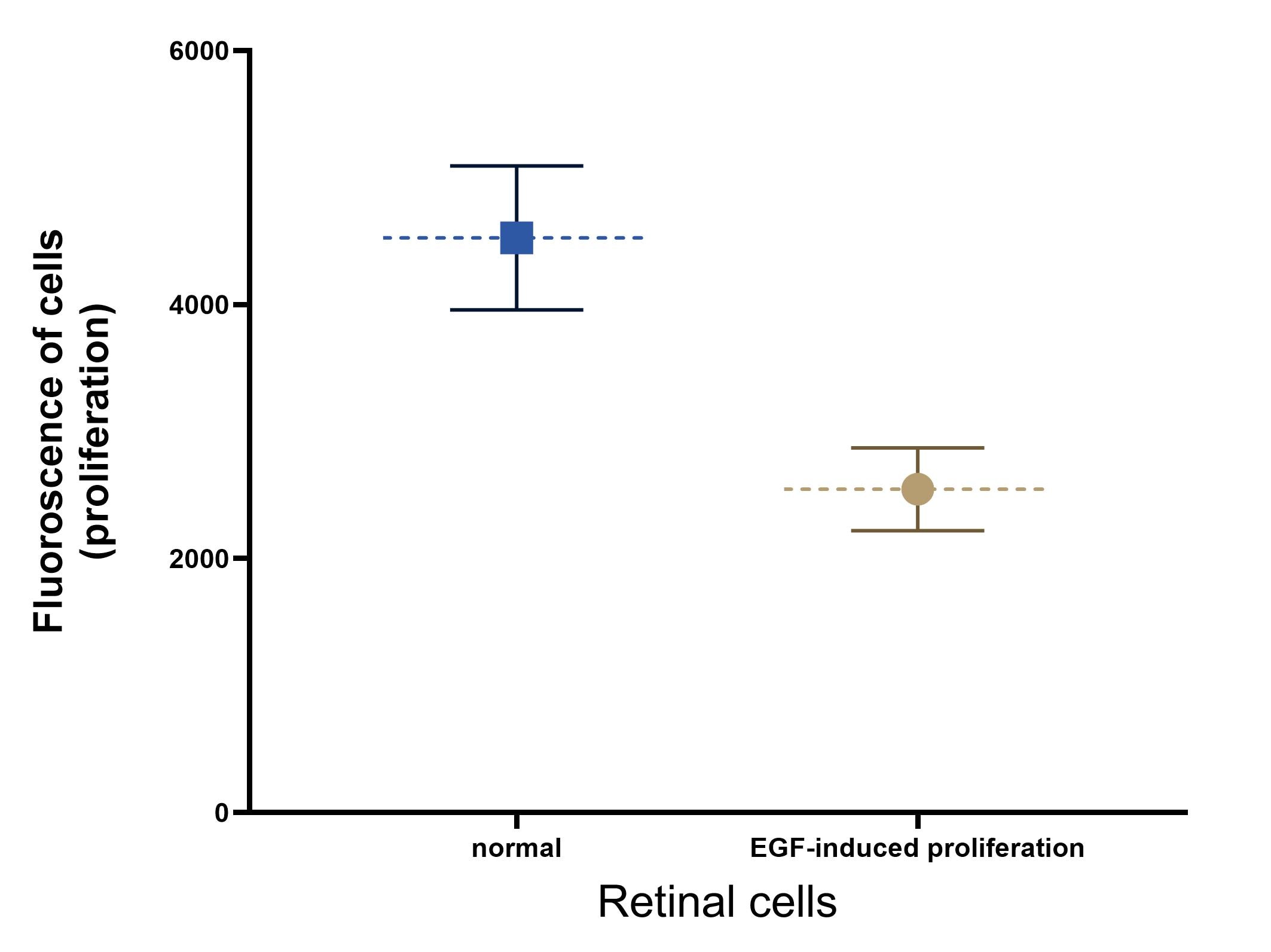

![FGF basic/FGF2/bFGF [Unconjugated]](/sites/all/modules/enterprise-tech/et_datasheets/images/novus_guarantee.png "FGF basic/FGF2/bFGF [Unconjugated]")
![Bioactivity IGF-I/IGF-1 [Unconjugated]](https://images.novusbio.com/images/protein/IGF-I_291-G1_41.jpg)
![Mass Spectrometry IGF-I/IGF-1 [Unconjugated]](https://images.novusbio.com/images/protein/IGF-I_291-G1_42.jpg)
![SEC-MALS IGF-I/IGF-1 [Unconjugated]](https://images.novusbio.com/images/291-g1_recombinant-human-igf-i-igf-1-protein-cf-sec-mals-224202691859.jpg)
![Western Blot ERK2 Antibody [Unconjugated]](https://images.novusbio.com/images/antibody/ERK2_AF1230_Western_Blot_5097.jpg)
![Knockout Validated ERK2 Antibody [Unconjugated]](https://images.novusbio.com/images/antibody/ERK2_AF1230_Knockout_Validated_22864.jpg)
![Immunohistochemistry ERK2 Antibody [Unconjugated]](https://images.novusbio.com/images/antibody/ERK2_AF1230_Immunohistochemistry_20696.jpg)
![SDS-Page TNF-alpha [Unconjugated]](https://images.novusbio.com/images/protein/TNF-alpha_210-TA_256.jpg)
![Bioactivity TNF-alpha [Unconjugated]](https://images.novusbio.com/images/protein/TNFalpha_210TA_1658.jpg)
![SEC-MALS TNF-alpha [Unconjugated]](https://images.novusbio.com/images/210-ta_recombinant-human-tnf-alpha-protein-sec-mals-35202312244..jpg)
![N/A IL-6 [HRP]](https://images.novusbio.com/images/elisa/DATA_IL6_M6000_ELISA_936.jpg)
![N/A IL-6 [HRP]](https://images.novusbio.com/images/elisa/IL-6_M6000_ELISA_415.jpg)
![N/A IL-6 [HRP]](https://images.novusbio.com/images/m6000b_mouse-il-6-quantikine-elisa-kit-1752025024034.jpg)
![Bioactivity ErbB2/Her2 [Unconjugated]](https://images.novusbio.com/images/protein/1129-er_recombinant-human-erbb2-her2-fc-chimera-protein-cf-bioactivity-7122020142841.jpg)
![SDS-Page ErbB2/Her2 [Unconjugated]](https://images.novusbio.com/images/protein/ErbB2_1129-ER_70.jpg)
![Bioactivity HGF [Unconjugated]](https://images.novusbio.com/images/protein/HGF_294HG_3253.jpg)
![Cell Culture HGF [Unconjugated]](https://images.novusbio.com/images/294-hg_recombinant-human-hgf-protein-cell-culture-252023111456.jpg)
![N/A VEGF [HRP]](https://images.novusbio.com/images/elisa/VEGF_DVE00_ELISA_208.jpg)
![N/A VEGF [HRP]](https://images.novusbio.com/images/elisa/DATA_VEGF_DVE00_ELISA_871.jpg)
![N/A VEGF [HRP]](https://images.novusbio.com/images/elisa/DATA_VEGF_DVE00_ELISA_872.jpg)
![SEC-MALS TGF-alpha [Unconjugated]](https://images.novusbio.com/images/239-a_recombinant-human-tgf-alpha-protein-cf-sec-mals-2810202585510.jpg)
![SDS-Page: Human EGF Protein [NBP2-34952]](https://images.novusbio.com/images/Human-EGF-Protein-SDS-Page-NBP2-34952-img0003.jpg "SDS-Page: Human EGF Protein [NBP2-34952]")


