CTS008) and counterstained with hematoxylin (blue). Lower panel shows a lack of labeling if primary antibodies are omitted and tissue is stained only with secondary antibody followed by incubation with detection reagents. View our protocol for Chromogenic IHC Staining of Paraffin-embedded Tissue Sections." title="p63/TP73L was detected in immersion fixed paraffin-embedded sections of human prostate array using Goat Anti-Human p63/TP73L Biotinylated Antigen Affinity-purified Polyclonal Antibody (Catalog # BAF1916) at 15 µg/mL overnight at 4 °C. Tissue was stained using the Anti-Goat HRP-DAB Cell & Tissue Staining Kit (brown; Catalog # CTS008) and counterstained with hematoxylin (blue). Lower panel shows a lack of labeling if primary antibodies are omitted and tissue is stained only with secondary antibody followed by incubation with detection reagents. View our protocol for Chromogenic IHC Staining of Paraffin-embedded Tissue Sections." />
| Reactivity | HuSpecies Glossary |
| Applications | WB, IHC, ICC/IF |
| Clonality | Polyclonal |
| Host | Goat |
| Conjugate | Biotin |
| Immunogen | E. coli-derived recombinant human p63/TP73L Met40-Cys339 Accession # Q9H3D4 |
| Specificity | Detects p63/TP73L in Western blots. |
| Source | N/A |
| Isotype | IgG |
| Clonality | Polyclonal |
| Host | Goat |
| Gene | TP63 |
| Purity Statement | Antigen Affinity-purified |
| Innovator's Reward | Test in a species/application not listed above to receive a full credit towards a future purchase. |
| Dilutions |
|
|
| Readout System | ||
| Publications |
|
| Storage | Use a manual defrost freezer and avoid repeated freeze-thaw cycles.
|
| Buffer | Lyophilized from a 0.2 μm filtered solution in PBS with BSA as a carrier protein. |
| Preservative | No Preservative |
| Reconstitution Instructions | Reconstitute at 0.2 mg/mL in sterile PBS. |
Tumor Protein 63 (p63), also named TP73L, TP63, p51, p40 or KET, is a p53 homolog. It is one of several proteins that are produced from a single gene using two promoters and alternative splicing of the primary RNA transcript. p63 is highly expressed in embryonic ectoderm and in the nuclei of basal regenerative cells of many epithelial tissues in the adult. p63 is suggested to play a role in development, epithelial cell maintenance and tumorigenesis (1‑3).






![p63/TP73L Antibody [Biotin]](/sites/all/modules/enterprise-tech/et_datasheets/images/novus_guarantee.png "p63/TP73L Antibody [Biotin]")
Secondary Antibodies |
Isotype Controls |
|
Spheroids vs. Organoids: Which 3D Cell Culture Model is Best for You? By Jennifer Jones, M.S.Spheroids and organoids are two words that, like “butter” and “margarine”, are often referred to interchangeably but have distinct meanings. The progression and adopt... Read full blog post. |
The concentration calculator allows you to quickly calculate the volume, mass or concentration of your vial. Simply enter your mass, volume, or concentration values for your reagent and the calculator will determine the rest.


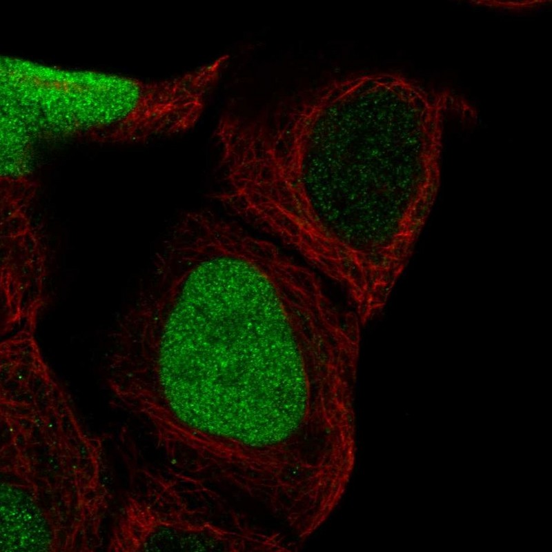
 NL999) and counterstained with DAPI (blue). Specific staining was localized to nuclei. View our protocol for
 ![Simple Western CKAP4/p63 Antibody [Unconjugated]](https://images.novusbio.com/images/antibody/af7355_human-ckap4-p63-affinity-purified-polyclonal-ab-simple-western-157202493849.jpg)
![Western Blot CKAP4/p63 Antibody [Unconjugated]](https://images.novusbio.com/images/antibody/CKAP4_AF7355_Western_Blot_11997.jpg)
![Immunocytochemistry CKAP4/p63 Antibody [Unconjugated]](https://images.novusbio.com/images/antibody/CKAP4_AF7355_Immunocytochemistry__Immunofluorescence_17000.jpg)
![SEC-MALS IL-23A/IL-23 P19 [Unconjugated]](https://images.novusbio.com/images/1290-il_recombinant-human-il-23-protein-sec-mals-1012202411154..jpg)
![SDS-Page TNF-alpha [Unconjugated]](https://images.novusbio.com/images/protein/TNF-alpha_210-TA_256.jpg)
![Bioactivity TNF-alpha [Unconjugated]](https://images.novusbio.com/images/protein/TNFalpha_210TA_1658.jpg)
![SEC-MALS TNF-alpha [Unconjugated]](https://images.novusbio.com/images/210-ta_recombinant-human-tnf-alpha-protein-sec-mals-35202312244..jpg)
![N/A IL-10 [Biotin]](https://images.novusbio.com/images/elisa/DATA_IL10_DY417_ELISA_2014.jpg)
![SEC-MALS IFN-gamma [Unconjugated]](https://images.novusbio.com/images/485-mi_recombinant-mouse-ifn-gamma-protein-sec-mals-1612202583245.jpg)
![Bioactivity IFN-gamma [Unconjugated]](https://images.novusbio.com/images/protein/IFN-gamma_485-MI_455.jpg)
![SDS-Page IFN-gamma [Unconjugated]](https://images.novusbio.com/images/protein/IFN-gamma_485-MI_407.jpg)
![SDS-Page IL-12 [Unconjugated]](https://images.novusbio.com/images/protein/IL-12_419-ML_403.jpg)
![Bioactivity IL-12 [Unconjugated]](https://images.novusbio.com/images/protein/IL-12_419-ML_405.jpg)
![Simple Western Annexin A1 Antibody [Unconjugated]](https://images.novusbio.com/images/antibody/Annexin_A1_AF3770_Simple_Western_16649.jpg)
![Immunohistochemistry Annexin A1 Antibody [Unconjugated]](https://images.novusbio.com/images/antibody/Annexin_A1_AF3770_Immunohistochemistry_10404.jpg)
![Western Blot Annexin A1 Antibody [Unconjugated]](https://images.novusbio.com/images/antibody/Annexin_A1_AF3770_Western_Blot_5334.jpg)