 followed by HRP-conjugated Anti-Mouse IgG Secondary Antibody (Catalog # HAF007). A specific band was detected for N-Cadherin at approximately 130 kDa (as indicated). This experiment was conducted under reducing conditions and using Immunoblot Buffer Group 1.")
| Reactivity | HuSpecies Glossary |
| Applications | WB, Flow, IHC, CyTOF-ready |
| Clone | 691723 |
| Clonality | Monoclonal |
| Host | Mouse |
| Conjugate | Unconjugated |
| Concentration | LYOPH |
| Immunogen | Mouse myeloma cell line NS0-derived recombinant mouse N-Cadherin Asp160-Ala724 Accession # P19022.4 |
| Specificity | Detects mouse N-Cadherin in direct ELISAs and Western blots. In direct ELISAs approximately 50% cross-reactivity
with recombinant mouse N-Cadherin is observed, and no cross-reactivity with recombinant
human (rh) E-Cadherin, rhP-Cadherin, rhVE-Cadherin, rhCadherin-4, -8, -11, -12,
or -13 is observed. |
| Source | N/A |
| Isotype | IgM |
| Clonality | Monoclonal |
| Host | Mouse |
| Gene | CDH2 |
| Purity Statement | Protein A or G purified from hybridoma culture supernatant |
| Innovator's Reward | Test in a species/application not listed above to receive a full credit towards a future purchase. |
| Dilutions |
|
|
| Publications |
|
| Storage | Use a manual defrost freezer and avoid repeated freeze-thaw cycles.
|
| Buffer | Lyophilized from a 0.2 μm filtered solution in PBS with Trehalose. *Small pack size (SP) is supplied either lyophilized or as a 0.2 µm filtered solution in PBS. |
| Preservative | No Preservative |
| Concentration | LYOPH |
| Reconstitution Instructions | Sterile PBS to a final concentration of 0.5 mg/mL. |


![Immunocytochemistry E-Cadherin Antibody [Unconjugated]](https://images.novusbio.com/images/antibody/ECadherin_AF748_Immunocytochemistry_16573.jpg)
![Western Blot E-Cadherin Antibody [Unconjugated]](https://images.novusbio.com/images/antibody/ECadherin_AF748_Western_Blot_19846.jpg)
![Immunocytochemistry E-Cadherin Antibody [Unconjugated]](https://images.novusbio.com/images/antibody/ECadherin_AF748_Immunocytochemistry_16572.jpg)
![Western Blot beta-Catenin Antibody [Unconjugated]](https://images.novusbio.com/images/af1329_human-mouse-rat-beta-catenin-affinity-purified-polyclonal-ab-western-blot-12122025922481.jpg)
![Western Blot beta-Catenin Antibody [Unconjugated]](https://images.novusbio.com/images/af2649_mouse-chitinase-3-like-1-affinity-purified-pab-western-blot-12122025848165.jpg)
![Western Blot beta-Catenin Antibody [Unconjugated]](https://images.novusbio.com/images/af1329_human-mouse-rat-beta-catenin-affinity-purified-polyclonal-ab-western-blot-121220258282812.jpg)


![N-Cadherin Antibody (691723) [Unconjugated]](/sites/all/modules/enterprise-tech/et_datasheets/images/novus_guarantee.png "N-Cadherin Antibody (691723) [Unconjugated]")
Secondary Antibodies |
Isotype Controls |
|
Read full blog post. |
|
Bad news for stomach cancer: BAMBI protein inhibits gastric carcinoma via TGF-beta/epithelial-mesenchymal transition signaling By Jamshed Arslan Pharm.D. Gastric carcinoma is the second leading cause of cancer-related deaths worldwide. One of the key features of gastric carcinoma is acidosis, which promotes growth and metastasis of gastric ... Read full blog post. |
|
Beta Tubulin III and neurogenesis Beta tubulin III, also known as Tuj-1, is a class III member of the beta tubulin protein family. Beta tubulins are one of two structural components that form our microtubule network. While general tubulins play a role in a wide range of cellular pr... Read full blog post. |
|
Epithelial-Mesenchymal Transition (EMT) Markers Epithelial-Mesenchymal Transition (EMT) is the trans-differentiation of stationary epithelial cells into motile mesenchymal cells. During EMT, epithelial cells lose their junctions and apical-basal polarity, reorganize their cytoskeleton, undergo a... Read full blog post. |
|
CD63: is it pro-metastatic or anti-metastatic? CD63 is a type II membrane protein belonging to tetraspanin superfamily and it play key roles in the activation of several cellular signaling cascades along with acting as TIMP1 receptor. It is expressed by activated platelets, monocytes,... Read full blog post. |
|
MMP24 (Matrix metalloproteinase-24, matrix metalloproteinase-25, MT5-MMP) MMP24 is an extracellular matrix (ECM) degradative peptidase enzyme that is a member of the large family of matrix metalloproteinases (MMP). Each MMP has a different substrate specificity, and the aberrant or derailed expression of these is strongly c... Read full blog post. |
The concentration calculator allows you to quickly calculate the volume, mass or concentration of your vial. Simply enter your mass, volume, or concentration values for your reagent and the calculator will determine the rest.


 or isotype control antibody mouse IgM (open histogram), followed by Phycoerythrin-conjugated Anti-Mouse IgM Secondary Antibody (Catalog # F0116).")
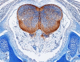
 at 15 µg/mL overnight at 4 °C. Before incubation with the primary antibody, tissue was subjected to heat-induced epitope retrieval using Antigen Retrieval Reagent-Basic (Catalog # CTS013). Tissue was stained using the Anti-Mouse HRP-DAB Cell & Tissue Staining Kit (brown; Catalog # CTS002) and counterstained with hematoxylin (blue). Specific staining was localized to cytoplasm of neurons. View our protocol for Chromogenic IHC Staining of Paraffin-embedded Tissue Sections.")
 and ERK1/2 (pERK1/2) was suppressed in DCN-overexpressing IBC cell lines. Total ERK1/2 (tERK1/2) remains unchanged. GAPDH served as a loading control. b Treatment of IBC cells with DCN protein (4 or 8 μg/mL) for 2 h suppresses E-cadherin expression and EGFR pathway activation. Tubulin served as a loading control. c and d Western blot validation of E-cadherin and EGFR downregulation in tumor samples obtained from mammary fatpad transplantation of control or DCN-overexpressing MDA-IBC3 (c) or SUM149 (d) cells. e and f Immunohistochemical staining validation of E-cadherin and EGFR downregulation in tumor samples obtained from mammary fatpad transplantation of control or DCN-overexpressing MDA-IBC3 (e) or SUM149 (f) cells. Scale bar: 100 μm. g DCN inhibits EGFR signaling in IBC cells independently of EGF stimulation. DCN-overexpressing and control IBC cell lines were stimulated with 50 ng/mL EGF for the indicated number of hours, and total cell lysates were analyzed by western blotting. Both the total levels and the phosphorylation levels of EGFR and ERK1/2 were detected by western blotting. Tubulin served as a loading control. h DCN-mediated inhibition of E-cadherin does not affect expression of epithelial–mesenchymal transition markers. Cell lysates containing 40 μg of total protein were analyzed by western blotting with anti-E-cadherin, fibronectin, vimentin, and DCN antibodies. GAPDH served as a loading control. Image collected and cropped by CiteAb from the following open publication (//pubmed.ncbi.nlm.nih.gov/33452400), licensed under a CC-BY license. Not internally tested by R&D Systems.")
![Bioactivity CTLA-4 [Unconjugated]](https://images.novusbio.com/images/protein/CTLA4_7268CT_2293.jpg)


![N/A MMP-9 [HRP]](https://images.novusbio.com/images/elisa/DATA_MMP9_DMP900_ELISA_770.jpg)
![N/A MMP-9 [HRP]](https://images.novusbio.com/images/elisa/MMP-9_DMP900_ELISA_148.jpg)
![N/A MMP-2 [HRP]](https://images.novusbio.com/images/elisa/DATA_MMP2_MMP200_ELISA_1003.jpg)
![N/A MMP-2 [HRP]](https://images.novusbio.com/images/elisa/MMP2_MMP200_ELISA_474.jpg)