 HAF017). A specific band was detected for Midkine at approximately 17 kDa (as indicated). This experiment was conducted under reducing conditions and using Immunoblot Buffer Group 1." title="Western blot shows lysates of SH-SY5Y human neuroblastoma cell line. PVDF membrane was probed with 2 µg/mL of Goat Anti-Human Midkine Antigen Affinity-purified Polyclonal Antibody (Catalog # AF-258-PB) followed by HRP-conjugated Anti-Goat IgG Secondary Antibody (HAF017). A specific band was detected for Midkine at approximately 17 kDa (as indicated). This experiment was conducted under reducing conditions and using Immunoblot Buffer Group 1." />
| Reactivity | HuSpecies Glossary |
| Applications | WB, Simple Western, IHC, IP, KO |
| Clonality | Polyclonal |
| Host | Goat |
| Conjugate | Unconjugated |
| Concentration | LYOPH |
| Immunogen | E. coli-derived recombinant human Midkine Lys23-Asp143 Accession # P21741 |
| Specificity | Detects human Midkine in direct ELISAs and Western blots. In direct ELISAs, approximately 10% cross-reactivity with recombinant mouse Midkine is observed, and less than 1% cross-reactivity with recombinant human Pleiotrophin is observed. |
| Source | N/A |
| Isotype | IgG |
| Clonality | Polyclonal |
| Host | Goat |
| Gene | MDK |
| Purity Statement | Antigen Affinity-purified |
| Innovator's Reward | Test in a species/application not listed above to receive a full credit towards a future purchase. |
| Dilutions |
|
|
| Publications |
|
| Storage | Use a manual defrost freezer and avoid repeated freeze-thaw cycles.
|
| Buffer | Lyophilized from a 0.2 μm filtered solution in PBS with Trehalose. *Small pack size (SP) is supplied either lyophilized or as a 0.2 µm filtered solution in PBS. |
| Preservative | No Preservative |
| Concentration | LYOPH |
| Reconstitution Instructions | Reconstitute at 0.2 mg/mL in sterile PBS. |
Midkine (MK) is a 15 kDa heparin-binding molecule originally cloned during a search for genes preferentially transcribed during retinoic acid (RA)-induced differentiation. Midkine belongs to a family of neurotrophic and developmentally-regulated heparin-binding molecules consisting of midkine, pleiotrophin (PTN/HBNF/OSF-1/HNGF-8) and the avian midkine homolog, RI-HB (for retinoic acid-inducible heparin-binding protein).
Midkine is a highly basic, nonglycosylated polypeptide that contains five intrachain disulfide bonds. The predicted molecular weight is approximately 13.3 kDa, based on a mature peptide length of 118 amino acid residues in the mouse and 121 amino acid residues in the human. Across species, MK shows 87% identity between the human and murine proteins. Between family members, human MK is approximately 50% identical to human PTN, with conservation of all 10 cysteines. Initial structure-function studies indicate that the C-terminal half of MK contains the principal heparin-binding site plus the molecule’s antigenicity and neurite-promoting sequences; while both the C- and N-termini are necessary for the molecule’s neurotrophic effects. Cells known to produce MK include endothelial cells, fetal astrocytes, renal proximal tubule epithelial cells and Wilms’ (kidney) tumor cells. MK has also been identified in the senile plaques of patients with Alzheimer’s disease. The pattern of expression of midkine during development strongly suggests a role for this factor both in epithelial-mesenchymal interactions and in development of the nervous system.


![Immunohistochemistry ACE/CD143 Antibody [Unconjugated]](https://images.novusbio.com/images/antibody/ACE_AF1513_Immunohistochemistry_6703.jpg)
![Western Blot ACE/CD143 Antibody [Unconjugated]](https://images.novusbio.com/images/antibody/ACE_AF1513_Flow_Cytometry_20028.jpg)
![Simple Western ACE/CD143 Antibody [Unconjugated]](https://images.novusbio.com/images/antibody/ACE_AF1513_Simple_Western_20109.jpg)


Secondary Antibodies |
Isotype Controls |
The concentration calculator allows you to quickly calculate the volume, mass or concentration of your vial. Simply enter your mass, volume, or concentration values for your reagent and the calculator will determine the rest.


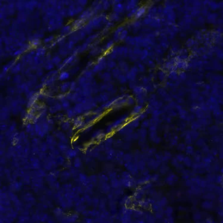
 CTS013). Tissue was stained using the Anti-Goat HRP-DAB Cell & Tissue Staining Kit (brown
  CTS008) and counterstained with hematoxylin (blue). View our protocol for
  HAF109). This experiment was conducted under reducing conditions and using the 12-230 kDa separation system." title="Simple Western lane view shows lysates of SH-SY5Y human neuroblastoma cell line, loaded at 0.2 mg/mL. A specific band was detected for Midkine at approximately 26 kDa (as indicated) using 100 µg/mL of Goat Anti-Human Midkine Antigen Affinity-purified Polyclonal Antibody (Catalog # AF-258-PB) followed by 1:50 dilution of HRP-conjugated Anti-Goat IgG Secondary Antibody (
 or treated (+) with 3.0 ug/ml Brefeldin A for 18 hr and Midkine knockout HAP1 cell line (KO). Nitrocellulose membrane was probed with 2 µg/mL of Goat Anti-Human Midkine Antigen Affinity-purified Polyclonal Antibody (Catalog # AF-258-PB) followed by HRP-conjugated Anti-Goat IgG Secondary Antibody. A specific band was detected for Midkine at approximately 11 kDa (as indicated) in the parental HAP1 cell line, but is not detectable in knockout HAP1 cell line. The Ponceau stained transfer of the blot is shown. This experiment was conducted under reducing conditions. Image, protocol, and testing courtesy of YCharOS Inc. See ycharos.com for additional details.")
 pre-coupled to protein G or protein A beads. Immunoprecipitated Midkine was detected with Rabbit Anti-Human Midkine Monoclonal Antibody (NBP2-66948 ) at 1/500. The Ponceau stained transfers of each blot are shown. SM=10% starting material; UB=10% unbound fraction; IP=immunoprecipitated. Image, protocol, and testing courtesy of YCharOS Inc. (ycharos.com).")
![Immunohistochemistry Pleiotrophin/PTN Antibody [Unconjugated]](https://images.novusbio.com/images/antibody/Pleiotrophin_AF252PB_Immunohistochemistry_23024.jpg)
![Bioactivity Thrombopoietin/THPO [Unconjugated]](https://images.novusbio.com/images/protein/288-tpncf_recombinant-human-thrombopoietin-ns0-expressed-protein-cf-bioactivity-272020135047.jpg)
![SEC-MALS IL-3 [Unconjugated]](https://images.novusbio.com/images/203-il_recombinant-human-il-3-protein-sec-mals-133202482656..jpg)
![Bioactivity IL-3 [Unconjugated]](https://images.novusbio.com/images/protein/IL-3_203-IL_347.jpg)
![Data IL-3 [Unconjugated]](https://images.novusbio.com/images/203-il_recombinant-human-il-3-protein-data-253202610550.jpg)
![N/A IL-6 [HRP]](https://images.novusbio.com/images/elisa/DATA_IL6_M6000_ELISA_936.jpg)
![N/A IL-6 [HRP]](https://images.novusbio.com/images/elisa/IL-6_M6000_ELISA_415.jpg)
![N/A IL-6 [HRP]](https://images.novusbio.com/images/m6000b_mouse-il-6-quantikine-elisa-kit-1752025024034.jpg)
![SCF/c-kit Ligand [Unconjugated]](/sites/all/modules/enterprise-tech/et_datasheets/images/novus_guarantee.png "SCF/c-kit Ligand [Unconjugated]")


![N/A VEGF [HRP]](https://images.novusbio.com/images/elisa/VEGF_DVE00_ELISA_208.jpg)
![N/A VEGF [HRP]](https://images.novusbio.com/images/elisa/DATA_VEGF_DVE00_ELISA_871.jpg)
![N/A VEGF [HRP]](https://images.novusbio.com/images/elisa/DATA_VEGF_DVE00_ELISA_872.jpg)
![SDS-Page TNF-alpha [Unconjugated]](https://images.novusbio.com/images/protein/TNF-alpha_210-TA_256.jpg)
![Bioactivity TNF-alpha [Unconjugated]](https://images.novusbio.com/images/protein/TNFalpha_210TA_1658.jpg)
![SEC-MALS TNF-alpha [Unconjugated]](https://images.novusbio.com/images/210-ta_recombinant-human-tnf-alpha-protein-sec-mals-35202312244..jpg)
![N/A Erythropoietin/EPO [HRP]](https://images.novusbio.com/images/elisa/Erythropoietin_MEP00_ELISA_421.jpg)
![N/A Erythropoietin/EPO [HRP]](https://images.novusbio.com/images/elisa/DATA_Erythropoietin_MEP00_ELISA_970.jpg)
![Bioactivity EGF [Unconjugated]](https://images.novusbio.com/images/protein/EGF_236EG_1570.jpg)
![Cell Culture EGF [Unconjugated]](https://images.novusbio.com/images/236-eg_recombinant-human-egf-protein-cf-25202394526.jpg)
![Cell Culture EGF [Unconjugated]](https://images.novusbio.com/images/236-eg_recombinant-human-egf-protein-cf-bioactivity-25202394053.jpg)
![Bioactivity CTLA-4 [Unconjugated]](https://images.novusbio.com/images/protein/CTLA4_7268CT_2293.jpg)
![Immunocytochemistry/ Immunofluorescence Renin Antibody [Unconjugated] - 1](https://images.novusbio.com/images/af4277_mouse-renin-1-affinity-purified-polyclonal-ab-41202412394940.jpg)
![Immunohistochemistry Renin Antibody [Unconjugated] - 1](https://images.novusbio.com/images/antibody/Renin_AF4277_Immunohistochemistry_17351.jpg)
![ELISA Renin Antibody [Unconjugated] - 1](https://images.novusbio.com/images/af4277_mouse-renin-1-affinity-purified-polyclonal-ab-elisa-639201778625485467.jpg)
 or Normal Goat IgG Isotype Control Antibody (Catalog # AB-108-C, open histogram), followed by Phycoerythrin-conjugated Anti-Goat IgG Secondary Antibody (Catalog # F0107).")