![Western Blot: LC3 Antibody Pack [NB910-40752] - LC3/MAP1LC3A Antibody Pack [NB910-40752] - Detection of LC3B in treated U87-MG (human glioblastoma astrocytoma) lysates using NB600-1384.](http://images.novusbio.com/fullsize/LC3-MAP1LC3A-Antibody-Pack-Western-Blot-NB910-40752-img0026.jpg "Western Blot: LC3 Antibody Pack [NB910-40752] - LC3/MAP1LC3A Antibody Pack [NB910-40752] - Detection of LC3B in treated U87-MG (human glioblastoma astrocytoma) lysates using NB600-1384.")
| Reactivity | Hu, Mu, Rt, Po, Ba, Bv, Ca, Pm, ZeSpecies Glossary |
| Applications | WB, Simple Western, ELISA, Flow, ICC/IF, IHC, IP, WB, ChIP, KO, Mycoplasma |
| Description | This pack contains 1 vial each of: NB100-2220 (0.1 mL), NB100-2331 (0.1 mL) and NB600-1384 (0.1 mL). |
| Immunogen | For NB600-1384: Polyclonal LC3B Antibody was made to a synthetic peptide made to the N-terminal region of the human LC3B protein [Uniprot: Q9GZQ8]. For NB100-2331: A synthetic peptide made to an internal portion of the human LC3 protein sequence (between residues 25-121) [Uniprot: Q9H492]. For NB100-2220: Polyclonal LC3B Antibody was made to a synthetic peptide made to an N-terminal portion of the human LC3B protein sequence (between residues 1-100) [UniProt# Q9GZQ8]. |
| Gene | MAP1LC3B |
| Dilutions |
|
|
| Application Notes | Antibodies in this pack are validated for the following applications: NB600-1384: Electron Microscopy, Flow Cytometry, Immunoblotting, Immunocytochemistry/Immunofluorescence, Immunohistochemistry, Immunohistochemistry Free-Floating, Immunohistochemistry-Frozen, Immunohistochemistry-Paraffin, Immunoprecipitation, Knockdown Validated, Knockout Validated, Simple Western, Western Blot NB100-2331: Chromatin Immunoprecipitation, ELISA, Flow Cytometry, Immunoblotting, Immunocytochemistry/Immunofluorescence, Immunohistochemistry, Immunohistochemistry Whole-Mount, Immunohistochemistry-Frozen, Immunohistochemistry-Paraffin, Immunoprecipitation, Simple Western, Southern Blot, Western Blot NB100-2220: Chromatin Immunoprecipitation (ChIP), ELISA, Flow Cytometry, Immunoblotting, Immunocytochemistry/Immunofluorescence, Immunohistochemistry, Immunohistochemistry-Frozen, Immunohistochemistry-Paraffin, Immunoprecipitation, Knockdown Validated, Knockout Validated, Proximity Ligation Assay, SDS-Page, Simple Western, Western Blot |
|
| Publications |
|
| Storage | Store at 4C short term. Aliquot and store at -20C long term. Avoid freeze-thaw cycles. |


![Immunohistochemistry ATG7 Antibody (683906) [Unconjugated]](https://images.novusbio.com/images/antibody/ATG7_MAB6608_Immunohistochemistry_10631.jpg)
![Simple Western ATG7 Antibody (683906) [Unconjugated]](https://images.novusbio.com/images/antibody/ATG7_MAB6608_Simple_Western_16409.jpg)
![Intracellular Staining by Flow Cytometry AKT [p Ser473] Antibody [Unconjugated] - Pan Specific](https://images.novusbio.com/images/antibody/Akt3_AF887_Flow_Cytometry_8283.jpg)
![Western Blot AKT [p Ser473] Antibody [Unconjugated] - Pan Specific](https://images.novusbio.com/images/af887_phospho-akt-s473-pan-specific-affinity-purified-pab-41202410485440.jpg)
![Western Blot AKT [p Ser473] Antibody [Unconjugated] - Pan Specific](https://images.novusbio.com/images/af887_phospho-akt-s473-pan-specific-affinity-purified-pab-8120255552843.jpg)


Research Areas for LC3 Antibody Pack (NB910-40752)Find related products by research area.
|
|
Autophagy and Metastasis By Christina Towers, PhD The majority of cancer patients die from metastatic disease at secondary sites. The threshold to undergo metastasis is high. Only a minority of cancer cells acquire invasive phenotypes... Read full blog post. |
|
Optogenetic Control of Mitophagy: AMBRA1 based mitophagy switch By Christina Towers, PhD Mitophagy in the BrainSelective autophagic degradation of damaged mitochondria, known as mitophagy, has been described as a cyto-protective process. Accordingly, defects in mitophagy h... Read full blog post. |
|
Read full blog post. |
|
Read full blog post. |
|
Read full blog post. |
|
How to visualize autophagy by microscopy By Christina Towers, PhD Autophagy is a recycling process that relies on the formation of a unique organelle termed an autophagosome. An elegant way to monitor autophagy is through various microscopy techniques to... Read full blog post. |
|
Microglia: pruning shears for homeostatic maintenance in the brain By Jennifer Sokolowski, MD, PhD.Microglia play a critical role in pruning neurons and synapses during homeostatic maintenance in the adult brain.1 A recent study by Ayata et al. (2018) identified regional differe... Read full blog post. |
|
Read full blog post. |
|
Read full blog post. |
|
Read full blog post. |
| Gene Symbol | MAP1LC3B |


![Immunocytochemistry/Immunofluorescence: LC3 Antibody Pack [NB910-40752] - LC3/MAP1LC3A Antibody Pack [NB910-40752] - Immunofluorescent staining of treated U373-MG cells using NB600-1384. The nuclei were stained with DAPI.](http://images.novusbio.com/fullsize/LC3-MAP1LC3A-Antibody-Pack-Immunocytochemistry-Immunofluorescence-NB910-40752-img0017.jpg "Immunocytochemistry/Immunofluorescence: LC3 Antibody Pack [NB910-40752] - LC3/MAP1LC3A Antibody Pack [NB910-40752] - Immunofluorescent staining of treated U373-MG cells using NB600-1384. The nuclei were stained with DAPI.")
![Western Blot: LC3 Antibody Pack [NB910-40752] - Lysates of HeLa parental cell line and LC3B knockout HeLa cell line (KO) untreated (-) or treated (+) with 50 uM Chloroquine for 18 hours. PVDF (Polyvinylidene difluoride) membrane was probed with 0.5 ug/mL of Rabbit Anti-LC3B Polyclonal Antibody (Catalog # NB100-2220) followed by HRP-conjugated Anti-Rabbit IgG Secondary Antibody (Catalog# HAF008). A specific band was detected for LC3B at a molecular weight of approximately 15 kDa (as indicated) in the parental HeLa cell line, but is not detectable in the knockout HeLa cell line. GAPDH is shown as a loading control. This experiment was conducted under reducing conditions. LC3B Antibody [NB100-2220]](http://images.novusbio.com/fullsize/LC3-Antibody-Pack-Knockout-Validated-NB910-40752-img0027.jpg "Western Blot: LC3 Antibody Pack [NB910-40752] - Lysates of HeLa parental cell line and LC3B knockout HeLa cell line (KO) untreated (-) or treated (+) with 50 uM Chloroquine for 18 hours. PVDF (Polyvinylidene difluoride) membrane was probed with 0.5 ug/mL of Rabbit Anti-LC3B Polyclonal Antibody (Catalog # NB100-2220) followed by HRP-conjugated Anti-Rabbit IgG Secondary Antibody (Catalog# HAF008). A specific band was detected for LC3B at a molecular weight of approximately 15 kDa (as indicated) in the parental HeLa cell line, but is not detectable in the knockout HeLa cell line. GAPDH is shown as a loading control. This experiment was conducted under reducing conditions. LC3B Antibody [NB100-2220]")
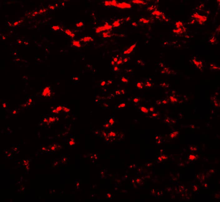
![Immunohistochemistry: LC3 Antibody Pack [NB910-40752] - Autophagosomes accumulate in neurons of the VH gray matter at day 1 after SCI. Representative images of IHC staining for LC3 (green) and neuronal marker NeuN (red) in VH of gray matter from sham and SCI animals. Stronger co-localization between NeuN and LC3 is apparent at day 1 after SCI. Scale bar is 20 um. Image collected and cropped by CiteAb from the following publication (//www.nature.com/articles/cddis2014527) licensed under a CC-BY license. LC3B Antibody [NB100-2220]](http://images.novusbio.com/fullsize/LC3-Antibody-Pack-Immunohistochemistry-NB910-40752-img0029.jpg "Immunohistochemistry: LC3 Antibody Pack [NB910-40752] - Autophagosomes accumulate in neurons of the VH gray matter at day 1 after SCI. Representative images of IHC staining for LC3 (green) and neuronal marker NeuN (red) in VH of gray matter from sham and SCI animals. Stronger co-localization between NeuN and LC3 is apparent at day 1 after SCI. Scale bar is 20 um. Image collected and cropped by CiteAb from the following publication (//www.nature.com/articles/cddis2014527) licensed under a CC-BY license. LC3B Antibody [NB100-2220]")
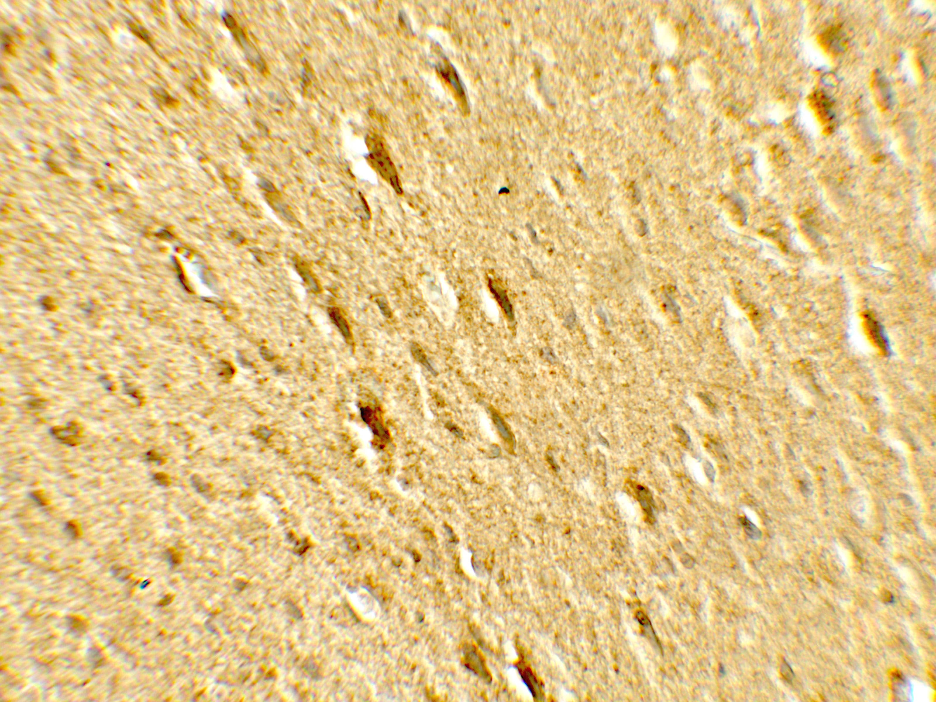
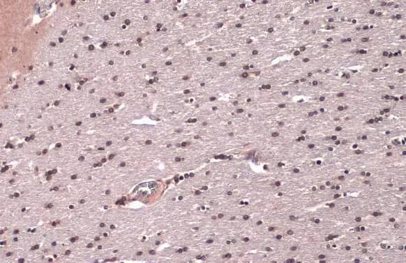
![Immunohistochemistry: LC3 Antibody Pack [NB910-40752] - Analysis in mouse renal tissue. Image from verifed customer review. LC3A Antibody [NB100-2331]](http://images.novusbio.com/fullsize/LC3-Antibody-Pack-Immunohistochemistry-NB910-40752-img0030.jpg "Immunohistochemistry: LC3 Antibody Pack [NB910-40752] - Analysis in mouse renal tissue. Image from verifed customer review. LC3A Antibody [NB100-2331]")
![Western Blot: LC3/MAP1LC3A Antibody Pack [NB910-40752] - Detection of autophagic LC3 in mouse ES cell lysate using NB 100-2331. The atg5-/- lane (ES cells, cultured to form embryonic bodies, that are deficient in conversion of LC3-1 to LC3-11) demonstrates the specificity of NB 100-2331, as there is no detection of LC3-11. Photo courtesy of Dr. Beth Levine, UT Southwestern Medical Center.](http://images.novusbio.com/fullsize/LC3-MAP1LC3A-Antibody-Pack-Western-Blot-NB910-40752-img0025.jpg "Western Blot: LC3/MAP1LC3A Antibody Pack [NB910-40752] - Detection of autophagic LC3 in mouse ES cell lysate using NB 100-2331. The atg5-/- lane (ES cells, cultured to form embryonic bodies, that are deficient in conversion of LC3-1 to LC3-11) demonstrates the specificity of NB 100-2331, as there is no detection of LC3-11. Photo courtesy of Dr. Beth Levine, UT Southwestern Medical Center.")
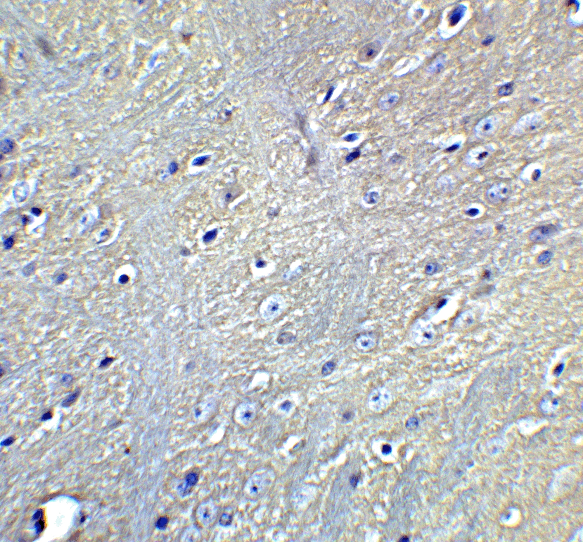
![Immunohistochemistry: LC3/MAP1LC3A Antibody Pack [NB910-40752] - Staining of treated U373-MG (human glioblastoma) cells using NB600-1384.](http://images.novusbio.com/fullsize/LC3-MAP1LC3A-Antibody-Pack-Immunohistochemistry-NB910-40752-img0021.jpg "Immunohistochemistry: LC3/MAP1LC3A Antibody Pack [NB910-40752] - Staining of treated U373-MG (human glioblastoma) cells using NB600-1384.")


![Immunohistochemistry TOR/mTOR [p Ser2448] Antibody - BSA Free](https://images.novusbio.com/images/nb600-607_rabbit-polyclonal-tor-mtor-p-ser2448-antibody-235202318143142.jpg)
![Western Blot TOR/mTOR [p Ser2448] Antibody - BSA Free](https://images.novusbio.com/images/nb600-607_rabbit-polyclonal-tor-mtor-p-ser2448-antibody-31020241535647.jpg)
![Data TOR/mTOR [p Ser2448] Antibody - BSA Free](https://images.novusbio.com/images/TOR-mTOR-[p-Ser2448]-Antibody-N-A-NB600-607-img0008.jpg)
![Immunohistochemistry Caspase-3 Antibody [Unconjugated] - Active](https://images.novusbio.com/images/af835_human-mouse-active-caspase-3-affinity-purified-pab-41202410331943.jpg)
![Western Blot Caspase-3 Antibody [Unconjugated] - Active](https://images.novusbio.com/images/af835_human-mouse-active-caspase-3-affinity-purified-pab-812025554170.jpg)
![Western Blot Caspase-3 Antibody [Unconjugated] - Active](https://images.novusbio.com/images/af835_human-mouse-active-caspase-3-affinity-purified-pab-8120255534731.jpg)


