Immunoblot Buffer Group 1." title="Western blot shows lysates of HEK293T human embryonic kidney cell line either mock transfected or transfected with human 4-1BB/TNFRSF9/ CD137-eGFP Fusion. PVDF membrane was probed with 0.5 µg/mL of Goat Anti-Human 4-1BB/ TNFRSF9/CD137 Antigen Affinity-purified Polyclonal Antibody (Catalog # AF838) followed by HRP-conjugated Anti-Goat IgG Secondary Antibody ([catalogNumber:HAF017). A specific band was detected for 4-1BB/ TNFRSF9/CD137-eGFP Fusion at approximately 50-65 kDa (as indicated). This experiment was conducted under reducing conditions and using Immunoblot Buffer Group 1." />
| Reactivity | HuSpecies Glossary |
| Applications | WB, Simple Western, Flow, IHC, CyTOF-ready, ELISA(Cap), Func, ICC/IF |
| Clonality | Polyclonal |
| Host | Goat |
| Conjugate | Unconjugated |
| Concentration | LYOPH |
| Immunogen | Mouse myeloma cell line NS0-derived recombinant human 4‑1BB/TNFRSF9/CD137 Leu24-Gln186 Accession # Q07011 |
| Specificity | Detects 4‑1BB/TNFRSF9/CD137 in direct ELISAs and Western blots. |
| Source | N/A |
| Isotype | IgG |
| Clonality | Polyclonal |
| Host | Goat |
| Gene | TNFRSF9 |
| Purity Statement | Antigen Affinity-purified |
| Endotoxin Note | <0.10 EU per 1 μg of the antibody by the LAL method. |
| Innovator's Reward | Test in a species/application not listed above to receive a full credit towards a future purchase. |
| Dilutions |
|
|
| Application Notes | ELISA Capture: Human 4-1BB/TNFRSF9/CD137 Antibody (Catalog # AF838) ELISA Detection: Human 4-1BB/TNFRSF9/CD137 Biotinylated Antibody (Catalog # BAF838) Standard: Recombinant Human 4-1BB/TNFRSF9/CD137 Fc Chimera (Catalog # 838-4B) |
|
| Publications |
|
| Storage | Use a manual defrost freezer and avoid repeated freeze-thaw cycles.
|
| Buffer | Lyophilized from a 0.2 μm filtered solution in PBS with Trehalose. See Certificate of Analysis for details. *Small pack size (-SP) is supplied either lyophilized or as a 0.2 µm filtered solution in PBS. |
| Preservative | No Preservative |
| Concentration | LYOPH |
| Reconstitution Instructions | Reconstitute at 0.2 mg/mL in sterile PBS. For liquid material, refer to CoA for concentration. |
4-1BB is an inducible T cell surface protein belonging to the TNF receptor superfamily. It is alternatively known as TNFRSF9, CD137, and ILA. The 255 amino acid human 4-1BB is a type I transmembrane protein having in its extracellular domain four of the cysteine-rich motifs that are characteristic of the TNF receptor superfamily. The 30 kDa glycoprotein exists both as a monomer and as a dimer on T cells. The human and mouse proteins share 60% amino acid identity. 4-1BB is absent from naive T cells, but it is upregulated and continually expressed following T cell activation. The natural ligand, 4-1BBL, is a member of the TNF superfamily and is expressed on activated antigen presenting cells including dendritic cells, macrophages, and B cells. Cross-linking of 4-1BB by 4-1BBL or by agonistic antibodies transmits a potent co-stimulatory signal that enhances the effect of other activating signals such as PHA or anti-CD3 antibodies. 4-1BB signals through the
TFAF2‑NIK pathway resulting in activation of NF-kappa B and ultimately promoting proliferation and survival of T cells.


![Neutralization B7-1/CD80 Antibody (37711) [Unconjugated]](https://images.novusbio.com/images/antibody/B7-1_MAB140_Block_Neutralize_9024.jpg)
![Flow Cytometry B7-1/CD80 Antibody (37711) [Unconjugated]](https://images.novusbio.com/images/antibody/B71_MAB140_Flow_Cytometry_16025.jpg)
![Immunohistochemistry B7-1/CD80 Antibody (37711) [Unconjugated]](https://images.novusbio.com/images/mab140_human-b7-1-cd80-mab-clone-37711-4120241048542.jpg)


Secondary Antibodies |
Isotype Controls |
The concentration calculator allows you to quickly calculate the volume, mass or concentration of your vial. Simply enter your mass, volume, or concentration values for your reagent and the calculator will determine the rest.


 CTS008) and counterstained with hematoxylin (blue). Specific staining was localized to lymphocytes. View our protocol for
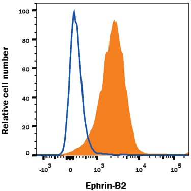
  F0108) and PE-conjugated anti-human CD3 (Catalog # FAB100P). Quad marker was set based on control antibody (
 NL001) and counterstained with DAPI (blue). Specific staining was localized to cytoplasm of PHA stimulated cells. View our protocol for
 838-4B) was serially diluted and captured by Goat Anti-Human 4‑1BB/TNFRSF9/CD137 Antigen Affinity-purified Polyclonal Antibody (Catalog # AF838) coated on a Clear Polystyrene Microplate (Catalog #
  838-4B) was serially diluted and captured by Goat Anti-Human 4‑1BB/TNFRSF9/CD137 Antigen Affinity-purified Polyclonal Antibody (Catalog #
  838-4B) was serially diluted and captured by Goat Anti-Human 4‑1BB/TNFRSF9/CD137 Antigen Affinity-purified Polyclonal Antibody (Catalog #
 ![Bioactivity CTLA-4 [Unconjugated]](https://images.novusbio.com/images/protein/CTLA4_7268CT_2293.jpg)
![SDS-Page TNF-alpha [Unconjugated]](https://images.novusbio.com/images/protein/TNF-alpha_210-TA_256.jpg)
![Bioactivity TNF-alpha [Unconjugated]](https://images.novusbio.com/images/protein/TNFalpha_210TA_1658.jpg)
![SEC-MALS TNF-alpha [Unconjugated]](https://images.novusbio.com/images/210-ta_recombinant-human-tnf-alpha-protein-sec-mals-35202312244..jpg)
![Western Blot 4-1BB Ligand/TNFSF9 Antibody [Unconjugated]](https://images.novusbio.com/images/af1246_mouse-4-1bb-ligand-tnfsf9-affinity-purified-polyclonal-ab-western-blot-12122025817121.jpg)
![Agonist Activity CD28 Antibody (37407) [Unconjugated]](https://images.novusbio.com/images/antibody/CD28_MAB342_Functional_Assay_2393.jpg)
![Immunocytochemistry CD28 Antibody (37407) [Unconjugated]](https://images.novusbio.com/images/mab342_human-cd28-mab-clone-37407-immunocytochemistry-31102022134634.jpg)
![Bioactivity IL-2 [Unconjugated]](https://images.novusbio.com/images/202-il_recombinant-human-il-2-protein-bioactivity-174202314946.jpg)


![SEC-MALS IFN-gamma [Unconjugated]](https://images.novusbio.com/images/485-mi_recombinant-mouse-ifn-gamma-protein-sec-mals-1612202583245.jpg)
![Bioactivity IFN-gamma [Unconjugated]](https://images.novusbio.com/images/protein/IFN-gamma_485-MI_455.jpg)
![SDS-Page IFN-gamma [Unconjugated]](https://images.novusbio.com/images/protein/IFN-gamma_485-MI_407.jpg)
![Bioactivity IL-4 [Unconjugated]](https://images.novusbio.com/images/protein/6507-ilcf_recombinant-human-il-4-cho-expressed-protein-cf-bioactivity-272020133214.jpg)
![N/A IL-10 [Biotin]](https://images.novusbio.com/images/elisa/DATA_IL10_DY417_ELISA_2014.jpg)
![N/A CD40 Ligand/TNFSF5 [HRP]](https://images.novusbio.com/images/elisa/CD40_Ligand_DCDL40_ELISA_60.jpg)
![N/A CD40 Ligand/TNFSF5 [HRP]](https://images.novusbio.com/images/elisa/DATA_CD40_Ligand_DCDL40_ELISA_624.jpg)
![Western Blot CD40/TNFRSF5 Antibody [Unconjugated]](https://images.novusbio.com/images/af632_human-cd40-tnfrsf5-affinity-purified-polyclonal-ab-822024950191.jpg)
![Western Blot CD40/TNFRSF5 Antibody [Unconjugated]](https://images.novusbio.com/images/af632_human-cd40-tnfrsf5-affinity-purified-polyclonal-ab-44202415425047.jpg)
![Simple Western CD40/TNFRSF5 Antibody [Unconjugated]](https://images.novusbio.com/images/antibody/af632_human-cd40-tnfrsf5-affinity-purified-polyclonal-ab-simple-western-58202111562.jpg)
![N/A IL-6 [HRP]](https://images.novusbio.com/images/elisa/DATA_IL6_M6000_ELISA_936.jpg)
![N/A IL-6 [HRP]](https://images.novusbio.com/images/elisa/IL-6_M6000_ELISA_415.jpg)
![N/A IL-6 [HRP]](https://images.novusbio.com/images/m6000b_mouse-il-6-quantikine-elisa-kit-1752025024034.jpg)