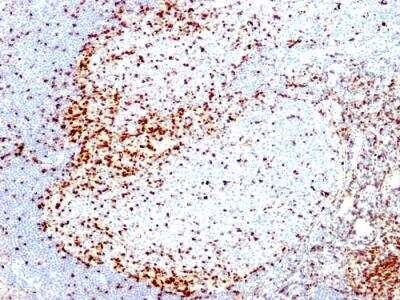
![Immunohistochemistry-Paraffin: CD3 Antibody (SP7) [NB600-1441] - FFPE human tonsil stained with CD3 antibody.](http://images.novusbio.com/fullsize/CD3-Antibody-SP7-Immunohistochemistry-Paraffin-NB600-1441-img0001.jpg "Immunohistochemistry-Paraffin: CD3 Antibody (SP7) [NB600-1441] - FFPE human tonsil stained with CD3 antibody.")
| Reactivity | Hu, Mu, Po, CaSpecies Glossary |
| Applications | Flow, ICC/IF, IHC, Mycoplasma |
| Clone | SP7 |
| Clonality | Monoclonal |
| Host | Rabbit |
| Conjugate | Unconjugated |
| Immunogen | This CD3 antibody was developed against a synthetic peptide: KAKAKPVTRGAGA, corresponding to amino acids 156-168 of Human CD3 epsilon chain. |
| Epitope | aa 156-168 of epsilon chain of human CD3 protein (intracytoplasmic). |
| Localization | Type I membrane protein. |
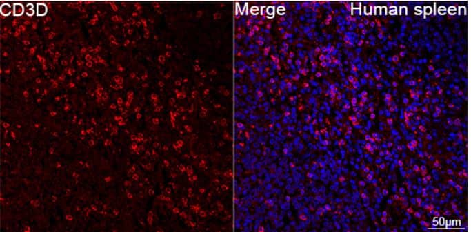
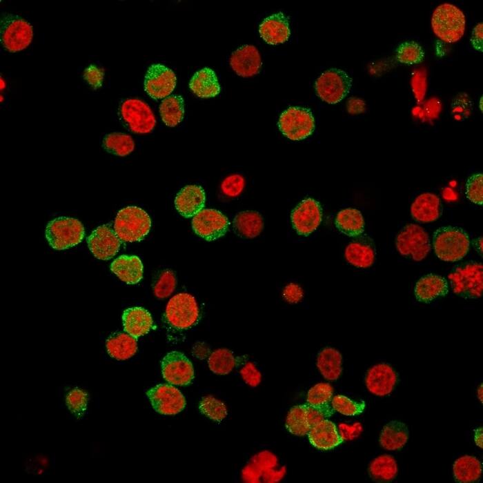
| Specificity | The CD3 antigen is present on early thymocytes and mature T cells and is generally regarded as a pan-T cell marker. This antibody will help detect CD3 expression in normal and neoplastic tissues. This antibody reacts with the intracytoplasmic portion of the CD3 antigen expressed by T cells. It stains human T cells in both the cortex and medulla of the thymus and in peripheral lymphoid tissues. This antibody is suitable for staining normal and neoplastic T cells in formalin-fixed, paraffin-embedded tissues. |
| Isotype | IgG |
| Clonality | Monoclonal |
| Host | Rabbit |
| Gene | CD3E |
| Purity | Tissue culture supernatant |
| Innovator's Reward | Test in a species/application not listed above to receive a full credit towards a future purchase. |
| Dilutions |
|
||
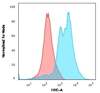
| Application Notes | IHC-P: recommended pretreatment of citrate buffer, pH 6.0. Recommended incubation time of 30-60 min at RT. Use in Immunocytochemistry/immunofluorescence reported in scientific literature (PMID 29037255). Use in FLOW reported in scientific literature (PMID: 31079916). |
||
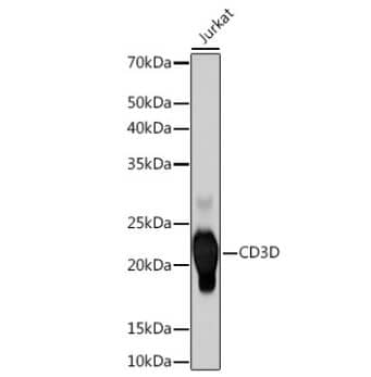
| Theoretical MW | 23.1 kDa. Disclaimer note: The observed molecular weight of the protein may vary from the listed predicted molecular weight due to post translational modifications, post translation cleavages, relative charges, and other experimental factors. |
||
| Control |
|
||
| Reviewed Applications |
|
||
| Publications |
|
| Storage | Store at 4C short term. Aliquot and store at -20C long term. Avoid freeze-thaw cycles. |
| Buffer | Tissue culture supernatant |
| Preservative | 0.05% Sodium Azide |
| Purity | Tissue culture supernatant |


![Western Blot NCAM-1/CD56 Antibody [Unconjugated]](https://images.novusbio.com/images/antibody/NCAM1_AF2408_Western_Blot_20725.jpg)
![Immunocytochemistry NCAM-1/CD56 Antibody [Unconjugated]](https://images.novusbio.com/images/antibody/NCAM1_AF2408_Immunocytochemistry_16574.jpg)
![Knockout Validated NCAM-1/CD56 Antibody [Unconjugated]](https://images.novusbio.com/images/antibody/af2408_human-mouse-ncam-1-cd56-affinity-purified-polyclonal-ab-knockout-validated-156202115045.jpg)


| Images | Ratings | Applications | Species | Date | Details | ||||||||||
|---|---|---|---|---|---|---|---|---|---|---|---|---|---|---|---|

Enlarge |
reviewed by:
Verified Customer |
IHC-P | Mouse | 03/19/2024 |
Summary
Comments
|
||||||||||

Enlarge |
reviewed by:
Kevin Thorburn |
IHC-P | Dog | 04/06/2023 |
Summary
Comments
|
||||||||||

Enlarge |
reviewed by:
Tyler Crowe |
IHC-Fr | Human | 08/20/2018 |
Summary
Comments
|
||||||||||

Enlarge |
reviewed by:
Xuenong Bo |
IHC-Fr | Mouse | 02/21/2018 |
Summary
Comments
|
||||||||||

Enlarge |
reviewed by:
Hans Snyder |
IHC-P | Canine and Human | 08/25/2017 |
Summary
Comments
|
Secondary Antibodies |
Isotype Controls |
Research Areas for CD3 Antibody (NB600-1441)Find related products by research area.
|
|
Transferrin and the blood brain barrier Transferrin, an iron binding protein that facilitates iron uptake in cells, is an integral part of a mechanism that may introduce antibody therapies to the central nervous system. Currently, the brain’s ability to take in antibody therapies i... Read full blog post. |
|
MHC Class I and the Herpes Simplex Virus MHC molecules (also known as major histocompatibility complex molecules) assist in the presentation of antigens to T cells in order to eradicate foreign pathogens. These molecules are highly polymorphic, meaning that they exist in multiple varian... Read full blog post. |
|
Cluster of Differentiation 3 (CD3) (OKT3 clone) as a Marker of Immune Response Efficiency Our immune system is a powerful defense mechanism against infection, however different variables can cause our immune response to work for or against us. CD3 (cluster of differentiation 3) is one component of our immune signal response that is co... Read full blog post. |
|
The CD4 Antibody: More than Just a Cellular Marker CD4 is a member of the cluster of differentiation family of proteins, mainly expressed on the surface of thymocytes and a specific subset of mature T-cells. CD4 antibody studies have also shown it expressed on monocytes, cortical cells, microglial cel... Read full blog post. |
The concentration calculator allows you to quickly calculate the volume, mass or concentration of your vial. Simply enter your mass, volume, or concentration values for your reagent and the calculator will determine the rest.
5 | |
4 | |
3 | |
2 | |
1 |
| Verified Customer 03/19/2024 |
||
| Application: | IHC-P | |
| Species: | Mouse |
| Kevin Thorburn 04/06/2023 |
||
| Application: | IHC-P | |
| Species: | Dog |
| Tyler Crowe 08/20/2018 |
||
| Application: | IHC-Fr | |
| Species: | Human |


![Immunohistochemistry-Frozen: CD3 Antibody (SP7) [NB600-1441] - T-cells and microglia in mouse spinal cord with acute EAE. Mouse was perfused with 4% paraformaldehyde. Spinal cord was post-fixed overnight, followed by cryoprotection with 30% sucrose for 24h. Spinal cord sections were treated with antigen-retrieval buffer (pH 6.0) at 60C for 10 min, then cooled down to room temperature (this step should not be omitted). Anti-CD3 antibody was diluted at 1:200. Incubation: room temperature overnight. IHC-Fr image submitted by a verified customer review.](http://images.novusbio.com/fullsize/CD3-Antibody-SP7-Immunohistochemistry-Frozen-NB600-1441-img0002.jpg "Immunohistochemistry-Frozen: CD3 Antibody (SP7) [NB600-1441] - T-cells and microglia in mouse spinal cord with acute EAE. Mouse was perfused with 4% paraformaldehyde. Spinal cord was post-fixed overnight, followed by cryoprotection with 30% sucrose for 24h. Spinal cord sections were treated with antigen-retrieval buffer (pH 6.0) at 60C for 10 min, then cooled down to room temperature (this step should not be omitted). Anti-CD3 antibody was diluted at 1:200. Incubation: room temperature overnight. IHC-Fr image submitted by a verified customer review.")
![Immunohistochemistry-Paraffin: CD3 Antibody (SP7) [NB600-1441] - Mouse lung tissue stained against C3aR normal conditions (left) and knockdown conditions (right). IHC-P image submitted by a verified customer review.](http://images.novusbio.com/fullsize/CD3-Antibody-SP7-Immunohistochemistry-Paraffin-NB600-1441-img0003.jpg "Immunohistochemistry-Paraffin: CD3 Antibody (SP7) [NB600-1441] - Mouse lung tissue stained against C3aR normal conditions (left) and knockdown conditions (right). IHC-P image submitted by a verified customer review.")
![Bioactivity IL-2 [Unconjugated]](https://images.novusbio.com/images/202-il_recombinant-human-il-2-protein-bioactivity-174202314946.jpg)
![Bioactivity CTLA-4 [Unconjugated]](https://images.novusbio.com/images/protein/CTLA4_7268CT_2293.jpg)
![Immunocytochemistry Lck [p Tyr394] Antibody (755103)](https://images.novusbio.com/images/antibody/Lck_MAB7500_Immunocytochemistry_12319.jpg)
![Western Blot Lck [p Tyr394] Antibody (755103)](https://images.novusbio.com/images/antibody/Lck_MAB7500_Western_Blot_12455.jpg)
![SEC-MALS IFN-gamma [Unconjugated]](https://images.novusbio.com/images/485-mi_recombinant-mouse-ifn-gamma-protein-sec-mals-1612202583245.jpg)
![Bioactivity IFN-gamma [Unconjugated]](https://images.novusbio.com/images/protein/IFN-gamma_485-MI_455.jpg)
![SDS-Page IFN-gamma [Unconjugated]](https://images.novusbio.com/images/protein/IFN-gamma_485-MI_407.jpg)
![Immunohistochemistry CD45 Antibody [Unconjugated]](https://images.novusbio.com/images/antibody/CD45_AF114_Immunohistochemistry_23525.jpg)
![Immunocytochemistry CD45 Antibody [Unconjugated]](https://images.novusbio.com/images/antibody/af114_mouse-cd45-affinity-purified-polyclonal-ab-immunocytochemistry-6122021145449.jpg)
![SDS-Page TNF-alpha [Unconjugated]](https://images.novusbio.com/images/protein/TNF-alpha_210-TA_256.jpg)
![Bioactivity TNF-alpha [Unconjugated]](https://images.novusbio.com/images/protein/TNFalpha_210TA_1658.jpg)
![SEC-MALS TNF-alpha [Unconjugated]](https://images.novusbio.com/images/210-ta_recombinant-human-tnf-alpha-protein-sec-mals-35202312244..jpg)
![Western Blot: Goat anti-Rabbit IgG (H+L) Secondary Antibody [HRP] [NB7160] - Western blot showing vemurafenib treatment in BRAFV600E CRC cells inhibits fission mediator DRP1 with no significant effect on fusion proteins (Mfn1 & 2) using MFN-1 antibody (NBP1-51841) and corresponding secondary antibody, goat anti-rabbit IgG-HRP (NB7160). Image collected and cropped by CiteAb from the following publication (https://pubmed.ncbi.nlm.nih.gov/33738242).](https://images.novusbio.com/images/Goat-anti-Rabbit-IgG-H+L-Secondary-Antibody-HRP-Western-Blot-NB7160-img0001.jpg "Western Blot: Goat anti-Rabbit IgG (H+L) Secondary Antibody [HRP] [NB7160] - Western blot showing vemurafenib treatment in BRAFV600E CRC cells inhibits fission mediator DRP1 with no significant effect on fusion proteins (Mfn1 & 2) using MFN-1 antibody (NBP1-51841) and corresponding secondary antibody, goat anti-rabbit IgG-HRP (NB7160). Image collected and cropped by CiteAb from the following publication (https://pubmed.ncbi.nlm.nih.gov/33738242).")
![Flow Cytometry: Rabbit IgG Isotype Control [NBP2-24891] - An intracellular stain was performed on Raji cells with Adiponectin antibody NB100-65810 (blue) and a matched isotype control NBP2-24893 (orange). Cells were fixed with 4% PFA and then permeablized with 0.1% saponin. Cells were incubated in an antibody dilution of 1 ug/mL for 30 minutes at room temperature, followed by Dylight488-conjugated anti-rabbit secondary antibody. Image using the Azide Free form of this antibody.](https://images.novusbio.com/images/Rabbit--Mouse-IgG-Isotype-Control-Flow-Cytometry-NBP2-24891-img0006.jpg "Flow Cytometry: Rabbit IgG Isotype Control [NBP2-24891] - An intracellular stain was performed on Raji cells with Adiponectin antibody NB100-65810 (blue) and a matched isotype control NBP2-24893 (orange). Cells were fixed with 4% PFA and then permeablized with 0.1% saponin. Cells were incubated in an antibody dilution of 1 ug/mL for 30 minutes at room temperature, followed by Dylight488-conjugated anti-rabbit secondary antibody. Image using the Azide Free form of this antibody.")


