| Molina, T;Fabre, P;Garcia, P;Kim, TY;Gurlekian, Z;Tellier, L;Rieger, L;Dallaire, J;Collette, É;Desaulniers, R;Greffard, K;Pellerito, O;McGraw, S;Bilodeau, JF;Dumont, NA; Fibroadipogenic progenitor-secreted prostaglandin E2 coordinates stem cell fate via autocrine and paracrine crosstalk in healthy and dystrophic muscle Cell death and differentiation 2026-06-12 [PMID: 42286262] (Immunohistochemistry, Mouse) |
Immunohistochemistry |
Mouse |
| Morikawa, S;Yoda, M;Ouchi, T;Yasui, T;Fukuda, K;Sugimoto, M;Harada, R;Matsumoto, T;Mabuchi, Y;Tsuji, O;Nagoshi, N;Kase, Y;Okano, H;Kitagawa, Y;Nakamura, M;Nakagawa, T;Nakayama, Y; PDGFR?-positive mesenchymal stem/stromal cells contribute to autonomous vascular formation through in-body tissue architecture PloS one 2026-04-16 [PMID: 41990006] (Immunohistochemistry, Mouse) |
Immunohistochemistry |
Mouse |
| Gasperini, C;Holton, KM;Limone, F;Juttu, M;DeMeo, CC;Kekrtova, K;Patankar, S;Wells, KM;Giadone, RM;Driss, LB;Wei, G;Kiem, A;Xu, Q;Lee, RT;Friedlander, M;Scadden, DT;Rubin, LL; Microbiome depletion rejuvenates the aging brain bioRxiv : the preprint server for biology 2026-02-15 [PMID: 41727000] (Immunohistochemistry, Mouse) |
Immunohistochemistry |
Mouse |
| Thompson, SM;Yaple, VS;Searle, GH;Phan, QM;Makkar, J;Cheng, X;Liu, R;Pulawska-Czub, A;Yanke, C;Williams, NM;Busch, IV;Duong, TT;Corneto, MV;Jordan, ZS;Roy, D;Salmon, AB;Slayden, OD;Hermann, BP;Stoltz, DA;Welsh, MJ;UW Birth Defects Research Laboratory, ;Glass, IA;Kobielak, K;Nie, Q;Jin, S;Jansen, HT;Ciccarelli, M;Plikus, MV;Driskell, IM;Driskell, RR; Rete ridges form via evolutionarily distinct mechanisms in mammalian skin Nature 2026-03-01 [PMID: 41639458] (Immunohistochemistry, Mouse) |
Immunohistochemistry |
Mouse |
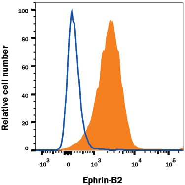
| Feng, Z;Gao, Y;Gao, H;Sun, S;Na, W;Huang, X;Li, S;Tan, C;Min, S;Lu, Y;Zhuang, Q;Lin, S;Ma, X;Liu, Y;Shou, W;Wang, M;Wang, J;Gu, Z;Sheng, W;Wu, F;Huang, G; Essential role of NONO-HOXA1-Wnt axis in cardiomyocyte differentiation Nature communications 2026-01-24 [PMID: 41580430] (Flow Cytometry, Human) |
Flow Cytometry |
Human |
| Le Ray, MA;Larralde, C;Legouez, L;Marret, S;Muller, JB;Gonzalez, BJ;Cleren, C; Short- and Long-Term Effects of Sodium Phenylbutyrate on White Matter and Sensorimotor and Cognitive Behavior in a Mild Murine Model of Encephalopathy of Prematurity International journal of molecular sciences 2025-12-16 [PMID: 41465524] (Immunohistochemistry, Mouse) |
Immunohistochemistry |
Mouse |
| Nolt, GL;Golden, LR;Thorpe, SP;Funnell, JL;Stephens, IO;Hernandez, G;MacLean, SM;Lucido, CC;Brock, CR;Pallerla, AV;Adreon, DR;Williams, HC;Morganti, JM;Johnson, LA; Microglia-derived APOE2 improves remyelination even in the presence of endogenous APOE4 Journal of neuroinflammation 2025-12-11 [PMID: 41382275] (Immunohistochemistry, Mouse) |
Immunohistochemistry |
Mouse |
| Bejar, MT;Jimenez-Gomez, P;Moutsopoulos, I;Ajith, H;Colom, B;McGinn, J;Han, S;Skrupskelyte, G;Calero-Nieto, FJ;Göttgens, B;Mohorianu, II;Simons, BD;Alcolea, MP; Lifting regenerative barriers promotes epithelial cell fate plasticity supporting lineage conversion Nature communications 2025-11-27 [PMID: 41309646] (Immunohistochemistry, Mouse) |
Immunohistochemistry |
Mouse |
| Wang, S;Niizuma, K;Liu, DD;Suchy, FP;Sato, H;Yanagida, A;Masaki, H;Miyauchi, M;Tabatabaee, S;Hidajat, N;Bhadury, J;Charlesworth, CT;Zhang, J;Weissman, IL;Nakauchi, H; Xenophagocytosis blockade enhances interspecies chimerism bioRxiv : the preprint server for biology 2025-10-15 [PMID: 41279269] (Immunohistochemistry, Xenograft) |
Immunohistochemistry |
Xenograft |
| Miao, L;Nusrat, A;Li, J;Yin, C;Lu, Y;Kuang, X;Sun, D;Lo, E;Song, R;Huang, W;Long, X;McConnell, BK;Singer, HA;Schwartz, RJ;Gunaratne, P;Fan, ZC;Kumar, A;Zhou, B;Munshi, N;Evans, SM;Wu, M; Distinct populations of vascular smooth muscle and sinoatrial progenitors are localized adjacent to pro-epicardium Cell reports 2025-11-18 [PMID: 41259201] (Immunohistochemistry, Transgenic Mouse) |
Immunohistochemistry |
Transgenic Mouse |
| Radomska, KJ;Onfroy, A;Lecerf, L;Job, B;Beaude, A;Sanz, LS;El Jalkh, T;Thieffry, D;Charnay, P;Wolkenstein, P;Ortonne, N;Coulpier, F;Topilko, P; Glial-to-mesenchymal transition of tumor Schwann cells drives the genetic burden in MPNSTs from neurofibromatosis type 1 mouse model Science advances 2025-11-14 [PMID: 41223283] (Immunohistochemistry, Transgenic Mouse) |
Immunohistochemistry |
Transgenic Mouse |
| Kuwata, Y;Yasuda, K;Tsukita, K;Kikuya, A;Takayama, N;Yanagida, N;Kimura, K;Takahashi, R;Matsumoto, R;Maki, T; Characterizing hypoxia-orchestrated post-stroke changes in oligodendrocyte precursor cells for optimized cell therapy Stem cell reports 2025-10-30 [PMID: 41173004] (Immunohistochemistry, Mouse) |
Immunohistochemistry |
Mouse |
| Radulovic, J;Zhang, H;Petrovic, Z;Wood, E;Cicvaric, A;Krispil-Alon, M;Jovasevic, V;Parker, K;Bassett, T;Carboncino, A;Guedea, A;Yi, P;Richter-Levin, G;Goncalves, J; Retrosplenial Cortical Reorganization During Late Adolescence Introduces Instability of Episodic Memory Circuits Research square 2025-09-10 [PMID: 40964004] (Immunohistochemistry, Transgenic Mouse) |
Immunohistochemistry |
Transgenic Mouse |
| Qin, Y;Choi, KY;El-Hamdi, N;McGregor, A; Selective knockout of key CMV receptors in fetal cells blocks direct and endocytic pathways of entry in the guinea pig bioRxiv : the preprint server for biology 2025-08-05 [PMID: 40799542] (Western Blot, Guinea Pig) |
Western Blot |
Guinea Pig |
| Wang, W;Lian, Y;Li, J;Wu, Y;Li, M;Wu, Y;Xu, R;Zhao, X;Kang, J;Guo, X; Zbtb16 determines the fate plasticity of cardiovascular progenitors through IGF2BP3-mediated mRNA stabilization Cell reports 2025-08-07 [PMID: 40779394] (Immunocytochemistry, Mouse) |
Immunocytochemistry |
Mouse |
| Mapkar, SA;Bliss, SA;Perez Carbajal, EE;Murray, SH;Li, Z;Wilson, AK;Piprode, V;Lee, YJ;Kirsch, T;Petroff, KS;Liu, F;Wosczyna, MN; Nuclear morphometrics coupled with machine learning identifies dynamic states of senescence across age Nature communications 2025-07-07 [PMID: 40624008] (Immunocytochemistry, Mouse) |
Immunocytochemistry |
Mouse |
| Okuno, N;Yamamoto, S;Hamashima, T;Dang, TS;Okita, N;Fujikawa, M;Kunisawa, T;Takakura, N;Fujimori, T;Mori, H;Betsholtz, C;Takata, K;Sasahara, M; Brain macrophages and pial fibroblasts promote inflammation in a hypomyelination model Acta neuropathologica communications 2025-07-04 [PMID: 40615895] (Immunohistochemistry, Transgenic Mouse) |
Immunohistochemistry |
Transgenic Mouse |
| Chen, Q;Yu, Y;Zhang, R;Zhao, Q;Yu, D;Feng, C;Zhou, J;Luo, M;Yang, M;Sun, S;Zhang, L;Jin, M; Commitment of adipose-resident c-kit+ progenitors to brown adipocytes contributes to adipose tissue homeostasis and remodeling Nature communications 2025-07-01 [PMID: 40593548] (Immunocytochemistry, Transgenic Mouse) |
Immunocytochemistry |
Transgenic Mouse |
| Ye, D;Feng, S;Yang, X;Su, Y;Zhang, J;Feng, H;Zhou, M;Zhou, B;Duan, L;Peng, T;Wang, C; Hedgehog-interacting protein orchestrates alveologenesis and protects against bronchopulmonary dysplasia and emphysema Science advances 2025-05-09 [PMID: 40333979] (Immunohistochemistry, Mouse) |
Immunohistochemistry |
Mouse |
| Abeysundara, N;Rasnitsyn, A;Fong, V;Bahcheli, A;Van Ommeren, R;Juraschka, K;Vladoiu, M;Ong, W;Livingston, B;de Antonellis, P;Ly, M;Holgado, BL;Sirbu, O;Bahrampour, S;Min, HK;Fan, J;Nor, C;Visvanathan, A;Zhang, J;Wang, H;Qin, L;Huang, N;Pallotta, J;Douglas, T;Mak, E;Su, H;Ng, K;Zhang, KY;Daniels, C;Lucas, CG;Eberhart, CG;Liu, H;Jiang, T;Notta, F;Ramaswamy, V;Reimand, J;Gallo, M;Rich, JN;Wu, X;Huang, X;Taylor, MD; Metastatic medulloblastoma remodels the local leptomeningeal microenvironment to promote further metastatic colonization and growth Nature cell biology 2025-04-22 [PMID: 40263572] (In vivo assay, Mouse) |
In vivo assay |
Mouse |
| Schiffer, M;Wagner, K;Carls, E;Nicke, J;Hesse, M;Fratila, RM;Hildebrand, S;Eberbeck, D;Mohr, T;Mohammadi, MM;de la Fuente, JM;Fleischmann, BK;Roell, W; Nanoparticle-assisted targeting of heart lesions with cardiac myofibroblasts: Combined gene and cell therapy Theranostics 2025-03-18 [PMID: 40225585] (Immunohistochemistry, Mouse) |
Immunohistochemistry |
Mouse |
| Xu, X;Zhu, P;Wang, H;Chen, K;Liu, L;Du, L;Jiang, L;Hu, Y;Zhou, X;Zhang, B;Pu, X;Hu, X;Xu, Q;Zhang, L;Li, W; CD34+ PI16+ fibroblast progenitors aggravate neointimal lesions of allograft arteries via CCL11/CCR3-PI3K/AKT pathway Theranostics 2025-01-20 [PMID: 39990233] (Immunohistochemistry, Mouse) |
Immunohistochemistry |
Mouse |
| Pellon-Cardenas, O;Rout, P;Hassan, S;Fokas, E;Ping, H;Patel, I;Patel, J;Plotsker, O;Wu, A;Kumar, R;Akther, M;Logerfo, A;Wu, S;Wagner, DE;Boffelli, D;Walton, KD;Manieri, E;Tong, K;Spence, JR;Bessman, NJ;Shivdasani, RA;Verzi, MP; Dynamic Reprogramming of Stromal Pdgfra-expressing cells during WNT-Mediated Transformation of the Intestinal Epithelium bioRxiv : the preprint server for biology 2025-01-25 [PMID: 39896606] (Immunohistochemistry, Mouse) |
Immunohistochemistry |
Mouse |
| Beiter, RM;Raghavan, TP;Suchocki, O;Ennerfelt, HE;Rivet-Noor, CR;Merchak, AR;Phillips, JL;Bathe, T;Lukens, JR;Prokop, S;Dupree, JL;Gaultier, A; Clusterin induced by OPC phagocytosis blocks IL-9 secretion to inhibit myelination in a model of Alzheimer's disease Heliyon 2025-01-15 [PMID: 39866464] (Immunohistochemistry, Mouse, Transgenic Mouse) |
Immunohistochemistry |
Mouse, Transgenic Mouse |
| Transient upregulation of procaspase-3 during oligodendrocyte fate decisions [PMID: 39837665] (Immunohistochemistry, Mouse) |
Immunohistochemistry |
Mouse |
| Willis, A;Jeong, D;Liu, Y;Lithopoulos, MA;Yuzwa, SA;Frankland, PW;Kaplan, DR;Miller, FD; Single cell approaches define neural stem cell niches and identify microglial ligands that can enhance precursor-mediated oligodendrogenesis Cell reports 2025-01-15 [PMID: 39823226] (Immunocytochemistry, Mouse) |
Immunocytochemistry |
Mouse |
| Lu, C;Zhu, H;Lu, H;Xie, X;Tong, L;Li, Y;Qian, Z; Colonic dysmotility regulated by downregulation of PDGFR?+ cells / SK3 channel in DSS-induced colitis mice PloS one 2024-12-17 [PMID: 39689119] (Immunohistochemistry, Mouse) |
Immunohistochemistry |
Mouse |
| Onji, M;Sigl, V;Lendl, T;Novatchkova, M;Ullate-Agote, A;Andersson-Rolf, A;Kozieradzki, I;Koglgruber, R;Pai, TP;Lichtscheidl, D;Nayak, K;Zilbauer, M;Carranza García, NA;Sievers, LK;Falk-Paulsen, M;Cronin, SJF;Hagelkruys, A;Sawa, S;Osborne, LC;Rosenstiel, P;Pasparakis, M;Ruland, J;Takayanagi, H;Clevers, H;Koo, BK;Penninger, JM; RANK drives structured intestinal epithelial expansion during pregnancy Nature 2024-12-04 [PMID: 39633049] (Immunohistochemistry, Mouse) |
Immunohistochemistry |
Mouse |
| Forman, TE;Sajek, MP;Larson, ED;Mukherjee, N;Fantauzzo, KA; PDGFR? signaling regulates Srsf3 transcript binding to affect PI3K signaling and endosomal trafficking eLife 2024-12-04 [PMID: 39630148] (Immunocytochemistry, Rat) |
Immunocytochemistry |
Rat |
| Horiuchi, M;Watanabe, S;Komine, O;Takahashi, E;Kaneko, K;Itohara, S;Shimada, M;Ogi, T;Yamanaka, K; ALS-linked mutant TDP-43 in oligodendrocytes induces oligodendrocyte damage and exacerbates motor dysfunction in mice Acta neuropathologica communications 2024-11-27 [PMID: 39605053] (Immunohistochemistry, Transgenic Mouse) |
Immunohistochemistry |
Transgenic Mouse |
| Wu, T;Chen, Z;Zhang, Z;Zhou, X;Gu, Y;Dinenno, FA;Chen, J; RBPMS and RBPMS2 Cooperate to Safeguard Cardiac Splicing bioRxiv : the preprint server for biology 2024-11-09 [PMID: 39574760] (Immunohistochemistry, Transgenic Mouse) |
Immunohistochemistry |
Transgenic Mouse |
| Faust, HJ;Cheng, TY;Korsunsky, I;Watts, GFM;Gal-Oz, ST;Trim, WV;Kongthong, S;Jonsson, AH;Simmons, DP;Zhang, F;Padera, R;Chubinskaya, S;Accelerating Medicines Partnership: RA/SLE Network, ;Wei, K;Raychaudhuri, S;Lynch, L;Moody, DB;Brenner, MB; Adipocyte associated glucocorticoid signaling regulates normal fibroblast function which is lost in inflammatory arthritis Nature communications 2024-11-14 [PMID: 39543086] (Immunohistochemistry, Human) |
Immunohistochemistry |
Human |
| Spurgin, S;Nguimtsop, AM;Chaudhry, FN;Michki, SN;Salvador, J;Iruela-Arispe, ML;Zepp, JA;Mukhopadhyay, S;Cleaver, O; Spatiotemporal dynamics of primary and motile cilia throughout lung development bioRxiv : the preprint server for biology 2024-10-26 [PMID: 39484464] (Immunohistochemistry, Transgenic Mouse) |
Immunohistochemistry |
Transgenic Mouse |
| Nicole D Ulrich, Alex Vargo, Qianyi Ma, Yu-Chi Shen, Dominic Bazzano, D Ford Hannum, Stephen J Gurczynski, Bethany B Moore, Samantha Schon, Richard Lieberman, Ariella Shikanov, Erica E Marsh, Asgerally Fazleabas, Jun Z Li, Saher Sue Hammoud Cellular heterogeneity and dynamics of the human uterus in healthy premenopausal women Proc Natl Acad Sci U S A 2024-11-05 [PMID: 39471215] (Immunohistochemistry, Human) |
Immunohistochemistry |
Human |
| Kyung, DS;Lee, E;Chae, S;Son, Y;Moon, YJ;Hwang, D;Kim, JK;Lee, YH;Seong, JK; Single-cell transcriptomic analysis reveals dynamic activation of cellular signaling pathways regulating beige adipogenesis Experimental & molecular medicine 2024-10-28 [PMID: 39465352] (Flow Cytometry, Mouse) |
Flow Cytometry |
Mouse |
| Duncan, GJ;Ingram, SD;Emberley, K;Hill, J;Cordano, C;Abdelhak, A;McCane, M;Jenks, JE;Jabassini, N;Ananth, K;Ferrara, SJ;Stedelin, B;Sivyer, B;Aicher, SA;Scanlan, TS;Watkins, TA;Mishra, A;Nelson, JW;Green, AJ;Emery, B; Remyelination protects neurons from DLK-mediated neurodegeneration. Nature communications 2024-10-23 [PMID: 39443516] (Immunohistochemistry, Mouse) |
Immunohistochemistry |
Mouse |
| Drexler, R;Drinnenberg, A;Gavish, A;Yalcin, B;Shamardani, K;Rogers, A;Mancusi, R;Taylor, KR;Kim, YS;Woo, PJ;Ravel, A;Tatlock, E;Ramakrishnan, C;Ayala-Sarmiento, AE;Pacheco, DRF;Siverts, L;Daigle, TL;Tasic, B;Zeng, H;Breunig, JJ;Deisseroth, K;Monje, M; Cholinergic Neuronal Activity Promotes Diffuse Midline Glioma Growth through Muscarinic Signaling bioRxiv : the preprint server for biology 2024-09-24 [PMID: 39386427] (Immunohistochemistry, Mouse) |
Immunohistochemistry |
Mouse |
| Yoshida, S;Yoshida, T;Inukai, K;Kato, K;Yura, Y;Hattori, T;Enomoto, A;Ohashi, K;Okumura, T;Ouchi, N;Kawase, H;Wettschureck, N;Offermanns, S;Murohara, T;Takefuji, M; Protein kinase N promotes cardiac fibrosis in heart failure by fibroblast-to-myofibroblast conversion Nature communications 2024-09-12 [PMID: 39266515] (Immunocytochemistry, Mouse, Transgenic Mouse) |
Immunocytochemistry |
Mouse, Transgenic Mouse |
| Fujikawa, Y;Kato, K;Unno, K;Narita, S;Okuno, Y;Sato, Y;Takefuji, M;Murohara, T; Dynamic upregulation of retinoic acid signal in the early postnatal murine heart promotes cardiomyocyte cell cycle exit and maturation Scientific reports 2024-08-30 [PMID: 39215116] (Immunohistochemistry, Mouse) |
Immunohistochemistry |
Mouse |
| Liu, Y;Yin, M;Mao, X;Wu, S;Wei, S;Heng, S;Yang, Y;Huang, J;Guo, Z;Li, C;Ji, C;Hu, L;Liu, W;Zhang, LJ; Defining cell type-specific immune responses in a mouse model of allergic contact dermatitis by single-cell transcriptomics eLife 2024-08-30 [PMID: 39213029] (Immunohistochemistry, Mouse) |
Immunohistochemistry |
Mouse |
| Jörg, LM;Schlötzer-Schrehardt, U;Lefebvre, V;Sock, E;Wegner, M; Transcription Factors Sox8 and Sox10 Contribute with Different Importance to the Maintenance of Mature Oligodendrocytes International journal of molecular sciences 2024-08-11 [PMID: 39201442] (Immunohistochemistry-Frozen, Mouse) |
Immunohistochemistry-Frozen |
Mouse |
| Ahuja, K;Vandenabeele, M;Nami, F;Lefevere, E;Van Hoecke, J;Bergmans, S;Claes, M;Vervliet, T;Neyrinck, K;Burg, T;De Herdt, D;Bhaskar, P;Zhu, Y;Looser, ZJ;Loncke, J;Gsell, W;Plaas, M;Agostinis, P;Swinnen, JV;Van Den Bosch, L;Bultynck, G;Saab, AS;Wolfs, E;Chai, YC;Himmelreich, U;Verfaillie, C;Moons, L;De Groef, L; A deep phenotyping study in mouse and iPSC models to understand the role of oligodendroglia in optic neuropathy in Wolfram syndrome Acta neuropathologica communications 2024-08-28 [PMID: 39198924] (Immunohistochemistry, Mouse) |
Immunohistochemistry |
Mouse |
| Bame, X;Hill, RA; Mitochondrial network reorganization and transient expansion during oligodendrocyte generation Nature communications 2024-08-14 [PMID: 39143079] (Immunohistochemistry, Mouse) |
Immunohistochemistry |
Mouse |
| Rodrigues-Amorim, D;Bozzelli, PL;Kim, T;Liu, L;Gibson, O;Yang, CY;Murdock, MH;Galiana-Melendez, F;Schatz, B;Davison, A;Islam, MR;Shin Park, D;Raju, RM;Abdurrob, F;Nelson, AJ;Min Ren, J;Yang, V;Stokes, MP;Tsai, LH; Multisensory gamma stimulation mitigates the effects of demyelination induced by cuprizone in male mice Nature communications 2024-08-08 [PMID: 39112447] (Immunohistochemistry, Mouse) |
Immunohistochemistry |
Mouse |
| Godoy, MI;Pandey, V;Wohlschlegel, JA;Zhang, Y; Secretome analysis of oligodendrocytes and precursors reveals their roles as contributors to the extracellular matrix and potential regulators of inflammation bioRxiv : the preprint server for biology 2024-07-23 [PMID: 39091874] (Immunocytochemistry, Rat) |
Immunocytochemistry |
Rat |
| Janeckova, L;Stastna, M;Hrckulak, D;Berkova, L;Kubovciak, J;Onhajzer, J;Kriz, V;Dostalikova, S;Mullerova, T;Vecerkova, K;Tenglerova, M;Coufal, S;Kostovcikova, K;Blumberg, RS;Filipp, D;Basler, K;Valenta, T;Kolar, M;Korinek, V; The fate of secretory cells during intestinal homeostasis, regeneration, and tumor formation is regulated by Tcf4 bioRxiv : the preprint server for biology 2024-07-11 [PMID: 39026783] (Immunohistochemistry, Mouse) |
Immunohistochemistry |
Mouse |
| Nevelchuk, S;Brawek, B;Schwarz, N;Valiente-Gabioud, A;Wuttke, TV;Kovalchuk, Y;Koch, H;Höllig, A;Steiner, F;Figarella, K;Griesbeck, O;Garaschuk, O; Morphotype-specific calcium signaling in human microglia Journal of neuroinflammation 2024-07-17 [PMID: 39020359] (Immunohistochemistry, Transgenic Mouse) |
Immunohistochemistry |
Transgenic Mouse |
| Widjaja, AA;Lim, WW;Viswanathan, S;Chothani, S;Corden, B;Dasan, CM;Goh, JWT;Lim, R;Singh, BK;Tan, J;Pua, CJ;Lim, SY;Adami, E;Schafer, S;George, BL;Sweeney, M;Xie, C;Tripathi, M;Sims, NA;Hübner, N;Petretto, E;Withers, DJ;Ho, L;Gil, J;Carling, D;Cook, SA; Inhibition of IL-11 signalling extends mammalian healthspan and lifespan Nature 2024-07-17 [PMID: 39020175] (Immunohistochemistry, Mouse, Transgenic Mouse) |
Immunohistochemistry |
Mouse, Transgenic Mouse |
| Peters, C;Aberle, T;Sock, E;Brunner, J;Küspert, M;Hillgärtner, S;Wüst, HM;Wegner, M; Voltage-Gated Ion Channels Are Transcriptional Targets of Sox10 during Oligodendrocyte Development Cells 2024-07-07 [PMID: 38995010] (Immunohistochemistry, Mouse, Transgenic Mouse) |
Immunohistochemistry |
Mouse, Transgenic Mouse |
| Beiter, RM;Raghavan, TP;Suchocki, O;Ennerfelt, HE;Rivet-Noor, CR;Merchak, AR;Phillips, JL;Bathe, T;Lukens, JR;Prokop, S;Dupree, JL;Gaultier, A; Oligomeric amyloid beta prevents myelination in a clusterin-dependent manner Research square 2024-05-30 [PMID: 38853911] (Flow Cytometry, Mouse) |
Flow Cytometry |
Mouse |
| Pan, Y;Hysinger, JD;Yalç?n, B;Lennon, JJ;Byun, YG;Raghavan, P;Schindler, NF;Anastasaki, C;Chatterjee, J;Ni, L;Xu, H;Malacon, K;Jahan, SM;Ivec, AE;Aghoghovwia, BE;Mount, CW;Nagaraja, S;Scheaffer, S;Attardi, LD;Gutmann, DH;Monje, M; Nf1 mutation disrupts activity-dependent oligodendroglial plasticity and motor learning in mice Nature neuroscience 2024-05-30 [PMID: 38816530] (Immunohistochemistry, Transgenic Mouse) |
Immunohistochemistry |
Transgenic Mouse |
| Arreguin, AJ;Shao, Z;Colognato, H; Dmd mdx mice have defective oligodendrogenesis, delayed myelin compaction and persistent hypomyelination Disease models & mechanisms 2024-04-01 [PMID: 38721692] (Immunohistochemistry, Mouse) |
Immunohistochemistry |
Mouse |
| Ishibashi, S;Kamei, N;Tsuchikawa, Y;Nakamae, T;Akimoto, T;Miyaki, S;Adachi, N; Myelin-Specific microRNA-23a/b Cluster Deletion Inhibits Myelination in the Central Nervous System during Postnatal Growth and Aging Genes 2024-03-25 [PMID: 38674338] (Immunohistochemistry, Transgenic Mouse) |
Immunohistochemistry |
Transgenic Mouse |
| Vishlaghi, N;Guo, L;Griswold-Wheeler, D;Sun, Y;Booker, C;Crossley, JL;Bancroft, AC;Juan, C;Korlakunta, S;Ramesh, S;Pagani, CA;Xu, L;James, AW;Tower, RJ;Dellinger, M;Levi, B; Vegfc-expressing cells form heterotopic bone after musculoskeletal injury Cell reports 2024-04-03 [PMID: 38573853] (Immunohistochemistry, Mouse, Transgenic Mouse) |
Immunohistochemistry |
Mouse, Transgenic Mouse |
| Halder, SK;Sapkota, A;Milner, R; ?1 integrins play a critical role maintaining vascular integrity in the hypoxic spinal cord, particularly in white matter Acta neuropathologica communications 2024-03-20 [PMID: 38509621] (Immunohistochemistry, Mouse) |
Immunohistochemistry |
Mouse |
| Lai, AY;Almanza, DLV;Ribeiro, JA;Hill, ME;Mandrozos, M;Koletar, MM;Stefanovic, B;McLaurin, J; Obesity Facilitates Sex-Specific Improvement In Cognition And Neuronal Function In A Rat Model Of Alzheimer's Disease bioRxiv : the preprint server for biology 2024-01-22 [PMID: 38328066] (Immunohistochemistry, Rat) |
Immunohistochemistry |
Rat |
| Shumliakivska, M;Luxán, G;Hemmerling, I;Scheller, M;Li, X;Müller-Tidow, C;Schuhmacher, B;Sun, Z;Dendorfer, A;Debes, A;Glaser, SF;Muhly-Reinholz, M;Kirschbaum, K;Hoffmann, J;Nagel, E;Puntmann, VO;Cremer, S;Leuschner, F;Abplanalp, WT;John, D;Zeiher, AM;Dimmeler, S; DNMT3A clonal hematopoiesis-driver mutations induce cardiac fibrosis by paracrine activation of fibroblasts Nature communications 2024-01-19 [PMID: 38242884] (ICC, Rat) |
ICC |
Rat |
| Zhang, D;Ruan, J;Peng, S;Li, J;Hu, X;Zhang, Y;Zhang, T;Ge, Y;Zhu, Z;Xiao, X;Zhu, Y;Li, X;Li, T;Zhou, L;Gao, Q;Zheng, G;Zhao, B;Li, X;Zhu, Y;Wu, J;Li, W;Zhao, J;Ge, WP;Xu, T;Jia, JM; Synaptic-like transmission between neural axons and arteriolar smooth muscle cells drives cerebral neurovascular coupling Nature neuroscience 2024-01-02 [PMID: 38168932] (Immunocytochemistry, Transgenic Mouse) |
Immunocytochemistry |
Transgenic Mouse |
| Nguyen, LT;Aprico, A;Nwoke, E;Walsh, AD;Blades, F;Avneri, R;Martin, E;Zalc, B;Kilpatrick, TJ;Binder, MD; Mertk-expressing microglia influence oligodendrogenesis and myelin modelling in the CNS Journal of neuroinflammation 2023-11-06 [PMID: 37926818] (IHC, Transgenic Mouse) |
IHC |
Transgenic Mouse |
| Tripathi, A;Rai, N;Perles, A;Jones, C;Dutta, R; Dicer deficiency in microglia leads to accelerated demyelination and failed remyelination bioRxiv : the preprint server for biology 2023-10-19 [PMID: 37905110] (IHC, Transgenic Mouse) |
IHC |
Transgenic Mouse |
| He, S;Guo, Z;Zhou, M;Wang, H;Zhang, Z;Shi, M;Li, X;Yang, X;He, L; Spatial-temporal proliferation of hepatocytes during pregnancy revealed by genetic lineage tracing Cell stem cell 2023-09-27 [PMID: 37794588] (IHC, Transgenic Mouse) |
IHC |
Transgenic Mouse |
| Mary Jo Talley, Diana Nardini, Lisa A. Ehrman, Q. Richard Lu, Ronald R. Waclaw Distinct requirements for Tcf3 and Tcf12 during oligodendrocyte development in the mouse telencephalon Neural Dev 2023-09-08 [PMID: 37684687] (Immunohistochemistry, Mouse, Transgenic Mouse) |
Immunohistochemistry |
Mouse, Transgenic Mouse |
| Mary Jo Talley, Diana Nardini, Lisa A. Ehrman, Q. Richard Lu, Ronald R. Waclaw Distinct requirements for Tcf3 and Tcf12 during oligodendrocyte development in the mouse telencephalon Neural Dev 9/8/2023 [PMID: 37684687] |
|
|
| Bowers SLK, Meng Q, Kuwabara Y et al. Col1a2-Deleted Mice Have Defective Type I Collagen and Secondary Reactive Cardiac Fibrosis with Altered Hypertrophic Dynamics Cells 8/30/2023 [PMID: 37681905] |
|
|
| Bowers SLK, Meng Q, Kuwabara Y et al. Col1a2-Deleted Mice Have Defective Type I Collagen and Secondary Reactive Cardiac Fibrosis with Altered Hypertrophic Dynamics Cells 2023-08-30 [PMID: 37681905] |
|
|
| Odell, ID;Agrawal, K;Sefik, E;Odell, AV;Caves, E;Kirkiles-Smith, NC;Horsley, V;Hinchcliff, M;Pober, JS;Kluger, Y;Flavell, RA; IL-6 trans-signaling in a humanized mouse model of scleroderma Proceedings of the National Academy of Sciences of the United States of America 2023-09-12 [PMID: 37669366] (IHC, Xenograft) |
IHC |
Xenograft |
| Jessica A. Kahng, Andre M. Xavier, Austin Ferro, Yohan S.S. Auguste, Lucas Cheadle Integrated high-confidence and high-throughput approaches for quantifying synapse engulfment by oligodendrocyte precursor cells bioRxiv 8/31/2023 [PMID: 37662250] |
|
|
| Whisler, J;Shahreza, S;Schlegelmilch, K;Ege, N;Javanmardi, Y;Malandrino, A;Agrawal, A;Fantin, A;Serwinski, B;Azizgolshani, H;Park, C;Shone, V;Demuren, OO;Del Rosario, A;Butty, VL;Holroyd, N;Domart, MC;Hooper, S;Szita, N;Boyer, LA;Walker-Samuel, S;Djordjevic, B;Sheridan, GK;Collinson, L;Calvo, F;Ruhrberg, C;Sahai, E;Kamm, R;Moeendarbary, E; Emergent mechanical control of vascular morphogenesis Science advances 2023-08-11 [PMID: 37566656] (Immunohistochemistry, Mouse) |
Immunohistochemistry |
Mouse |
| Tsutsumi, R;Ueberheide, B;Liang, FX;Neel, BG;Sakai, R;Saito, Y; Endocytic vesicles act as vehicles for glucose uptake in response to growth factor stimulation bioRxiv : the preprint server for biology 2023-07-24 [PMID: 37546742] (Immunocytochemistry, Mouse) |
Immunocytochemistry |
Mouse |
| Cabrera-Cabrera F, Tull H, Capuana R et al. Cell type-specific labeling of newly synthesized proteins by puromycin inactivation Journal of Biological Chemistry 8/4/2023 [PMID: 37543363] |
|
|
| Cabrera-Cabrera F, Tull H, Capuana R et al. Cell type-specific labeling of newly synthesized proteins by puromycin inactivation Journal of Biological Chemistry 2023-08-04 [PMID: 37543363] |
|
|
| Halder, SK;Milner, R; Spinal Cord Blood Vessels in Aged Mice Show Greater Levels of Hypoxia-Induced Vascular Disruption and Microglial Activation International journal of molecular sciences 2023-07-08 [PMID: 37510999] (Immunohistochemistry, Mouse) |
Immunohistochemistry |
Mouse |
| Bianca Rusu, Bharti Kukreja, Taiyi Wu, Sophie J. Dan, Min Yi Feng, Brian T. Kalish Single-Nucleus Profiling Identifies Accelerated Oligodendrocyte Precursor Cell Senescence in a Mouse Model of Down Syndrome eNeuro 7/25/2023 [PMID: 37491366] |
|
|
| Bianca Rusu, Bharti Kukreja, Taiyi Wu, Sophie J. Dan, Min Yi Feng, Brian T. Kalish Single-Nucleus Profiling Identifies Accelerated Oligodendrocyte Precursor Cell Senescence in a Mouse Model of Down Syndrome eNeuro 2023-07-25 [PMID: 37491366] (Immunohistochemistry, Mouse) |
Immunohistochemistry |
Mouse |
| Huycke, TR;Miyazaki, H;Häkkinen, TJ;Srivastava, V;Barruet, E;McGinnis, CS;Kalantari, A;Cornwall-Scoones, J;Vaka, D;Zhu, Q;Jo, H;DeGrado, WF;Thomson, M;Garikipati, K;Boffelli, D;Klein, OD;Gartner, ZJ; Patterning and folding of intestinal villi by active mesenchymal dewetting bioRxiv : the preprint server for biology 2023-06-26 [PMID: 37425793] (Bioassay, Xenograft) |
Bioassay |
Xenograft |
| Stec, MJ;Su, Q;Adler, C;Zhang, L;Golann, DR;Khan, NP;Panagis, L;Villalta, SA;Ni, M;Wei, Y;Walls, JR;Murphy, AJ;Yancopoulos, GD;Atwal, GS;Kleiner, S;Halasz, G;Sleeman, MW; A cellular and molecular spatial atlas of dystrophic muscle Proceedings of the National Academy of Sciences of the United States of America 2023-07-18 [PMID: 37410813] (IHC, Mouse) |
IHC |
Mouse |
| Yun-Yun Hu, Xuan-Si Ding, Gang Yang, Xue-Song Liang, Lei Feng, Yan-Yun Sun et al. Analysis of the influences of social isolation on cognition and the therapeutic potential of deep brain stimulation in a mouse model Frontiers in Psychiatry 6/20/2023 [PMID: 37409161] |
|
|
| Waldhauser, V;Baroti, T;Fröb, F;Wegner, M; PBAF Subunit Pbrm1 Selectively Influences the Transition from Progenitors to Pre-Myelinating Cells during Oligodendrocyte Development Cells 2023-06-06 [PMID: 37371026] (IHC, Mouse) |
IHC |
Mouse |
| Norris, AM;Appu, AB;Johnson, CD;Zhou, LY;McKellar, DW;Renault, MA;Hammers, D;Cosgrove, BD;Kopinke, D; Hedgehog signaling via its ligand DHH acts as cell fate determinant during skeletal muscle regeneration Nature communications 2023-06-24 [PMID: 37355632] (IHC, Mouse) |
IHC |
Mouse |
| Sun, L;Zhang, X;Wu, S;Liu, Y;Guerrero-Juarez, CF;Liu, W;Huang, J;Yao, Q;Yin, M;Li, J;Ramos, R;Liao, Y;Wu, R;Xia, T;Zhang, X;Yang, Y;Li, F;Heng, S;Zhang, W;Yang, M;Tzeng, CM;Ji, C;Plikus, MV;Gallo, RL;Zhang, LJ; Dynamic interplay between IL-1 and WNT pathways in regulating dermal adipocyte lineage cells during skin development and wound regeneration Cell reports 2023-06-16 [PMID: 37330908] |
|
|
Wu J, Jing X, Du Q et al. Disruption of the Clock Component Bmal1 in Mice Promotes Cancer Metastasis through the PAI-1-TGF-?-myoCAF-Dependent Mechanism Advanced science (Weinheim, Baden-Wurttemberg, Germany) 6/17/2023 [PMID: 37330661]
Details:
IHC-P: antibdy dilution: 1:200. Antigen retrieval: citrate-based solution pH 6.0 (H-3300, Vector) |
|
|
| Alber, AB;Marquez, HA;Ma, L;Kwong, G;Thapa, BR;Villacorta-Martin, C;Lindstrom-Vautrin, J;Bawa, P;Wang, F;Luo, Y;Ikonomou, L;Shi, W;Kotton, DN; Directed differentiation of mouse pluripotent stem cells into functional lung-specific mesenchyme Nature communications 2023-06-13 [PMID: 37311756] (Immunohistochemistry, Transgenic Mouse) |
Immunohistochemistry |
Transgenic Mouse |
| Kalgudde Gopal, S;Dai, R;Stefanska, AM;Ansari, M;Zhao, J;Ramesh, P;Bagnoli, JW;Correa-Gallegos, D;Lin, Y;Christ, S;Angelidis, I;Lupperger, V;Marr, C;Davies, LC;Enard, W;Machens, HG;Schiller, HB;Jiang, D;Rinkevich, Y; Wound infiltrating adipocytes are not myofibroblasts Nature communications 2023-05-25 [PMID: 37230982] (IHC, Mouse) |
IHC |
Mouse |
| Wang, K;Papadopoulos, N;Hamidi, A;Lennartsson, J;Heldin, CH; SUMOylation of PDGF receptor ? affects signaling via PLC? and STAT3, and cell proliferation BMC molecular and cell biology 2023-05-16 [PMID: 37193980] (Immunoprecipitation, Western Blot, Immunocytochemistry, Human, Porcine, Primate - Chlorocebus aethiops (African Green Monkey)) |
Immunoprecipitation, Western Blot, Immunocytochemistry |
Human, Porcine, Primate - Chlorocebus aethiops (African Green Monkey) |
| Lukas Menges, Jan Giesen, Kerem Yilmaz, Evanthia Mergia, Annette Füchtbauer, Ernst-Martin Füchtbauer et al. It takes two to tango: cardiac fibroblast-derived NO-induced cGMP enters cardiac myocytes and increases cAMP by inhibiting PDE3 Communications Biology 5/10/2023 [PMID: 37165086] |
|
|
| Chaen, T;Kurosawa, T;Kishi, K;Kaji, N;Ikemoto-Uezumi, M;Uezumi, A;Hori, M; Transcriptome analysis of mesenchymal stromal cells of the large and small intestinal smooth muscle layers reveals a unique gastrontestinal stromal signature Biochemistry and biophysics reports 2023-07-01 [PMID: 37153863] (IHC, Mouse) |
IHC |
Mouse |
| Wei, H;Wu, X;Withrow, J;Cuevas-Diaz Duran, R;Singh, S;Chaboub, LS;Rakshit, J;Mejia, J;Rolfe, A;Herrera, JJ;Horner, PJ;Wu, JQ; Glial progenitor heterogeneity and key regulators revealed by single-cell RNA sequencing provide insight to regeneration in spinal cord injury Cell reports 2023-05-06 [PMID: 37149868] (Immunohistochemistry, Transgenic Mouse) |
Immunohistochemistry |
Transgenic Mouse |
| Yuji Tsuchikawa, Naosuke Kamei, Yohei Sanada, Toshio Nakamae, Takahiro Harada, Kazunori Imaizumi et al. Deficiency of MicroRNA-23-27-24 Clusters Exhibits the Impairment of Myelination in the Central Nervous System Neural Plasticity 3/22/2023 [PMID: 37006814] |
|
|
| Chengcheng Fu, Britney Chin-Young, GaYoung Park, Mariana Guzmán-Seda, Damien Laudier, Woojin M. Han WNT7A suppresses adipogenesis of skeletal muscle mesenchymal stem cells and fatty infiltration through the alternative Wnt-Rho-YAP/TAZ signaling axis Stem Cell Reports 4/11/2023 [PMID: 37001514] |
|
|
| Yao Lulu Xing, Jasmine Poh, Bernard H.A. Chuang, Kaveh Moradi, Stanislaw Mitew, William D. Richardson et al. High-efficiency pharmacogenetic ablation of oligodendrocyte progenitor cells in the adult mouse CNS Cell Reports Methods 2/27/2023 [PMID: 36936074] |
|
|
| A Kolaj, SK Zahr, BS Wang, T Krawec, H Kazan, G Yang, DR Kaplan, FD Miller The P-body protein 4E-T represses translation to regulate the balance between cell genesis and establishment of the postnatal NSC pool Cell Reports, 2023-03-15;42(3):112242. 2023-03-15 [PMID: 36924490] (ICC, Mouse) |
ICC |
Mouse |
| Salome M. Brütsch, Elizabeta Madzharova, Sophia Pantasis, Till Wüstemann, Selina Gurri, Heiko Steenbock et al. Mesenchyme-derived vertebrate lonesome kinase controls lung organogenesis by altering the matrisome Cellular and Molecular Life Sciences 4/1/2023 [PMID: 36920550] |
|
|
| TT Lemmetyine, EW Viitala, L Wartiovaar, T Kaprio, J Hagström, C Haglund, P Katajisto, TC Wang, E Domènech-M, S Ollila Fibroblast-derived EGF ligand Neuregulin-1 induces fetal-like reprogramming of the intestinal epithelium without supporting tumorigenic growth Disease Models & Mechanisms, 2023-04-03;0(0):. 4/3/2023 [PMID: 36912192] |
|
|
| Q Liu, C Li, B Deng, P Gao, L Wang, Y Li, M Shiri, F Alkaifi, J Zhao, JM Stephens, CA Simintiras, J Francis, J Sun, X Fu Tcf21 marks visceral adipose mesenchymal progenitors and functions as a rate-limiting factor during visceral adipose tissue development Cell Reports, 2023-02-28;42(3):112166. 2/28/2023 [PMID: 36857185] |
|
|
| K Hoeft, GJL Schaefer, H Kim, D Schumacher, T Bleckwehl, Q Long, BM Klinkhamme, F Peisker, L Koch, J Nagai, M Halder, S Ziegler, E Liehn, C Kuppe, J Kranz, S Menzel, I Costa, A Wahida, P Boor, RK Schneider, S Hayat, R Kramann Platelet-instructed SPP1+ macrophages drive myofibroblast activation in fibrosis in a CXCL4-dependent manner Cell Reports, 2023-02-17;42(2):112131. 2023-02-17 [PMID: 36807143] (IHC, Mouse) |
IHC |
Mouse |
| ME Mohr, S Li, AM Trouten, RA Stairley, PL Roddy, C Liu, M Zhang, HM Sucov, G Tao Cardiomyocyte-fibroblast interaction regulates ferroptosis and fibrosis after myocardial injury bioRxiv : the preprint server for biology, 2023-02-08;0(0):. 2023-02-08 [PMID: 36798323] (IHC, Mouse) |
IHC |
Mouse |
| E Manieri, G Tie, D Seruggia, S Madha, A Maglieri, K Huang, Y Fujiwara, K Zhang, S Orkin, R He, N McCarthy, RA Shivdasani Structure, signals, and cellular elements of the mouse gastric mesenchymal niche bioRxiv : the preprint server for biology, 2023-02-24;0(0):. 2023-02-24 [PMID: 36798304] (IHC, Mouse) |
IHC |
Mouse |
| NL Jacobsen, AB Morton, SS Segal Angiogenesis precedes myogenesis during regeneration following biopsy injury of skeletal muscle Skeletal Muscle, 2023-02-14;13(1):3. 2023-02-14 [PMID: 36788624] (IHC, Mouse) |
IHC |
Mouse |
| L Pan, A Trimarco, AJ Zhang, K Fujimori, Y Urade, LO Sun, C Taveggia, Y Zhang Oligodendrocyte-lineage cell exocytosis and L-type prostaglandin D synthase promote oligodendrocyte development and myelination Elife, 2023-02-13;12(0):. 2/13/2023 [PMID: 36779701] |
|
|
| Z Li, K Chen, Q Shao, H Lu, X Zhang, Y Pu, X Sun, H He, L Cao Nanoparticulate MgH2 ameliorates anxiety/depression-like behaviors in a mouse model of multiple sclerosis by regulating microglial polarization and oxidative stress Journal of Neuroinflammation, 2023-01-30;20(1):16. 2023-01-30 [PMID: 36710351] (ICC, IHC, Mouse) |
ICC, IHC |
Mouse |
| L Legouez, B Le Dieu-Lu, S Feillet, G Riou, M Yeddou, T Plouchart, N Dourmap, MA Le Ray, S Marret, BJ Gonzalez, C Cleren Effects of MgSO4 Alone or Associated with 4-PBA on Behavior and White Matter Integrity in a Mouse Model of Cerebral Palsy: A Sex- and Time-Dependent Study International Journal of Molecular Sciences, 2022-12-15;23(24):. 2022-12-15 [PMID: 36555591] (IHC, Mouse) |
IHC |
Mouse |
| CA Pagani, AC Bancroft, RJ Tower, N Livingston, Y Sun, JY Hong, RN Kent, AL Strong, JH Nunez, JMR Medrano, N Patel, BA Nanes, KM Dean, Z Li, C Ge, BM Baker, AW James, SJ Weiss, RT Franceschi, B Levi Discoidin domain receptor 2 regulates aberrant mesenchymal lineage cell fate and matrix organization Science Advances, 2022-12-21;8(51):eabq6152. 2022-12-21 [PMID: 36542719] (Immunohistochemistry, Mouse) |
Immunohistochemistry |
Mouse |
| X Hong, Y Jian, S Ding, J Zhou, X Zheng, H Zhang, B Zhou, C Zhuang, J Wan, X Tong Kir4.1 channel activation in NG2 glia contributes to remyelination in ischemic stroke EBioMedicine, 2022-12-15;87(0):104406. 2022-12-15 [PMID: 36527899] (IHC, Mouse) |
IHC |
Mouse |
| Francesca Boscolo Sesillo, Varsha Rajesh, Michelle Wong, Pamela Duran, John B. Rudell, Courtney P. Rundio et al. Muscle stem cells and fibro-adipogenic progenitors in female pelvic floor muscle regeneration following birth injury npj Regenerative Medicine 12/16/2022 [PMID: 36526635] |
|
|
| Xiangdong Liu, Liang Zhao, Yao Gao, Yanting Chen, Qiyu Tian, Jun Seok Son et al. AMP‐activated protein kinase inhibition in fibro‐adipogenic progenitors impairs muscle regeneration and increases fibrosis Journal of Cachexia, Sarcopenia and Muscle 2/1/2023 [PMID: 36513394] |
|
|
| Gretchen A Meyer, Stavros Thomopoulos, Yousef Abu-Amer, Karen C Shen Tenotomy-induced muscle atrophy is sex-specific and independent of NF kappa B eLife 12/12/2022 [PMID: 36508247] |
|
|
| Zifeng Dai, Jingwei Li, Ying Li, Rui Wang, Huili Yan, Ziyu Xiong et al. Role of pericytes in the development of cerebral cavernous malformations iScience 12/22/2022 [PMID: 36465134] |
|
|
| P Cattaneo, MGB Hayes, N Baumgarten, D Hecker, S Peruzzo, GS Aslan, P Kunderfran, V Larcher, L Zhang, R Contu, G Fonseca, S Spinozzi, J Chen, G Condorelli, S Dimmeler, MH Schulz, S Heinz, N Guimarães-, SM Evans DOT1L regulates chamber-specific transcriptional networks during cardiogenesis and mediates postnatal cell cycle withdrawal Nature Communications, 2022-12-02;13(1):7444. 2022-12-02 [PMID: 36460641] (Immunohistochemistry, Mouse) |
Immunohistochemistry |
Mouse |
| K Ono, H Gotoh, T Nomura, T Morita, O Baba, M Matsumoto, S Saitoh, N Ohno Ultrastructural characteristics of oligodendrocyte precursor cells in the early postnatal mouse optic nerve observed by serial block-face scanning electron microscopy PLoS ONE, 2022-12-01;17(12):e0278118. 2022-12-01 [PMID: 36454994] (IHC, Mouse) |
IHC |
Mouse |
Yamakawa D, Tsuboi J, Kasahara K et al. Cilia-Mediated Insulin/Akt and ST2/JNK Signaling Pathways Regulate the Recovery of Muscle Injury Advanced science (Weinheim, Baden-Wurttemberg, Germany) 11/14/2022 [PMID: 36373718]
Details:
Dilution used in WB 1:1000 |
|
|
| C Bernhardt, E Sock, F Fröb, S Hillgärtne, M Nemer, M Wegner KLF9 and KLF13 transcription factors boost myelin gene expression in oligodendrocytes as partners of SOX10 and MYRF Nucleic Acids Research, 2022-11-11;0(0):. 2022-11-11 [PMID: 36318265] (IHC, Mouse) |
IHC |
Mouse |
| Jorge Alonso-Pérez, Ana Carrasco-Rozas, Maria Borrell-Pages, Esther Fernández-Simón, Patricia Piñol-Jurado, Lina Badimon et al. Nintedanib Reduces Muscle Fibrosis and Improves Muscle Function of the Alpha-Sarcoglycan-Deficient Mice Biomedicines 10/19/2022 [PMID: 36289891] |
|
|
| T Kaya, N Mattugini, L Liu, H Ji, L Cantuti-Ca, J Wu, M Schifferer, J Groh, R Martini, S Besson-Gir, S Kaji, A Liesz, O Gokce, M Simons CD8+ T cells induce interferon-responsive oligodendrocytes and microglia in white matter aging Nature Neuroscience, 2022-10-24;25(11):1446-1457. 2022-10-24 [PMID: 36280798] (IHC, Mouse) |
IHC |
Mouse |
| David W. Hammers NOX4 inhibition promotes the remodeling of dystrophic muscle JCI Insight 10/24/2022 [PMID: 36278481] |
|
|
| Hannah Ennerfelt, Elizabeth L. Frost, Daniel A. Shapiro, Coco Holliday, Kristine E. Zengeler, Gabrielle Voithofer et al. SYK coordinates neuroprotective microglial responses in neurodegenerative disease Cell 10/27/2022 [PMID: 36257314] |
|
|
| Madeline Nicholson, Rhiannon J. Wood, David G. Gonsalvez, Anthony J. Hannan, Jessica L. Fletcher, Junhua Xiao et al. Remodelling of myelinated axons and oligodendrocyte differentiation is stimulated by environmental enrichment in the young adult brain European Journal of Neuroscience 12/1/2022 [PMID: 36217300] |
|
|
| O'Shea TM, Ao Y, Wang S et al. Lesion environments direct transplanted neural progenitors towards a wound repair astroglial phenotype in mice Nature Communications 2022-09-28 [PMID: 36171203] |
|
|
| O'Shea TM, Ao Y, Wang S et al. Lesion environments direct transplanted neural progenitors towards a wound repair astroglial phenotype in mice Nature Communications 9/28/2022 [PMID: 36171203] |
|
|
| M Sobecki, J Chen, E Krzywinska, S Nagarajan, Z Fan, E Nelius, JM Monné Rodr, F Seehusen, A Hussein, G Moschini, EY Hajam, R Kiran, D Gotthardt, J Debbache, C Badoual, T Sato, T Isagawa, N Takeda, C Tanchot, E Tartour, A Weber, S Werner, J Loffing, L Sommer, V Sexl, C Münz, C Feghali-Bo, E Pachera, O Distler, J Snedeker, C Jamora, C Stockmann Vaccination-based immunotherapy to target profibrotic cells in liver and lung Cell Stem Cell, 2022-09-15;0(0):. 2022-09-15 [PMID: 36113462] (Flow Cytometry, IHC, Mouse) |
Flow Cytometry, IHC |
Mouse |
| Kasra Khalaj, Rebeca Lopes Figueira, Lina Antounians, Sree Gandhi, Matthew Wales, Louise Montalva et al. Treatment with Amniotic Fluid Stem Cell Extracellular Vesicles Promotes Fetal Lung Branching and Cell Differentiation at Canalicular and Saccular Stages in Experimental Pulmonary Hypoplasia Secondary to Congenital Diaphragmatic Hernia Stem Cells Translational Medicine 10/21/2022 [PMID: 36103370] |
|
|
| Alessia Caramello, Christophe Galichet, Miriam Llorian Sopena, Robin Lovell‐Badge, Karine Rizzoti The cortical hem lacks stem cell potential despite expressing SOX9 and HOPX Developmental Neurobiology 10/1/2022 [PMID: 36067402] |
|
|
Li Y, Li C, Liu Q et al. Loss of Acta2 in cardiac fibroblasts does not prevent the myofibroblast differentiation or affect the cardiac repair after myocardial infarction Journal of molecular and cellular cardiology 8/22/2022 [PMID: 36007455]
Details:
IHC-Fr dilution 1:100 |
|
|
| Iris K. Lee, Estela Noguera-Ortega, Zebin Xiao, Leslie Todd, John Scholler, Decheng Song et al. Monitoring Therapeutic Response to Anti-Fibroblast Activation Protein (FAP) CAR T Cells using [18F]AlF-FAPI-74 Clinical Cancer Research 2022-12-15 [PMID: 35972732] (Immunohistochemistry, Mouse) |
Immunohistochemistry |
Mouse |
| Iris K. Lee, Estela Noguera-Ortega, Zebin Xiao, Leslie Todd, John Scholler, Decheng Song et al. Monitoring Therapeutic Response to Anti-Fibroblast Activation Protein (FAP) CAR T Cells using [18F]AlF-FAPI-74 Clinical Cancer Research 12/15/2022 [PMID: 35972732] |
|
|
| Palikuqi B, Rispal J, Reyes EA et al. Lymphangiocrine signals are required for proper intestinal repair after cytotoxic injury Cell stem cell 8/4/2022 [PMID: 35931034] |
|
|
| RM Beiter, C Rivet-Noor, AR Merchak, R Bai, DM Johanson, E Slogar, K Sol-Church, CC Overall, A Gaultier Evidence for oligodendrocyte progenitor cell heterogeneity in the adult mouse brain Scientific Reports, 2022-07-28;12(1):12921. 2022-07-28 [PMID: 35902669] (IHC, Mouse) |
IHC |
Mouse |
| Jongryul Hong, Yeonji Jeong, Won Do Heo The Neurotrophic Receptor Tyrosine Kinase in MEC-mPFC Neurons Contributes to Remote Memory Consolidation The Journal of Neuroscience 8/24/2022 [PMID: 35863892] |
|
|
| Jongryul Hong, Yeonji Jeong, Won Do Heo The Neurotrophic Receptor Tyrosine Kinase in MEC-mPFC Neurons Contributes to Remote Memory Consolidation The Journal of Neuroscience 2022-08-24 [PMID: 35863892] (Immunohistochemistry, Mouse) |
Immunohistochemistry |
Mouse |
Hamed AA, Kunz DJ, El-Hamamy I et al. A brain precursor atlas reveals the acquisition of developmental-like states in adult cerebral tumours Nature communications 7/19/2022 [PMID: 35853870]
Details:
Sup. Fig 11 |
|
|
| Rayanne B Burl, Elizabeth Ann Rondini, Hongguang Wei, Roger Pique-Regi, James G Granneman Deconstructing cold-induced brown adipocyte neogenesis in mice eLife 2022-07-18 [PMID: 35848799] (Immunohistochemistry, Transgenic Mouse) |
Immunohistochemistry |
Transgenic Mouse |
| Rayanne B Burl, Elizabeth Ann Rondini, Hongguang Wei, Roger Pique-Regi, James G Granneman Deconstructing cold-induced brown adipocyte neogenesis in mice eLife 7/18/2022 [PMID: 35848799] |
|
|
| Yingzi Liu, Christian F. Guerrero-Juarez, Fei Xiao, Nitish Udupi Shettigar, Raul Ramos, Chen-Hsiang Kuan et al. Hedgehog signaling reprograms hair follicle niche fibroblasts to a hyper-activated state Developmental Cell 7/1/2022 [PMID: 35777353] |
|
|
| Yingzi Liu, Christian F. Guerrero-Juarez, Fei Xiao, Nitish Udupi Shettigar, Raul Ramos, Chen-Hsiang Kuan et al. Hedgehog signaling reprograms hair follicle niche fibroblasts to a hyper-activated state Developmental Cell 2022-07-01 [PMID: 35777353] (Immunohistochemistry, Mouse) |
Immunohistochemistry |
Mouse |
| Anthony Fernández-Castañeda, Peiwen Lu, Anna C. Geraghty, Eric Song, Myoung-Hwa Lee, Jamie Wood et al. Mild respiratory COVID can cause multi-lineage neural cell and myelin dysregulation Cell 7/1/2022 [PMID: 35768006] |
|
|
| Cheuk Wang Fung, Shaopu Zhou, Han Zhu, Xiuqing Wei, Zhenguo Wu, Angela Ruohao Wu Cell fate determining molecular switches and signaling pathways in Pax7-expressing somitic mesoderm Cell Discovery 6/28/2022 [PMID: 35764624] |
|
|
| Florent Marguet, Mélanie Brosolo, Gaëlle Friocourt, Fanny Sauvestre, Pascale Marcorelles, Céline Lesueur et al. Oligodendrocyte lineage is severely affected in human alcohol-exposed foetuses Acta Neuropathologica Communications 12/1/2022 [PMID: 35568959] |
|
|
| SE Ha, B Jin, BG Jorgensen, H Zogg, L Wei, R Singh, C Park, M Kurahashi, S Kim, G Baek, SM Poudrier, MY Lee, KM Sanders, S Ro Transcriptome profiling of subepithelial PDGFRalpha cells in colonic mucosa reveals several cell-selective markers PLoS ONE, 2022-05-13;17(5):e0261743. 2022-05-13 [PMID: 35560163] (IHC, Mouse) |
IHC |
Mouse |
| JM Jacob, SE Di Carlo, I Stzepourgi, A Lepelletie, PD Ndiaye, H Varet, R Legendre, E Kornobis, A Benabid, G Nigro, L Peduto PDGFRalpha-induced stromal maturation is required to restrain postnatal intestinal epithelial stemness and promote defense mechanisms Cell Stem Cell, 2022-05-05;29(5):856-868.e5. 2022-05-05 [PMID: 35523143] (Flow Cytometry, Mouse) |
Flow Cytometry |
Mouse |
| S Ghorbani, E Jelinek, R Jain, B Buehner, C Li, BM Lozinski, S Sarkar, DK Kaushik, Y Dong, TN Wight, S Karimi-Abd, GJ Schenk, EM Strijbis, J Geurts, P Zhang, CC Ling, VW Yong Versican promotes T helper 17 cytotoxic inflammation and impedes oligodendrocyte precursor cell remyelination Nature Communications, 2022-05-04;13(1):2445. 2022-05-04 [PMID: 35508608] (IHC, Mouse) |
IHC |
Mouse |
| JK Knowles, H Xu, C Soane, A Batra, T Saucedo, E Frost, LT Tam, D Fraga, L Ni, K Villar, S Talmi, JR Huguenard, M Monje Maladaptive myelination promotes generalized epilepsy progression Nature Neuroscience, 2022-05-02;25(5):596-606. 2022-05-02 [PMID: 35501379] (IHC, Rat) |
IHC |
Rat |
| Y Zhang, E Manouchehr, M Herre, J Cedervall, Q Qiao, Z Miao, A Hamidi, L Hellman, M Kamali-Mog, AK Olsson Platelet-Derived PDGFB Promotes Recruitment of Cancer-Associated Fibroblasts, Deposition of Extracellular Matrix and Tgfbeta Signaling in the Tumor Microenvironment Cancers, 2022-04-12;14(8):. 2022-04-12 [PMID: 35454853] (Mouse) |
|
Mouse |
| T Potluri, MJ Taylor, JJ Stulberg, RL Lieber, H Zhao, SE Bulun An estrogen-sensitive fibroblast population drives abdominal muscle fibrosis in an inguinal hernia mouse model JCI Insight, 2022-04-19;7(9):. 2022-04-19 [PMID: 35439171] (ICC, Mouse) |
ICC |
Mouse |
| Xiao-Ru Ma, Shu-Ying Yang, Shuang-Shuang Zheng, Huan-Huan Yan, Hui-Min Gu, Fan Wang et al. Inhibition of RIPK1 by ZJU-37 promotes oligodendrocyte progenitor proliferation and remyelination via NF-kappa B pathway Cell Death Discovery 4/1/2022 [PMID: 35365618] |
|
|
| Saclier M, Angelini G, Bonfanti C et al. Selective ablation of Nfix in macrophages attenuates muscular dystrophy by inhibiting fibro-adipogenic progenitor-dependent fibrosis The Journal of pathology 3/17/2022 [PMID: 35297529] |
|
|
| Li-Pao Fang, Na Zhao, Laura C Caudal, Hsin-Fang Chang, Renping Zhao, Ching-Hsin Lin, Nadine Hainz, Carola Meier, Bernhard Bettler, Wenhui Huang, Anja Scheller, Frank Kirchhoff, Xianshu Bai Impaired bidirectional communication between interneurons and oligodendrocyte precursor cells affects social cognitive behavior Nat Commun 2022-03-16 [PMID: 35296664] (Immunohistochemistry, Mouse) |
Immunohistochemistry |
Mouse |
| S Pantasis, J Friemel, SM Brütsch, Z Hu, S Krautbauer, G Liebisch, J Dengjel, A Weber, S Werner, MR Bordoli Vertebrate lonesome kinase modulates the hepatocyte secretome to prevent perivascular liver fibrosis and inflammation Journal of Cell Science, 2022-04-12;0(0):. 2022-04-12 [PMID: 35293576] (IHC, Mouse) |
IHC |
Mouse |
| XR Ma, X Zhu, Y Xiao, HM Gu, SS Zheng, L Li, F Wang, ZJ Dong, DX Wang, Y Wu, C Yang, W Jiang, K Yao, Y Yin, Y Zhang, C Peng, L Gao, Z Meng, Z Hu, C Liu, L Li, HZ Chen, Y Shu, Z Ju, JW Zhao Restoring nuclear entry of Sirtuin 2 in oligodendrocyte progenitor cells promotes remyelination during ageing Nature Communications, 2022-03-09;13(1):1225. 2022-03-09 [PMID: 35264567] (IF, Mouse) |
IF |
Mouse |
| Jason T Long, Abigail Leinroth, Yihan Liao, Yinshi Ren, Anthony J Mirando, Tuyet Nguyen et al. Hypertrophic chondrocytes serve as a reservoir for marrow-associated skeletal stem and progenitor cells, osteoblasts, and adipocytes during skeletal development eLife 2/18/2022 [PMID: 35179487] |
|
|
| D Bugg, LRJ Bailey, RC Bretherton, KE Beach, IM Reichardt, KZ Robeson, AC Reese, J Gunaje, G Flint, CA DeForest, A Stempien-O, J Davis MBNL1 drives dynamic transitions between fibroblasts and myofibroblasts in cardiac wound healing Cell Stem Cell, 2022-02-16;29(3):419-433.e10. 2/16/2022 [PMID: 35176223] |
|
|
| Fiona Eitner, Beatrice Richter, Saskia Schwänen, Malgorzata Szaroszyk, Isabel Vogt, Andrea Grund et al. Comprehensive Expression Analysis of Cardiac Fibroblast Growth Factor 23 in Health and Pressure-induced Cardiac Hypertrophy Frontiers in Cell and Developmental Biology 1/18/2022 [PMID: 35118076] |
|
|
| Marta Celorrio, Kirill Shumilov, Camryn Payne, Sangeetha Vadivelu, Stuart H. Friess Acute minocycline administration reduces brain injury and improves long-term functional outcomes after delayed hypoxemia following traumatic brain injury Acta Neuropathologica Communications 12/1/2022 [PMID: 35090569] |
|
|
| Adwiteeya Misra, Cameron D. Baker, Elizabeth M. Pritchett, Kimberly N. Burgos Villar, John M. Ashton, Eric M. Small Characterizing Neonatal Heart Maturation, Regeneration, and Scar Resolution Using Spatial Transcriptomics Journal of Cardiovascular Development and Disease 12/21/2021 [PMID: 35050211] |
|
|
| A Fernández-, P Lu, AC Geraghty, E Song, MH Lee, J Wood, B Yalç?n, KR Taylor, S Dutton, L Acosta-Alv, L Ni, D Contreras-, JR Gehlhausen, J Klein, C Lucas, T Mao, J Silva, MA Peña-Herná, A Tabachniko, T Takahashi, L Tabacof, J Tosto-Manc, E Breyman, A Kontorovic, D McCarthy, M Quezado, M Hefti, D Perl, R Folkerth, D Putrino, A Nath, A Iwasaki, M Monje Mild respiratory SARS-CoV-2 infection can cause multi-lineage cellular dysregulation and myelin loss in the brain bioRxiv : the preprint server for biology, 2022-01-10;0(0):. 2022-01-10 [PMID: 35043113] (IHC, Mouse) |
IHC |
Mouse |
| O Méndez-Sol, M Bendjennat, J Naipauer, PR Theodoridi, JJD Ho, RE Verdun, JM Hare, E Cesarman, S Lee, EA Mesri Kaposi's sarcoma herpesvirus activates the hypoxia response to usurp HIF2alpha-dependent translation initiation for replication and oncogenesis Cell Reports, 2021-12-28;37(13):110144. 12/28/2021 [PMID: 34965440] |
|
|
| Jing Tang, Xin Liang, Xiaoyun Dou, Yingqiang Qi, Chunmao Yang, Yanmin Luo et al. Exercise rather than fluoxetine promotes oligodendrocyte differentiation and myelination in the hippocampus in a male mouse model of depression Translational Psychiatry 12/8/2021 [PMID: 34880203] |
|
|
| V Reyes Nico, JM Allaire, AB Alfonso, D Pupo Gómez, V Pomerleau, V Giroux, F Boudreau, N Perreault Altered Mucus Barrier Integrity and Increased Susceptibility to Colitis in Mice upon Loss of Telocyte Bone Morphogenetic Protein Signalling Cells, 2021-10-29;10(11):. 2021-10-29 [PMID: 34831177] (IHC, Mouse) |
IHC |
Mouse |
| YJ Li, JP Ao, X Huang, HL Lu, HY Fu, NN Song, WX Xu, J Chen Involvement of PAR2 in platelet-derived growth factor receptor-alpha-positive cell proliferation in the colon of diabetic mice Physiological Reports, 2021-11-01;9(21):e15099. 2021-11-01 [PMID: 34755491] (IHC, Mouse) |
IHC |
Mouse |
| Yiran Liu, David Castano, Francesco Girolamo, Laia Trigueros-Motos, Han-Gyu Bae, Suat Peng Neo et al. Loss of ABCA8B decreases myelination by reducing oligodendrocyte precursor cells in mice Journal of Lipid Research 1/1/2022 [PMID: 34752805] |
|
|
| Adam A A Nabeebaccus, Sharwari Verma, Anna Zoccarato, Giulia Emanuelli, Celio XC. Santos, Katrin Streckfuss-Bömeke et al. Cardiomyocyte protein O-GlcNAcylation is regulated by GFAT1 not GFAT2 Biochemical and Biophysical Research Communications 12/17/2021 [PMID: 34735873] |
|
|
| F Progatzky, M Shapiro, SH Chng, B Garcia-Cas, CH Classon, S Sevgi, A Laddach, AC Bon-Frauch, R Lasrado, M Rahim, EM Amaniti, S Boeing, K Shah, LJ Entwistle, A Suárez-Bon, MS Wilson, B Stockinger, V Pachnis Regulation of intestinal immunity and tissue repair by enteric glia Nature, 2021-10-20;599(7883):125-130. 2021-10-20 [PMID: 34671159] (IHC, Mouse) |
IHC |
Mouse |
| N Cohen, H Massalha, S Ben-Moshe, A Egozi, M Rozenberg, K Bahar Halp, S Itzkovitz Spatial gene expression maps of the intestinal lymphoid follicle and associated epithelium identify zonated expression programs PloS Biology, 2021-10-11;19(10):e3001214. 2021-10-11 [PMID: 34634036] (IF, Mouse) |
IF |
Mouse |
| Y Wang, Y Zhang, S Zhang, B Kim, VL Hull, J Xu, P Prabhu, M Gregory, V Martinez-C, X Zhan, W Deng, F Guo PARP1-mediated PARylation activity is essential for oligodendroglial differentiation and CNS myelination Cell Reports, 2021-10-05;37(1):109695. 2021-10-05 [PMID: 34610310] (IHC, Mouse) |
IHC |
Mouse |
| X Zhang, Y Liu, X Hong, X Li, CK Meshul, C Moore, Y Yang, Y Han, WG Li, X Qi, H Lou, S Duan, TL Xu, X Tong NG2 glia-derived GABA release tunes inhibitory synapses and contributes to stress-induced anxiety Nature Communications, 2021-09-30;12(1):5740. 2021-09-30 [PMID: 34593806] (IHC, Mouse) |
IHC |
Mouse |
| H Shin, HD Kawai Sensitive timing of undifferentiation in oligodendrocyte progenitor cells and their enhanced maturation in primary visual cortex of binocularly enucleated mice PLoS ONE, 2021-09-17;16(9):e0257395. 2021-09-17 [PMID: 34534256] (IHC, Mouse) |
IHC |
Mouse |
| Y Takanezawa, S Tanabe, D Kato, R Ozeki, M Komoda, T Suzuki, H Baba, R Muramatsu Microglial ASD-related genes are involved in oligodendrocyte differentiation Scientific Reports, 2021-09-08;11(1):17825. 2021-09-08 [PMID: 34497307] (ICC, IHC, Mouse) |
ICC, IHC |
Mouse |
| K Lee, SO Park, PC Choi, SB Ryoo, H Lee, LE Peri, T Zhou, RD Corrigan, AC Yanez, SB Moon, BA Perrino, KM Sanders, SD Koh Molecular and functional characterization of detrusor PDGFRalpha positive cells in spinal cord injury-induced detrusor overactivity Scientific Reports, 2021-08-11;11(1):16268. 2021-08-11 [PMID: 34381120] (IHC, Mouse) |
IHC |
Mouse |
| L Cordero-Es, AM Dowbaj, TN Kohler, B Strauss, O Sarlidou, G Belenguer, C Pacini, NP Martins, R Dobie, JR Wilson-Kan, R Butler, N Prior, P Serup, F Jug, NC Henderson, F Hollfelder, M Huch Dynamic cell contacts between periportal mesenchyme and ductal epithelium act as a rheostat for liver cell proliferation Cell Stem Cell, 2021-08-02;0(0):. 2021-08-02 [PMID: 34343491] (IHC, Mouse) |
IHC |
Mouse |
| Hyeryun Shin, Hideki Derek Kawai Visual deprivation induces transient upregulation of oligodendrocyte progenitor cells in the subcortical white matter of mouse visual cortex IBRO Neuroscience Reports 12/1/2021 [PMID: 34286312] |
|
|
| I De, V Maklakova, S Litscher, MM Boyd, LC Klemm, Z Wang, C Kendziorsk, LS Collier Microglial responses to CSF1 overexpression do not promote the expansion of other glial lineages Journal of Neuroinflammation, 2021-07-19;18(1):162. 2021-07-19 [PMID: 34281564] (IHC, Mouse) |
IHC |
Mouse |
| KK Sivaraj, HW Jeong, B Dharmaling, D Zeuschner, S Adams, M Potente, RH Adams Regional specialization and fate specification of bone stromal cells in skeletal development Cell Reports, 2021-07-13;36(2):109352. 2021-07-13 [PMID: 34260921] (IHC, Mouse) |
IHC |
Mouse |
| John T. Walker, Lauren E. Flynn, Douglas W. Hamilton Lineage tracing of Foxd1‐expressing embryonic progenitors to assess the role of divergent embryonic lineages on adult dermal fibroblast function FASEB BioAdvances 3/30/2021 [PMID: 34258523] |
|
|
| Mary Jo Talley, Diana Nardini, Nisha Shabbir, Lisa A. Ehrman, Carlos E. Prada, Ronald R. Waclaw Generation of a Mouse Model to Study the Noonan Syndrome Gene Lztr1 in the Telencephalon Frontiers in Cell and Developmental Biology 2021-06-16 [PMID: 34222248] (Immunohistochemistry, Mouse) |
Immunohistochemistry |
Mouse |
| Mary Jo Talley, Diana Nardini, Nisha Shabbir, Lisa A. Ehrman, Carlos E. Prada, Ronald R. Waclaw Generation of a Mouse Model to Study the Noonan Syndrome Gene Lztr1 in the Telencephalon Frontiers in Cell and Developmental Biology 6/16/2021 [PMID: 34222248] |
|
|
| Venkata Thulabandu, Timothy Nehila, James W. Ferguson, Radhika P. Atit Dermal EZH2 orchestrates dermal differentiation and epidermal proliferation during murine skin development Developmental Biology 10/1/2021 [PMID: 34166654] |
|
|
| SC Schüler, JM Kirkpatric, M Schmidt, D Santinha, P Koch, S Di Sanzo, E Cirri, M Hemberg, A Ori, J von Maltza Extensive remodeling of the extracellular matrix during aging contributes to age-dependent impairments of muscle stem cell functionality Cell Reports, 2021-06-08;35(10):109223. 2021-06-08 [PMID: 34107247] (IHC, Mouse) |
IHC |
Mouse |
| Toshiro Hara, Rony Chanoch-Myers, Nathan D. Mathewson, Chad Myskiw, Lyla Atta, Lillian Bussema et al. Interactions between cancer cells and immune cells drive transitions to mesenchymal-like states in glioblastoma Cancer Cell 6/3/2021 [PMID: 34087162] |
|
|
| A Waisman, AM Norris, M Elías Cost, D Kopinke Automatic and unbiased segmentation and quantification of myofibers in skeletal muscle Scientific Reports, 2021-06-03;11(1):11793. 2021-06-03 [PMID: 34083673] (IHC, Mouse) |
IHC |
Mouse |
| F Pfeiffer, A Sherafat, A Nishiyama The Impact of Fixation on the Detection of Oligodendrocyte Precursor Cell Morphology and Vascular Associations Cells, 2021-05-24;10(6):. 2021-05-24 [PMID: 34073801] (IHC, Mouse) |
IHC |
Mouse |
| Y Pan, JD Hysinger, T Barron, NF Schindler, O Cobb, X Guo, B Yalç?n, C Anastasaki, SB Mulinyawe, A Ponnuswami, S Scheaffer, Y Ma, KC Chang, X Xia, JA Toonen, JJ Lennon, EM Gibson, JR Huguenard, LM Liau, JL Goldberg, M Monje, DH Gutmann NF1 mutation drives neuronal�activity-dependent initiation of optic glioma Nature, 2021-05-26;594(7862):277-282. 2021-05-26 [PMID: 34040258] (ICC, IHC, Mouse) |
ICC, IHC |
Mouse |
| O Contreras, A Córdova-Ca, E Brandan PDGF-PDGFR network differentially regulates the fate, migration, proliferation, and cell cycle progression of myogenic cells Cellular Signalling, 2021-05-08;84(0):110036. 2021-05-08 [PMID: 33971280] (ICC/IF, Western Blot, Mouse) |
ICC/IF, Western Blot |
Mouse |
| Akitoshi Hara, Katsuhiro Kato, Toshikazu Ishihara, Hiroki Kobayashi, Naoya Asai, Shinji Mii et al. Meflin defines mesenchymal stem cells and/or their early progenitors with multilineage differentiation capacity Genes to Cells 2021-07-01 [PMID: 33960573] (Immunohistochemistry, Transgenic Mouse) |
Immunohistochemistry |
Transgenic Mouse |
| Akitoshi Hara, Katsuhiro Kato, Toshikazu Ishihara, Hiroki Kobayashi, Naoya Asai, Shinji Mii et al. Meflin defines mesenchymal stem cells and/or their early progenitors with multilineage differentiation capacity Genes to Cells 7/1/2021 [PMID: 33960573] |
|
|
| MN Wosczyna, EE Perez Carb, MW Wagner, S Paredes, CT Konishi, L Liu, TT Wang, RA Walsh, Q Gan, CS Morrissey, TA Rando Targeting microRNA-mediated gene repression limits adipogenic conversion of skeletal muscle mesenchymal stromal cells Cell Stem Cell, 2021-05-03;0(0):. 2021-05-03 [PMID: 33945794] (Flow Cytometry, IHC, Mouse) |
Flow Cytometry, IHC |
Mouse |
| K Kimura, K Ramirez, TAV Nguyen, Y Yamashiro, A Sada, H Yanagisawa Contribution of PDGFR&alpha-positive cells in maintenance and injury responses in mouse large vessels Scientific Reports, 2021-04-21;11(1):8683. 2021-04-21 [PMID: 33883668] (IHC, Mouse) |
IHC |
Mouse |
| Gesine M. Dittrich, Natali Froese, Xue Wang, Hannah Kroeger, Honghui Wang, Malgorzata Szaroszyk et al. Fibroblast GATA-4 and GATA-6 promote myocardial adaptation to pressure overload by enhancing cardiac angiogenesis Basic Research in Cardiology 12/1/2021 [PMID: 33876316] |
|
|
| W Mu, S Li, J Xu, X Guo, H Wu, Z Chen, L Qiao, G Helfer, F Lu, C Liu, QF Wu Hypothalamic Rax+ tanycytes contribute to tissue repair and tumorigenesis upon oncogene activation in mice Nature Communications, 2021-04-16;12(1):2288. 2021-04-16 [PMID: 33863883] (IHC, Mouse) |
IHC |
Mouse |
| L Mészáros, MJ Riemenschn, H Gassner, F Marxreiter, S von Hörste, A Hoffmann, J Winkler Human alpha-synuclein overexpressing MBP29 mice mimic functional and structural hallmarks of the cerebellar subtype of multiple system atrophy Acta neuropathologica communications, 2021-04-14;9(1):68. 2021-04-14 [PMID: 33853667] (IHC, Mouse) |
IHC |
Mouse |
| Anthony R. Angueira, Alexander P. Sakers, Corey D. Holman, Lan Cheng, Michelangella N. Arbocco, Farnaz Shamsi et al. Defining the lineage of thermogenic perivascular adipose tissue Nature Metabolism 4/12/2021 [PMID: 33846639] |
|
|
| T Hiroshige, KI Uemura, S Hirashima, K Hino, A Togo, K Ohta, T Igawa, KI Nakamura Identification of PDGFR&alpha-positive interstitial cells in the distal segment of the murine vas deferens Scientific Reports, 2021-04-06;11(1):7553. 2021-04-06 [PMID: 33824385] (IHC, Mouse) |
IHC |
Mouse |
| K Moore, D Fulmer, L Guo, N Koren, J Glover, R Moore, C Gensemer, T Beck, J Morningsta, R Stairley, RA Norris PDGFR&alpha: Expression and Function during Mitral Valve Morphogenesis Journal of cardiovascular development and disease, 2021-03-13;8(3):. 2021-03-13 [PMID: 33805717] (IHC, Mouse) |
IHC |
Mouse |
| S Choi, B Zhang, S Ma, M Gonzalez-C, D Stein, X Jin, ST Kim, YL Kang, A Besnard, A Rezza, L Grisanti, JD Buenrostro, M Rendl, M Nahrendorf, A Sahay, YC Hsu Corticosterone inhibits GAS6 to govern hair follicle stem-cell quiescence Nature, 2021-03-31;0(0):. 2021-03-31 [PMID: 33790465] (IHC, Mouse) |
IHC |
Mouse |
| Mary Jo Talley, Diana Nardini, Shenyue Qin, Carlos E. Prada, Lisa A. Ehrman, Ronald R. Waclaw A role for sustained MAPK activity in the mouse ventral telencephalon Developmental Biology 8/1/2021 [PMID: 33775695] |
|
|
| D Yamakawa, D Katoh, K Kasahara, T Shiromizu, M Matsuyama, C Matsuda, Y Maeno, M Watanabe, Y Nishimura, M Inagaki Primary cilia-dependent lipid raft/caveolin dynamics regulate adipogenesis Cell Reports, 2021-03-09;34(10):108817. 2021-03-09 [PMID: 33691104] (ICC, IHC, Mouse) |
ICC, IHC |
Mouse |
| LL Boshans, H Soh, WM Wood, TM Nolan, II Mandoiu, Y Yanagawa, AV Tzingounis, A Nishiyama Direct reprogramming of oligodendrocyte precursor cells into GABAergic inhibitory neurons by a single homeodomain transcription factor Dlx2 Scientific Reports, 2021-02-11;11(1):3552. 2021-02-11 [PMID: 33574458] (IHC, Mouse) |
IHC |
Mouse |
| Brya G Matthews, Sanja Novak, Francesca V Sbrana, Jessica L Funnell, Ye Cao, Emma J Buckels et al. Heterogeneity of murine periosteum progenitors involved in fracture healing eLife 2/9/2021 [PMID: 33560227] |
|
|
| Osvaldo Contreras, Fabio M. Rossi, Enrique Brandan Adherent muscle connective tissue fibroblasts are phenotypically and biochemically equivalent to stromal fibro/adipogenic progenitors Matrix Biology Plus 5/1/2019 [PMID: 33543006] |
|
|
| Osvaldo Contreras, Fabio M. Rossi, Enrique Brandan Adherent muscle connective tissue fibroblasts are phenotypically and biochemically equivalent to stromal fibro/adipogenic progenitors Matrix Biology Plus 2019-05-01 [PMID: 33543006] (Western Blot, Immunocytochemistry, Mouse) |
Western Blot, Immunocytochemistry |
Mouse |
| G Giuliani, S Vumbaca, C Fuoco, C Gargioli, E Giorda, G Massacci, A Palma, A Reggio, F Riccio, M Rosina, M Vinci, L Castagnoli, G Cesareni SCA-1 micro-heterogeneity in the fate decision of dystrophic fibro/adipogenic progenitors Cell Death & Disease, 2021-01-25;12(1):122. 2021-01-25 [PMID: 33495447] (ICC, Mouse) |
ICC |
Mouse |
| Hajime Takase, Gen Hamanaka, Ryo Ohtomo, Ji Hyun Park, Kelly K. Chung, Irwin H. Gelman et al. Roles of A-kinase Anchor Protein 12 in Astrocyte and Oligodendrocyte Precursor Cell in Postnatal Corpus Callosum Stem Cell Reviews and Reports 8/1/2021 [PMID: 33492625] |
|
|
| CL Cullen, RE Pepper, MT Clutterbuc, KA Pitman, V Oorschot, L Auderset, AD Tang, G Ramm, B Emery, J Rodger, RB Jolivet, KM Young Periaxonal and nodal plasticities modulate action potential conduction in the adult mouse brain Cell Reports, 2021-01-19;34(3):108641. 2021-01-19 [PMID: 33472075] (IHC, Mouse, Transgenic Mouse) |
IHC |
Mouse, Transgenic Mouse |
| MD Brügger, T Valenta, H Fazilaty, G Hausmann, K Basler Distinct populations of crypt-associated fibroblasts act as signaling hubs to control colon homeostasis PloS Biology, 2020-12-11;18(12):e3001032. 2020-12-11 [PMID: 33306673] (IHC, Mouse) |
IHC |
Mouse |
| Loic Auderset, Kimberley A. Pitman, Carlie L. Cullen, Renee E. Pepper, Bruce V. Taylor, Lisa Foa et al. Low-Density Lipoprotein Receptor-Related Protein 1 (LRP1) Is a Negative Regulator of Oligodendrocyte Progenitor Cell Differentiation in the Adult Mouse Brain Frontiers in Cell and Developmental Biology 11/13/2020 [PMID: 33282858] |
|
|
| Xin Jin, Sean K. Simmons, Amy Guo, Ashwin S. Shetty, Michelle Ko, Lan Nguyen et al. In vivo Perturb-Seq reveals neuronal and glial abnormalities associated with autism risk genes Science 2020-11-27 [PMID: 33243861] (Immunohistochemistry, Transgenic Mouse) |
Immunohistochemistry |
Transgenic Mouse |
| Xin Jin, Sean K. Simmons, Amy Guo, Ashwin S. Shetty, Michelle Ko, Lan Nguyen et al. In vivo Perturb-Seq reveals neuronal and glial abnormalities associated with autism risk genes Science 11/27/2020 [PMID: 33243861] |
|
|
| EM Floriddia, T Lourenço, S Zhang, D van Brugge, MM Hilscher, P Kukanja, JP Gonçalves, M Alt?nkök, C Yokota, E Llorens-Bo, SB Mulinyawe, M Grãos, LO Sun, J Frisén, M Nilsson, G Castelo-Br Distinct oligodendrocyte populations have spatial preference and different responses to spinal cord injury Nat Commun, 2020-11-17;11(1):5860. 2020-11-17 [PMID: 33203872] (IHC, Mouse) |
IHC |
Mouse |
| M Luo, Y Yin, D Li, W Tang, Y Liu, L Pan, L Yu, B Tan Neuronal activity-dependent myelin repair promotes motor function recovery after contusion spinal cord injury Brain Res Bull, 2020-11-14;166(0):73-81. 2020-11-14 [PMID: 33197536] (IHC, Mouse) |
IHC |
Mouse |
| D Jiang, S Christ, D Correa-Gal, P Ramesh, S Kalgudde G, J Wannemache, CH Mayr, V Lupperger, Q Yu, H Ye, M Mück-Häusl, V Rajendran, L Wan, J Liu, U Mirastschi, T Volz, C Marr, HB Schiller, Y Rinkevich Injury triggers fascia fibroblast collective cell migration to drive scar formation through N-cadherin Nat Commun, 2020-11-06;11(1):5653. 2020-11-06 [PMID: 33159076] (IHC, Mouse) |
IHC |
Mouse |
| Kyaw Thu Aung, Kentaro Akiyama, Masayoshi Kunitomo, Aung Ye Mun, Ikue Tosa, Ha Thi Thu Nguyen et al. Aging-Affected MSC Functions and Severity of Periodontal Tissue Destruction in a Ligature-Induced Mouse Periodontitis Model International Journal of Molecular Sciences 10/30/2020 [PMID: 33143068] |
|
|
| La Li, Shiqi Xiang, Bing Wang, Hang Lin, Guorui Cao, Peter G. Alexander et al. Dead muscle tissue promotes dystrophic calcification by lowering circulating TGF-beta 1 level Bone & Joint Research 11/2/2020 [PMID: 33135464] |
|
|
| Jelena Medved, William M. Wood, Michael D. van Heyst, Amin Sherafat, Ju‐Young Song, Sagune Sakya et al. Novel guanidine compounds inhibit platelet‐derived growth factor receptor alpha transcription and oligodendrocyte precursor cell proliferation Glia 3/1/2021 [PMID: 33098183] |
|
|
| K Song, Y Qing, Q Guo, EK Peden, C Chen, WE Mitch, L Truong, J Cheng PDGFRA in vascular adventitial MSCs promotes neointima formation in arteriovenous fistula in chronic kidney disease JCI Insight, 2020-11-05;0(0):. 2020-11-05 [PMID: 33001865] (ICC, IHC, Western Blot, Mouse) |
ICC, IHC, Western Blot |
Mouse |
| Quan M Phan, Gracelyn M Fine, Lucia Salz, Gerardo G Herrera, Ben Wildman, Iwona M Driskell et al. Lef1 expression in fibroblasts maintains developmental potential in adult skin to regenerate wounds eLife 2020-09-29 [PMID: 32990218] (Immunohistochemistry, Mouse) |
Immunohistochemistry |
Mouse |
| Quan M Phan, Gracelyn M Fine, Lucia Salz, Gerardo G Herrera, Ben Wildman, Iwona M Driskell et al. Lef1 expression in fibroblasts maintains developmental potential in adult skin to regenerate wounds eLife 9/29/2020 [PMID: 32990218] |
|
|
| L Zhao, JS Son, B Wang, Q Tian, Y Chen, X Liu, JM de Avila, MJ Zhu, M Du Retinoic acid signalling in fibro/adipogenic progenitors robustly enhances muscle regeneration EBioMedicine, 2020-09-24;60(0):103020. 2020-09-24 [PMID: 32980698] (IHC, Mouse) |
IHC |
Mouse |
| Himari Ogino, Tsuzumi Nakajima, Yuki Hirota, Kohki Toriuchi, Mineyoshi Aoyama, Kazunori Nakajima et al. The Secreted Glycoprotein Reelin Suppresses the Proliferation and Regulates the Distribution of Oligodendrocyte Progenitor Cells in the Embryonic Neocortex The Journal of Neuroscience 9/30/2020 [PMID: 32913108] |
|
|
| SP Hong, MJ Yang, H Cho, I Park, H Bae, K Choe, SH Suh, RH Adams, K Alitalo, D Lim, GY Koh Distinct fibroblast subsets regulate lacteal integrity through YAP/TAZ-induced VEGF-C in intestinal villi Nat Commun, 2020-08-14;11(1):4102. 2020-08-14 [PMID: 32796823] (IHC, Mouse) |
IHC |
Mouse |
| S Abbasi, S Sinha, E Labit, NL Rosin, G Yoon, W Rahmani, A Jaffer, N Sharma, A Hagner, P Shah, R Arora, J Yoon, A Islam, A Uchida, CK Chang, JA Stratton, RW Scott, FMV Rossi, TM Underhill, J Biernaskie Distinct Regulatory Programs Control the Latent Regenerative Potential of Dermal Fibroblasts during Wound Healing Cell Stem Cell, 2020-08-04;27(3):396-412.e6. 2020-08-04 [PMID: 32755548] (IHC, Transgenic Mouse) |
IHC |
Transgenic Mouse |
| AF Asega, MC Menezes, D Trevisan-S, D Cajado-Car, L Bertholim, AK Oliveira, A Zelanis, SMT Serrano Cleavage of proteoglycans, plasma proteins and the platelet-derived growth factor receptor in the hemorrhagic process induced by snake venom metalloproteinases Sci Rep, 2020-07-31;10(1):12912. 2020-07-31 [PMID: 32737331] (Western Blot, Mouse) |
Western Blot |
Mouse |
| S Kaji, T Maki, J Ueda, T Ishimoto, Y Inoue, K Yasuda, M Sawamura, R Hikawa, T Ayaki, H Yamakado, R Takahashi BCAS1-positive immature oligodendrocytes are affected by the &alpha-synuclein-induced pathology of multiple system atrophy Acta Neuropathol Commun, 2020-07-29;8(1):120. 2020-07-29 [PMID: 32727582] (IHC, Rat) |
IHC |
Rat |
| A Pu, MK Mishra, Y Dong, S Ghorbaniga, EL Stephenson, KS Rawji, C Silva, H Kitagawa, S Sawcer, VW Yong The glycosyltransferase EXTL2 promotes proteoglycan deposition and injurious neuroinflammation following demyelination J Neuroinflammation, 2020-07-23;17(1):220. 2020-07-23 [PMID: 32703234] (IHC, Mouse) |
IHC |
Mouse |
| Yulia Shwartz, Meryem Gonzalez-Celeiro, Chih-Lung Chen, H. Amalia Pasolli, Shu-Hsien Sheu, Sabrina Mai-Yi Fan et al. Cell Types Promoting Goosebumps Form a Niche to Regulate Hair Follicle Stem Cells Cell 8/1/2020 [PMID: 32679029] |
|
|
| Matthew S. Elitt, Lilianne Barbar, H. Elizabeth Shick, Berit E. Powers, Yuka Maeno-Hikichi, Mayur Madhavan et al. Suppression of proteolipid protein rescues Pelizaeus–Merzbacher disease Nature 9/17/2020 [PMID: 32610343] |
|
|
| Matthew S. Elitt, Lilianne Barbar, H. Elizabeth Shick, Berit E. Powers, Yuka Maeno-Hikichi, Mayur Madhavan et al. Suppression of proteolipid protein rescues Pelizaeus–Merzbacher disease Nature 2020-09-17 [PMID: 32610343] (Immunohistochemistry, Mouse) |
Immunohistochemistry |
Mouse |
| Alerie G. de la Fuente, Rayner M.L. Queiroz, Tanay Ghosh, Christopher E. McMurran, Juan F. Cubillos, Dwight E. Bergles et al. Changes in the Oligodendrocyte Progenitor Cell Proteome with Ageing Molecular & Cellular Proteomics 11/23/2020 [PMID: 32434922] |
|
|
| O Contreras, H Soliman, M Theret, FMV Rossi, E Brandan TGF-beta-driven downregulation of the transcription factor TCF7L2 affects Wnt/beta-catenin signaling in PDGFRalpha+ fibroblasts J. Cell. Sci., 2020-06-19;133(12):. 2020-06-19 [PMID: 32434871] (Western Blot, Mouse, Transgenic Mouse) |
Western Blot |
Mouse, Transgenic Mouse |
| S Davidson, M Efremova, A Riedel, B Mahata, J Pramanik, J Huuhtanen, G Kar, R Vento-Torm, T Hagai, X Chen, MA Haniffa, JD Shields, SA Teichmann Single-Cell RNA Sequencing Reveals a Dynamic Stromal Niche That Supports Tumor Growth Cell Rep, 2020-05-19;31(7):107628. 2020-05-19 [PMID: 32433953] (Flow Cytometry, Mouse) |
Flow Cytometry |
Mouse |
| Nathan Grainger, Ryan S. Freeman, Cameron C. Shonnard, Bernard T. Drumm, Sang Don Koh, Sean M. Ward et al. Identification and classification of interstitial cells in the mouse renal pelvis The Journal of Physiology 8/1/2020 [PMID: 32415739] |
|
|
| CM Marsters, D Nesan, R Far, N Klenin, QJ Pittman, DM Kurrasch Embryonic microglia influence developing hypothalamic glial populations J Neuroinflammation, 2020-05-06;17(1):146. 2020-05-06 [PMID: 32375817] (IHC, Mouse) |
IHC |
Mouse |
| N Micali, SK Kim, M Diaz-Busta, G Stein-O'Br, S Seo, JH Shin, BG Rash, S Ma, Y Wang, NA Olivares, JI Arellano, KR Maynard, EJ Fertig, AJ Cross, RW Bürli, NJ Brandon, DR Weinberger, JG Chenoweth, DJ Hoeppner, N Sestan, P Rakic, C Colantuoni, RD McKay Variation of Human Neural Stem Cells Generating Organizer States In�Vitro before Committing to Cortical Excitatory or Inhibitory Neuronal Fates Cell Rep, 2020-05-05;31(5):107599. 2020-05-05 [PMID: 32375049] (IF, Human) |
IF |
Human |
| J Camps, N Breuls, A Sifrim, N Giarratana, M Corvelyn, L Danti, H Grosemans, S Vanuytven, I Thiry, M Belicchi, M Meregalli, K Platko, ME MacDonald, RC Austin, R Gijsbers, G Cossu, Y Torrente, T Voet, M Sampaolesi Interstitial Cell Remodeling Promotes Aberrant Adipogenesis in Dystrophic Muscles Cell Rep, 2020-05-05;31(5):107597. 2020-05-05 [PMID: 32375047] (Human) |
|
Human |
| Nicole K. Biltz, Kelsey H. Collins, Karen C. Shen, Kendall Schwartz, Charles A. Harris, Gretchen A. Meyer Infiltration of intramuscular adipose tissue impairs skeletal muscle contraction The Journal of Physiology 7/1/2020 [PMID: 32358797] |
|
|
| Nicole K. Biltz, Kelsey H. Collins, Karen C. Shen, Kendall Schwartz, Charles A. Harris, Gretchen A. Meyer Infiltration of intramuscular adipose tissue impairs skeletal muscle contraction The Journal of Physiology 2020-07-01 [PMID: 32358797] (Immunohistochemistry, Mouse) |
Immunohistochemistry |
Mouse |
| Jeremy S. Toma, Konstantina Karamboulas, Matthew J. Carr, Adelaida Kolaj, Scott A. Yuzwa, Neemat Mahmud et al. Peripheral Nerve Single-Cell Analysis Identifies Mesenchymal Ligands that Promote Axonal Growth eNeuro 4/29/2020 [PMID: 32349983] |
|
|
| K Bahar Halp, H Massalha, RK Zwick, AE Moor, D Castillo-A, M Rozenberg, L Farack, A Egozi, DR Miller, I Averbukh, Y Harnik, N Weinberg-C, FJ de Sauvage, I Amit, OD Klein, M Shoshkes-C, S Itzkovitz Lgr5+�telocytes are a signaling source at the intestinal villus tip Nat Commun, 2020-04-22;11(1):1936. 2020-04-22 [PMID: 32321913] (Flow Cytometry, Mouse) |
Flow Cytometry |
Mouse |
| Luciano Fiore, Nozomu Takata, Sandra Acosta, Wanshu Ma, Tanushree Pandit, Michael Oxendine et al. Optic vesicle morphogenesis requires primary cilia Developmental Biology 6/1/2020 [PMID: 32169553] |
|
|
| V Chitu, F Biundo, GGL Shlager, ES Park, P Wang, ME Gulinello, ? Gokhan, HC Ketchum, K Saha, MA DeTure, DW Dickson, ZK Wszolek, D Zheng, AL Croxford, B Becher, D Sun, MF Mehler, ER Stanley Microglial Homeostasis Requires Balanced CSF-1/CSF-2 Receptor Signaling Cell Rep, 2020-03-03;30(9):3004-3019.e5. 3/3/2020 [PMID: 32130903] |
|
|
| Masaaki Kurahashi, Yoshihiko Kito, Salah A. Baker, Libby K. Jennings, James G. R. Dowers, Sang Don Koh et al. A novel post-synaptic signal pathway of sympathetic neural regulation of murine colonic motility The FASEB Journal 4/1/2020 [PMID: 32086857] |
|
|
| Masaaki Kurahashi, Yoshihiko Kito, Salah A. Baker, Libby K. Jennings, James G. R. Dowers, Sang Don Koh et al. A novel post-synaptic signal pathway of sympathetic neural regulation of murine colonic motility The FASEB Journal 2020-04-01 [PMID: 32086857] (Immunohistochemistry, Mouse) |
Immunohistochemistry |
Mouse |
| N McCarthy, E Manieri, EE Storm, A Saadatpour, AM Luoma, VN Kapoor, S Madha, LT Gaynor, C Cox, S Keerthivas, K Wucherpfen, GC Yuan, FJ de Sauvage, SJ Turley, RA Shivdasani Distinct Mesenchymal Cell Populations Generate the Essential Intestinal BMP Signaling Gradient Cell Stem Cell, 2020-02-20;0(0):. 2020-02-20 [PMID: 32084389] (IHC, Mouse) |
IHC |
Mouse |
| Rawji KS, Young AMH, Ghosh T et al. Niacin-mediated rejuvenation of macrophage/microglia enhances remyelination of the aging central nervous system Acta Neuropathol. 2/6/2020 [PMID: 32030468] |
|
|
| Reggio A, Rosina M, Krahmer N et al. Metabolic reprogramming of fibro/adipogenic progenitors facilitates muscle regeneration Life Sci Alliance. NULL [PMID: 32019766] |
|
|
| Wenjuan Pu, Ximeng Han, Lingjuan He, Yan Li, Xiuzhen Huang, Mingjun Zhang et al. A genetic system for tissue-specific inhibition of cell proliferation Development 1/1/2020 [PMID: 31988189] |
|
|
| Huan Xu, Yulia Dzhashiashvili, Ankeeta Shah, Rejani B. Kunjamma, Yi-lan Weng, Benayahu Elbaz et al. m6A mRNA Methylation Is Essential for Oligodendrocyte Maturation and CNS Myelination Neuron 1/1/2020 [PMID: 31901304] |
|
|
| Vivianne E. Morrison, Victoria N. Smith, Jeffrey K. Huang Retinoic Acid Is Required for Oligodendrocyte Precursor Cell Production and Differentiation in the Postnatal Mouse Corpus Callosum eNeuro 12/26/2019 [PMID: 31879367] |
|
|
| I Wojahn, TH Lüdtke, VM Christoffe, MO Trowe, A Kispert TBX2-positive cells represent a multi-potent mesenchymal progenitor pool in the developing lung Respir. Res., 2019-12-23;20(1):292. 2019-12-23 [PMID: 31870435] (IHC-P, Mouse) |
IHC-P |
Mouse |
| Charles Hwang, Simone Marini, Amanda K. Huber, David M. Stepien, Michael Sorkin, Shawn Loder et al. Mesenchymal VEGFA induces aberrant differentiation in heterotopic ossification Bone Research 12/10/2019 [PMID: 31840004] |
|
|
| Hilgendorf KI, Johnson CT, Mezger A et al. Omega-3 Fatty Acids Activate Ciliary FFAR4 to Control Adipogenesis Cell 11/27/2019 [PMID: 31761534] |
|
|
| Patrick E. Steadman, Frances Xia, Moriam Ahmed, Andrew J. Mocle, Amber R.A. Penning, Anna C. Geraghty et al. Disruption of Oligodendrogenesis Impairs Memory Consolidation in Adult Mice Neuron 1/1/2020 [PMID: 31753579] |
|
|
| Christopher W Mount, Belgin Yalçın, Kennedy Cunliffe-Koehler, Shree Sundaresh, Michelle Monje Monosynaptic tracing maps brain-wide afferent oligodendrocyte precursor cell connectivity eLife 10/18/2019 [PMID: 31625910] |
|
|
| Anthony Fernández-Castañeda, Megan S. Chappell, Dorian A Rosen, Scott M. Seki, Rebecca M. Beiter, David M. Johanson et al. The active contribution of OPCs to neuroinflammation is mediated by LRP1 Acta Neuropathologica 2/1/2020 [PMID: 31552482] |
|
|
| Anthony Fernández-Castañeda, Megan S. Chappell, Dorian A Rosen, Scott M. Seki, Rebecca M. Beiter, David M. Johanson et al. The active contribution of OPCs to neuroinflammation is mediated by LRP1 Acta Neuropathologica 2020-02-01 [PMID: 31552482] (Flow Cytometry, Mouse) |
Flow Cytometry |
Mouse |
| Huynh T. H. Nguyen, Rhiannon J. Wood, Alexa R. Prawdiuk, Sebastian G. B. Furness, Junhua Xiao, Simon S. Murray et al. TrkB Agonist LM22A-4 Increases Oligodendroglial Populations During Myelin Repair in the Corpus Callosum Frontiers in Molecular Neuroscience 8/27/2019 [PMID: 31507374] |
|
|
| A Grund, M Szaroszyk, M Korf-Kling, M Malek Moha, FA Trogisch, U Schrameck, A Gigina, C Tiedje, M Gaestel, T Kraft, J Hegermann, S Batkai, T Thum, A Perrot, CD Remedios, E Riechert, M Völkers, S Doroudgar, A Jungmann, R Bauer, X Yin, M Mayr, KC Wollert, A Pich, H Xiao, HA Katus, J Bauersachs, OJ Müller, J Heineke TIP30 counteracts cardiac hypertrophy and failure by inhibiting translational elongation EMBO Mol Med, 2019-08-30;0(0):e10018. 2019-08-30 [PMID: 31468715] (ICC, Rat) |
ICC |
Rat |
| Osvaldo Contreras, Meilyn Cruz-Soca, Marine Theret, Hesham Soliman, Lin Wei Tung, Elena Groppa et al. Cross-talk between TGF-beta and PDGFR alpha signaling pathways regulates the fate of stromal fibro-adipogenic progenitors Journal of Cell Science 3/21/2019 [PMID: 31434718] |
|
|
| Osvaldo Contreras, Meilyn Cruz-Soca, Marine Theret, Hesham Soliman, Lin Wei Tung, Elena Groppa et al. Cross-talk between TGF-beta and PDGFR alpha signaling pathways regulates the fate of stromal fibro-adipogenic progenitors Journal of Cell Science 2019-03-21 [PMID: 31434718] (Immunohistochemistry, Western Blot, Mouse) |
Immunohistochemistry, Western Blot |
Mouse |
| X Zhang, X Wang, H Yin, L Zhang, A Feng, QX Zhang, Y Lin, B Bao, LL Hernandez, GP Shi, J Liu Functional Inactivation of Mast Cells Enhances Subcutaneous Adipose Tissue Browning in Mice Cell Rep, 2019-07-16;28(3):792-803.e4. 2019-07-16 [PMID: 31315055] (IHC-P, Mouse) |
IHC-P |
Mouse |
| Gesine M. Scharf, Katja Kilian, Julio Cordero, Yong Wang, Andrea Grund, Melanie Hofmann et al. Inactivation of Sox9 in fibroblasts reduces cardiac fibrosis and inflammation JCI Insight 8/8/2019 [PMID: 31310588] |
|
|
| SS Welc, I Flores, M Wehling-He, J Ramos, Y Wang, C Bertoni, JG Tidball Targeting a therapeutic LIF transgene to muscle via the immune system ameliorates muscular dystrophy Nat Commun, 2019-06-26;10(1):2788. 2019-06-26 [PMID: 31243277] (IHC, Mouse, Transgenic Mouse) |
IHC |
Mouse, Transgenic Mouse |
| Eaton AF, Clayton DR, Ruiz WG et al. Expansion and Contraction of the Umbrella Cell Apical Junctional Ring in Response to Bladder Filling and Voiding Mol. Biol. Cell 6/5/2019 [PMID: 31166831] |
|
|
| MW Hogarth, A Defour, C Lazarski, E Gallardo, JD Manera, TA Partridge, K Nagaraju, JK Jaiswal Fibroadipogenic progenitors are responsible for muscle loss in limb girdle muscular dystrophy 2B Nat Commun, 2019-06-03;10(1):2430. 2019-06-03 [PMID: 31160583] (IHC, Mouse) |
IHC |
Mouse |
| Xi Wang, Brya G Matthews, Jungeun Yu, Sanja Novak, Danka Grcevic, Archana Sanjay et al. PDGF Modulates BMP2-Induced Osteogenesis in Periosteal Progenitor Cells JBMR Plus 5/1/2019 [PMID: 31131345] |
|
|
| Anna C. Geraghty, Erin M. Gibson, Reem A. Ghanem, Jacob J. Greene, Alfonso Ocampo, Andrea K. Goldstein et al. Loss of Adaptive Myelination Contributes to Methotrexate Chemotherapy-Related Cognitive Impairment Neuron 7/1/2019 [PMID: 31122677] |
|
|
| Anna C. Geraghty, Erin M. Gibson, Reem A. Ghanem, Jacob J. Greene, Alfonso Ocampo, Andrea K. Goldstein et al. Loss of Adaptive Myelination Contributes to Methotrexate Chemotherapy-Related Cognitive Impairment Neuron 2019-07-01 [PMID: 31122677] (Immunohistochemistry, Mouse) |
Immunohistochemistry |
Mouse |
| MN Wosczyna, CT Konishi, EE Perez Carb, TT Wang, RA Walsh, Q Gan, MW Wagner, TA Rando Mesenchymal Stromal Cells Are Required for Regeneration and Homeostatic Maintenance of Skeletal Muscle Cell Rep, 2019-05-14;27(7):2029-2035.e5. 2019-05-14 [PMID: 31091443] (ICC, IHC frozen fixed, Mouse) |
ICC, IHC frozen fixed |
Mouse |
| Gopakumar Changarathil, Karina Ramirez, Hiroko Isoda, Aiko Sada, Hiromi Yanagisawa Wild-type and SAMP8 mice show age-dependent changes in distinct stem cell compartments of the interfollicular epidermis PLOS ONE 5/15/2019 [PMID: 31091266] |
|
|
| Machika Hamaguchi, Rieko Muramatsu, Harutoshi Fujimura, Hideki Mochizuki, Hirotoshi Kataoka, Toshihide Yamashita Circulating transforming growth factor-beta 1 facilitates remyelination in the adult central nervous system eLife 5/9/2019 [PMID: 31071011] |
|
|
| T. Mahlakõiv, A.-L. Flamar, L. K. Johnston, S. Moriyama, G. G. Putzel, P. J. Bryce et al. Stromal cells maintain immune cell homeostasis in adipose tissue via production of interleukin-33 Science Immunology 2019-05-24 [PMID: 31053655] (Immunohistochemistry, Mouse) |
Immunohistochemistry |
Mouse |
| T. Mahlakõiv, A.-L. Flamar, L. K. Johnston, S. Moriyama, G. G. Putzel, P. J. Bryce et al. Stromal cells maintain immune cell homeostasis in adipose tissue via production of interleukin-33 Science Immunology 5/24/2019 [PMID: 31053655] |
|
|
| Anna R. Bryniarski, Gretchen A. Meyer Brown Fat Promotes Muscle Growth During Regeneration Journal of Orthopaedic Research 8/1/2019 [PMID: 31042310] |
|
|
| Alistair E. Govier-Cole, Rhiannon J. Wood, Jessica L. Fletcher, David G. Gonsalvez, Daniel Merlo, Holly S. Cate et al. Inhibiting Bone Morphogenetic Protein 4 Type I Receptor Signaling Promotes Remyelination by Potentiating Oligodendrocyte Differentiation eNeuro 4/26/2019 [PMID: 31028086] |
|
|
| JM Goldstein, M Tabebordba, K Zhu, LD Wang, KA Messemer, B Peacker, S Ashrafi Ka, M Gonzalez-C, Y Shwartz, JKW Cheng, R Xiao, T Barungi, C Albright, YC Hsu, LH Vandenberg, AJ Wagers In Situ Modification of Tissue Stem and Progenitor Cell Genomes Cell Rep, 2019-04-23;27(4):1254-1264.e7. 2019-04-23 [PMID: 31018138] (IHC, Mouse) |
IHC |
Mouse |
| MD Hartley, T Banerji, IJ Tagge, LL Kirkemo, P Chaudhary, E Calkins, D Galipeau, MD Shokat, MJ DeBell, S Van Leuven, H Miller, G Marracci, E Pocius, T Banerji, SJ Ferrara, JM Meinig, B Emery, D Bourdette, TS Scanlan Myelin repair stimulated by CNS-selective thyroid hormone action JCI Insight, 2019-04-18;4(8):. 2019-04-18 [PMID: 30996143] (IHC, Mouse) |
IHC |
Mouse |
| PA Joshi, PD Waterhouse, K Kasaian, H Fang, O Gulyaeva, HS Sul, PC Boutros, R Khokha PDGFR?+ stromal adipocyte progenitors transition into epithelial cells during lobulo-alveologenesis in the murine mammary gland Nat Commun, 2019-04-15;10(1):1760. 2019-04-15 [PMID: 30988300] (IHC, Mouse) |
IHC |
Mouse |
| Qiao‐Yan Shen, Shuai Yu, Ying Zhang, Zhe Zhou, Zhen‐Shuo Zhu, Qin Pan et al. Characterization of porcine extraembryonic endoderm cells Cell Proliferation 5/1/2019 [PMID: 30896067] |
|
|
| X Han, Y Wang, W Pu, X Huang, L Qiu, Y Li, W Yu, H Zhao, X Liu, L He, L Zhang, Y Ji, J Lu, KO Lui, B Zhou Lineage Tracing Reveals the Bipotency of SOX9+ Hepatocytes during Liver Regeneration Stem Cell Reports, 2019-02-14;12(3):624-638. 2019-02-14 [PMID: 30773487] (IHC, Mouse) |
IHC |
Mouse |
| CF Guerrero-J, PH Dedhia, S Jin, R Ruiz-Vega, D Ma, Y Liu, K Yamaga, O Shestova, DL Gay, Z Yang, K Kessenbroc, Q Nie, WS Pear, G Cotsarelis, MV Plikus Single-cell analysis reveals fibroblast heterogeneity and myeloid-derived adipocyte progenitors in murine skin wounds Nat Commun, 2019-02-08;10(1):650. 2019-02-08 [PMID: 30737373] (IHC-P, Mouse) |
IHC-P |
Mouse |
| Timo H. Lüdtke, Carsten Rudat, Jennifer Kurz, Regine Häfner, Franziska Greulich, Irina Wojahn et al. Mesothelial mobilization in the developing lung and heart differs in timing, quantity, and pathway dependency American Journal of Physiology-Lung Cellular and Molecular Physiology 5/1/2019 [PMID: 30702346] |
|
|
| Timo H. Lüdtke, Carsten Rudat, Jennifer Kurz, Regine Häfner, Franziska Greulich, Irina Wojahn et al. Mesothelial mobilization in the developing lung and heart differs in timing, quantity, and pathway dependency American Journal of Physiology-Lung Cellular and Molecular Physiology 2019-05-01 [PMID: 30702346] (Immunohistochemistry, Mouse) |
Immunohistochemistry |
Mouse |
| L Lukjanenko, S Karaz, P Stuelsatz, U Gurriaran-, J Michaud, G Dammone, F Sizzano, O Mashinchia, S Ancel, E Migliavacc, S Liot, G Jacot, S Metairon, F Raymond, P Descombes, A Palini, B Chazaud, MA Rudnicki, CF Bentzinger, JN Feige Aging Disrupts Muscle Stem Cell Function by Impairing Matricellular WISP1 Secretion from Fibro-Adipogenic Progenitors Cell Stem Cell, 2019-01-24;0(0):. 2019-01-24 [PMID: 30686765] (IHC, Mouse) |
IHC |
Mouse |
| Juwon Park, Malina J. Ivey, Yanik Deana, Kara L. Riggsbee, Emelie Sörensen, Veronika Schwabl et al. The Tcf21 lineage constitutes the lung lipofibroblast population American Journal of Physiology-Lung Cellular and Molecular Physiology 5/1/2019 [PMID: 30675802] |
|
|
| Juwon Park, Malina J. Ivey, Yanik Deana, Kara L. Riggsbee, Emelie Sörensen, Veronika Schwabl et al. The Tcf21 lineage constitutes the lung lipofibroblast population American Journal of Physiology-Lung Cellular and Molecular Physiology 2019-05-01 [PMID: 30675802] (Immunohistochemistry, Transgenic Mouse) |
Immunohistochemistry |
Transgenic Mouse |
| D Aran, AP Looney, L Liu, E Wu, V Fong, A Hsu, S Chak, RP Naikawadi, PJ Wolters, AR Abate, AJ Butte, M Bhattachar Reference-based analysis of lung single-cell sequencing reveals a transitional profibrotic macrophage Nat. Immunol., 2019-01-14;20(2):163-172. 2019-01-14 [PMID: 30643263] (IHC-Fr, Mouse) |
IHC-Fr |
Mouse |
| Mouton AJ, Ma Y, Rivera Gonzalez OJ et al. Fibroblast polarization over the myocardial infarction time continuum shifts roles from inflammation to angiogenesis. Basic Res. Cardiol. 1/11/2019 [PMID: 30635789] |
|
|
| K Kretzschma, Y Post, M Bannier-Hé, A Mattiotti, J Drost, O Basak, VSW Li, M van den Bo, QD Gunst, D Versteeg, L Kooijman, S van der El, JH van Es, E van Rooij, MJB van den Ho, H Clevers Profiling proliferative cells and their progeny in damaged murine hearts Proc. Natl. Acad. Sci. U.S.A., 2018-12-07;0(0):. 2018-12-07 [PMID: 30530645] (IHC-Fr, IHC-P, Mouse) |
IHC-Fr, IHC-P |
Mouse |
| Erin M. Gibson, Surya Nagaraja, Alfonso Ocampo, Lydia T. Tam, Lauren S. Wood, Praveen N. Pallegar et al. Methotrexate Chemotherapy Induces Persistent Tri-glial Dysregulation that Underlies Chemotherapy-Related Cognitive Impairment Cell 1/1/2019 [PMID: 30528430] |
|
|
| S Oishi, D Harkins, ND Kurniawan, M Kasherman, L Harris, O Zalucki, RM Gronostajs, THJ Burne, M Piper Heterozygosity for Nuclear Factor One X in mice models features of Malan syndrome EBioMedicine, 2018-11-29;0(0):. 2018-11-29 [PMID: 30503862] (IHC, Mouse) |
IHC |
Mouse |
| MJ Carr, JS Toma, APW Johnston, PE Steadman, SA Yuzwa, N Mahmud, PW Frankland, DR Kaplan, FD Miller Mesenchymal Precursor Cells in Adult Nerves Contribute to Mammalian Tissue Repair and Regeneration Cell Stem Cell, 2018-11-29;0(0):. 2018-11-29 [PMID: 30503141] (IHC, Mouse) |
IHC |
Mouse |
| G Juban, M Saclier, H Yacoub-You, A Kernou, L Arnold, C Boisson, S Ben Larbi, M Magnan, S Cuvellier, M Théret, BJ Petrof, I Desguerre, J Gondin, R Mounier, B Chazaud AMPK Activation Regulates LTBP4-Dependent TGF-?1 Secretion by Pro-inflammatory Macrophages and Controls Fibrosis in Duchenne Muscular Dystrophy Cell Rep, 2018-11-20;25(8):2163-2176.e6. 2018-11-20 [PMID: 30463013] (IHC-Fr, Mouse) |
IHC-Fr |
Mouse |
| Jill L. Chang, Mirrah Bashir, Christiana Santiago, Kathryn Farrow, Camille Fung, Ashley S. Brown et al. Intrauterine Growth Restriction and Hyperoxia as a Cause of White Matter Injury Developmental Neuroscience 11/14/2018 [PMID: 30428455] |
|
|
| SK Jensen, NJ Michaels, S Ilyntskyy, MB Keough, O Kovalchuk, VW Yong Multimodal Enhancement of Remyelination by Exercise with a Pivotal Role for Oligodendroglial PGC1? Cell Rep, 2018-09-18;24(12):3167-3179. 2018-09-18 [PMID: 30232000] (IHC, Mouse) |
IHC |
Mouse |
| Sisu Han, Daniel J. Dennis, Anjali Balakrishnan, Rajiv Dixit, Olivier Britz, Dawn Zinyk et al. A non-canonical role for the proneural gene Neurog1 as a negative regulator of neocortical neurogenesis Development 10/1/2018 [PMID: 30201687] |
|
|
| Philip T. T. Ly, Craig Stewart, Catherine J. Pallen PTP alpha is required for laminin-2-induced Fyn-Akt signaling to drive oligodendrocyte differentiation Journal of Cell Science 1/1/2018 [PMID: 29991512] |
|
|
| Jessica L. Fletcher, Rhiannon J. Wood, Jacqueline Nguyen, Eleanor M.L. Norman, Christine M.K. Jun, Alexa R. Prawdiuk et al. Targeting TrkB with a Brain-Derived Neurotrophic Factor Mimetic Promotes Myelin Repair in the Brain The Journal of Neuroscience 8/8/2018 [PMID: 29976621] |
|
|
| K Kato, R Diéguez-Hu, DY Park, SP Hong, S Kato-Azuma, S Adams, M Stehling, B Trappmann, JL Wrana, GY Koh, RH Adams Pulmonary pericytes regulate lung morphogenesis Nat Commun, 2018-06-22;9(1):2448. 2018-06-22 [PMID: 29934496] (IHC-P, Mouse) |
IHC-P |
Mouse |
| M Maden, JO Brant, A Rubiano, AGW Sandoval, C Simmons, R Mitchell, H Collin-Hoo, J Jacobson, S Omairi, K Patel Perfect chronic skeletal muscle regeneration in adult spiny mice, Acomys cahirinus Sci Rep, 2018-06-11;8(1):8920. 2018-06-11 [PMID: 29892004] (IHC, Mouse) |
IHC |
Mouse |
| SN Kim, S Akindehin, HJ Kwon, YH Son, A Saha, YS Jung, JK Seong, KM Lim, JH Sung, KR Maddipati, YH Lee Anti-inflammatory role of 15-lipoxygenase contributes to the maintenance of skin integrity in mice Sci Rep, 2018-06-11;8(1):8856. 2018-06-11 [PMID: 29891910] (IHC-P, Mouse) |
IHC-P |
Mouse |
| LN Collins, DL Hill, PC Brunjes Myelination of the developing lateral olfactory tract and anterior commissure J. Comp. Neurol., 2018-04-26;0(0):. 2018-04-26 [PMID: 29665005] (IHC, Mouse) |
IHC |
Mouse |
| Zhang S, RaSai A, Wang Y et al. The Stem Cell Factor Sox2 Is a Positive Timer of Oligodendrocyte Development in the Postnatal Murine Spinal Cord. Mol. Neurobiol. 4/5/2018 [PMID: 29623612] |
|
|
| Stephen E. McGowan, Diann M. McCoy Neuropilin-1 and platelet-derived growth factor receptors cooperatively regulate intermediate filaments and mesenchymal cell migration during alveolar septation American Journal of Physiology-Lung Cellular and Molecular Physiology 7/1/2018 [PMID: 29543041] |
|
|
| S Tanabe, T Yamashita B-1a lymphocytes promote oligodendrogenesis during brain development Nat. Neurosci., 2018-03-05;0(0):. 2018-03-05 [PMID: 29507409] (ICC, IHC, Mouse) |
ICC, IHC |
Mouse |
| DO Dias, H Kim, D Holl, B Werne Soln, J Lundeberg, M Carlén, C Göritz, J Frisén Reducing Pericyte-Derived Scarring Promotes Recovery after Spinal Cord Injury Cell, 2018-03-01;173(1):153-165.e22. 3/1/2018 [PMID: 29502968] |
|
|
| PP Gonzalez, J Kim, RP Galvao, N Cruickshan, R Abounader, H Zong p53 and NF 1 loss plays distinct but complementary roles in glioma initiation and progression Glia, 2018-02-02;0(0):. 2/2/2018 [PMID: 29392777] |
|
|
| W Yu, WG Hill, SC Robson, ML Zeidel Role of P2XReceptor in Mouse Voiding Function Sci Rep, 2018-01-30;8(1):1838. 2018-01-30 [PMID: 29382907] (IHC-Fr, Mouse) |
IHC-Fr |
Mouse |
| S Mitew, I Gobius, LR Fenlon, SJ McDougall, D Hawkes, YL Xing, H Bujalka, AL Gundlach, LJ Richards, TJ Kilpatrick, TD Merson, B Emery Pharmacogenetic stimulation of neuronal activity increases myelination in an axon-specific manner Nat Commun, 2018-01-22;9(1):306. 2018-01-22 [PMID: 29358753] (IHC, Mouse) |
IHC |
Mouse |
| S Begolly, JA Olschowka, T Love, JP Williams, MK O'Banion Fractionation enhances acute oligodendrocyte progenitor cell radiation sensitivity and leads to long term depletion Glia, 2017-12-30;0(0):. 2017-12-30 [PMID: 29288597] (IHC, Mouse) |
IHC |
Mouse |
| Y Chen, H Xu, G Lin Generation of iPSC-derived limb progenitor-like cells for stimulating phalange regeneration in the adult mouse Cell Discov, 2017-12-19;3(0):17046. 12/19/2017 [PMID: 29263795] |
|
|
| Y Chen, W Zhen, T Guo, Y Zhao, A Liu, JP Rubio, D Krull, JC Richardson, H Lu, R Wang Histamine Receptor 3 negatively regulates oligodendrocyte differentiation and remyelination PLoS ONE, 2017-12-18;12(12):e0189380. 2017-12-18 [PMID: 29253893] (IHC-Fr, Mouse) |
IHC-Fr |
Mouse |
| Fabian Marc Schmid, Kenneth Bødtker Schou, Martin Juel Vilhelm, Maria Schrøder Holm, Loretta Breslin, Pietro Farinelli et al. IFT20 modulates ciliary PDGFR alpha signaling by regulating the stability of Cbl E3 ubiquitin ligases Journal of Cell Biology 1/2/2018 [PMID: 29237719] |
|
|
| James Ferguson, Mahima Devarajan, Gregg DiNuoscio, Alina Saiakhova, Chia-Feng Liu, Veronique Lefebvre et al. PRC2 Is Dispensable in Vivo for beta -Catenin-Mediated Repression of Chondrogenesis in the Mouse Embryonic Cranial Mesenchyme G3 Genes|Genomes|Genetics 2/1/2018 [PMID: 29223978] |
|
|
| Jing Qin, Arend H. Sikkema, Kristine van der Bij, Jenny C. de Jonge, Karin Klappe, Vera Nies et al. GD1a Overcomes Inhibition of Myelination by Fibronectin via Activation of Protein Kinase A: Implications for Multiple Sclerosis The Journal of Neuroscience 10/11/2017 [PMID: 28899916] |
|
|
| H Lu, C Zhang, N Song, C Lu, L Tong, X Huang, YC Kim, J Chen, W Xu Colonic PDGFR? Overexpression Accompanied Forkhead Transcription Factor FOXO3 Up-Regulation in STZ-Induced Diabetic Mice Cell. Physiol. Biochem., 2017-08-28;43(1):158-171. 2017-08-28 [PMID: 28848093] (IHC, Mouse) |
IHC |
Mouse |
| SE Ha, MY Lee, M Kurahashi, L Wei, BG Jorgensen, C Park, PJ Park, D Redelman, KC Sasse, LS Becker, KM Sanders, S Ro Transcriptome analysis of PDGFR?+ cells identifies T-type Ca2+ channel CACNA1G as a new pathological marker for PDGFR?+ cell hyperplasia PLoS ONE, 2017-08-14;12(8):e0182265. 2017-08-14 [PMID: 28806761] (IHC-Fr, Mouse) |
IHC-Fr |
Mouse |
| Enming Joseph Su, Chunzhang Cao, Linda Fredriksson, Ingrid Nilsson, Christina Stefanitsch, Tamara K. Stevenson et al. Microglial-mediated PDGF-CC activation increases cerebrovascular permeability during ischemic stroke Acta Neuropathologica 10/1/2017 [PMID: 28725968] |
|
|
| H Aghajanian, YK Cho, NW Rizer, Q Wang, L Li, K Degenhardt, R Jain Pdgfr? functions in endothelial-derived cells to regulate neural crest cells and development of the great arteries Dis Model Mech, 2017-07-14;0(0):. 2017-07-14 [PMID: 28714851] (IHC, Mouse) |
IHC |
Mouse |
| Daniel Kopinke, Elle C. Roberson, Jeremy F. Reiter Ciliary Hedgehog Signaling Restricts Injury-Induced Adipogenesis Cell 7/13/2017 [PMID: 28709001] |
|
|
| D Gonzalez, O Contreras, DL Rebolledo, JP Espinoza, B van Zunder, E Brandan ALS skeletal muscle shows enhanced TGF-? signaling, fibrosis and induction of fibro/adipogenic progenitor markers PLoS ONE, 2017-05-16;12(5):e0177649. 2017-05-16 [PMID: 28520806] (IHC, Western Blot, Mouse) |
IHC, Western Blot |
Mouse |
| Q Guo, J Minnier, J Burchard, K Chiotti, P Spellman, P Schedin Physiologically activated mammary fibroblasts promote postpartum mammary cancer JCI Insight, 2017-03-23;2(6):e89206. 2017-03-23 [PMID: 28352652] (IHC, Mouse) |
IHC |
Mouse |
| N Guimar?es-, P Cattaneo, Y Sun, T Moore-Morr, Y Gu, ND Dalton, E Rockenstei, E Masliah, KL Peterson, WB Stallcup, J Chen, SM Evans Pericytes of Multiple Organs Do Not Behave as Mesenchymal Stem Cells In�Vivo Cell Stem Cell, 2017-01-19;0(0):. 2017-01-19 [PMID: 28111199] (IHC-Fr, Mouse) |
IHC-Fr |
Mouse |
| K Matoba, R Muramatsu, T Yamashita Leptin sustains spontaneous remyelination in the adult central nervous system Sci Rep, 2017-01-16;7(0):40397. 2017-01-16 [PMID: 28091609] (IHC, Mouse) |
IHC |
Mouse |
| Efficient derivation of extraembryonic endoderm stem cell lines from mouse postimplantation embryos Sci Rep, 2016-12-19;6(0):39457. 2016-12-19 [PMID: 27991575] (ICC, Mouse) |
ICC |
Mouse |
| Emergence of a Wave of Wnt Signaling that Regulates Lung Alveologenesis by Controlling Epithelial Self-Renewal and Differentiation Cell Rep, 2016-11-22;17(9):2312-2325. 2016-11-22 [PMID: 27880906] (IHC-P, Mouse) |
IHC-P |
Mouse |
| Dong-An Wang Real-time and Non-invasive Monitoring of Embryonic Stem Cell Survival during the Development of Embryoid Bodies with Smart Nanosensor Acta Biomater, 2016-11-11;0(0):. 2016-11-11 [PMID: 27845273] (ICC, Mouse) |
ICC |
Mouse |
| Michael Piper USP9X deletion elevates the density of oligodendrocytes within the postnatal dentate gyrus Neurogenesis (Austin), 2016-09-30;3(1):e1235524. 2016-09-30 [PMID: 27830160] (IHC, Mouse) |
IHC |
Mouse |
| Sage Begolly, Peter G. Shrager, John A. Olschowka, Jacqueline P. Williams, M. Kerry O'Banion Fractionation Spares Mice From Radiation-Induced Reductions in Weight Gain But Does Not Prevent Late Oligodendrocyte Lineage Side Effects International Journal of Radiation Oncology*Biology*Physics 10/1/2016 [PMID: 27478169] |
|
|
| Sage Begolly, Peter G. Shrager, John A. Olschowka, Jacqueline P. Williams, M. Kerry O'Banion Fractionation Spares Mice From Radiation-Induced Reductions in Weight Gain But Does Not Prevent Late Oligodendrocyte Lineage Side Effects International Journal of Radiation Oncology*Biology*Physics 2016-10-01 [PMID: 27478169] (Immunohistochemistry, Mouse) |
Immunohistochemistry |
Mouse |
| Loic Auderset, Carlie L. Cullen, Kaylene M. Young Low Density Lipoprotein-Receptor Related Protein 1 Is Differentially Expressed by Neuronal and Glial Populations in the Developing and Mature Mouse Central Nervous System PLOS ONE 6/9/2016 [PMID: 27280679] |
|
|
| Sean M. Gross, Peter Rotwein Mapping growth-factor-modulated Akt signaling dynamics Development 6/1/2016 [PMID: 27246716] |
|
|
| K Noda, M Kitami, K Kitami, M Kaku, Y Komatsu Canonical and noncanonical intraflagellar transport regulates craniofacial skeletal development Proc Natl Acad Sci USA, 2016-04-26;0(0):. 2016-04-26 [PMID: 27118846] (IHC, Western Blot, Mouse) |
IHC, Western Blot |
Mouse |
| Benjamin Ettle, Bilal E. Kerman, Elvira Valera, Clarissa Gillmann, Johannes C. M. Schlachetzki, Simone Reiprich et al. alpha -Synuclein-induced myelination deficit defines a novel interventional target for multiple system atrophy Acta Neuropathologica 7/1/2016 [PMID: 27059609] |
|
|
| I Tagge, A O'Connor, P Chaudhary, J Pollaro, Y Berlow, M Chalupsky, D Bourdette, R Woltjer, M Johnson, W Rooney Spatio-Temporal Patterns of Demyelination and Remyelination in the Cuprizone Mouse Model PLoS ONE, 2016-04-07;11(4):e0152480. 2016-04-07 [PMID: 27054832] (IHC-P, Mouse) |
IHC-P |
Mouse |
| JH Shin, SH Lee, YN Kim, IY Kim, YJ Kim, DS Kyeong, HJ Lim, SY Cho, J Choi, YJ Wi, JH Choi, YS Yoon, YS Bae, JK Seong AHNAK deficiency promotes browning and lipolysis in mice via increased responsiveness to ?-adrenergic signalling Sci Rep, 2016-03-18;6(0):23426. 2016-03-18 [PMID: 26987950] (IHC-P, Mouse) |
IHC-P |
Mouse |
| Beate M. Lichtenberger, Maria Mastrogiannaki, Fiona M. Watt Epidermal beta -catenin activation remodels the dermis via paracrine signalling to distinct fibroblast lineages Nature Communications 2/3/2016 [PMID: 26837596] |
|
|
| Brya G. Matthews, Elena Torreggiani, Emilie Roeder, Igor Matic, Danka Grcevic, Ivo Kalajzic Osteogenic potential of alpha smooth muscle actin expressing muscle resident progenitor cells Bone 3/1/2016 [PMID: 26721734] |
|
|
| Brya G. Matthews, Elena Torreggiani, Emilie Roeder, Igor Matic, Danka Grcevic, Ivo Kalajzic Osteogenic potential of alpha smooth muscle actin expressing muscle resident progenitor cells Bone 2016-03-01 [PMID: 26721734] (Immunohistochemistry, Transgenic Mouse) |
Immunohistochemistry |
Transgenic Mouse |
| Hayakawa K, Pham LDD, Seo JH et al. CD200 restrains macrophage attack on oligodendrocyte precursors via toll-like receptor 4 downregulation. J Cereb Blood Flow Metab. 9/1/2015 [PMID: 26661156] |
|
|
| Christina Stefanitsch, Anna-Lisa E. Lawrence, Anna Olverling, Ingrid Nilsson, Linda Fredriksson tPA Deficiency in Mice Leads to Rearrangement in the Cerebrovascular Tree and Cerebroventricular Malformations Frontiers in Cellular Neuroscience 11/30/2015 [PMID: 26648843] |
|
|
| Maretoshi Hirai, Ju Chen, Sylvia M. Evans Tissue-Specific Cell Cycle Indicator Reveals Unexpected Findings for Cardiac Myocyte Proliferation Circulation Research 1/8/2016 [PMID: 26472817] |
|
|
| Nurnberg ST, Cheng K, Raiesdana A et al. Coronary Artery Disease Associated Transcription Factor TCF21 Regulates Smooth Muscle Precursor Cells That Contribute to the Fibrous Cap. PLoS Genet 5/1/2015 [PMID: 26020946] |
|
|
| Zou H, Feng R, Huang Y, Tripodi J, Najfeld V, Tsankova N, Jahanshahi M, Olson L, Soriano P, Friedel R Double minute amplification of mutant PDGF receptor alpha in a mouse glioma model. Sci Rep, 2015-02-16;5(0):8468. 2015-02-16 [PMID: 25683249] (IHC, Mouse) |
IHC |
Mouse |
| Iinuma S, Aikawa E, Tamai K, Fujita R, Kikuchi Y, Chino T, Kikuta J, McGrath J, Uitto J, Ishii M, Iizuka H, Kaneda Y Transplanted bone marrow-derived circulating PDGFRalpha+ cells restore type VII collagen in recessive dystrophic epidermolysis bullosa mouse skin graft. J Immunol, 2015-01-19;194(4):1996-2003. 2015-01-19 [PMID: 25601922] (IHC, Mouse) |
IHC |
Mouse |
| Daquinag , A C, Tseng , C, Salameh , A, Zhang , Y, Amaya-Manzanares , F, Dadbin , A, Florez , F, Xu , Y, Tong , Q, Kolonin , M G Depletion of white adipocyte progenitors induces beige adipocyte differentiation and suppresses obesity development. Cell Death Differ, 2014-10-24;22(2):351-63. 2014-10-24 [PMID: 25342467] (IHC, Mouse) |
IHC |
Mouse |
| Pinto , Mauricio, Dye , Wendy W, Jacobsen , Britta M, Horwitz , Kathryn Malignant stroma increases luminal breast cancer cell proliferation and angiogenesis through platelet-derived growth factor signaling. BMC Cancer, 2014-10-01;14(0):735. 2014-10-01 [PMID: 25274034] (IHC, Mouse) |
IHC |
Mouse |
| Robert A. Hill, Kiran D. Patel, Christopher M. Goncalves, Jaime Grutzendler, Akiko Nishiyama Modulation of oligodendrocyte generation during a critical temporal window after NG2 cell division Nature Neuroscience 2014-11-01 [PMID: 25262495] (Immunohistochemistry, Transgenic Mouse) |
Immunohistochemistry |
Transgenic Mouse |
| Robert A. Hill, Kiran D. Patel, Christopher M. Goncalves, Jaime Grutzendler, Akiko Nishiyama Modulation of oligodendrocyte generation during a critical temporal window after NG2 cell division Nature Neuroscience 11/1/2014 [PMID: 25262495] |
|
|
| Ettle B, Reiprich S, Deusser J, Schlachetzki J, Xiang W, Prots I, Masliah E, Winner B, Wegner M, Winkler J Intracellular alpha-synuclein affects early maturation of primary oligodendrocyte progenitor cells. Mol Cell Neurosci, 2014-07-11;62(0):68-78. 2014-07-11 [PMID: 25019582] (ICC, Western Blot, Rat) |
ICC, Western Blot |
Rat |
| Moore-Morris T, Guimaraes-Camboa N, Banerjee I, Zambon A, Kisseleva T, Velayoudon A, Stallcup W, Gu Y, Dalton N, Cedenilla M, Gomez-Amaro R, Zhou B, Brenner D, Peterson K, Chen J, Evans S Resident fibroblast lineages mediate pressure overload-induced cardiac fibrosis. J Clin Invest, 2014-06-17;124(7):2921-34. 2014-06-17 [PMID: 24937432] (IHC, Mouse) |
IHC |
Mouse |
| Gibson , Erin M, Purger , David, Mount , Christop, Goldstein , Andrea K, Lin , Grant L, Wood , Lauren S, Inema , Ingrid, Miller , Sarah E, Bieri , Gregor, Zuchero , J Bradle, Barres , Ben A, Woo , Pamelyn, Vogel , Hannes, Monje , Michelle Neuronal activity promotes oligodendrogenesis and adaptive myelination in the mammalian brain. Science, 2014-04-10;344(6183):1252304. 2014-04-10 [PMID: 24727982] (IHC, Mouse) |
IHC |
Mouse |
| Duan L, Takeda K, Fong G Hypoxia inducible factor-2alpha regulates the development of retinal astrocytic network by maintaining adequate supply of astrocyte progenitors. PLoS ONE, 2014-01-27;9(1):e84736. 2014-01-27 [PMID: 24475033] (IHC, Mouse) |
IHC |
Mouse |
| Fan J, Ponferrada V, Sato T, Vemaraju S, Fruttiger M, Gerhardt H, Ferrara N, Lang R Crim1 maintains retinal vascular stability during development by regulating endothelial cell Vegfa autocrine signaling. Development, 2013-12-18;141(2):448-59. 2013-12-18 [PMID: 24353059] (IHC, Mouse) |
IHC |
Mouse |
| Driskell R, Lichtenberger B, Hoste E, Kretzschmar K, Simons B, Charalambous M, Ferron S, Herault Y, Pavlovic G, Ferguson-Smith A, Watt F Distinct fibroblast lineages determine dermal architecture in skin development and repair. Nature, 2013-12-12;504(7479):277-81. 2013-12-12 [PMID: 24336287] (IHC-Fr, Mouse) |
IHC-Fr |
Mouse |
| Cordani N, Pisa V, Pozzi L, Sciorati C, Clementi E Nitric oxide controls fat deposition in dystrophic skeletal muscle by regulating fibro-adipogenic precursor differentiation. Stem Cells, 2014-04-01;32(4):874-85. 2014-04-01 [PMID: 24170326] (IHC, Mouse) |
IHC |
Mouse |
| Kornélia Baghy, Zsolt Horváth, Eszter Regős, Katalin Kiss, Zsuzsa Schaff, Renato V. Iozzo et al. Decorin interferes with platelet-derived growth factor receptor signaling in experimental hepatocarcinogenesis FEBS Journal 2013-05-01 [PMID: 23448253] (Immunohistochemistry, Western Blot, Transgenic Mouse) |
Immunohistochemistry, Western Blot |
Transgenic Mouse |
| Kornélia Baghy, Zsolt Horváth, Eszter Regős, Katalin Kiss, Zsuzsa Schaff, Renato V. Iozzo et al. Decorin interferes with platelet-derived growth factor receptor signaling in experimental hepatocarcinogenesis FEBS Journal 5/1/2013 [PMID: 23448253] |
|
|
| Carter C, Vogel T, Zhang Q, Seo S, Swiderski R, Moninger T, Cassell M, Thedens D, Keppler-Noreuil K, Nopoulos P, Nishimura D, Searby C, Bugge K, Sheffield V Abnormal development of NG2+PDGFR-alpha+ neural progenitor cells leads to neonatal hydrocephalus in a ciliopathy mouse model. Nat Med, 2012-11-18;18(12):1797-804. 2012-11-18 [PMID: 23160237] (Immunoprecipitation, Western Blot, Mouse) |
Immunoprecipitation, Western Blot |
Mouse |
| Koh BH, Roy R, Hollywood MAet al. PDGFRa cells in mouse urinary bladder: A new class of interstitial cells. J Cell Mol Med 12/12/2011 [PMID: 22151424] |
|
|
| Malea M. Murphy, Jennifer A. Lawson, Sam J. Mathew, David A. Hutcheson, Gabrielle Kardon Satellite cells, connective tissue fibroblasts and their interactions are crucial for muscle regeneration Development 9/1/2011 [PMID: 21828091] |
|
|
| Tang Z, Arjunan P, Lee C, Li Y, Kumar A, Hou X, Wang B, Wardega P, Zhang F, Dong L, Zhang Y, Zhang SZ, Ding H, Fariss RN, Becker KG, Lennartsson J, Nagai N, Cao Y, Li X Survival effect of PDGF-CC rescues neurons from apoptosis in both brain and retina by regulating GSK3beta phosphorylation. J. Exp. Med., 2010-03-15;207(4):867-80. 2010-03-15 [PMID: 20231377] (Neutralization, Mouse) |
Neutralization |
Mouse |
| Su EJ, Fredriksson L, Geyer M, Folestad E, Cale J, Andrae J, Gao Y, Pietras K, Mann K, Yepes M, Strickland DK, Betsholtz C, Eriksson U, Lawrence DA Activation of PDGF-CC by tissue plasminogen activator impairs blood-brain barrier integrity during ischemic stroke. Nat. Med., 2008-06-22;14(7):731-7. 2008-06-22 [PMID: 18568034] (Western Blot, Mouse) |
Western Blot |
Mouse |
| Makar TK, Trisler D, Bever CT, Goolsby JE, Sura KT, Balasubramanian S, Sultana S, Patel N, Ford D, Singh IS, Gupta A, Valenzuela RM, Dhib-Jalbut S Stem cell based delivery of IFN-beta reduces relapses in experimental autoimmune encephalomyelitis. J. Neuroimmunol., 2008-05-08;196(1):67-81. 2008-05-08 [PMID: 18471898] (IHC-P, Mouse) |
IHC-P |
Mouse |
| Luyt K, Varadi A, Durant CF, Molnar E Oligodendroglial metabotropic glutamate receptors are developmentally regulated and involved in the prevention of apoptosis. J. Neurochem., 2006-07-06;99(2):641-56. 2006-07-06 [PMID: 16836654] (ICC, Mouse) |
ICC |
Mouse |
| Tejada ML, Yu L, Dong J, Jung K, Meng G, Peale FV, Frantz GD, Hall L, Liang X, Gerber HP, Ferrara N Tumor-driven paracrine platelet-derived growth factor receptor alpha signaling is a key determinant of stromal cell recruitment in a model of human lung carcinoma. Clin. Cancer Res., 2006-05-01;12(9):2676-88. 2006-05-01 [PMID: 16675559] (IHC-P, Human) |
IHC-P |
Human |
|
Show All 361 Publications.
Collapse Publications.
|
 HAF017). Specific bands were detected for PDGF Ra at approximately 160-200 kDa (as indicated). This experiment was conducted under reducing conditions and using Immunoblot Buffer Group 1." title="Western blot shows lysates of mouse uterus tissue and mouse lung tissue. PVDF membrane was probed with 1 µg/mL of Goat Anti-Mouse PDGF Ra Antigen Affinity-purified Polyclonal Antibody (Catalog # AF1062) followed by HRP-conjugated Anti-Goat IgG Secondary Antibody (HAF017). Specific bands were detected for PDGF Ra at approximately 160-200 kDa (as indicated). This experiment was conducted under reducing conditions and using Immunoblot Buffer Group 1." />
![Flow Cytometry CD117/c-kit Antibody [Unconjugated]](https://images.novusbio.com/images/af1356_human-mouse-cd117-c-kit-affinity-purified-polyclonal-ab-81202555532.jpg)
![Immunohistochemistry CD117/c-kit Antibody [Unconjugated]](https://images.novusbio.com/images/antibody/af1356_human-mouse-cd117-c-kit-affinity-purified-polyclonal-ab-immunohistochemistry-812202575731.png)
![Immunocytochemistry/ Immunofluorescence CD117/c-kit Antibody [Unconjugated]](https://images.novusbio.com/images/af1356_human-mouse-cd117-c-kit-affinity-purified-polyclonal-ab-41202410481199.jpg)
![Immunocytochemistry EGFR Antibody [Unconjugated]](https://images.novusbio.com/images/antibody/EGF_R_AF231_Immunocytochemistry__Immunofluorescence_21143.jpg)
![Flow Cytometry EGFR Antibody [Unconjugated]](https://images.novusbio.com/images/antibody/EGF_R_AF231_Flow_Cytometry_20401.jpg)
![Western Blot EGFR Antibody [Unconjugated]](https://images.novusbio.com/images/antibody/EGF_R_AF231_Western_Blot_19925.jpg)

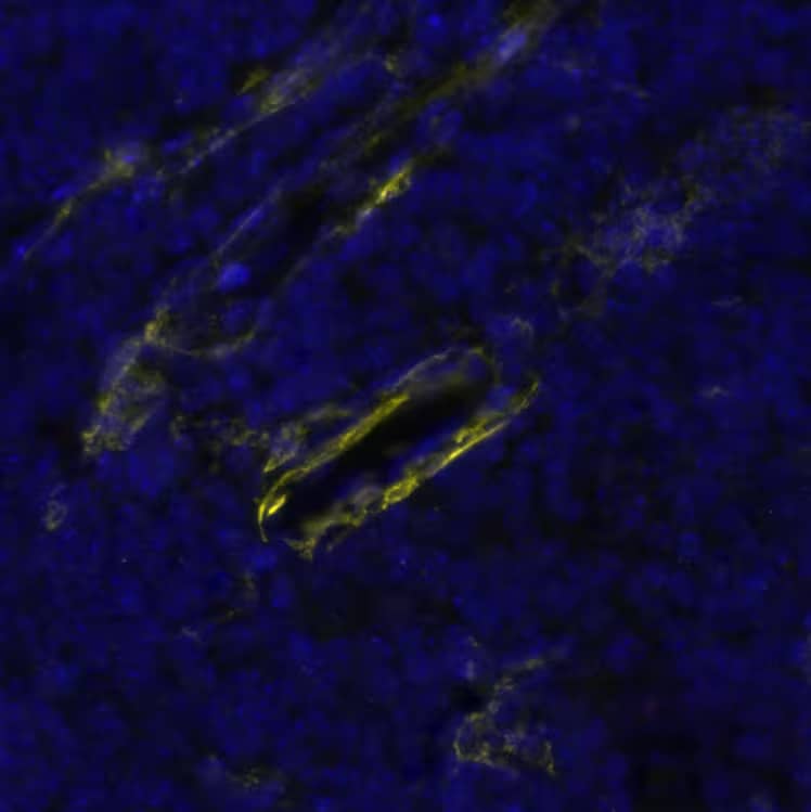


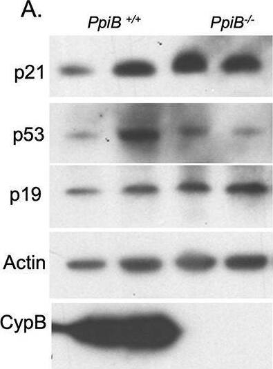
![Immunohistochemistry VEGFR2/KDR/Flk-1 Antibody [Unconjugated]](https://images.novusbio.com/images/antibody/VEGF_R2_AF644_Immunohistochemistry_6888.jpg)
![Immunohistochemistry VEGFR2/KDR/Flk-1 Antibody [Unconjugated]](https://images.novusbio.com/images/af644_mouse-vegf-r2-flk-1-affinity-purified-polyclonal-ab-4120241048248.jpg)
![In-situ Hybridization VEGFR2/KDR/Flk-1 Antibody [Unconjugated]](https://images.novusbio.com/images/antibody/af644_mouse-vegf-r2-flk-1-affinity-purified-polyclonal-ab-in-situ-hybridization-235202421442..jpg)


![Intracellular Staining by Flow Cytometry AKT [p Ser473] Antibody [Unconjugated] - Pan Specific](https://images.novusbio.com/images/antibody/Akt3_AF887_Flow_Cytometry_8283.jpg)
![Western Blot AKT [p Ser473] Antibody [Unconjugated] - Pan Specific](https://images.novusbio.com/images/af887_phospho-akt-s473-pan-specific-affinity-purified-pab-41202410485440.jpg)
![Western Blot AKT [p Ser473] Antibody [Unconjugated] - Pan Specific](https://images.novusbio.com/images/af887_phospho-akt-s473-pan-specific-affinity-purified-pab-8120255552843.jpg)
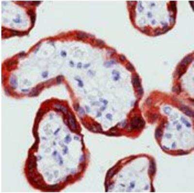


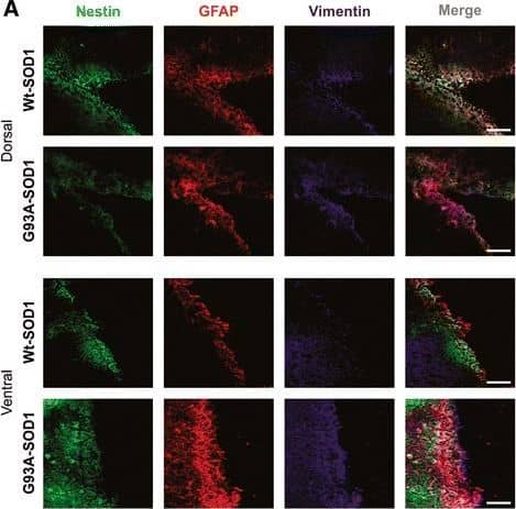

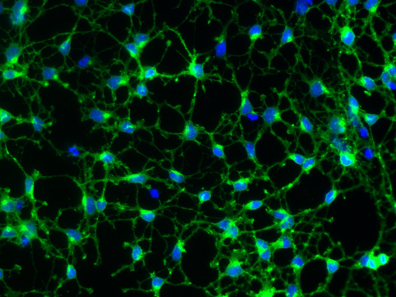


 CTS008) and counterstained with hematoxylin (blue). View our protocol for
  CTS008) and counterstained with hematoxylin (blue). Specific labeling was localized to the plasma membrane of mesenchymal cells. View our protocol for
  221-AA) stimulates proliferation in the NR6R-3T3 mouse fibroblast cell line in a dose-dependent manner (orange line), as measured by Resazurin (


 F0108). View our protocol for


![Immunohistochemistry PDGF R beta Antibody [Unconjugated]](https://images.novusbio.com/images/af1042_mouse-pdgf-r-beta-affinity-purified-polyclonal-ab-immunohistochemistry-121220259315424.jpg)
![Immunohistochemistry PDGF R beta Antibody [Unconjugated]](https://images.novusbio.com/images/af1042_mouse-pdgf-r-beta-affinity-purified-polyclonal-ab-immunohistochemistry-121220258463418.jpg)
![Immunohistochemistry PDGF R beta Antibody [Unconjugated]](https://images.novusbio.com/images/af1042_mouse-pdgf-r-beta-affinity-purified-polyclonal-ab-immunohistochemistry-12122025857822.jpg)
![Cell Culture PDGF-AA [Unconjugated]](https://images.novusbio.com/images/221-aa_recombinant-human-pdgf-aa-protein-cf-cell-culture-1052024153816..jpg)
![Bioactivity PDGF-AA [Unconjugated]](https://images.novusbio.com/images/protein/221-aa_recombinant-human-pdgf-aa-protein-cf-bioactivity-1410202017288.jpg)
![SCF/c-kit Ligand [Unconjugated]](/sites/all/modules/enterprise-tech/et_datasheets/images/novus_guarantee.png "SCF/c-kit Ligand [Unconjugated]")


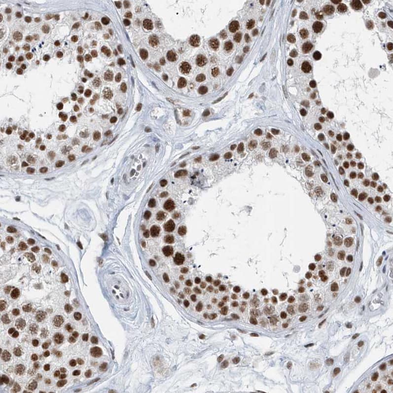
![Immunohistochemistry Desmin Antibody [Unconjugated]](https://images.novusbio.com/images/antibody/Desmin_AF3844_Immunohistochemistry_10689.jpg)
![Immunohistochemistry Desmin Antibody [Unconjugated]](https://images.novusbio.com/images/antibody/Desmin_AF3844_Immunohistochemistry_6808.jpg)
![Immunocytochemistry/ Immunofluorescence Desmin Antibody [Unconjugated]](https://images.novusbio.com/images/af3844_human-mouse-desmin-affinity-purified-polyclonal-ab-41202410331920.jpg)
![N/A VEGF [HRP]](https://images.novusbio.com/images/elisa/VEGF_DVE00_ELISA_208.jpg)
![N/A VEGF [HRP]](https://images.novusbio.com/images/elisa/DATA_VEGF_DVE00_ELISA_871.jpg)
![N/A VEGF [HRP]](https://images.novusbio.com/images/elisa/DATA_VEGF_DVE00_ELISA_872.jpg)
![Neutralization PDGF-C Antibody [Unconjugated]](https://images.novusbio.com/images/antibody/PDGF-C_AF1447_Block_Neutralize_8780.jpg)
![Immunocytochemistry/ Immunofluorescence PDGF-C Antibody [Unconjugated]](https://images.novusbio.com/images/af1447_mouse-pdgf-c-affinity-purified-polyclonal-ab-44202415425060.jpg)