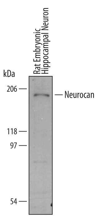
 HAF016). A specific band was detected for Neurocan at approximately 200 kDa (as indicated). This experiment was conducted under reducing conditions and using Immunoblot Buffer Group 8." title="Western blot shows lysates of rat embryonic hippocampal neurons. PVDF membrane was probed with 1 µg/mL of Mouse/Rat Neurocan Antigen Affinity-purified Polyclonal Antibody (Catalog # AF5800) followed by HRP-conjugated Anti-Sheep IgG Secondary Antibody (Catalog # HAF016). A specific band was detected for Neurocan at approximately 200 kDa (as indicated). This experiment was conducted under reducing conditions and using Immunoblot Buffer Group 8." />
| Reactivity | Mu, RtSpecies Glossary |
| Applications | WB, IHC |
| Clonality | Polyclonal |
| Host | Sheep |
| Conjugate | Unconjugated |
| Concentration | LYOPH |
| Immunogen | Chinese hamster ovary cell line CHO-derived recombinant mouse Neurocan Asp23-Asp637 Accession # NP_031815 |
| Specificity | Detects mouse and rat Neurocan in direct ELISAs and Western blots. |
| Source | N/A |
| Isotype | IgG |
| Clonality | Polyclonal |
| Host | Sheep |
| Gene | Ncan |
| Purity Statement | Antigen Affinity-purified |
| Innovator's Reward | Test in a species/application not listed above to receive a full credit towards a future purchase. |
| Dilutions |
|
|
| Publications |
|
| Storage | Use a manual defrost freezer and avoid repeated freeze-thaw cycles.
|
| Buffer | Lyophilized from a 0.2 μm filtered solution in PBS with Trehalose. *Small pack size (SP) is supplied either lyophilized or as a 0.2 µm filtered solution in PBS. |
| Preservative | No Preservative |
| Concentration | LYOPH |
| Reconstitution Instructions | Reconstitute at 0.2 mg/mL in sterile PBS. |


![Immunoprecipitation CD44 Antibody (2C5) [Unconjugated] - s Pan Specific](https://images.novusbio.com/images/antibody/bba10_human-cd44s-pan-specific-mab-clone-2c5-immunoprecipitation-2852025105258..png)
![Knockout Validated CD44 Antibody (2C5) [Unconjugated] - s Pan Specific](https://images.novusbio.com/images/antibody/bba10_human-cd44s-pan-specific-mab-clone-2c5-knockout-validated-285202510529..png)
![Western Blot CD44 Antibody (2C5) [Unconjugated] - s Pan Specific](https://images.novusbio.com/images/antibody/CD44_BBA10_Western_Blot_21459.jpg)


![Western Blot Decorin Antibody [Unconjugated]](https://images.novusbio.com/images/antibody/Decorin_AF1060_Western_Blot_19006.jpg)
Secondary Antibodies |
Isotype Controls |
The concentration calculator allows you to quickly calculate the volume, mass or concentration of your vial. Simply enter your mass, volume, or concentration values for your reagent and the calculator will determine the rest.


 CTS019) and counterstained with hematoxylin (blue). View our protocol for
  Immunoblot of total brain extracts for neurocan, NCAM, PSA-NCAM, and GAPDH (loading control). (B) Co-localization of tdTomato puncta (red) with the presynaptic marker GAD65 (green) around a neuronal soma (blue). Scale bar = 5 μm. (C) Immunofluorescent staining of neurocan (green) around PV+ tdTomato-labeled interneurons (red) in mouse frontal cortex at P28 and P60. Scale bar = 50 μm. (D) Confocal images of neurocan (green) around perisomatic synaptic puncta (red) onto MATH-2 expressing pyramidal neurons (blue nucleus, outlined with white border). A magnified inset is indicated with a white box. Scale bars = 2.5 μm. (E) Electron micrograph of immunogold labeling for neurocan along the membrane of a neuronal soma (black arrows point to examples). Scale bar = 1 μm. (F) Electron micrograph of an inhibitory axon terminal onto a neuronal soma. Neurocan immunogold labeling is indicated with black arrows, and the axon terminal (AT) is labeled with a white arrowhead. Cytoplasm and nucleus of the soma are identified with text. Scale bar = 1 μm. (G) Verification of neurocan antibody specificity by immunostaining of COS7 cells transfected with Neurocan-AP (+control) or AP (−control), and a no primary antibody control. Scale bar = 50 μm.(H) Immunofluorescent localization of NCAM and neurocan in GABA+ and GABA− cortical neuron cultures. Confocal images of GABA (green), NCAM (blue), and neurocan (red) in cortical neuron cultures. White arrow indicates a GABA-negative neuron positive for NCAM and neurocan, and the green neuron represents a GABAergic interneuron. Scale bar = 10 μm. Image collected and cropped by CiteAb from the following open publication (//pubmed.ncbi.nlm.nih.gov/29670169), licensed under a CC-BY license. Not internally tested by R&D Systems.")


![Immunohistochemistry Brevican Antibody [Unconjugated]](https://images.novusbio.com/images/antibody/Brevican_AF4009_Immunohistochemistry_19694.jpg)
![Western Blot Brevican Antibody [Unconjugated]](https://images.novusbio.com/images/af4009_human-rat-brevican-affinity-purified-polyclonal-ab-western-blot-121220259302820.jpg)
![Immunocytochemistry Brevican Antibody [Unconjugated]](https://images.novusbio.com/images/antibody/Brevican_AF4009_Immunocytochemistry__Immunofluorescence_19520.jpg)

![Western Blot Tenascin R Antibody (619) [Unconjugated]](https://images.novusbio.com/images/mab1624_mouse-rat-tenascin-r-mab-clone-619-western-blot-35202385637..jpg)
![Immunohistochemistry Tenascin R Antibody (619) [Unconjugated]](https://images.novusbio.com/images/antibody/Tenascin_R_MAB1624_Immunohistochemistry_10138.jpg)


![Immunohistochemistry Contactin-2/TAG1 Antibody [Unconjugated]](https://images.novusbio.com/images/antibody/Contactin-2_AF4439_Immunohistochemistry_10516.jpg)
![Simple Western Contactin-2/TAG1 Antibody [Unconjugated]](https://images.novusbio.com/images/antibody/Contactin2_AF4439_Simple_Western_17121.jpg)
![Western Blot Contactin-2/TAG1 Antibody [Unconjugated]](https://images.novusbio.com/images/antibody/Contactin-2_AF4439_Western_Blot_9589.jpg)
![N/A MMP-2 [HRP]](https://images.novusbio.com/images/elisa/DATA_MMP2_MMP200_ELISA_1003.jpg)
![N/A MMP-2 [HRP]](https://images.novusbio.com/images/elisa/MMP2_MMP200_ELISA_474.jpg)


) induced by Homo-BacPROTAC 8 (UdSBI-0545) compared to its enantiomer 8a (UdSBI-0966), matching monomer 5 and dCymC (3 independent experiments done in triplicates). b WES visualization of ClpC1-NTD degradation after titration of Homo-BacPROTACs, developed with anti-His antibody recognizing His6-tagged ClpC1-NTD and processed, His4-tagged ClpP1P2. Concentration-dependent degradation of ClpC1-NTD can be observed for 8 (UdSBI-0545) (lanes 10–13) but not for 8a (UdSBI-0966) (lanes 2–8). c Analogous to a, except that exit vector 7-based Homo-BacPROTAC 12 (UdSBI-4377), enantiomer 12a (UdSBI-0117) and monomer 10 were used. d WES-derived gel picture visualizing ClpC1-NTD degradation (lane 2–5) from representative experiment summarized in c. e SYPROTM Ruby-stained SDS-PAGE gel from exemplary cell-free degradation assay depicting efficient degradation (lane 3,4) of ClpC1-NTD by Homo-BacPROTAC 8 (UdSBI-0545), while full length ClpC1 is not significantly degraded. Green vertical lines indicate the DC50 (for 8,12). Error bars represent mean ± SD of n = 3 independent experiments in triplicates. The actual mean DC50 values for all cell-free degradation experiments conducted for this study are summarized in Supplementary Table 8. Source data are provided as a Source Data file. Image collected and cropped by CiteAb from the following open publication (https://www.nature.com/articles/s41467-024-46218-7), licensed under a CC-BY license. Not internally tested by R&D Systems.")