![Western Blot: LC3 Antibody Pack [NB910-40435] - Lysates of HeLa parental cell line and LC3B knockout HeLa cell line (KO) untreated (-) or treated (+) with 50 uM Chloroquine for 18 hours. PVDF (Polyvinylidene difluoride) membrane was probed with 0.5 ug/mL of Rabbit Anti-LC3B Polyclonal Antibody (Catalog # NB100-2220) followed by HRP-conjugated Anti-Rabbit IgG Secondary Antibody (Catalog# HAF008). A specific band was detected for LC3B at a molecular weight of approximately 15 kDa (as indicated) in the parental HeLa cell line, but is not detectable in the knockout HeLa cell line. GAPDH is shown as a loading control. This experiment was conducted under reducing conditions. LC3B Antibody [NB100-2220]](http://images.novusbio.com/fullsize/LC3-Antibody-Pack-Knockout-Validated-NB910-40435-img0013.jpg "Western Blot: LC3 Antibody Pack [NB910-40435] - Lysates of HeLa parental cell line and LC3B knockout HeLa cell line (KO) untreated (-) or treated (+) with 50 uM Chloroquine for 18 hours. PVDF (Polyvinylidene difluoride) membrane was probed with 0.5 ug/mL of Rabbit Anti-LC3B Polyclonal Antibody (Catalog # NB100-2220) followed by HRP-conjugated Anti-Rabbit IgG Secondary Antibody (Catalog# HAF008). A specific band was detected for LC3B at a molecular weight of approximately 15 kDa (as indicated) in the parental HeLa cell line, but is not detectable in the knockout HeLa cell line. GAPDH is shown as a loading control. This experiment was conducted under reducing conditions. LC3B Antibody [NB100-2220]")
| Reactivity | Hu, Mu, Rt, Po, Al, Am, Av, Ba, Bv, Ca, Ch, ChHa, Fi, Gp, Ha, In, Pl, Pm, Pm, Rb, SyHa, ZeSpecies Glossary |
| Applications | WB, Simple Western, ELISA, Flow, Func, ICC/IF, IHC, IP, NULL, PAGE, WB, ChIP, IHC, KO, Mycoplasma |
| Description | This pack contains 1 vial each of: NB100-2220 (0.1 mL) and NB100-2331 (0.1 mL). |
| Immunogen | For NB100-2331: A synthetic peptide made to an internal portion of the human LC3 protein sequence (between residues 25-121) [Uniprot: Q9H492]. For NB100-2220: Polyclonal LC3B Antibody was made to a synthetic peptide made to an N-terminal portion of the human LC3B protein sequence (between residues 1-100) [UniProt# Q9GZQ8]. |
| Marker | Autophagosome Marker |
| Gene | MAP1LC3B |
| Dilutions |
|
|
| Application Notes | Antibodies in this pack are validated for the following applications: NB100-2331: Chromatin Immunoprecipitation, ELISA, Flow Cytometry, Immunoblotting, Immunocytochemistry/Immunofluorescence, Immunohistochemistry, Immunohistochemistry Whole-Mount, Immunohistochemistry-Frozen, Immunohistochemistry-Paraffin, Immunoprecipitation, Simple Western, Southern Blot, Western Blot NB100-2220: Chromatin Immunoprecipitation (ChIP), ELISA, Flow Cytometry, Immunoblotting, Immunocytochemistry/Immunofluorescence, Immunohistochemistry, Immunohistochemistry-Frozen, Immunohistochemistry-Paraffin, Immunoprecipitation, Knockdown Validated, Knockout Validated, Proximity Ligation Assay, SDS-Page, Simple Western, Western Blot |
|
| Reviewed Applications |
|
|
| Publications |
|
| Storage | Store at 4C short term. Aliquot and store at -20C long term. Avoid freeze-thaw cycles. |


![Immunohistochemistry ATG7 Antibody (683906) [Unconjugated]](https://images.novusbio.com/images/antibody/ATG7_MAB6608_Immunohistochemistry_10631.jpg)
![Simple Western ATG7 Antibody (683906) [Unconjugated]](https://images.novusbio.com/images/antibody/ATG7_MAB6608_Simple_Western_16409.jpg)
![Intracellular Staining by Flow Cytometry AKT [p Ser473] Antibody [Unconjugated] - Pan Specific](https://images.novusbio.com/images/antibody/Akt3_AF887_Flow_Cytometry_8283.jpg)
![Western Blot AKT [p Ser473] Antibody [Unconjugated] - Pan Specific](https://images.novusbio.com/images/af887_phospho-akt-s473-pan-specific-affinity-purified-pab-41202410485440.jpg)
![Western Blot AKT [p Ser473] Antibody [Unconjugated] - Pan Specific](https://images.novusbio.com/images/af887_phospho-akt-s473-pan-specific-affinity-purified-pab-8120255552843.jpg)


| Images | Ratings | Applications | Species | Date | Details | ||||||||
|---|---|---|---|---|---|---|---|---|---|---|---|---|---|
|
reviewed by:
Christiana Doulami |
ICC | Mouse | 02/12/2019 |
Summary
|
|||||||||

Enlarge |
reviewed by:
Verified Customer |
WB | Mouse | 07/11/2014 |
Summary
|
||||||||

Enlarge |
reviewed by:
Verified Customer |
IHC | Mouse | 04/29/2014 |
Summary
|
Research Areas for LC3 Antibody Pack (NB910-40435)Find related products by research area.
|
|
Autophagy and Metastasis By Christina Towers, PhD The majority of cancer patients die from metastatic disease at secondary sites. The threshold to undergo metastasis is high. Only a minority of cancer cells acquire invasive phenotypes... Read full blog post. |
|
Optogenetic Control of Mitophagy: AMBRA1 based mitophagy switch By Christina Towers, PhD Mitophagy in the BrainSelective autophagic degradation of damaged mitochondria, known as mitophagy, has been described as a cyto-protective process. Accordingly, defects in mitophagy h... Read full blog post. |
|
Read full blog post. |
|
Read full blog post. |
|
Read full blog post. |
|
How to visualize autophagy by microscopy By Christina Towers, PhD Autophagy is a recycling process that relies on the formation of a unique organelle termed an autophagosome. An elegant way to monitor autophagy is through various microscopy techniques to... Read full blog post. |
|
Microglia: pruning shears for homeostatic maintenance in the brain By Jennifer Sokolowski, MD, PhD.Microglia play a critical role in pruning neurons and synapses during homeostatic maintenance in the adult brain.1 A recent study by Ayata et al. (2018) identified regional differe... Read full blog post. |
|
Read full blog post. |
|
Read full blog post. |
|
Read full blog post. |
5 | |
4 | |
3 | |
2 | |
1 |
| Christiana Doulami 02/12/2019 |
||
| Application: | ICC | |
| Species: | Mouse |
| Verified Customer 07/11/2014 |
||
| Application: | WB | |
| Species: | Mouse |
| Verified Customer 04/29/2014 |
||
| Application: | IHC | |
| Species: | Mouse |
| Gene Symbol | MAP1LC3B |


![Immunohistochemistry: LC3 Antibody Pack [NB910-40435] - Rapamycin increases autophagy in brains of PDAPP mice. Representative epifluorescent (c200x) image of hippocampal CA1 in control- and rapamycin-fed transgenic PDAPP mice stained with an anti-LC3 antibody. An increase in LC3-immunoreactive puncta was observed in CA1 projections of transgenic PDAPP mice following rapamycin administration. Image collected and cropped by CiteAb from the following publication (//dx.plos.org/10.1371/journal.pone.0009979) licensed under a CC-BY license. LC3A Antibody [NB100-2331]](http://images.novusbio.com/fullsize/LC3-Antibody-Pack-Immunohistochemistry-NB910-40435-img0009.jpg "Immunohistochemistry: LC3 Antibody Pack [NB910-40435] - Rapamycin increases autophagy in brains of PDAPP mice. Representative epifluorescent (c200x) image of hippocampal CA1 in control- and rapamycin-fed transgenic PDAPP mice stained with an anti-LC3 antibody. An increase in LC3-immunoreactive puncta was observed in CA1 projections of transgenic PDAPP mice following rapamycin administration. Image collected and cropped by CiteAb from the following publication (//dx.plos.org/10.1371/journal.pone.0009979) licensed under a CC-BY license. LC3A Antibody [NB100-2331]")
![Immunocytochemistry/ Immunofluorescence: LC3 Antibody Pack [NB910-40435] - LC3B was detected in immersion fixed Cloroquine treated HeLa cells (left) but was not detected in LC3B knockout HeLa cells (right) using rabbit anti-human LC3B polyclonal antibody (Catalog #NB100-2220) at 0.3 ug/mL for 3 hours at room temperature. Cells were stained using the NorthernLights(TM) 557-conjugated anti-Rabbit IgG Secondary Antibody (red; Catalog # NL004) and counterstained with DAPI (blue). Specific staining was localized to cytoplasm.](http://images.novusbio.com/fullsize/LC3-Antibody-Pack-Knockout-Validated-NB910-40435-img0014.jpg "Immunocytochemistry/ Immunofluorescence: LC3 Antibody Pack [NB910-40435] - LC3B was detected in immersion fixed Cloroquine treated HeLa cells (left) but was not detected in LC3B knockout HeLa cells (right) using rabbit anti-human LC3B polyclonal antibody (Catalog #NB100-2220) at 0.3 ug/mL for 3 hours at room temperature. Cells were stained using the NorthernLights(TM) 557-conjugated anti-Rabbit IgG Secondary Antibody (red; Catalog # NL004) and counterstained with DAPI (blue). Specific staining was localized to cytoplasm.")
![Western Blot: LC3 Antibody Pack [NB910-40435] - High autophagosome concentration is consumed during early immortalized human mesenchymal stem cell differentiation. Immortalized human mesenchymal stem cells were differentiated under osteogenic conditions (see Materials and methods) and assayed for changes in LC3I and LC3II during a 72-hour window. Cells were differentiated under standard conditions (top) or with addition of 5 uM rapamycin (middle) or 5 nM bafilomycin (bottom) for the first 3 hours of differentiation to modulate autophagy. Immunoblots were performed for LC3 at the indicated time points to assess autophagosome degradation via relative changes in LC3II (lower band; 17 kDa). Studies were repeated three times with similar trends seen consistently. Image collected and cropped by CiteAb from the following publication (//stemcellres.com/content/5/6/140), licensed under a CC-BY license. LC3A Antibody [NB100-2331]](http://images.novusbio.com/fullsize/LC3-Antibody-Pack-Western-Blot-NB910-40435-img0011.jpg "Western Blot: LC3 Antibody Pack [NB910-40435] - High autophagosome concentration is consumed during early immortalized human mesenchymal stem cell differentiation. Immortalized human mesenchymal stem cells were differentiated under osteogenic conditions (see Materials and methods) and assayed for changes in LC3I and LC3II during a 72-hour window. Cells were differentiated under standard conditions (top) or with addition of 5 uM rapamycin (middle) or 5 nM bafilomycin (bottom) for the first 3 hours of differentiation to modulate autophagy. Immunoblots were performed for LC3 at the indicated time points to assess autophagosome degradation via relative changes in LC3II (lower band; 17 kDa). Studies were repeated three times with similar trends seen consistently. Image collected and cropped by CiteAb from the following publication (//stemcellres.com/content/5/6/140), licensed under a CC-BY license. LC3A Antibody [NB100-2331]")
![Immunohistochemistry: LC3 Antibody Pack [NB910-40435] - Localization of LC3 by immunohistochemical method. The immunohistochemical staining shows that LC3, CRF, and HIF-1alpha have an overlapping localization in villous and extravillous trophoblast. d: decidua; v: villi; DeVe: decidual vessel. Original magnification 40x. Image collected and cropped by CiteAb from the following publication (//www.hindawi.com/journals/bmri/2013/689768/) licensed under a CC-BY license. LC3B Antibody [NB100-2220]](http://images.novusbio.com/fullsize/LC3-Antibody-Pack-Immunohistochemistry-NB910-40435-img0015.jpg "Immunohistochemistry: LC3 Antibody Pack [NB910-40435] - Localization of LC3 by immunohistochemical method. The immunohistochemical staining shows that LC3, CRF, and HIF-1alpha have an overlapping localization in villous and extravillous trophoblast. d: decidua; v: villi; DeVe: decidual vessel. Original magnification 40x. Image collected and cropped by CiteAb from the following publication (//www.hindawi.com/journals/bmri/2013/689768/) licensed under a CC-BY license. LC3B Antibody [NB100-2220]")
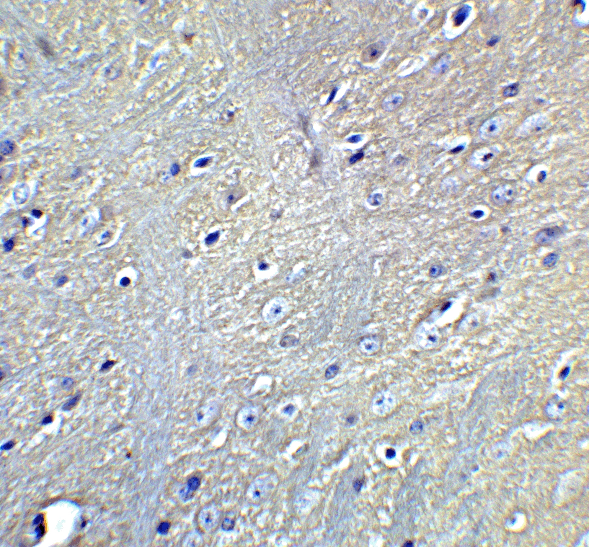
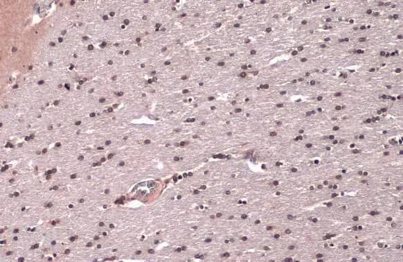
![Immunohistochemistry: LC3 Antibody Pack [NB910-40435] - Brain, Cerebral Cortex, Cell Processes in Gray Matter 40x.](http://images.novusbio.com/fullsize/LC3-Antibody-Pack-Immunohistochemistry-NB910-40435-img0008.jpg "Immunohistochemistry: LC3 Antibody Pack [NB910-40435] - Brain, Cerebral Cortex, Cell Processes in Gray Matter 40x.")
![Immunohistochemistry: LC3 Antibody Pack [NB910-40435] - Analysis in PFA fixed NIH/3T3 cells using anti-LC3A antibody. Image from verified customer review. LC3A Antibody [NB100-2331]](http://images.novusbio.com/fullsize/LC3-Antibody-Pack-Immunohistochemistry-NB910-40435-img0012.jpg "Immunohistochemistry: LC3 Antibody Pack [NB910-40435] - Analysis in PFA fixed NIH/3T3 cells using anti-LC3A antibody. Image from verified customer review. LC3A Antibody [NB100-2331]")
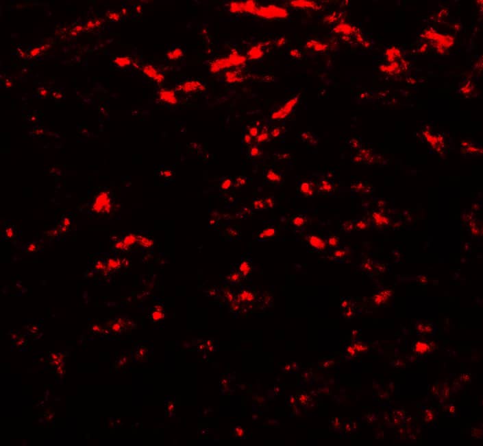
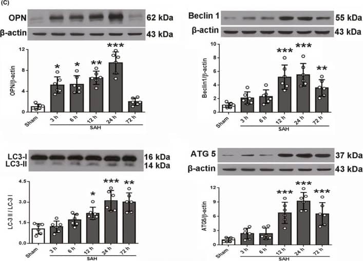
![Immunocytochemistry/ Immunofluorescence: LC3 Antibody Pack [NB910-40435] - The autophagy level was increased in degenerated mouse SGNs. (a-c) Western blot results revealed that the levels of the autophagy-related proteins LC3 & BECN1 were increased in the degenerated SGNs on the 5th, 15th & 30th day after ototoxic drug administration & were significantly different from those in the normal mice. *, P < 0.05. (d) Immunofluorescence staining of LC3 puncta (red) also demonstrated that the LC3 level in the degenerated SGNs (green) was significantly increased on the 30th day after drug administration. Con, normal mice without drug treatment; 30D, 30 days after drug administration. Images of immunofluorescence staining were taken from the middle turn of cochlea. Scale bar: 10 µm. Image collected & cropped by CiteAb from the following publication (//pubmed.ncbi.nlm.nih.gov/30706760), licensed under a CC-BY license. Not internally tested by Novus Biologicals.](http://images.novusbio.com/fullsize/nb910-40435_lc3-antibody-pack-31020241538249.jpg "Immunocytochemistry/ Immunofluorescence: LC3 Antibody Pack [NB910-40435] - The autophagy level was increased in degenerated mouse SGNs. (a-c) Western blot results revealed that the levels of the autophagy-related proteins LC3 & BECN1 were increased in the degenerated SGNs on the 5th, 15th & 30th day after ototoxic drug administration & were significantly different from those in the normal mice. *, P < 0.05. (d) Immunofluorescence staining of LC3 puncta (red) also demonstrated that the LC3 level in the degenerated SGNs (green) was significantly increased on the 30th day after drug administration. Con, normal mice without drug treatment; 30D, 30 days after drug administration. Images of immunofluorescence staining were taken from the middle turn of cochlea. Scale bar: 10 µm. Image collected & cropped by CiteAb from the following publication (//pubmed.ncbi.nlm.nih.gov/30706760), licensed under a CC-BY license. Not internally tested by Novus Biologicals.")
![Western Blot: LC3 Antibody Pack [NB910-40435] - The autophagy level was increased in degenerated mouse SGNs. (a-c) Western blot results revealed that the levels of the autophagy-related proteins LC3 & BECN1 were increased in the degenerated SGNs on the 5th, 15th & 30th day after ototoxic drug administration & were significantly different from those in the normal mice. *, P < 0.05. (d) Immunofluorescence staining of LC3 puncta (red) also demonstrated that the LC3 level in the degenerated SGNs (green) was significantly increased on the 30th day after drug administration. Con, normal mice without drug treatment; 30D, 30 days after drug administration. Images of immunofluorescence staining were taken from the middle turn of cochlea. Scale bar: 10 µm. Image collected & cropped by CiteAb from the following publication (//pubmed.ncbi.nlm.nih.gov/30706760), licensed under a CC-BY license. Not internally tested by Novus Biologicals.](http://images.novusbio.com/fullsize/nb910-40435_lc3-antibody-pack-310202415284514.jpg "Western Blot: LC3 Antibody Pack [NB910-40435] - The autophagy level was increased in degenerated mouse SGNs. (a-c) Western blot results revealed that the levels of the autophagy-related proteins LC3 & BECN1 were increased in the degenerated SGNs on the 5th, 15th & 30th day after ototoxic drug administration & were significantly different from those in the normal mice. *, P < 0.05. (d) Immunofluorescence staining of LC3 puncta (red) also demonstrated that the LC3 level in the degenerated SGNs (green) was significantly increased on the 30th day after drug administration. Con, normal mice without drug treatment; 30D, 30 days after drug administration. Images of immunofluorescence staining were taken from the middle turn of cochlea. Scale bar: 10 µm. Image collected & cropped by CiteAb from the following publication (//pubmed.ncbi.nlm.nih.gov/30706760), licensed under a CC-BY license. Not internally tested by Novus Biologicals.")
![Western Blot: LC3 Antibody Pack [NB910-40435] - CCI-779 significantly rescued the impaired autophagy-lysosomal pathway in degenerated SGNs of mice. (a) The levels of Ctsb, Ctsd, & Lamp1 & of the autophagic genes Becn1 & Lc3b were significantly higher, & Sqstm1 was lower in the experimental group than in the negative control group, which was determined by quantitative real-time PCR. (b & c) The LAMP1 & CTSD levels determined by western blotting were consistent with the quantitative real-time PCR results. (d & e) Compared with those in the negative control group, the LC3 & BECN1 levels in the experimental groups were significantly increased, as determined by western blot assays. (f & g) The western blot results revealed that the levels of the autophagic cargo receptor SQSTM1 & ubiquitinated proteins were decreased significantly in the experimental group compared with those in the negative control groups. *, the difference between the experimental group & the blank control group was significant (P < 0.05); #, the difference between the experimental group & the negative control group was significant (P < 0.05); CCI-779, experimental group; 30D, negative control group; Con, blank control group. Image collected & cropped by CiteAb from the following publication (//pubmed.ncbi.nlm.nih.gov/30706760), licensed under a CC-BY license. Not internally tested by Novus Biologicals.](http://images.novusbio.com/fullsize/nb910-40435_lc3-antibody-pack-310202416163727.jpg "Western Blot: LC3 Antibody Pack [NB910-40435] - CCI-779 significantly rescued the impaired autophagy-lysosomal pathway in degenerated SGNs of mice. (a) The levels of Ctsb, Ctsd, & Lamp1 & of the autophagic genes Becn1 & Lc3b were significantly higher, & Sqstm1 was lower in the experimental group than in the negative control group, which was determined by quantitative real-time PCR. (b & c) The LAMP1 & CTSD levels determined by western blotting were consistent with the quantitative real-time PCR results. (d & e) Compared with those in the negative control group, the LC3 & BECN1 levels in the experimental groups were significantly increased, as determined by western blot assays. (f & g) The western blot results revealed that the levels of the autophagic cargo receptor SQSTM1 & ubiquitinated proteins were decreased significantly in the experimental group compared with those in the negative control groups. *, the difference between the experimental group & the blank control group was significant (P < 0.05); #, the difference between the experimental group & the negative control group was significant (P < 0.05); CCI-779, experimental group; 30D, negative control group; Con, blank control group. Image collected & cropped by CiteAb from the following publication (//pubmed.ncbi.nlm.nih.gov/30706760), licensed under a CC-BY license. Not internally tested by Novus Biologicals.")


![Immunohistochemistry TOR/mTOR [p Ser2448] Antibody - BSA Free](https://images.novusbio.com/images/nb600-607_rabbit-polyclonal-tor-mtor-p-ser2448-antibody-235202318143142.jpg)
![Western Blot TOR/mTOR [p Ser2448] Antibody - BSA Free](https://images.novusbio.com/images/nb600-607_rabbit-polyclonal-tor-mtor-p-ser2448-antibody-31020241535647.jpg)
![Data TOR/mTOR [p Ser2448] Antibody - BSA Free](https://images.novusbio.com/images/TOR-mTOR-[p-Ser2448]-Antibody-N-A-NB600-607-img0008.jpg)
![Immunohistochemistry Caspase-3 Antibody [Unconjugated] - Active](https://images.novusbio.com/images/af835_human-mouse-active-caspase-3-affinity-purified-pab-41202410331943.jpg)
![Western Blot Caspase-3 Antibody [Unconjugated] - Active](https://images.novusbio.com/images/af835_human-mouse-active-caspase-3-affinity-purified-pab-812025554170.jpg)
![Western Blot Caspase-3 Antibody [Unconjugated] - Active](https://images.novusbio.com/images/af835_human-mouse-active-caspase-3-affinity-purified-pab-8120255534731.jpg)


