After August 17, 2026, Novus Biologicals products and services will no longer be available on this website; you will access all products and services on rndsystems.com. Create your R&D Systems online account today.
Western Blot: LAP (TGF-beta 1) Antibody (7F6) [NBP2-22114] - Western blot analysis using TGF beta 1 mAb against human LAP (TGF-beta 1) recombinant protein. (Expected MW is 41 kDa)
Immunohistochemistry-Paraffin: LAP (TGF-beta 1) Antibody (7F6) [NBP2-22114] - Immunohistochemical analysis of paraffin-embedded lymphoid tissue tissues using LAP (TGF-beta 1) mouse mAb with DAB staining.
Flow Cytometry: LAP (TGF-beta 1) Antibody (7F6) [NBP2-22114] - Flow cytometric analysis of A549 cells using LAP (TGF-beta 1) mouse mAb (green) and negative control (red).
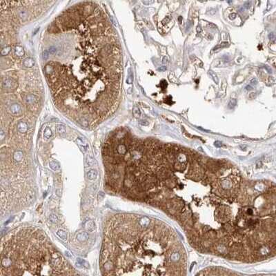
Western Blot: LAP (TGF-beta 1) Antibody (7F6) [NBP2-22114] - Western blot analysis of human stomach tissue (A) and human small intestine tissue (B) using TGF beta 1 antibody (NBP2-22114) at 2 ug/ml.
Immunohistochemistry-Paraffin: LAP (TGF-beta 1) Antibody (7F6) [NBP2-22114] - Human tonsil. Image from verified customer review.
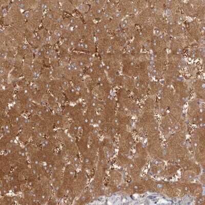
Immunohistochemistry-Paraffin: LAP (TGF-beta 1) Antibody (7F6) [NBP2-22114] - Immunohistochemical analysis of paraffin-embedded lung cancer tissues using LAP (TGF-beta 1) mouse mAb with DAB staining.
CD47 regulates thrombospondin-1, TGF-beta 1, and collagen deposition after injury. a Whole cell lysates from freshly isolated intestinal epithelial cells from Cd47f/f and Cd47 delta IEC mice were subjected to SDS–PAGE ...read more
Inhibition of ERK1/2 activation attenuates myocardial remodeling and inflammation in permanent MI.Trametinib- and vehicle-treated mice were studied for 7 days after permanent LAD ligation. (A) Relative mRNA expression ...read more
Inhibition of ERK1/2 activation attenuates myocardial remodeling and inflammation in permanent MI.Trametinib- and vehicle-treated mice were studied for 7 days after permanent LAD ligation. (A) Relative mRNA expression ...read more
The upregulation of TGF beta 1/IGF-1/BDNF expression in the brain of AD mice by PBMT extended to promote adult hippocampal neurogenesis. A, B Western blotting analysis (A) and quantification (B) of TGF beta 1/IGF-1/BDNF ...read more
TF cytoplasmic domain phosphorylation–dependent increased TGF-beta 1 activation in clinical setting of MI.(A) Representative confocal images of phosphorylation status of TF in infarcted myocardium obtained from WT or ...read more
A profibrotic MEK1/2–TGF-beta 1 pathway is linked to PAR2-mediated ROS signaling in monocytes.(A and B) Protein expression analysis of monocytes isolated from WT mice and pretreated in vitro with trametinib (10 μM) ...read more
A profibrotic MEK1/2–TGF-beta 1 pathway is linked to PAR2-mediated ROS signaling in monocytes.(A and B) Protein expression analysis of monocytes isolated from WT mice and pretreated in vitro with trametinib (10 μM) ...read more
Myeloid cell–derived TF-PAR2 complex is required for TGF-beta 1 activation.(A) Confocal microscopy of myocardial cryosections obtained from n = 5 sham-operated and n = 5 LAD-ligated WT (C57BL/6J) mice at day 7. ...read more
Myeloid cell TF cytoplasmic domain phosphorylation mediates ERK1/2–TGF-beta 1–dependent cardiac remodeling in permanent LAD ligation.(A) Confocal microscopy of myocardial cryosections obtained from WT (C57BL/6J) and ...read more
A profibrotic MEK1/2–TGF-beta 1 pathway is linked to PAR2-mediated ROS signaling in monocytes.(A and B) Protein expression analysis of monocytes isolated from WT mice and pretreated in vitro with trametinib (10 μM) ...read more
Immunohistochemistry reported in scientific literature (PMID 30147399)
Immunohistochemistry-Paraffin reported by customer review
Western Blot 1:500-1:2000
Theoretical MW
44.3 kDa. Disclaimer note: The observed molecular weight of the protein may vary from the listed predicted molecular weight due to post translational modifications, post translation cleavages, relative charges, and other experimental factors.
Reviewed Applications
Read 3 Reviews rated 3.7 using NBP2-22114 in the following applications:
Mouse reactivity reported in scientific literature (PMID: 26851347). Rat reactivity reported in scientific literature (PMID: 30147399).
Packaging, Storage & Formulations
Storage
Store at 4C short term. Aliquot and store at -20C long term. Avoid freeze-thaw cycles.
Buffer
PBS
Preservative
0.02% Sodium Azide
Concentration
1 mg/ml
Purity
Ammonium sulfate precipitation
Alternate Names for LAP (TGF-beta 1) Antibody (7F6) - BSA Free
CED
DPD1
LAP (TGFbeta 1)
LAP (TGF-beta 1)
LAP
TGFB
TGFB1
TGFbeta
transforming growth factor beta 1
Background
This gene encodes a member of the transforming growth factor beta (TGFB) family of cytokines, which are multifunctional peptides that regulate proliferation, differentiation, adhesion, migration, and other functions in many cell types. Many cells have TGFB receptors, and the protein positively and negatively regulates many other growth factors. The secreted protein is cleaved into a latency-associated peptide (LAP) and a mature TGFB1 peptide, and is found in either a latent form composed of a TGFB1 homodimer, a LAP homodimer, and a latent TGFB1-binding protein, or in an active form composed of a TGFB1 homodimer. The mature peptide may also form heterodimers with other TGFB family members. This gene is frequently upregulated in tumor cells, and mutations in this gene result in Camurati-Engelmann disease
Limitations
This product is for research use only and is not approved for use in humans or in clinical diagnosis. Primary Antibodies are guaranteed for 1 year from date of receipt.
The concentration calculator allows you to quickly calculate the volume, mass or concentration of your vial. Simply enter your mass, volume, or concentration values for your reagent and the calculator will determine the rest.
![Western Blot: LAP (TGF-beta 1) Antibody (7F6) [NBP2-22114] - Western blot analysis using TGF beta 1 mAb against human LAP (TGF-beta 1) recombinant protein. (Expected MW is 41 kDa)](http://images.novusbio.com/fullsize/TGF-beta-1-Antibody-7F6-Western-Blot-NBP2-22114-img0002.jpg "Western Blot: LAP (TGF-beta 1) Antibody (7F6) [NBP2-22114] - Western blot analysis using TGF beta 1 mAb against human LAP (TGF-beta 1) recombinant protein. (Expected MW is 41 kDa)")
![Western Blot Smad3 [p Ser423, p Ser425] Antibody - BSA Free](https://images.novusbio.com/images/Smad3-[p-Ser423--p-Ser425]-Antibody-Western-Blot-NBP1-77836-img0005.jpg)
![Immunocytochemistry/ Immunofluorescence Smad3 [p Ser423, p Ser425] Antibody - BSA Free](https://images.novusbio.com/images/Smad3-[p-Ser423--p-Ser425]-Antibody-Immunocytochemistry-Immunofluorescence-NBP1-77836-img0007.jpg)
![Immunohistochemistry Smad3 [p Ser423, p Ser425] Antibody - BSA Free](https://images.novusbio.com/images/Smad3-[p-Ser423--p-Ser425]-Antibody-Immunohistochemistry-NBP1-77836-img0006.jpg)


-(01-ml)_NBP2-22114_6771.jpg)


![Immunohistochemistry-Paraffin: LAP (TGF-beta 1) Antibody (7F6) [NBP2-22114] - Immunohistochemical analysis of paraffin-embedded lymphoid tissue tissues using LAP (TGF-beta 1) mouse mAb with DAB staining.](http://images.novusbio.com/fullsize/TGF-beta-1-Antibody-7F6-Immunohistochemistry-Paraffin-NBP2-22114-img0005.jpg "Immunohistochemistry-Paraffin: LAP (TGF-beta 1) Antibody (7F6) [NBP2-22114] - Immunohistochemical analysis of paraffin-embedded lymphoid tissue tissues using LAP (TGF-beta 1) mouse mAb with DAB staining.")
![Flow Cytometry: LAP (TGF-beta 1) Antibody (7F6) [NBP2-22114] - Flow cytometric analysis of A549 cells using LAP (TGF-beta 1) mouse mAb (green) and negative control (red).](http://images.novusbio.com/fullsize/TGF-beta-1-Antibody-7F6-Flow-Cytometry-NBP2-22114-img0003.jpg "Flow Cytometry: LAP (TGF-beta 1) Antibody (7F6) [NBP2-22114] - Flow cytometric analysis of A549 cells using LAP (TGF-beta 1) mouse mAb (green) and negative control (red).")
![Western Blot: LAP (TGF-beta 1) Antibody (7F6) [NBP2-22114] - Western blot analysis of human stomach tissue (A) and human small intestine tissue (B) using TGF beta 1 antibody (NBP2-22114) at 2 ug/ml.](http://images.novusbio.com/fullsize/TGF-beta-1-Antibody-7F6-Western-Blot-NBP2-22114-img0007.jpg "Western Blot: LAP (TGF-beta 1) Antibody (7F6) [NBP2-22114] - Western blot analysis of human stomach tissue (A) and human small intestine tissue (B) using TGF beta 1 antibody (NBP2-22114) at 2 ug/ml.")
![Immunohistochemistry-Paraffin: LAP (TGF-beta 1) Antibody (7F6) [NBP2-22114] - Human tonsil. Image from verified customer review.](http://images.novusbio.com/fullsize/TGF-beta-1-Antibody-7F6-Immunohistochemistry-Paraffin-NBP2-22114-img0006.jpg "Immunohistochemistry-Paraffin: LAP (TGF-beta 1) Antibody (7F6) [NBP2-22114] - Human tonsil. Image from verified customer review.")
![Immunohistochemistry-Paraffin: LAP (TGF-beta 1) Antibody (7F6) [NBP2-22114] - Immunohistochemical analysis of paraffin-embedded lung cancer tissues using LAP (TGF-beta 1) mouse mAb with DAB staining.](http://images.novusbio.com/fullsize/TGF-beta-1-Antibody-7F6-Immunohistochemistry-Paraffin-NBP2-22114-img0004.jpg "Immunohistochemistry-Paraffin: LAP (TGF-beta 1) Antibody (7F6) [NBP2-22114] - Immunohistochemical analysis of paraffin-embedded lung cancer tissues using LAP (TGF-beta 1) mouse mAb with DAB staining.")
![ELISA: LAP (TGF-beta 1) Antibody (7F6) [NBP2-22114] - Red: Control Antigen (100ng); Purple: Antigen (10ng); Green: Antigen (50ng); Blue: Antigen (100ng).](http://images.novusbio.com/fullsize/TGF-beta-1-Antibody-7F6-ELISA-NBP2-22114-img0001.jpg "ELISA: LAP (TGF-beta 1) Antibody (7F6) [NBP2-22114] - Red: Control Antigen (100ng); Purple: Antigen (10ng); Green: Antigen (50ng); Blue: Antigen (100ng).")
, licensed under a CC-BY license. Not internally tested by Novus Biologicals.")
 Relative mRNA expression analysis of Il6, Tnf, Ccl2, and Ccr2 from the infarcted myocardium. (B) Flow cytometry analysis of the infarcted myocardium obtained from vehicle- or trametinib-treated mice normalized to heart weight. Representative gating strategies for quantification of CD45+ leukocytes: CD45+CD90.2–B220–NK1.1–CD11b+ myelomonocytic cells, CD45+CD90.2–B220–NK1.1–CD11b+Ly6G–F4/80–Ly6Chi monocytes, and CD45+CD90.2–B220–NK1.1–CD11b+Ly6G–F4/80–Ly6Clo macrophages. (C) Protein expression analysis of p-ERK1/2 (normalized to total ERK1/2) and activated TGF-beta 1 (normalized to GAPDH) in infarcted myocardium obtained from vehicle- or trametinib-treated mice. IOD, integrated optical density. (D) Protein expression analysis of p-ERK1/2 (normalized to ERK1/2) and activated TGF-beta 1 (normalized to GAPDH) in PBMCs isolated from vehicle- or trametinib-treated mice. Ordinary 1-way ANOVA, Šidak’s multiple-comparison test; n = 4–6 animals per group. Data are shown as mean +/- SEM. *P < 0.05, **P < 0.01, ***P < 0.001, ****P < 0.0001. Image collected and cropped by CiteAb from the following open publication (//pubmed.ncbi.nlm.nih.gov/36548062), licensed under a CC-BY license. Not internally tested by Novus Biologicals.")
 Relative mRNA expression analysis of Il6, Tnf, Ccl2, and Ccr2 from the infarcted myocardium. (B) Flow cytometry analysis of the infarcted myocardium obtained from vehicle- or trametinib-treated mice normalized to heart weight. Representative gating strategies for quantification of CD45+ leukocytes: CD45+CD90.2–B220–NK1.1–CD11b+ myelomonocytic cells, CD45+CD90.2–B220–NK1.1–CD11b+Ly6G–F4/80–Ly6Chi monocytes, and CD45+CD90.2–B220–NK1.1–CD11b+Ly6G–F4/80–Ly6Clo macrophages. (C) Protein expression analysis of p-ERK1/2 (normalized to total ERK1/2) and activated TGF-beta 1 (normalized to GAPDH) in infarcted myocardium obtained from vehicle- or trametinib-treated mice. IOD, integrated optical density. (D) Protein expression analysis of p-ERK1/2 (normalized to ERK1/2) and activated TGF-beta 1 (normalized to GAPDH) in PBMCs isolated from vehicle- or trametinib-treated mice. Ordinary 1-way ANOVA, Šidak’s multiple-comparison test; n = 4–6 animals per group. Data are shown as mean +/- SEM. *P < 0.05, **P < 0.01, ***P < 0.001, ****P < 0.0001. Image collected and cropped by CiteAb from the following open publication (//pubmed.ncbi.nlm.nih.gov/36548062), licensed under a CC-BY license. Not internally tested by Novus Biologicals.")
 and quantification (B) of TGF beta 1/IGF-1/BDNF expression in APP/PS1 and 3xTg-AD mouse brain after PBMT, (n = 3–4 per group). C The concentration of TGF beta 1/IGF-1/BDNF in brain tissues were measured by enzyme linked immunosorbent assay (ELISA), (n = 3–6 per group). D Representative images of Nestin+ (neural stem cell staining) and neuronal class-III beta -tubulin (Tuj1)+ (newborn neurons staining) expression cells in APP/PS1 and 3xTg-AD mouse hippocampal dentate gyrus (DG) at the end of PBMT. Scale bars, 50 μm. E Quantitative analyses of Nestin+ and Tuj1+ area in the hippocampal DG of each group, (n = 4 per group). F Quantitative analyses of the Nestin and Tuj1 mean fluorescence (MFI) in the brain tissues of each group after PBMT. The Nestin and Tuj1 MFI were detected by flow cytometer, (n = 5–6 per group). G Tuj1 antibody was used to staining the newborn neurons, and then, the expression of alpha -amino-3-hydroxy-5-methyl-4-isoxazole-propionic acid receptors (AMPAR) and postsynaptic density protein 95 (PSD95) on Tuj1+ neurons were detected by flow cytometer. All quantifications are presented as mean +/- SEM and were analyzed by one-way ANOVA test; ***p < 0.001, **p < 0.01, *p < 0.05 versus WT group; ###p < 0.001, ##p < 0.01, #p < 0.05 versus indicated group Image collected and cropped by CiteAb from the following open publication (//pubmed.ncbi.nlm.nih.gov/36217178), licensed under a CC-BY license. Not internally tested by Novus Biologicals.")
 Representative confocal images of phosphorylation status of TF in infarcted myocardium obtained from WT or TF delta CT mice after 7 days. Representative images and quantification of biological replicates. Kruskal-Wallis test and Dunn’s multiple-comparison test; n = 3–4 animals per group. Scale bars: 50 μm. (B) Representative immunofluorescence confocal microscopy images of CD45+ and CD45/p-TF double-positive cells in human myocardium specimens obtained from n = 5 nonischemic (NI) donor hearts and n = 7 IHF patients. Quantification of biological replicates. Mann-Whitney test. Scale bars: 50 μm. (C and D) Western blot analysis and quantification of human LV tissue obtained from n = 5 nonischemic donor hearts and n = 9 IHF patients for p-TF (normalized to total TF) and TF (C) or TGF-beta 1 (normalized to GAPDH) and p-SMAD2 (normalized to total SMAD2) (D). Mann-Whitney test. Data are shown as mean +/- SEM. *P < 0.05. Image collected and cropped by CiteAb from the following open publication (//pubmed.ncbi.nlm.nih.gov/36548062), licensed under a CC-BY license. Not internally tested by Novus Biologicals.")
 Protein expression analysis of monocytes isolated from WT mice and pretreated in vitro with trametinib (10 μM) for 1 hour (A), or isolated from PAR2–/– versus WT mice (B). Cells were stimulated with an inflammatory cytokine cocktail containing IL-6, TNF-alpha , and CCL2 at a concentration of 20 ng/mL with and without hypoxia for 4 hours. Western blotting of p-ERK1/2 (normalized to total ERK1/2) and activated TGF-beta 1 (normalized to GAPDH). Ordinary 1-way ANOVA, Šidak’s multiple-comparison test; n = 5 replicates (2–3 mice were pooled for each sample). (C–E) PAR2fl/fl and PAR2fl/fl LysMCre littermates were subjected to permanent LAD ligation and investigated after 7 days; n = 5–10 animals per group. (C) Western blot analysis of activated TGF-beta 1 (normalized to GAPDH) and p-SMAD2 (normalized to total SMAD2) in the infarcted myocardium. Representative blots and quantification of biological replicates. (D) High-frequency ultrasound echocardiography obtained from PAR2fl/fl LysMCre and PAR2fl/fl littermate control mice with measurement of LVEF (%) and LVEDV (μL). Mann-Whitney test. (E) Kaplan-Meier survival analysis of permanently LAD-ligated PAR2fl/fl LysMCre and PAR2fl/fl littermate control mice over 7 days. Log-rank (Mantel-Cox) test. (F) Sirius red staining and deconvoluted images of fibrotic area on paraffin-embedded heart sections 4 weeks after permanent LAD ligation to induce IHF. Representative images and quantification of fibrotic areas normalized to surface area. Unpaired, 2-sided t test; n = 5 animals per group. Data are shown as mean +/- SEM. *P < 0.05, **P < 0.01, ****P < 0.0001. Image collected and cropped by CiteAb from the following open publication (//pubmed.ncbi.nlm.nih.gov/36548062), licensed under a CC-BY license. Not internally tested by Novus Biologicals.")
 Protein expression analysis of monocytes isolated from WT mice and pretreated in vitro with trametinib (10 μM) for 1 hour (A), or isolated from PAR2–/– versus WT mice (B). Cells were stimulated with an inflammatory cytokine cocktail containing IL-6, TNF-alpha , and CCL2 at a concentration of 20 ng/mL with and without hypoxia for 4 hours. Western blotting of p-ERK1/2 (normalized to total ERK1/2) and activated TGF-beta 1 (normalized to GAPDH). Ordinary 1-way ANOVA, Šidak’s multiple-comparison test; n = 5 replicates (2–3 mice were pooled for each sample). (C–E) PAR2fl/fl and PAR2fl/fl LysMCre littermates were subjected to permanent LAD ligation and investigated after 7 days; n = 5–10 animals per group. (C) Western blot analysis of activated TGF-beta 1 (normalized to GAPDH) and p-SMAD2 (normalized to total SMAD2) in the infarcted myocardium. Representative blots and quantification of biological replicates. (D) High-frequency ultrasound echocardiography obtained from PAR2fl/fl LysMCre and PAR2fl/fl littermate control mice with measurement of LVEF (%) and LVEDV (μL). Mann-Whitney test. (E) Kaplan-Meier survival analysis of permanently LAD-ligated PAR2fl/fl LysMCre and PAR2fl/fl littermate control mice over 7 days. Log-rank (Mantel-Cox) test. (F) Sirius red staining and deconvoluted images of fibrotic area on paraffin-embedded heart sections 4 weeks after permanent LAD ligation to induce IHF. Representative images and quantification of fibrotic areas normalized to surface area. Unpaired, 2-sided t test; n = 5 animals per group. Data are shown as mean +/- SEM. *P < 0.05, **P < 0.01, ****P < 0.0001. Image collected and cropped by CiteAb from the following open publication (//pubmed.ncbi.nlm.nih.gov/36548062), licensed under a CC-BY license. Not internally tested by Novus Biologicals.")
 Confocal microscopy of myocardial cryosections obtained from n = 5 sham-operated and n = 5 LAD-ligated WT (C57BL/6J) mice at day 7. Representative images and quantification of TF+ cells costained for CD45. Unpaired, 2-sided t test. Scale bar: 50 μm. (B–D) TFfl/fl LysMCre and TFfl/fl littermates were subjected to permanent LAD ligation versus sham surgery and investigated after 7 days; n = 5–7 animals per group. (B) Protein expression analysis of p-ERK1/2 (normalized to total ERK1/2) in the infarcted myocardium. Ordinary 1-way ANOVA, Šidak’s multiple-comparison test. (C) Western blot analysis of activated TGF-beta 1 (normalized to GAPDH) and p-SMAD2 (normalized to total SMAD2) in the infarcted myocardium obtained from TFfl/fl LysMCre and TFfl/fl littermates. Representative blots and quantification of biological replicates. (D) High-frequency ultrasound echocardiography obtained from TFfl/fl LysMCre and TFfl/fl littermates. Ordinary 1-way ANOVA, Šidak’s multiple-comparison test. (E) Sirius red staining and deconvoluted images of fibrotic area on paraffin-embedded heart sections 4 weeks after permanent LAD ligation to induce IHF versus sham surgery. Representative images and quantification of fibrotic areas normalized to surface area. Ordinary 1-way ANOVA, Šidak’s multiple-comparison test; n = 5 animals per group. (F) Kaplan-Meier survival analysis of permanently LAD-ligated TFfl/fl LysMCre and TFfl/fl littermate mice over 4 weeks. Log-rank (Mantel-Cox) test; n = 10–15 animals per group. Data are shown as mean +/- SEM. *P < 0.05, **P < 0.01, ***P < 0.001. Image collected and cropped by CiteAb from the following open publication (//pubmed.ncbi.nlm.nih.gov/36548062), licensed under a CC-BY license. Not internally tested by Novus Biologicals.")
 Confocal microscopy of myocardial cryosections obtained from WT (C57BL/6J) and TF delta CT mice. Representative images and quantification of MFI of Ly6C+TGF beta -1+ and CD31+TGF-beta 1+ cells. Ordinary 1-way ANOVA, Šidak’s multiple-comparison test; n = 5 animals per group. Scale bars: 25 μm. (B) Mice with transplanted BM were subjected to permanent LAD ligation versus sham surgery and investigated 7 days later. Western blot analysis of NOX2 (normalized to GAPDH), p-ERK1/2 (normalized to total ERK1/2), and TGF-beta 1 (normalized to GAPDH) in infarcted myocardium obtained from chimeric mice. Ordinary 1-way ANOVA, Šidak’s multiple-comparison test; n = 5–7 animals per group. Data are shown as mean +/- SEM. *P < 0.05, **P < 0.01, ***P < 0.001. Image collected and cropped by CiteAb from the following open publication (//pubmed.ncbi.nlm.nih.gov/36548062), licensed under a CC-BY license. Not internally tested by Novus Biologicals.")
 Protein expression analysis of monocytes isolated from WT mice and pretreated in vitro with trametinib (10 μM) for 1 hour (A), or isolated from PAR2–/– versus WT mice (B). Cells were stimulated with an inflammatory cytokine cocktail containing IL-6, TNF-alpha , and CCL2 at a concentration of 20 ng/mL with and without hypoxia for 4 hours. Western blotting of p-ERK1/2 (normalized to total ERK1/2) and activated TGF-beta 1 (normalized to GAPDH). Ordinary 1-way ANOVA, Šidak’s multiple-comparison test; n = 5 replicates (2–3 mice were pooled for each sample). (C–E) PAR2fl/fl and PAR2fl/fl LysMCre littermates were subjected to permanent LAD ligation and investigated after 7 days; n = 5–10 animals per group. (C) Western blot analysis of activated TGF-beta 1 (normalized to GAPDH) and p-SMAD2 (normalized to total SMAD2) in the infarcted myocardium. Representative blots and quantification of biological replicates. (D) High-frequency ultrasound echocardiography obtained from PAR2fl/fl LysMCre and PAR2fl/fl littermate control mice with measurement of LVEF (%) and LVEDV (μL). Mann-Whitney test. (E) Kaplan-Meier survival analysis of permanently LAD-ligated PAR2fl/fl LysMCre and PAR2fl/fl littermate control mice over 7 days. Log-rank (Mantel-Cox) test. (F) Sirius red staining and deconvoluted images of fibrotic area on paraffin-embedded heart sections 4 weeks after permanent LAD ligation to induce IHF. Representative images and quantification of fibrotic areas normalized to surface area. Unpaired, 2-sided t test; n = 5 animals per group. Data are shown as mean +/- SEM. *P < 0.05, **P < 0.01, ****P < 0.0001. Image collected and cropped by CiteAb from the following open publication (//pubmed.ncbi.nlm.nih.gov/36548062), licensed under a CC-BY license. Not internally tested by Novus Biologicals.")
![SDS-Page TNF-alpha [Unconjugated]](https://images.novusbio.com/images/protein/TNF-alpha_210-TA_256.jpg)
![Bioactivity TNF-alpha [Unconjugated]](https://images.novusbio.com/images/protein/TNFalpha_210TA_1658.jpg)
![SEC-MALS TNF-alpha [Unconjugated]](https://images.novusbio.com/images/210-ta_recombinant-human-tnf-alpha-protein-sec-mals-35202312244..jpg)
![N/A IL-6 [HRP]](https://images.novusbio.com/images/elisa/DATA_IL6_M6000_ELISA_936.jpg)
![N/A IL-6 [HRP]](https://images.novusbio.com/images/elisa/IL-6_M6000_ELISA_415.jpg)
![N/A IL-6 [HRP]](https://images.novusbio.com/images/m6000b_mouse-il-6-quantikine-elisa-kit-1752025024034.jpg)
![N/A IL-10 [Biotin]](https://images.novusbio.com/images/elisa/DATA_IL10_DY417_ELISA_2014.jpg)
![Western Blot Smad2/3 Antibody [Unconjugated]](https://images.novusbio.com/images/antibody/Smad3_AF3797_Western_Blot_5347.jpg)
![Immunohistochemistry Smad2/3 Antibody [Unconjugated]](https://images.novusbio.com/images/antibody/af3797_human-mouse-smad2-3-affinity-purified-polyclonal-ab-immunohistochemistry-3082021153223.jpg)
![Simple Western Smad2/3 Antibody [Unconjugated]](https://images.novusbio.com/images/antibody/Smad2_3_AF3797_Simple_Western_20385.jpg)
![N/A VEGF [HRP]](https://images.novusbio.com/images/elisa/VEGF_DVE00_ELISA_208.jpg)
![N/A VEGF [HRP]](https://images.novusbio.com/images/elisa/DATA_VEGF_DVE00_ELISA_871.jpg)
![N/A VEGF [HRP]](https://images.novusbio.com/images/elisa/DATA_VEGF_DVE00_ELISA_872.jpg)
![FGF basic/FGF2/bFGF [Unconjugated]](/sites/all/modules/enterprise-tech/et_datasheets/images/novus_guarantee.png "FGF basic/FGF2/bFGF [Unconjugated]")
![Immunocytochemistry TGF-beta RI/ALK-5 Antibody [Unconjugated]](https://images.novusbio.com/images/antibody/TGF-beta_RI_AF3025_Immunocytochemistry_9948.jpg)
![Western Blot TGF-beta RI/ALK-5 Antibody [Unconjugated]](https://images.novusbio.com/images/af3025_human-tgf-beta-ri-alk-5-affinity-purified-polyclonal-ab-western-blot-12122025932372.jpg)
![Western Blot TGF-beta RI/ALK-5 Antibody [Unconjugated]](https://images.novusbio.com/images/af3025_human-tgf-beta-ri-alk-5-affinity-purified-polyclonal-ab-41202412402510.jpg)
![Bioactivity EGF [Unconjugated]](https://images.novusbio.com/images/protein/EGF_236EG_1570.jpg)
![Cell Culture EGF [Unconjugated]](https://images.novusbio.com/images/236-eg_recombinant-human-egf-protein-cf-25202394526.jpg)
![Cell Culture EGF [Unconjugated]](https://images.novusbio.com/images/236-eg_recombinant-human-egf-protein-cf-bioactivity-25202394053.jpg)
![Western Blot Smad4 Antibody [Unconjugated]](https://images.novusbio.com/images/antibody/Smad4_AF2097_Western_Blot_17168.jpg)
![Chromatin Immunoprecipitation (ChIP) Smad4 Antibody [Unconjugated]](https://images.novusbio.com/images/antibody/Smad4_AF2097_Chromatin_Immunoprecipitation_11225.jpg)
![Immunocytochemistry Smad4 Antibody [Unconjugated]](https://images.novusbio.com/images/antibody/Smad4_AF2097_Immunocytochemistry__Immunofluorescence_17807.jpg)
![Bioactivity TGF-beta 1 [Unconjugated]](https://images.novusbio.com/images/protein/7754-bhcf_recombinant-human-tgf-beta-1-human-cell-expressed-cf-bioactivity-1811202013946.jpg)
![Western Blot ERK2 Antibody [Unconjugated]](https://images.novusbio.com/images/antibody/ERK2_AF1230_Western_Blot_5097.jpg)
![Knockout Validated ERK2 Antibody [Unconjugated]](https://images.novusbio.com/images/antibody/ERK2_AF1230_Knockout_Validated_22864.jpg)
![Immunohistochemistry ERK2 Antibody [Unconjugated]](https://images.novusbio.com/images/antibody/ERK2_AF1230_Immunohistochemistry_20696.jpg)
![Bioactivity IL-4 [Unconjugated]](https://images.novusbio.com/images/protein/6507-ilcf_recombinant-human-il-4-cho-expressed-protein-cf-bioactivity-272020133214.jpg)
![Simple Western Smad7 Antibody (293039) [Unconjugated]](https://images.novusbio.com/images/antibody/Smad7_MAB2029_Simple_Western_20386.jpg)
![Western Blot Smad7 Antibody (293039) [Unconjugated]](https://images.novusbio.com/images/mab2029_human-mouse-rat-smad7-mab-clone-293039-41202410481187.jpg)
![Western Blot Smad7 Antibody (293039) [Unconjugated]](https://images.novusbio.com/images/antibody/Smad7_MAB2029_Western_Blot_6004.jpg)

 followed by 30 min incubation with Goat anti Mouse HRP conjugated secondary antibodies (Catalog # HAF007) at 1:20 dilution + DAB chromogen (brown). The tissue was counterstained with Hematoxylin (blue). Control was done by omitting primary antibody.")
![SDS-Page: Mouse IgG1 Isotype Control (MG1) [NBP1-97005] - Lane 1: , Non-reduced. M: Opal Pre-stained Ladder. Lane 2: , Reduced. Load: 1.0 ug per lane. Predicted/Observed: 120 kDa Non-reduced, 55 and 25 Reduced.](https://images.novusbio.com/images/Mouse-IgG1-Isotype-Control-MG1-SDS-Page-NBP1-97005-img0002.jpg "SDS-Page: Mouse IgG1 Isotype Control (MG1) [NBP1-97005] - Lane 1: , Non-reduced. M: Opal Pre-stained Ladder. Lane 2: , Reduced. Load: 1.0 ug per lane. Predicted/Observed: 120 kDa Non-reduced, 55 and 25 Reduced.")

-(01-ml)_NBP2-22114_6771.jpg)