
| Reactivity | Hu, Mu, Rt, Ca, EqSpecies Glossary |
| Applications | WB, Func, ICC/IF, IHC, IHC, Mycoplasma |
| Clone | SP6 |
| Clonality | Monoclonal |
| Host | Rabbit |
| Conjugate | Unconjugated |
| Format | Unpurified |
| Immunogen | The immunogen for this unpurified KI67/MKI67 Antibody (SP6) was made using a synthetic peptide from the C-Terminus of Human KI67/MKI67. |
| Epitope | C-terminus |
| Localization | Nuclear |
| Marker | Proliferation Marker |
| Isotype | IgG |
| Clonality | Monoclonal |
| Host | Rabbit |
| Gene | MKI67 |
| Purity | Tissue culture supernatant |
| Innovator's Reward | Test in a species/application not listed above to receive a full credit towards a future purchase. |
| Dilutions |
|
||
| Application Notes | Use in IHC-WHMT reported in scientific literature (PMID:35104247) IHC-P: recommended incubation time of 30-60 min at RT. Ki67/MKI67 Antibody (SP6) was used for ICC/IF (PMID: 20235278) and IHC-Fr reported in scientific literature (PMID: 23300752). Use in Western blot reported in scientific literature (PMID: 31078687). Use In vivo reported in scientific literature (PMID:31398954).. |
||
| Theoretical MW | 359 kDa. Disclaimer note: The observed molecular weight of the protein may vary from the listed predicted molecular weight due to post translational modifications, post translation cleavages, relative charges, and other experimental factors. |
||
| Control |
|
||
| Reviewed Applications |
|
||
| Publications |
|
| Storage | Store at 4C. |
| Buffer | Tissue culture supernatant |
| Preservative | 0.05% Sodium Azide |
| Purity | Tissue culture supernatant |


![Immunocytochemistry EGFR Antibody [Unconjugated]](https://images.novusbio.com/images/antibody/EGF_R_AF231_Immunocytochemistry__Immunofluorescence_21143.jpg)
![Flow Cytometry EGFR Antibody [Unconjugated]](https://images.novusbio.com/images/antibody/EGF_R_AF231_Flow_Cytometry_20401.jpg)
![Western Blot EGFR Antibody [Unconjugated]](https://images.novusbio.com/images/antibody/EGF_R_AF231_Western_Blot_19925.jpg)


![Immunohistochemistry PAK3 Antibody [Unconjugated]](https://images.novusbio.com/images/antibody/PAK3_AF6897_Immunohistochemistry_11144.jpg)
![Simple Western PAK3 Antibody [Unconjugated]](https://images.novusbio.com/images/antibody/af6897_human-mouse-pak3-affinity-purified-polyclonal-ab-simple-western-245202494437..jpg)
![Immunohistochemistry PAK3 Antibody [Unconjugated]](https://images.novusbio.com/images/antibody/PAK3_AF6897_Immunohistochemistry_11480.jpg)


| Images | Ratings | Applications | Species | Date | Details | ||||||||||
|---|---|---|---|---|---|---|---|---|---|---|---|---|---|---|---|

Enlarge |
reviewed by:
Verified Customer |
IHC-P | Human | 04/15/2024 |
Summary
Comments
|
||||||||||

Enlarge |
reviewed by:
Verified Customer |
IHC-P | Horse | 04/15/2024 |
Summary
Comments
|
||||||||||

Enlarge |
reviewed by:
Verified Customer |
IHC-P | Dog | 04/15/2024 |
Summary
Comments
|
||||||||||

Enlarge |
reviewed by:
Kevin Thorburn |
IHC-P | Mouse | 04/06/2023 |
Summary
Comments
|
||||||||||
Enlarge |
reviewed by:
Bhavin Shah |
IF | Mouse | 12/23/2021 |
Summary
Comments
|
||||||||||

Enlarge |
reviewed by:
Verified Customer |
IF-paraffin | Rat | 04/25/2019 |
Summary
|
||||||||||

Enlarge |
reviewed by:
Verified Customer |
IHC-P | Rat | 02/17/2018 |
Summary
Comments
|
||||||||||

Enlarge |
reviewed by:
Feng Li |
ICC | Mouse | 05/30/2017 |
Summary
|
Secondary Antibodies |
Isotype Controls |
Research Areas for Ki67/MKI67 Antibody (NB600-1252)Find related products by research area.
|
|
The relationship between Ki67 and HIF-1 in cancer Ki67, also known as MKI67, is best known as the leading marker of cellular proliferation. Ki67 is regulated by a balance between synthesis and degradation, and often carries a very short half-life. First discovered to be located to dividing cells,... Read full blog post. |
|
Ki67 - an established marker for labelling proliferating cells Ki-67/MKI67 is an antigen which is expressed during G1, S, G2, and M phases of the cell cycle (mitotically active cells), but not during G0 phase (resting cells). It is a large protein with expected molecular weight of about 395 kDa, and it has a v... Read full blog post. |
|
Ki67 - A Crucial Cellular Proliferation Marker The Ki67 antigen is a prototypic cell cycle-related protein expressed by proliferating cells in all phases of the active cell cycle (G1, S, G2 and M). It is a non-histone nuclear protein originally identified in a Hodgkin's lymphoma-derived cell line.... Read full blog post. |
|
The Ki67 Antibody in Cell Marker Studies The MK167, or Ki67 antibody recognizes a nuclear protein encoded by the MK167 gene. Ki167 is involved with RNA transcription and essential to cellular proliferation, being expressed by proliferating cells at all stages of the active cell cycle; it is ... Read full blog post. |
The concentration calculator allows you to quickly calculate the volume, mass or concentration of your vial. Simply enter your mass, volume, or concentration values for your reagent and the calculator will determine the rest.
5 | |
4 | |
3 | |
2 | |
1 |
| Verified Customer 04/15/2024 |
||
| Application: | IHC-P | |
| Species: | Human |
| Verified Customer 04/15/2024 |
||
| Application: | IHC-P | |
| Species: | Horse |
| Verified Customer 04/15/2024 |
||
| Application: | IHC-P | |
| Species: | Dog |


![Immunohistochemistry-Paraffin: Ki67/MKI67 Antibody (SP6) - Unpurified [NB600-1252] - Formalin fixed paraffin embedded human tonsil stained with Ki-67 antibody.](http://images.novusbio.com/fullsize/Ki67-MKI67-Antibody-SP6-Unpurified-Immunohistochemistry-Paraffin-NB600-1252-img0001.jpg "Immunohistochemistry-Paraffin: Ki67/MKI67 Antibody (SP6) - Unpurified [NB600-1252] - Formalin fixed paraffin embedded human tonsil stained with Ki-67 antibody.")
![Immunohistochemistry-Paraffin: Ki67/MKI67 Antibody (SP6) - Unpurified [NB600-1252] - Ki-67/MKI67 Antibody (SP6) [NB600-1252] - Formalin fixed paraffin embedded human tonsil stained with Ki-67 antibody.](http://images.novusbio.com/fullsize/Ki67-MKI67-Antibody-SP6-Unpurified-Immunohistochemistry-Paraffin-NB600-1252-img0002.jpg "Immunohistochemistry-Paraffin: Ki67/MKI67 Antibody (SP6) - Unpurified [NB600-1252] - Ki-67/MKI67 Antibody (SP6) [NB600-1252] - Formalin fixed paraffin embedded human tonsil stained with Ki-67 antibody.")
![Immunohistochemistry: Ki67/MKI67 Antibody (SP6) - Unpurified [NB600-1252] - Paraffin-embedded alcohol fixed rat spleen tissue (20x). Antigen retrieval pH 9. Ki67 dilution 1:100 incubation ON 4C. This image was submitted via customer review.](http://images.novusbio.com/fullsize/Ki67-MKI67-Antibody-SP6-Unpurified-Immunohistochemistry-NB600-1252-img0005.jpg "Immunohistochemistry: Ki67/MKI67 Antibody (SP6) - Unpurified [NB600-1252] - Paraffin-embedded alcohol fixed rat spleen tissue (20x). Antigen retrieval pH 9. Ki67 dilution 1:100 incubation ON 4C. This image was submitted via customer review.")

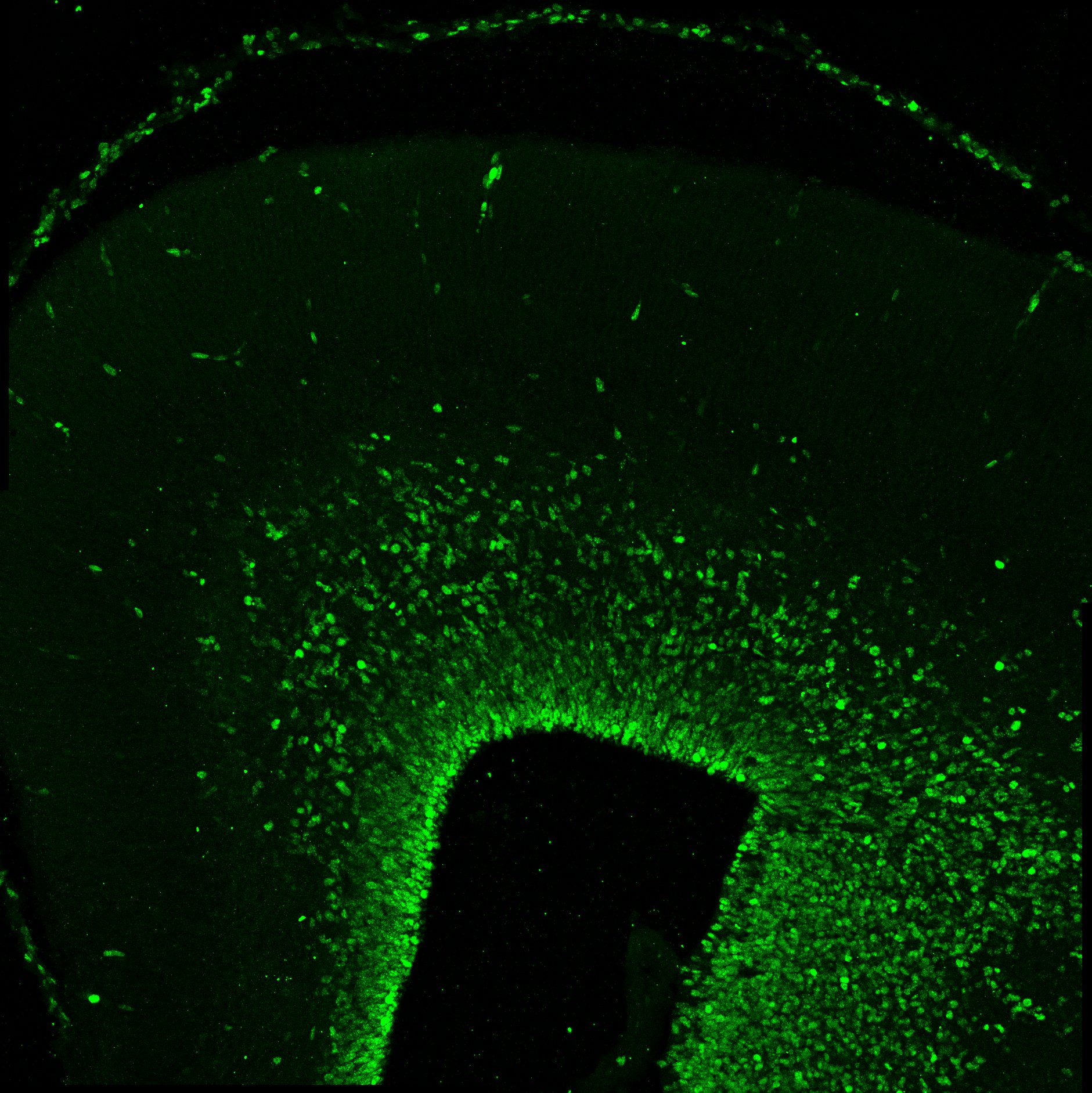
![Immunohistochemistry: Ki67/MKI67 Antibody (SP6) - Unpurified [NB600-1252] - Ki-67/MKI67 Antibody (SP6) [NB600-1252] - Ki67 staining in mouse thyroid tissue at pre-tumor stage (green). Dilution is 1:100. This image was submitted via customer Review.](http://images.novusbio.com/fullsize/Ki67-MKI67-Antibody-SP6-Unpurified-Immunohistochemistry-NB600-1252-img0003.jpg "Immunohistochemistry: Ki67/MKI67 Antibody (SP6) - Unpurified [NB600-1252] - Ki-67/MKI67 Antibody (SP6) [NB600-1252] - Ki67 staining in mouse thyroid tissue at pre-tumor stage (green). Dilution is 1:100. This image was submitted via customer Review.")
![Immunohistochemistry-Paraffin: Ki67/MKI67 Antibody (SP6) - Unpurified [NB600-1252] - Ki-67 (NB600-1252) immunoreactivity in an FFPE section of mouse small intestine. Primary antibody was diluted 1:100 and left on sections for 1h at room temperature. Secondary antibody was Horse Anti-Rabbit HRP. Image from verified customer review.](http://images.novusbio.com/fullsize/nb600-1252_rabbit-monoclonal-ki67-mki67-antibody-sp6-unpurified-immunohistochemistry-paraffin-204202311437.jpg "Immunohistochemistry-Paraffin: Ki67/MKI67 Antibody (SP6) - Unpurified [NB600-1252] - Ki-67 (NB600-1252) immunoreactivity in an FFPE section of mouse small intestine. Primary antibody was diluted 1:100 and left on sections for 1h at room temperature. Secondary antibody was Horse Anti-Rabbit HRP. Image from verified customer review.")
 and (cleaved caspase-3, arrows; D-F) were quantified by counting the number of positively stained cells in 4 fields of view at x40. Images are representative of untreated (A, D), trastuzumab at 100 µg/ml (B and E) and docetaxel at 100 nM (C and F). Images were digitally scanned ×40 (AlexaSoft X-PRO).")
![Immunohistochemistry-Paraffin: Rabbit Monoclonal Ki67/MKI67 Antibody (SP6) - Unpurified [NB600-1252] - Image depicting Ki67/MKI67 immunoreactivity in a FFPE section of human tonsil. NB600-1252 was diluted 1-100 and left on tissue sections for 30m at room temperature. Secondary was horse anti rabbit HRP polymer. Image from a verified customer review.](http://images.novusbio.com/fullsize/nb600-1252_rabbit-monoclonal-ki67-mki67-antibody-sp6-unpurified-immunohistochemistry-paraffin-2442024135233..png "Immunohistochemistry-Paraffin: Rabbit Monoclonal Ki67/MKI67 Antibody (SP6) - Unpurified [NB600-1252] - Image depicting Ki67/MKI67 immunoreactivity in a FFPE section of human tonsil. NB600-1252 was diluted 1-100 and left on tissue sections for 30m at room temperature. Secondary was horse anti rabbit HRP polymer. Image from a verified customer review.")
![Immunohistochemistry-Paraffin: Rabbit Monoclonal Ki67/MKI67 Antibody (SP6) - Unpurified [IMGENEX: IMG-80336] [NB600-1252] - Images depicting Ki67/MKI67 immunoreactivity in FFPE sections of canine cecum and skin. NB600-1252 was diluted 1-100 and left on tissue sections for 30m at room temperature. Image from a verified customer review.](http://images.novusbio.com/fullsize/nb600-1252_rabbit-monoclonal-ki67-mki67-antibody-sp6-unpurified-imgenex-img-80336-immunohistochemistry-paraffin-1052024152017..jpg "Immunohistochemistry-Paraffin: Rabbit Monoclonal Ki67/MKI67 Antibody (SP6) - Unpurified [IMGENEX: IMG-80336] [NB600-1252] - Images depicting Ki67/MKI67 immunoreactivity in FFPE sections of canine cecum and skin. NB600-1252 was diluted 1-100 and left on tissue sections for 30m at room temperature. Image from a verified customer review.")
 - Unpurified - Images depicting Ki67/MKI67 immunoreactivity in FFPE sections of equine intestine and spleen. NB600-1252 was diluted 1-100 and left on tissue sections for 30m at room temperature. Image from a verified customer review.")
 Representative images of immunohistological staining (brown) of SOX2+ olfactory receptor neurons (ORN) progenitor cells, Gap43+ immature ORNs, Ki67+ proliferating cells & cleaved caspase-3+ (Cas3+) apoptotic cells. Tissue sections were counterstained with the nuclear dye hematoxylin (blue). Arrowheads indicate Cas3+ apoptotic cells in the OE (n = 6). (B) Numbers of SOX2+ ORN progenitors & Ki67+ actively proliferating cells per mm of the basal layer, & Gap43+ immature ORNs & Cas3+ apoptotic cells per mm of the OE in saline or CSS-treated mice. Data represent means ± SEM (n = 6). *P < 0.05; **P < 0.01; ***P < 0.001 compared with saline-treated mice (one-way ANOVA). Image collected & cropped by CiteAb from the following publication (//pubmed.ncbi.nlm.nih.gov/29950987), licensed under a CC-BY license. Not internally tested by Novus Biologicals.")
![Immunohistochemistry-Paraffin: Ki67/MKI67 Antibody (SP6) - Unpurified [NB600-1252] - Immunohistochemistry of tissue slices treated with trastuzumab & docetaxel. (Ki-67, arrows; A-C) & (cleaved caspase-3, arrows; D-F) were quantified by counting the number of positively stained cells in 4 fields of view at x40. Images are representative of untreated (A, D), trastuzumab at 100 µg/ml (B & E) & docetaxel at 100 nM (C & F). Images were digitally scanned ×40 (AlexaSoft X-PRO). Image collected & cropped by CiteAb from the following publication (//www.spandidos-publications.com/10.3892/or.2015.4074), licensed under a CC-BY license. Not internally tested by Novus Biologicals.](http://images.novusbio.com/fullsize/nb600-1252_rabbit-monoclonal-ki67-mki67-antibody-sp6-unpurified-310202415371921.jpg "Immunohistochemistry-Paraffin: Ki67/MKI67 Antibody (SP6) - Unpurified [NB600-1252] - Immunohistochemistry of tissue slices treated with trastuzumab & docetaxel. (Ki-67, arrows; A-C) & (cleaved caspase-3, arrows; D-F) were quantified by counting the number of positively stained cells in 4 fields of view at x40. Images are representative of untreated (A, D), trastuzumab at 100 µg/ml (B & E) & docetaxel at 100 nM (C & F). Images were digitally scanned ×40 (AlexaSoft X-PRO). Image collected & cropped by CiteAb from the following publication (//www.spandidos-publications.com/10.3892/or.2015.4074), licensed under a CC-BY license. Not internally tested by Novus Biologicals.")
![Immunohistochemistry: Ki67/MKI67 Antibody (SP6) - Unpurified [NB600-1252] - (A–C) Inflammatory cell infiltration & cell division in the nasal RM were evaluated using immunohistochemical staining (brown). Tissue sections were counterstained with the nuclear dye hematoxylin (blue). Representative immunohistochemical images of neutrophils (A), CD3+ lymphocytes (B), & Ki67+ dividing cells (C) (400× magnification), & comparative charts of cell counts in each group. *P < 0.01; **P < 0.01; ****P < 0.0001 (n = 6, one-way analysis of variance). OVA, ovalbumin; CSS, cigarette smoke solution. Image collected & cropped by CiteAb from the following publication (//pubmed.ncbi.nlm.nih.gov/32132898), licensed under a CC-BY license. Not internally tested by Novus Biologicals.](http://images.novusbio.com/fullsize/nb600-1252_rabbit-monoclonal-ki67-mki67-antibody-sp6-unpurified-310202415284528.jpg "Immunohistochemistry: Ki67/MKI67 Antibody (SP6) - Unpurified [NB600-1252] - (A–C) Inflammatory cell infiltration & cell division in the nasal RM were evaluated using immunohistochemical staining (brown). Tissue sections were counterstained with the nuclear dye hematoxylin (blue). Representative immunohistochemical images of neutrophils (A), CD3+ lymphocytes (B), & Ki67+ dividing cells (C) (400× magnification), & comparative charts of cell counts in each group. *P < 0.01; **P < 0.01; ****P < 0.0001 (n = 6, one-way analysis of variance). OVA, ovalbumin; CSS, cigarette smoke solution. Image collected & cropped by CiteAb from the following publication (//pubmed.ncbi.nlm.nih.gov/32132898), licensed under a CC-BY license. Not internally tested by Novus Biologicals.")
![Immunohistochemistry: Ki67/MKI67 Antibody (SP6) - Unpurified [NB600-1252] - In vivo anti-tumor effect of Y-TR1 in the NOD/SCID mouse xenograft model using CD26 positive MM cell line JMN. (A) Y-TR1 administered intraperitoneally 4 mg/kg/dose, three times per week, for a total of nine doses from day zero of subcutaneous inoculation of 1 × 10⁷ JMN cells. Average estimated tumor volume on day 55 compared among three groups (control, YS110, Y-TR1, n = 10) w/ Fisher’s protected least protected difference multiple comparison test. Mean tumor volume of the Y-TR1 group significantly lower (* p < 0.05) than that of the control or YS110 group. Mean tumor volume of the YS110 group not significantly altered compared w/ the control. An experiment out of two w/ similar results is shown; (B) Y-TR1 administered intraperitoneally 8 mg/kg/dose, three times per week, for a total of nine doses. The average estimated tumor weight on day 42 compared among three groups (control, 14D10, YS110, Y-TR1, n = 10) w/ Fisher’s protected least protected difference multiple comparison test. Mean tumor weight of the YS110 or Y-TR1 groups significantly lower (* p < 0.05 or ** p < 0.025, respectively) than that of the control group. Mean tumor weight of the Y-TR1 group significantly lower (* p < 0.05) than that of the YS110 group. An experiment out of two w/ similar results is shown; (C) histological analysis of xenograft tumors of JMN cells. JMN-derived tumors show histopathology of sarcomatoid mesothelioma. (×20). a: Hematoxylin & eosin staining. b: Immunohistochemical staining w/ anti-human CD26 antibody revealed CD26 expression in tumor cells. c–e: MIB-1 (Ki67) staining showed a decreased number of MIB-1-positive cells in Y-TR1-treated tumors compared to IgG1- or YS110-treated tumors. Scale bar: 10 μm. Image collected & cropped by CiteAb from the following publication (//pubmed.ncbi.nlm.nih.gov/31398954), licensed under a CC-BY license. Not internally tested by Novus Biologicals.](http://images.novusbio.com/fullsize/nb600-1252_rabbit-monoclonal-ki67-mki67-antibody-sp6-unpurified-310202416165616.jpg "Immunohistochemistry: Ki67/MKI67 Antibody (SP6) - Unpurified [NB600-1252] - In vivo anti-tumor effect of Y-TR1 in the NOD/SCID mouse xenograft model using CD26 positive MM cell line JMN. (A) Y-TR1 administered intraperitoneally 4 mg/kg/dose, three times per week, for a total of nine doses from day zero of subcutaneous inoculation of 1 × 10⁷ JMN cells. Average estimated tumor volume on day 55 compared among three groups (control, YS110, Y-TR1, n = 10) w/ Fisher’s protected least protected difference multiple comparison test. Mean tumor volume of the Y-TR1 group significantly lower (* p < 0.05) than that of the control or YS110 group. Mean tumor volume of the YS110 group not significantly altered compared w/ the control. An experiment out of two w/ similar results is shown; (B) Y-TR1 administered intraperitoneally 8 mg/kg/dose, three times per week, for a total of nine doses. The average estimated tumor weight on day 42 compared among three groups (control, 14D10, YS110, Y-TR1, n = 10) w/ Fisher’s protected least protected difference multiple comparison test. Mean tumor weight of the YS110 or Y-TR1 groups significantly lower (* p < 0.05 or ** p < 0.025, respectively) than that of the control group. Mean tumor weight of the Y-TR1 group significantly lower (* p < 0.05) than that of the YS110 group. An experiment out of two w/ similar results is shown; (C) histological analysis of xenograft tumors of JMN cells. JMN-derived tumors show histopathology of sarcomatoid mesothelioma. (×20). a: Hematoxylin & eosin staining. b: Immunohistochemical staining w/ anti-human CD26 antibody revealed CD26 expression in tumor cells. c–e: MIB-1 (Ki67) staining showed a decreased number of MIB-1-positive cells in Y-TR1-treated tumors compared to IgG1- or YS110-treated tumors. Scale bar: 10 μm. Image collected & cropped by CiteAb from the following publication (//pubmed.ncbi.nlm.nih.gov/31398954), licensed under a CC-BY license. Not internally tested by Novus Biologicals.")
![Immunohistochemistry: Ki67/MKI67 Antibody (SP6) - Unpurified [NB600-1252] - Representative images of immunohistological staining (brown) of OMP-positive (OMP+) cells (A), SOX2+ ORN progenitor cells (B), GAP43+ immature ORNs (C), Ki67+ proliferating cells (D), & cleaved Cas3+ apoptotic cells (E). Each cell except for many OMP+ cells is indicated by arrows. Tissue sections were counterstained with the nuclear dye hematoxylin (blue). Numbers of SOX2+ ORN progenitors & Ki67+ actively proliferating cells per mm of the basal layer & OMP+ mature ORNs, GAP43+ immature ORNs, & Cas3+ apoptotic cells per mm of the OE in saline or rhIGF-1-treated mice. Open circles, rectangles, & triangles represent the values for each mouse in the saline, low-IGF-1, & high-IGF-1 treated groups (each n = 6), respectively. The horizontal lines represent the mean value for each group. ∗P < 0.05; ∗∗P < 0.01; ∗∗∗P < 0.001; & ∗∗∗∗P < 0.0001 (one-way ANOVA). Image collected & cropped by CiteAb from the following publication (//pubmed.ncbi.nlm.nih.gov/30515092), licensed under a CC-BY license. Not internally tested by Novus Biologicals.](http://images.novusbio.com/fullsize/nb600-1252_rabbit-monoclonal-ki67-mki67-antibody-sp6-unpurified-310202416182632.jpg "Immunohistochemistry: Ki67/MKI67 Antibody (SP6) - Unpurified [NB600-1252] - Representative images of immunohistological staining (brown) of OMP-positive (OMP+) cells (A), SOX2+ ORN progenitor cells (B), GAP43+ immature ORNs (C), Ki67+ proliferating cells (D), & cleaved Cas3+ apoptotic cells (E). Each cell except for many OMP+ cells is indicated by arrows. Tissue sections were counterstained with the nuclear dye hematoxylin (blue). Numbers of SOX2+ ORN progenitors & Ki67+ actively proliferating cells per mm of the basal layer & OMP+ mature ORNs, GAP43+ immature ORNs, & Cas3+ apoptotic cells per mm of the OE in saline or rhIGF-1-treated mice. Open circles, rectangles, & triangles represent the values for each mouse in the saline, low-IGF-1, & high-IGF-1 treated groups (each n = 6), respectively. The horizontal lines represent the mean value for each group. ∗P < 0.05; ∗∗P < 0.01; ∗∗∗P < 0.001; & ∗∗∗∗P < 0.0001 (one-way ANOVA). Image collected & cropped by CiteAb from the following publication (//pubmed.ncbi.nlm.nih.gov/30515092), licensed under a CC-BY license. Not internally tested by Novus Biologicals.")
![Immunohistochemistry: Ki67/MKI67 Antibody (SP6) - Unpurified [NB600-1252] - (A) Serum immunoglobin E (IgE) levels of the control & cigarette smoke solution (CSS)-treated mice were determined by enzyme linked immunosorbent assay. (B) Representative images of Sirius red staining for eosinophils & periodic acid-Schiff & Alcian blue (PAS/AB) staining for goblet cells in the nasal RM of the control mice & mice treated with 10 doses of CSS (CSS 10). (C) Representative immunohistochemical images of neutrophils & Ki67+ dividing cells in the nasal RM (400× magnification), & comparative charts of Ki67+ cell counts (n = 6, Mann–Whitney U test). (D) Representative images of olfactory marker protein (OMP)+ mature olfactory receptor neurons (ORNs) in two different areas of the olfactory epithelium: the nasal septum & upper lateral area. There were no significant differences in the number of OMP+ mature ORNs between the control & CSS-treated mice (n = 6, Mann–Whitney U test). OVA, ovalbumin. Image collected & cropped by CiteAb from the following publication (//pubmed.ncbi.nlm.nih.gov/32132898), licensed under a CC-BY license. Not internally tested by Novus Biologicals.](http://images.novusbio.com/fullsize/nb600-1252_rabbit-monoclonal-ki67-mki67-antibody-sp6-unpurified-310202416173541.jpg "Immunohistochemistry: Ki67/MKI67 Antibody (SP6) - Unpurified [NB600-1252] - (A) Serum immunoglobin E (IgE) levels of the control & cigarette smoke solution (CSS)-treated mice were determined by enzyme linked immunosorbent assay. (B) Representative images of Sirius red staining for eosinophils & periodic acid-Schiff & Alcian blue (PAS/AB) staining for goblet cells in the nasal RM of the control mice & mice treated with 10 doses of CSS (CSS 10). (C) Representative immunohistochemical images of neutrophils & Ki67+ dividing cells in the nasal RM (400× magnification), & comparative charts of Ki67+ cell counts (n = 6, Mann–Whitney U test). (D) Representative images of olfactory marker protein (OMP)+ mature olfactory receptor neurons (ORNs) in two different areas of the olfactory epithelium: the nasal septum & upper lateral area. There were no significant differences in the number of OMP+ mature ORNs between the control & CSS-treated mice (n = 6, Mann–Whitney U test). OVA, ovalbumin. Image collected & cropped by CiteAb from the following publication (//pubmed.ncbi.nlm.nih.gov/32132898), licensed under a CC-BY license. Not internally tested by Novus Biologicals.")
![Immunohistochemistry: Ki67/MKI67 Antibody (SP6) - Unpurified [NB600-1252] - (A,B) Representative images of hematoxylin & eosin (H&E)-stained sections of the olfactory epithelium from young adult mice (A, 40× magnification; B, 400× magnification). A black line in (A) indicates the range for counting the number of each cell type. The box in (A) indicates the region of the olfactory epithelium shown at a representative higher magnification in (B). Differences in the number of OMP+ mature olfactory receptor neurons (ORNs) (C), SOX2+ ORN progenitors (D), Ki-67+ proliferating cells (E), GAP43+ immature ORNs (F), & cleaved Cas3+ apoptotic cells (G) in the OE were evaluated by immunohistological staining (brown). Tissue sections were counterstained with the nuclear dye hematoxylin (blue). Representative images (400× magnification) of tissues stained with antibodies against olfactory marker protein (OMP), SRY (sex determining region Y)-box 2 (SOX2), Ki-67 (antigen identified by monoclonal antibody Ki-67), growth associated protein 43 (GAP43), & cleaved caspase 3 (CAS3) are shown. The number of cells per mm of basal layer length (C–G) was counted manually. Data represent the mean ± SD. **P < 0.01 (n = 6, Mann–Whitney U-test). Image collected & cropped by CiteAb from the following publication (//journal.frontiersin.org/article/10.3389/fnagi.2018.00086/full), licensed under a CC-BY license. Not internally tested by Novus Biologicals.](http://images.novusbio.com/fullsize/nb600-1252_rabbit-monoclonal-ki67-mki67-antibody-sp6-unpurified-310202416161841.jpg "Immunohistochemistry: Ki67/MKI67 Antibody (SP6) - Unpurified [NB600-1252] - (A,B) Representative images of hematoxylin & eosin (H&E)-stained sections of the olfactory epithelium from young adult mice (A, 40× magnification; B, 400× magnification). A black line in (A) indicates the range for counting the number of each cell type. The box in (A) indicates the region of the olfactory epithelium shown at a representative higher magnification in (B). Differences in the number of OMP+ mature olfactory receptor neurons (ORNs) (C), SOX2+ ORN progenitors (D), Ki-67+ proliferating cells (E), GAP43+ immature ORNs (F), & cleaved Cas3+ apoptotic cells (G) in the OE were evaluated by immunohistological staining (brown). Tissue sections were counterstained with the nuclear dye hematoxylin (blue). Representative images (400× magnification) of tissues stained with antibodies against olfactory marker protein (OMP), SRY (sex determining region Y)-box 2 (SOX2), Ki-67 (antigen identified by monoclonal antibody Ki-67), growth associated protein 43 (GAP43), & cleaved caspase 3 (CAS3) are shown. The number of cells per mm of basal layer length (C–G) was counted manually. Data represent the mean ± SD. **P < 0.01 (n = 6, Mann–Whitney U-test). Image collected & cropped by CiteAb from the following publication (//journal.frontiersin.org/article/10.3389/fnagi.2018.00086/full), licensed under a CC-BY license. Not internally tested by Novus Biologicals.")


![Bioactivity ErbB2/Her2 [Unconjugated]](https://images.novusbio.com/images/protein/1129-er_recombinant-human-erbb2-her2-fc-chimera-protein-cf-bioactivity-7122020142841.jpg)
![SDS-Page ErbB2/Her2 [Unconjugated]](https://images.novusbio.com/images/protein/ErbB2_1129-ER_70.jpg)


![N/A MMP-9 [HRP]](https://images.novusbio.com/images/elisa/DATA_MMP9_DMP900_ELISA_770.jpg)
![N/A MMP-9 [HRP]](https://images.novusbio.com/images/elisa/MMP-9_DMP900_ELISA_148.jpg)


![SDS-Page TNF-alpha [Unconjugated]](https://images.novusbio.com/images/protein/TNF-alpha_210-TA_256.jpg)
![Bioactivity TNF-alpha [Unconjugated]](https://images.novusbio.com/images/protein/TNFalpha_210TA_1658.jpg)
![SEC-MALS TNF-alpha [Unconjugated]](https://images.novusbio.com/images/210-ta_recombinant-human-tnf-alpha-protein-sec-mals-35202312244..jpg)
![Western Blot: Goat anti-Rabbit IgG (H+L) Secondary Antibody [HRP] [NB7160] - Western blot showing vemurafenib treatment in BRAFV600E CRC cells inhibits fission mediator DRP1 with no significant effect on fusion proteins (Mfn1 & 2) using MFN-1 antibody (NBP1-51841) and corresponding secondary antibody, goat anti-rabbit IgG-HRP (NB7160). Image collected and cropped by CiteAb from the following publication (https://pubmed.ncbi.nlm.nih.gov/33738242).](https://images.novusbio.com/images/Goat-anti-Rabbit-IgG-H+L-Secondary-Antibody-HRP-Western-Blot-NB7160-img0001.jpg "Western Blot: Goat anti-Rabbit IgG (H+L) Secondary Antibody [HRP] [NB7160] - Western blot showing vemurafenib treatment in BRAFV600E CRC cells inhibits fission mediator DRP1 with no significant effect on fusion proteins (Mfn1 & 2) using MFN-1 antibody (NBP1-51841) and corresponding secondary antibody, goat anti-rabbit IgG-HRP (NB7160). Image collected and cropped by CiteAb from the following publication (https://pubmed.ncbi.nlm.nih.gov/33738242).")
 followed by 30 min incubation with Goat anti Rabbit HRP conjugated secondary antibodies (Catalog # HAF008) at 1:20 dilution + DAB chromogen (brown). The tissue was counterstained with Hematoxylin (blue). Control was done by omitting primary antibody.")
![Flow Cytometry: Rabbit IgG Isotype Control [NBP2-24891] - An intracellular stain was performed on Raji cells with Adiponectin antibody NB100-65810 (blue) and a matched isotype control NBP2-24893 (orange). Cells were fixed with 4% PFA and then permeablized with 0.1% saponin. Cells were incubated in an antibody dilution of 1 ug/mL for 30 minutes at room temperature, followed by Dylight488-conjugated anti-rabbit secondary antibody. Image using the Azide Free form of this antibody.](https://images.novusbio.com/images/Rabbit--Mouse-IgG-Isotype-Control-Flow-Cytometry-NBP2-24891-img0006.jpg "Flow Cytometry: Rabbit IgG Isotype Control [NBP2-24891] - An intracellular stain was performed on Raji cells with Adiponectin antibody NB100-65810 (blue) and a matched isotype control NBP2-24893 (orange). Cells were fixed with 4% PFA and then permeablized with 0.1% saponin. Cells were incubated in an antibody dilution of 1 ug/mL for 30 minutes at room temperature, followed by Dylight488-conjugated anti-rabbit secondary antibody. Image using the Azide Free form of this antibody.")


