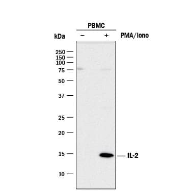
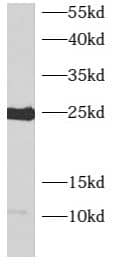
 HAF017). A specific band was detected for IL-2 at approximately 14 kDa (as indicated). This experiment was conducted under reducing conditions and using Immunoblot Buffer Group 1." title="Western blot shows lysates of monensin treated human peripheral blood mononuclear cells (PBMCs) with no additional treatment (-) or additionally treated (+) with 0.5 μg/mL calcium ionomycin (Iono) and 50 ng/mL PMA overnight. PVDF membrane was probed with 2 µg/mL of Goat Anti-Human/Rat IL-2 Antigen Affinity-purified Polyclonal Antibody (Catalog # AF-502-NA) followed by HRP-conjugated Anti-Goat IgG Secondary Antibody (HAF017). A specific band was detected for IL-2 at approximately 14 kDa (as indicated). This experiment was conducted under reducing conditions and using Immunoblot Buffer Group 1." />
| Reactivity | Hu, RtSpecies Glossary |
| Applications | WB, IHC, B/N, ELISA(Cap), ICC/IF |
| Clonality | Polyclonal |
| Host | Goat |
| Conjugate | Unconjugated |
| Concentration | LYOPH |
| Immunogen | E. coli-derived recombinant rat IL-2 Ala21-Gln155 Accession # P17108 |
| Specificity | Detects rat IL-2 in ELISAs. Detects human and rat IL-2 in Western blots. In sandwich immunoassays, less than 0.2% cross-reactivity with recombinant mouse IL‑2 is observed. |
| Source | N/A |
| Isotype | IgG |
| Clonality | Polyclonal |
| Host | Goat |
| Gene | IL2 |
| Purity Statement | Antigen Affinity-purified |
| Endotoxin Note | <0.10 EU per 1 μg of the antibody by the LAL method. |
| Innovator's Reward | Test in a species/application not listed above to receive a full credit towards a future purchase. |
| Dilutions |
|
|
| Application Notes | ELISA Capture: Rat IL-2 Antibody (Catalog # AF-502-NA) ELISA Detection: Rat IL-2 Biotinylated Antibody (Catalog # BAF502) Standard: Recombinant Rat IL-2 (Catalog # 502-RL) |
|
| Publications |
|
| Storage | Use a manual defrost freezer and avoid repeated freeze-thaw cycles.
|
| Buffer | Lyophilized from a 0.2 μm filtered solution in PBS with Trehalose. *Small pack size (SP) is supplied either lyophilized or as a 0.2 µm filtered solution in PBS. |
| Preservative | No Preservative |
| Concentration | LYOPH |
| Reconstitution Instructions | Reconstitute at 0.2 mg/mL in sterile PBS. |


Secondary Antibodies |
Isotype Controls |
|
The role of HIF-2 alpha in the progression and therapy of clear cell renal cell carcinoma HIF-2 alpha, also known as hypoxia-inducible factor 2, endothelial PAS domain protein-1, and member of PAS superfamily 2 is part of the HIF family of proteins. The HIF family is composed of HIF-1, HIF-2 and HIF-3, where HIF-2 is a dimeric protein ... Read full blog post. |
|
iNOS: The Nitric Oxide Boss Nitric oxide (NO) is an inorganic, gaseous, and reactive free radical that acts as a biologic mediator in processes such as neurotransmission, vasorelaxation, and cytotoxicity. In addition, it has antimicrobial and anti-tumoral activities. NO producti... Read full blog post. |
The concentration calculator allows you to quickly calculate the volume, mass or concentration of your vial. Simply enter your mass, volume, or concentration values for your reagent and the calculator will determine the rest.


 NL001) and counterstained with DAPI (blue). Specific staining was localized to cytoplasm. View our protocol for
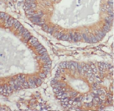
  CTS008) and counterstained with hematoxylin (blue). Specific staining was localized to cytoplasm in lymphocytes. View our protocol for
  502-RL) stimulates proliferation in the CTLL-2 mouse cytotoxic T cell line in a dose-dependent manner (orange line). Proliferation elicited by Recombinant Rat IL-2 (2 ng/mL) is neutralized (green line) by increasing concentrations of Goat Anti-Human/Rat IL-2 Antigen Affinity-purified Polyclonal Antibody (Catalog # AF-502-NA). The ND50 is typically 0.15-0.75 µg/mL." title="Recombinant Rat IL-2 (
 VC004). Before incubation with the primary antibody, tissue was subjected to heat-induced epitope retrieval using VisUCyte Antigen Retrieval Reagent-Basic (Catalog #
  502-RL) was serially diluted and captured by Goat Anti-Human/Rat IL‑2 Antigen Affinity-purified Polyclonal Antibody (Catalog # AF-502-NA) coated on a Clear Polystyrene Microplate (Catalog #
  502-RL) was serially diluted and captured by Goat Anti-Human/Rat IL‑2 Antigen Affinity-purified Polyclonal Antibody (Catalog #
 ![Bioactivity IL-4 [Unconjugated]](https://images.novusbio.com/images/protein/6507-ilcf_recombinant-human-il-4-cho-expressed-protein-cf-bioactivity-272020133214.jpg)
![SDS-Page TNF-alpha [Unconjugated]](https://images.novusbio.com/images/protein/TNF-alpha_210-TA_256.jpg)
![Bioactivity TNF-alpha [Unconjugated]](https://images.novusbio.com/images/protein/TNFalpha_210TA_1658.jpg)
![SEC-MALS TNF-alpha [Unconjugated]](https://images.novusbio.com/images/210-ta_recombinant-human-tnf-alpha-protein-sec-mals-35202312244..jpg)
![SEC-MALS IFN-gamma [Unconjugated]](https://images.novusbio.com/images/485-mi_recombinant-mouse-ifn-gamma-protein-sec-mals-1612202583245.jpg)
![Bioactivity IFN-gamma [Unconjugated]](https://images.novusbio.com/images/protein/IFN-gamma_485-MI_455.jpg)
![SDS-Page IFN-gamma [Unconjugated]](https://images.novusbio.com/images/protein/IFN-gamma_485-MI_407.jpg)
![N/A IL-10 [Biotin]](https://images.novusbio.com/images/elisa/DATA_IL10_DY417_ELISA_2014.jpg)
![N/A IL-6 [HRP]](https://images.novusbio.com/images/elisa/DATA_IL6_M6000_ELISA_936.jpg)
![N/A IL-6 [HRP]](https://images.novusbio.com/images/elisa/IL-6_M6000_ELISA_415.jpg)
![N/A IL-6 [HRP]](https://images.novusbio.com/images/m6000b_mouse-il-6-quantikine-elisa-kit-1752025024034.jpg)
![Bioactivity CTLA-4 [Unconjugated]](https://images.novusbio.com/images/protein/CTLA4_7268CT_2293.jpg)
![N/A CD25/IL-2R alpha [HRP]](https://images.novusbio.com/images/elisa/IL-2_R_alpha_DR2A00_ELISA_167.jpg)
![N/A CD25/IL-2R alpha [HRP]](https://images.novusbio.com/images/elisa/DATA_CD25_DR2A00_ELISA_800.jpg)
![Bioactivity IL-1 beta/IL-1F2 [Unconjugated]](https://images.novusbio.com/images/protein/IL-1_beta_201-LB_306.jpg)
![SDS-Page IL-1 beta/IL-1F2 [Unconjugated]](https://images.novusbio.com/images/protein/IL-1_beta_201-LB_304.jpg)
![SEC-MALS IL-1 beta/IL-1F2 [Unconjugated]](https://images.novusbio.com/images/201-lb_recombinant-human-il-1-beta-il-1f2-protein-sec-mals-123202491556.jpg)
![N/A IL-5 [HRP]](https://images.novusbio.com/images/elisa/IL-5_M5000_ELISA_237.jpg)
![N/A IL-5 [HRP]](https://images.novusbio.com/images/elisa/DATA_IL5_M5000_ELISA_935.jpg)


![Agonist Activity CD28 Antibody (37407) [Unconjugated]](https://images.novusbio.com/images/antibody/CD28_MAB342_Functional_Assay_2393.jpg)
![Immunocytochemistry CD28 Antibody (37407) [Unconjugated]](https://images.novusbio.com/images/mab342_human-cd28-mab-clone-37407-immunocytochemistry-31102022134634.jpg)


![SEC-MALS IL-15 [Unconjugated]](https://images.novusbio.com/images/247-ilb_recombinant-human-il-15-protein-sec-mals-2922024964..jpg)
![SEC-MALS IL-3 [Unconjugated]](https://images.novusbio.com/images/203-il_recombinant-human-il-3-protein-sec-mals-133202482656..jpg)
![Bioactivity IL-3 [Unconjugated]](https://images.novusbio.com/images/protein/IL-3_203-IL_347.jpg)
![Data IL-3 [Unconjugated]](https://images.novusbio.com/images/203-il_recombinant-human-il-3-protein-data-253202610550.jpg)