 HAF016). A specific band was detected for Glypican 3 at approximately 75 kDa (as indicated). This experiment was conducted under reducing conditions and using Immunoblot Buffer Group 1." title="Western blot shows lysates of HepG2 human hepatocellular carcinoma cell line. PVDF membrane was probed with 2 µg/mL of Sheep Anti-Human Glypican 3 Antigen Affinity-purified Polyclonal Antibody (Catalog # AF2119) followed by HRP-conjugated Anti-Sheep IgG Secondary Antibody (Catalog # HAF016). A specific band was detected for Glypican 3 at approximately 75 kDa (as indicated). This experiment was conducted under reducing conditions and using Immunoblot Buffer Group 1." />
| Applications | WB, ELISA, Flow, IHC, CyTOF-ready, ICC/IF |
| Clonality | Polyclonal |
| Host | Sheep |
| Conjugate | Unconjugated |
| Concentration | LYOPH |
| Immunogen | Mouse myeloma cell line NS0-derived recombinant human Glypican 3 Gln25-Val558 Accession # P51654 |
| Specificity | Detects human Glypican 3 in direct ELISAs and Western blots. In direct ELISAs, less than 10% cross-reactivity with recombinant mouse Glypican 3 is observed and less than 5% cross-reactivity with recombinant human (rh) Glypican 2, rhGlypican 5, and rhGlypican 6 is observed. |
| Source | N/A |
| Isotype | IgG |
| Clonality | Polyclonal |
| Host | Sheep |
| Gene | GPC3 |
| Purity Statement | Antigen Affinity-purified |
| Innovator's Reward | Test in a species/application not listed above to receive a full credit towards a future purchase. |
| Dilutions |
|
|
| Application Notes | Use in IHC-Fr at 1:50 dilution with IF detection for Rat frozen tissues is reported by Grozdanov et al. in PMID 17117158. |
|
| Publications |
|
| Storage | Use a manual defrost freezer and avoid repeated freeze-thaw cycles.
|
| Buffer | Lyophilized from a 0.2 μm filtered solution in PBS with Trehalose. *Small pack size (SP) is supplied either lyophilized or as a 0.2 µm filtered solution in PBS. |
| Preservative | No Preservative |
| Concentration | LYOPH |
| Reconstitution Instructions | Reconstitute at 0.2 mg/mL in sterile PBS. |
Glypicans (GPC) are a family of heparan sulfate proteoglycans that are attached to the cell surface by a glycosylphosphatidylinositol (GPI) anchor. Six members of this family have been identified in mammals (GPC1-GPC6). All glypican core proteins contain an N-terminal signal peptide, a large globular cysteine-rich domain (CRD) with 14 invariant cysteine residues, a stalk-like region containing the heparan sulfate attachment sites, and a C-terminal GPI attachment site. While glypican proteins do not share strong amino acid sequence identity (they range from 17-63%), the conserved cysteine residues in their CRDs suggests similarity in their three‑dimensional structure (1, 2).
Mutations in GPC3 cause a rare disorder in humans, Simpson-Golabi-Behmel Syndrome, which is characterized by pre and postnatal overgrowth of multiple tissues and organs and an increased risk for developing embryonic tumors (3). These features are also present in the mouse knock-out of GPC3 indicating that GPC3 regulates cell survival and inhibits cell proliferation during development (4). Glypican 3 has been implicated in regulating many different signaling pathways including: IGF, FGF, BMP, and Wnt. An endoproteolytic processing of GPC3 by proprotein convertases is required for the modulation of Wnt signaling (5). Direct interaction with FGF-basic has been observed and is mediated by the heparan sulfate chains (6).
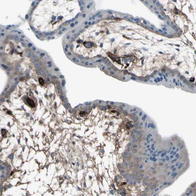
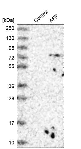
![Western Blot alpha-Fetoprotein/AFP Antibody (189502) [Unconjugated]](https://images.novusbio.com/images/antibody/alphaFetoprotein_MAB1368_Western_Blot_20276.jpg)
![Immunocytochemistry/ Immunofluorescence alpha-Fetoprotein/AFP Antibody (189502) [Unconjugated]](https://images.novusbio.com/images/mab1368_human-mouse-alpha-fetoprotein-mab-clone-189502-41202410325519.jpg)
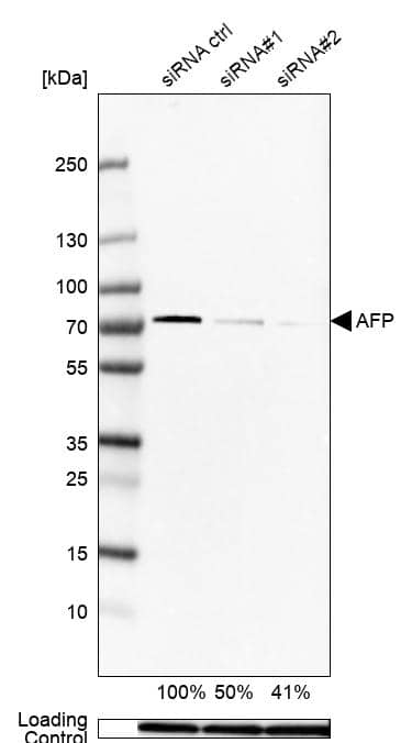
![Knockout Validated alpha-Fetoprotein/AFP Antibody (189502) [Unconjugated]](https://images.novusbio.com/images/antibody/alphaFetoprotein_MAB1368_Knockout_Validated_21503.jpg)


![Immunoprecipitation CD44 Antibody (2C5) [Unconjugated] - s Pan Specific](https://images.novusbio.com/images/antibody/bba10_human-cd44s-pan-specific-mab-clone-2c5-immunoprecipitation-2852025105258..png)
![Knockout Validated CD44 Antibody (2C5) [Unconjugated] - s Pan Specific](https://images.novusbio.com/images/antibody/bba10_human-cd44s-pan-specific-mab-clone-2c5-knockout-validated-285202510529..png)
![Western Blot CD44 Antibody (2C5) [Unconjugated] - s Pan Specific](https://images.novusbio.com/images/antibody/CD44_BBA10_Western_Blot_21459.jpg)


![Glypican 3 Antibody [Unconjugated]](/sites/all/modules/enterprise-tech/et_datasheets/images/novus_guarantee.png "Glypican 3 Antibody [Unconjugated]")
Secondary Antibodies |
Isotype Controls |
|
Glypican 3 as a biomarker for gastro-esophageal adenocarcinoma By Jamshed Arslan, Pharm. D., PhD. Gastroesophageal adenocarcinoma originates from the glandular epithelium of the esophagus, gastroesophageal junction and stomach. The incidence of gastroesophageal adenocarcinoma is ... Read full blog post. |
The concentration calculator allows you to quickly calculate the volume, mass or concentration of your vial. Simply enter your mass, volume, or concentration values for your reagent and the calculator will determine the rest.


 NL010) and counterstained with DAPI (blue). Specific staining was localized to cytoplasm and cell membranes. View our protocol for
  CTS019) and counterstained with hematoxylin (blue). View our protocol for
  CTS019) and counterstained with hematoxylin (blue). Lower panel shows a lack of labeling if primary antibodies are omitted and tissue is stained only with secondary antibody followed by incubation with detection reagents. View our protocol for
  MAB21191) coated on a Clear Polystyrene Microplate (
 2119-GP) was serially diluted and captured by Mouse Anti-Human Glypican 3 Monoclonal Antibody (Catalog #
 ![Flow Cytometry Syndecan-2/CD362 Antibody (305515) [Unconjugated]](https://images.novusbio.com/images/antibody/Syndecan2_MAB2965_Flow_Cytometry_20268.jpg)
![Flow Cytometry Glypican 1 Antibody [Unconjugated]](https://images.novusbio.com/images/antibody/Glypican_1_AF4519_Flow_Cytometry_8254.jpg)
![Immunocytochemistry/ Immunofluorescence Glypican 1 Antibody [Unconjugated]](https://images.novusbio.com/images/af4519_human-glypican-1-affinity-purified-polyclonal-ab-immunocytochemistry-immunofluorescence-1212202582239.jpg)
![Immunocytochemistry Glypican 1 Antibody [Unconjugated]](https://images.novusbio.com/images/antibody/Glypican_1_AF4519_Flow_Cytometry_17434.jpg)


![SDS-Page IGF-II/IGF2 [Unconjugated]](https://images.novusbio.com/images/protein/IGF-II_292-G2_668.jpg)
![Bioactivity IGF-II/IGF2 [Unconjugated]](https://images.novusbio.com/images/protein/IGF-II_292-G2_669.jpg)


![Western Blot Decorin Antibody [Unconjugated]](https://images.novusbio.com/images/antibody/Decorin_AF1060_Western_Blot_19006.jpg)


![Immunohistochemistry Insulin Antibody (182410) [Unconjugated]](https://images.novusbio.com/images/antibody/mab1417_human-bovine-mouse-insulin-mab-clone-182410-immunohistochemistry-308202115145.jpg)
![Immunocytochemistry Insulin Antibody (182410) [Unconjugated]](https://images.novusbio.com/images/antibody/Insulin_MAB1417_Immunocytochemistry_9376.jpg)


