 HAF016). A specific band was detected for Aminopeptidase N/CD13 at approximately 150 kDa (as indicated). This experiment was conducted under reducing conditions and using Immunoblot Buffer Group 1." title="Western blot shows lysates of human kidney tissue and human prostate tissue. PVDF membrane was probed with 0.5 µg/mL of Sheep Anti-Human Aminopeptidase N/CD13 Antigen Affinity-purified Polyclonal Antibody (Catalog # AF3815) followed by HRP-conjugated Anti-Sheep IgG Secondary Antibody (Catalog # HAF016). A specific band was detected for Aminopeptidase N/CD13 at approximately 150 kDa (as indicated). This experiment was conducted under reducing conditions and using Immunoblot Buffer Group 1." />
| Reactivity | HuSpecies Glossary |
| Applications | WB, Simple Western, Flow, IHC, IP, CyTOF-ready, KO |
| Clonality | Polyclonal |
| Host | Sheep |
| Conjugate | Unconjugated |
| Concentration | LYOPH |
| Immunogen | Mouse myeloma cell line NS0-derived recombinant human Aminopeptidase N/CD13 Lys69-Lys967 Accession # P15144 |
| Specificity | Detects human Aminopeptidase N/CD13 in the direct ELISAs and Western blots. In direct ELISAs and Western blots, approximately 20% cross-reactivity with recombinant mouse APN is observed. |
| Source | N/A |
| Isotype | IgG |
| Clonality | Polyclonal |
| Host | Sheep |
| Gene | ANPEP |
| Purity Statement | Antigen Affinity-purified |
| Innovator's Reward | Test in a species/application not listed above to receive a full credit towards a future purchase. |
| Dilutions |
|
|
| Publications |
|
| Storage | Use a manual defrost freezer and avoid repeated freeze-thaw cycles.
|
| Buffer | Lyophilized from a 0.2 μm filtered solution in PBS with Trehalose. *Small pack size (SP) is supplied either lyophilized or as a 0.2 µm filtered solution in PBS. |
| Preservative | No Preservative |
| Concentration | LYOPH |
| Reconstitution Instructions | Reconstitute at 0.2 mg/mL in sterile PBS. |
The human ANPEP gene encodes aminopeptidase N (APN), which is also known as microsomal aminopeptiase, alanyl aminopeptidase, aminopeptidase M, CD13, or membrane protein p161 (1‑3). The deduced amino acid sequence of human APN consists of a short cytoplasmic tail (residues 2 to 8), a transmembrane region (residue 9 to 32), a Ser/Thr rich region and a zinc metalloprotease domain (residues 69 to 966). Widely expressed in many cells, tissues and species, APN cleaves the N-terminal amino acids from bioactive peptides, leading to their inactivation or degradation. The roles of APN in many fields, such as neuroscience, hematopoeitic cells, immune system, angiogenesis, cancer and viral infection, have been reviewed (3).


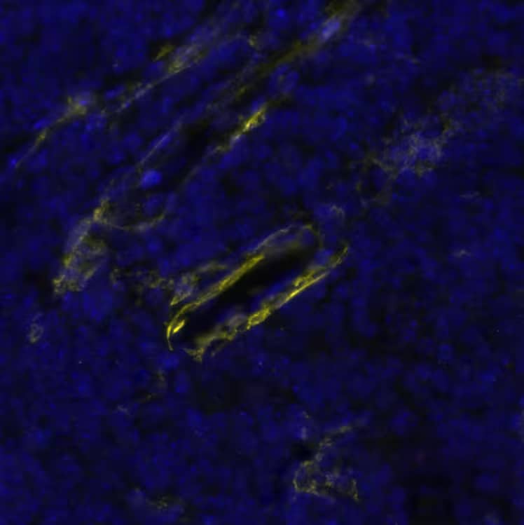


![Simple Western Myeloperoxidase/MPO Antibody [Unconjugated]](https://images.novusbio.com/images/antibody/Myeloperoxidase_AF3667_Simple_Western_16637.jpg)
![Immunohistochemistry Myeloperoxidase/MPO Antibody [Unconjugated]](https://images.novusbio.com/images/af3667_human-mouse-myeloperoxidase-affinity-purified-polyclonal-ab-8120255541727.jpg)
![Immunocytochemistry/ Immunofluorescence Myeloperoxidase/MPO Antibody [Unconjugated]](https://images.novusbio.com/images/af3667_human-mouse-myeloperoxidase-affinity-purified-polyclonal-ab-4120241032140.jpg)
![Aminopeptidase N/CD13 Antibody [Unconjugated]](/sites/all/modules/enterprise-tech/et_datasheets/images/novus_guarantee.png "Aminopeptidase N/CD13 Antibody [Unconjugated]")
Secondary Antibodies |
Isotype Controls |
The concentration calculator allows you to quickly calculate the volume, mass or concentration of your vial. Simply enter your mass, volume, or concentration values for your reagent and the calculator will determine the rest.


 5-001-A, open histogram), followed by APC-conjugated Anti-Sheep IgG Secondary Antibody (Catalog #
  CTS019) and counterstained with hematoxylin (blue). Specific staining was localized to convoluted tubules. View our protocol for
  HAF016). This experiment was conducted under reducing conditions and using the 12-230 kDa separation system." title="Simple Western lane view shows lysates of human small intestine tissue and human prostate tissue, loaded at 0.5 mg/mL. A specific band was detected for Aminopeptidase N/CD13 at approximately 177-198 kDa (as indicated) using 10 µg/mL of Sheep Anti-Human Aminopeptidase N/CD13 Antigen Affinity-purified Polyclonal Antibody (Catalog # AF3815) followed by 1:50 dilution of HRP-conjugated Anti-Sheep IgG Secondary Antibody (Catalog #
  HAF016). A specific band was detected for Aminopeptidase N/CD13 at approximately 150 kDa (as indicated) in the parental U937 human histiocytic lymphoma cell line, but is not detectable in knockout U937 human histiocytic lymphoma cell line. GAPDH (

![Bioactivity CTLA-4 [Unconjugated]](https://images.novusbio.com/images/protein/CTLA4_7268CT_2293.jpg)
![Immunohistochemistry Neprilysin/CD10 Antibody [Unconjugated]](https://images.novusbio.com/images/antibody/Neprilysin_AF1126_Immunohistochemistry_6679.jpg)
![Immunohistochemistry Neprilysin/CD10 Antibody [Unconjugated]](https://images.novusbio.com/images/antibody/af1126_mouse-neprilysin-affinity-purified-polyclonal-ab-immunohistochemistry-7520268331.png)
![N/A Neprilysin/CD10 Antibody [Unconjugated]](https://images.novusbio.com/images/antibody/af1126_mouse-neprilysin-affinity-purified-polyclonal-ab-7520268433.png)
![Immunocytochemistry/ Immunofluorescence DPPIV/CD26 Antibody [Unconjugated]](https://images.novusbio.com/images/af1180_human-dppiv-cd26-affinity-purified-polyclonal-ab-44202415425025.jpg)
![Western Blot DPPIV/CD26 Antibody [Unconjugated]](https://images.novusbio.com/images/af1180_human-dppiv-cd26-affinity-purified-polyclonal-ab-812025555283.jpg)
![Immunohistochemistry DPPIV/CD26 Antibody [Unconjugated]](https://images.novusbio.com/images/antibody/DPPIV_AF1180_Immunohistochemistry_17209.jpg)
![Flow Cytometry Integrin beta 2/CD18 Antibody [Unconjugated]](https://images.novusbio.com/images/antibody/af1730_human-integrin-beta-2-cd18-affinity-purified-polyclonal-ab-flow-cytometry-7220258626..jpg)
![Immunocytochemistry Integrin beta 2/CD18 Antibody [Unconjugated]](https://images.novusbio.com/images/antibody/Integrin_beta_2_AF1730_Immunocytochemistry_6474.jpg)
![Immunocytochemistry Integrin beta 2/CD18 Antibody [Unconjugated]](https://images.novusbio.com/images/antibody/af1730_human-integrin-beta-2-cd18-affinity-purified-polyclonal-ab-immunocytochemistry-1292023225343.jpg)
![N/A CD14 [HRP]](https://images.novusbio.com/images/elisa/DATA_CD14_DC140_ELISA_597.jpg)
![N/A CD14 [HRP]](https://images.novusbio.com/images/elisa/CD14_DC140_ELISA_42.jpg)
![Western Blot Integrin alpha L/CD11a Antibody (345913) [Unconjugated]](https://images.novusbio.com/images/antibody/Integrin_alpha_L_MAB3595_Western_Blot_10266.jpg)
![Simple Western Integrin alpha L/CD11a Antibody (345913) [Unconjugated]](https://images.novusbio.com/images/mab3595_human-integrin-alpha-l-cd11a-mab-clone-345913-simple-western-268202215744.jpg)
![Immunohistochemistry CD45 Antibody [Unconjugated]](https://images.novusbio.com/images/antibody/CD45_AF114_Immunohistochemistry_23525.jpg)
![Immunocytochemistry CD45 Antibody [Unconjugated]](https://images.novusbio.com/images/antibody/af114_mouse-cd45-affinity-purified-polyclonal-ab-immunocytochemistry-6122021145449.jpg)


![SDS-Page TNF-alpha [Unconjugated]](https://images.novusbio.com/images/protein/TNF-alpha_210-TA_256.jpg)
![Bioactivity TNF-alpha [Unconjugated]](https://images.novusbio.com/images/protein/TNFalpha_210TA_1658.jpg)
![SEC-MALS TNF-alpha [Unconjugated]](https://images.novusbio.com/images/210-ta_recombinant-human-tnf-alpha-protein-sec-mals-35202312244..jpg)


