 followed by HRP-conjugated Anti-Sheep IgG Secondary Antibody (Catalog # HAF016). A specific band was detected for Vitamin D BP at approximately 52-58 kDa (as indicated). This experiment was conducted under reducing conditions and using Immunoblot Buffer Group 1.")
| Reactivity | MuSpecies Glossary |
| Applications | WB, Simple Western |
| Clonality | Polyclonal |
| Host | Sheep |
| Conjugate | Unconjugated |
| Concentration | LYOPH |
| Immunogen | Mouse myeloma cell line NS0-derived recombinant mouse Vitamin D BP Leu17-Ser476 Accession # P21614 |
| Specificity | Detects mouse Vitamin D BP in direct ELISAs and Western blots. In direct ELISAs, approximately 15% cross-reactivity with recombinant human Vitamin D BP is observed. |
| Source | N/A |
| Isotype | IgG |
| Clonality | Polyclonal |
| Host | Sheep |
| Gene | GC |
| Purity Statement | Antigen Affinity-purified |
| Innovator's Reward | Test in a species/application not listed above to receive a full credit towards a future purchase. |
| Dilutions |
|
|
| Publications |
|
| Storage | Use a manual defrost freezer and avoid repeated freeze-thaw cycles.
|
| Buffer | Lyophilized from a 0.2 μm filtered solution in PBS with Trehalose. *Small pack size (SP) is supplied either lyophilized or as a 0.2 µm filtered solution in PBS. |
| Preservative | No Preservative |
| Concentration | LYOPH |
| Reconstitution Instructions | Reconstitute at 0.2 mg/mL in sterile PBS. |
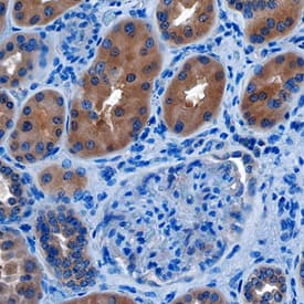
VDBP (Vitamin D binding protein; also group-specific component and GC-globulin) is a 52‑58 kDa, monomeric glycoprotein member of the ALB/AFP/VDB family of molecules. It is found in blood, urine and CSF, carries Vitamin D and its metabolites, and serves as an actin-scavenging protein. Mature mouse VDBP is 460 amino acids (aa) in length. It contains three albumin-type domains (aa 26‑476) that are accompanied by 14 intrachain disulfide bonds. There are three potential alternative splice forms. One shows a deletion of aa 346‑421, a second shows a 67 aa substitution for aa 345‑421, and a third shows a 34 aa substitution for aa 346‑423. All these variants involve the second and third albumin-like domains. Mature mouse VDBP (aa 17‑476) is 77% and 90% aa identical to human and rat VDBP, respectively.
![Immunohistochemistry Albumin Antibody (188835) [Unconjugated] - Serum](https://images.novusbio.com/images/antibody/Albumin_MAB1455_Immunohistochemistry_23317.jpg)
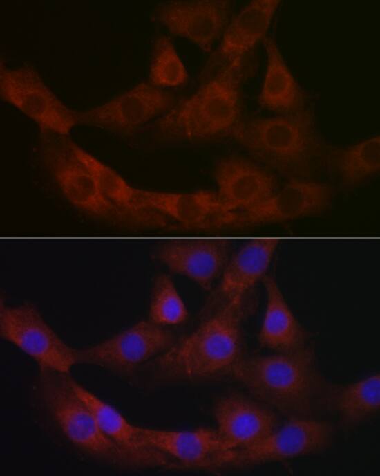
![Immunocytochemistry Albumin Antibody (188835) [Unconjugated] - Serum](https://images.novusbio.com/images/antibody/Albumin_MAB1455_Immunocytochemistry__Immunofluorescence_17777.jpg)
![Intracellular Staining by Flow Cytometry Albumin Antibody (188835) [Unconjugated] - Serum](https://images.novusbio.com/images/antibody/Albumin_MAB1455_Flow_Cytometry_15110.jpg)
![Immunohistochemistry ACE/CD143 Antibody [Unconjugated]](https://images.novusbio.com/images/antibody/ACE_AF1513_Immunohistochemistry_6703.jpg)
![Western Blot ACE/CD143 Antibody [Unconjugated]](https://images.novusbio.com/images/antibody/ACE_AF1513_Flow_Cytometry_20028.jpg)
![Simple Western ACE/CD143 Antibody [Unconjugated]](https://images.novusbio.com/images/antibody/ACE_AF1513_Simple_Western_20109.jpg)


![Vitamin D BP Antibody [Unconjugated]](/sites/all/modules/enterprise-tech/et_datasheets/images/novus_guarantee.png "Vitamin D BP Antibody [Unconjugated]")
Secondary Antibodies |
Isotype Controls |
The concentration calculator allows you to quickly calculate the volume, mass or concentration of your vial. Simply enter your mass, volume, or concentration values for your reagent and the calculator will determine the rest.


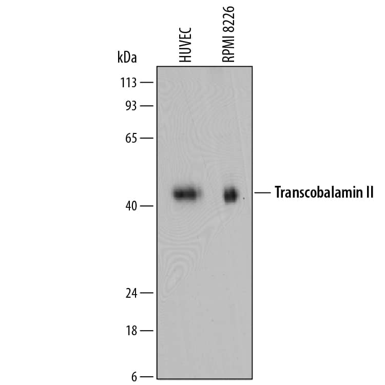
 using 5 µg/mL of Sheep Anti-Mouse Vitamin D BP Antigen Affinity-purified Polyclonal Antibody (Catalog # AF4188) followed by 1:50 dilution of HRP-conjugated Anti-Sheep IgG Secondary Antibody (Catalog # HAF016). This experiment was conducted under reducing conditions and using the 12-230 kDa separation system.")
![Immunohistochemistry Insulin Antibody (182410) [Unconjugated]](https://images.novusbio.com/images/antibody/mab1417_human-bovine-mouse-insulin-mab-clone-182410-immunohistochemistry-308202115145.jpg)
![Immunocytochemistry Insulin Antibody (182410) [Unconjugated]](https://images.novusbio.com/images/antibody/Insulin_MAB1417_Immunocytochemistry_9376.jpg)


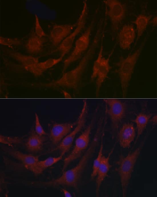
![Immunocytochemistry CD55/DAF Antibody [Unconjugated]](https://images.novusbio.com/images/antibody/CD55_AF2009_Immunocytochemistry__Immunofluorescence_23052.jpg)
![Immunohistochemistry CD55/DAF Antibody [Unconjugated]](https://images.novusbio.com/images/antibody/CD55_AF2009_Immunohistochemistry_6728.jpg)
![Immunocytochemistry/ Immunofluorescence Renin Antibody [Unconjugated] - 1](https://images.novusbio.com/images/af4277_mouse-renin-1-affinity-purified-polyclonal-ab-41202412394940.jpg)
![Immunohistochemistry Renin Antibody [Unconjugated] - 1](https://images.novusbio.com/images/antibody/Renin_AF4277_Immunohistochemistry_17351.jpg)
![N/A C-Reactive Protein/CRP [Biotin]](https://images.novusbio.com/images/elisa/DATA_CReactive_Protein_DY1707_ELISA_1701.jpg)
![Immunohistochemistry SHBG Antibody [Unconjugated]](https://images.novusbio.com/images/antibody/SHBG_AF2656_Immunohistochemistry_7324.jpg)
![Western Blot SHBG Antibody [Unconjugated]](https://images.novusbio.com/images/af2656_human-shbg-affinity-purified-pab-western-blot-2642023152834..jpg)
![Immunohistochemistry SHBG Antibody [Unconjugated]](https://images.novusbio.com/images/af2656_human-shbg-affinity-purified-pab-8120255541789.jpg)


 The chemotaxis in response to SDF-1 alpha (10 ng/ml for 12 h) was performed in the NP-MSCs and HP-MSCs treated with a neutralizing anti-CXCR4 antibody, an anti-CXCR7 antibody, and the respective isotype-matched control antibodies. *P<0.05, vs NP-MSCs; †P<0.05, vs the respective isotype-matched control antibodies. (B) NP-MSCs were transiently overexpressed with CXCR4 using pORF9-mCXCR4 vector or with CXCR7 using pORF9-mCXCR7 vector (n = 6). A negative control empty (pORF9-MCS) vector was used. (C) The transfected cells were subjected to chemotaxis in response to the indicated concentrations of SDF-1 alpha for 12 h. *P<0.05, vs the empty vector. (D and E) Western blot analysis (D) and FCM (E) were performed to determine the intracellular and extracellular expression of both CXCR4 and CXCR7 in the cells treated with or without SDF-1 alpha (50 ng/ml for 60 min). (F) The chemotaxis in response to SDF-1 alpha was performed in the cells treated with or without SDF-1 alpha . Image collected and cropped by CiteAb from the following open publication (https://pubmed.ncbi.nlm.nih.gov/22511954), licensed under a CC-BY license. Not internally tested by R&D Systems.")