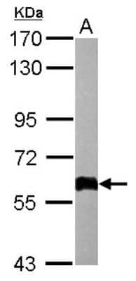
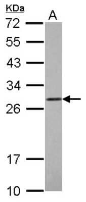
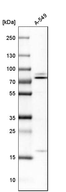
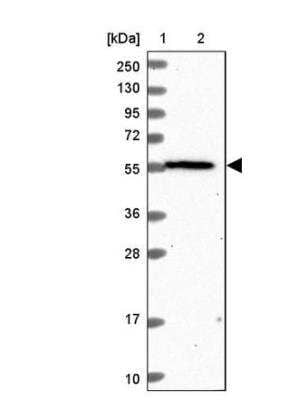
VAP-A Antibody [Unconjugated]

Images

 
Western blot shows lysates of HEK293 human embryonic kidney cell line and human heart tissue. PVDF membrane was probed with 1 µg/mL of Sheep Anti- Human VAP-A Antigen Affinity-purified Polyclonal Antibody (Catalog # ...read more

Product Details

Summary
Reactivity HuSpecies Glossary
Applications WB
Clonality
Polyclonal
Host
Sheep
Conjugate
Unconjugated
Concentration
LYOPH

Order Details

VAP-A Antibody [Unconjugated] Summary

Immunogen
E. coli-derived recombinant human VAP-A
Ala2-Met132
Accession # Q9P0L0
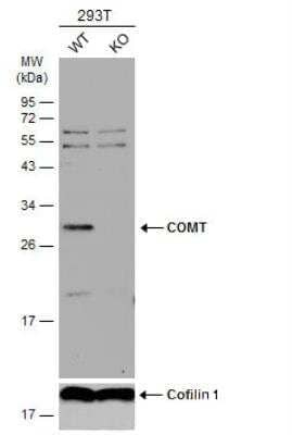
Specificity
Detects human VAP-A in direct ELISAs and Western blots. In direct ELISAs, less than 1% cross-reactivity with recombinant human VAP-B and recombinant rat VAP-B is observed.
Source
N/A
Isotype
IgG
Clonality
Polyclonal
Host
Sheep
Gene
VAPA
Purity Statement
Antigen Affinity-purified
Innovator's Reward
Test in a species/application not listed above to receive a full credit towards a future purchase.

Applications/Dilutions

Dilutions
  • Western Blot 1 ug/mL

Packaging, Storage & Formulations

Storage
Use a manual defrost freezer and avoid repeated freeze-thaw cycles.
  • 12 months from date of receipt, -20 to -70 °C as supplied.
  • 1 month, 2 to 8 °C under sterile conditions after reconstitution.
  • 6 months, -20 to -70 °C under sterile conditions after reconstitution.
Buffer
Lyophilized from a 0.2 μm filtered solution in PBS with Trehalose. *Small pack size (SP) is supplied either lyophilized or as a 0.2 µm filtered solution in PBS.
Preservative
No Preservative
Concentration
LYOPH
Reconstitution Instructions
Reconstitute at 0.2 mg/mL in sterile PBS.

Notes

This product is produced by and ships from R&D Systems, Inc., a Bio-Techne brand.

Alternate Names for VAP-A Antibody [Unconjugated]

  • hVAP-33
  • MGC3745,33 kDa VAMP-associated protein
  • VAMP (vesicle-associated membrane protein)-associated protein A, 33kDa
  • VAMP-A
  • VAP-33
  • VAP33VAMP (vesicle-associated membrane protein)-associated protein A (33kD)
  • VAPA
  • VAP-A
  • VAP-AVAMP-associated protein A
  • vesicle-associated membrane protein-associated protein A

Background

Vesicle-associated membrane protein (VAMP)-associated protein A (VAP-A; also VAMP-A and VAP-33) is a 33 kDa, ubiquitously expressed, type IV transmembrane protein belonging to the VAP family of proteins (1). It is found in plasma and ER membranes as well as in intracellular vesicles as a homodimer and a heterodimer with VAP-B. Human VAP-A is synthesized as a 249 amino acid (aa) precursor that contains a 227 aa cytoplasmic domain and a 21 aa transmembrane region. The cytoplasmic domain contains a mobile sperm protein (MSP) domain (aa 13-131) and a coiled-coil region (aa 169-205). Human VAP-A is 97% aa identical to mouse and rat VAP-A. VAP-A and VAP-B recruit FFAT (two phenylalanines in an acidic tract)-motif-containing proteins to the cytosolic surface of ER membranes through a conserved region within their MSP domain, and they have been implicated in regulation of membrane transport, phospholipid biosynthesis, and the unfolded protein response (2, 3). Their role in maintaining the identities of intracellular organelles has not been demonstrated, but their ability to interact with lipid-transfer/binding proteins (LT/BPs) may affect the lipid composition of certain cellular membranes (2, 4). One study shows that VAPs play a critical role in maintaining the structural and functional properties of the Golgi complex (2). Researchers found that knockdown of VAP reduces the levels of phosphatidylinositol‑4‑phosphate (PI4P), diacylglycerol (DAG), and sphingomyelin (SM) in Golgi membranes and exports pleiotropic effects in Golgi-mediated transport (2). The effects of VAPs are mediated by their interacting FFAT-motif-containing proteins Nir2, OSBP, and CERT (2). VAPs provide a scaffold for these LT/BPs at the ER-Golgi membrane contact sites, thereby affecting the lipid composition of the Golgi membranes and consequently their structural and functional identities (2). Most recently, researchers found that VAP-A associates and co-localizes with protrudin, a protein that promotes neurite formation, and found that it was an important regulator both of the subcellular localization of protrudin and of its ability to stimulate neurite outgrowth (5).
  1. Weir, M.L. et al. (1998) Biochem. J. 333:247.
  2. Peretti, D. et al. (2008) Mol. Biol. Cell 19:3871.
  3. Kaiser, S.E. et al. (2005) Structure 13:1035.
  4. Loewen, C.J. et al. (2003) EMBO J. 22:2025.
  5. Saita, S. et al. (2009) J. Biol. Chem. 284:13766.

    Limitations

    This product is for research use only and is not approved for use in humans or in clinical diagnosis. Primary Antibodies are guaranteed for 1 year from date of receipt.

    Customers Who Viewed This Item Also Viewed...

    NBP1-89112
    Species: Hu, Mu, Rt
    Applications: ICC/IF, IHC,  IHC-P, IP, WB
    NBP2-00935
    Species: Hu, Mu, Rt
    Applications: Flow, ICC/IF, IHC,  IHC-P, WB
    NBP1-31118
    Species: Hu, Mu
    Applications: ICC/IF, IHC,  IHC-P, WB
    NBP1-49533
    Species: Hu, Mu, Pl, Rt
    Applications: Flow-IC, Flow, ICC/IF, IHC,  IHC-P, KD, WB
    NBP1-33714
    Species: Hu, Mu, Rt, Ze
    Applications: ICC/IF, IHC-WhMt, IHC, IHC-Fr,  IHC-P, Simple Western, WB
    NBP1-83512
    Species: Hu
    Applications: IHC,  IHC-P, WB
    NBP2-03971
    Species: Bv, Ca, Ch, ChHa, Eq, Hu, Mu, Po, Pm, Rb, Rt
    Applications: IHC,  IHC-P, WB
    NBP1-82968
    Species: Hu
    Applications: ICC/IF, IHC,  IHC-P, WB
    NBP1-32870
    Species: Ch, Hu
    Applications: IHC,  IHC-P, IP, KD, WB
    NB100-1780
    Species: Hu, Mu
    Applications: Flow, PEP-ELISA, WB
    H00003845-M01
    Species: Hu, Mu
    Applications: ELISA, ICC/IF, IP, WB
    NBP1-92435
    Species: Hu
    Applications: ICC/IF, IHC,  IHC-P
    NBP1-85047
    Species: Hu, Mu, Po, Rt
    Applications: ICC/IF, IHC,  IHC-P, WB
    NBP1-87035
    Species: Hu, Rt
    Applications: ICC/IF, IHC,  IHC-P, WB
    NBP2-24915
    Species: Bv, Hu, Mu, Rt
    Applications: IHC,  IHC-P, WB
    NBP2-49320
    Species: Hu
    Applications: ICC/IF, IHC,  IHC-P, WB
    NBP1-88871
    Species: Hu
    Applications: ICC/IF, IHC,  IHC-P
    NBP1-92236
    Species: Hu
    Applications: ICC/IF, IHC,  IHC-P, WB
    NBP3-16756
    Species: Hu, Mu, Rt
    Applications: ICC/IF, IHC,  IHC-P, WB
    AF5820
    Species: Hu
    Applications: WB

    Publications for VAP-A Antibody (AF5820) (0)

    There are no publications for VAP-A Antibody (AF5820).
    By submitting your publication information earn gift cards and discounts for future purchases.

    Reviews for VAP-A Antibody (AF5820) (0)

    There are no reviews for VAP-A Antibody (AF5820). By submitting a review you will receive an Amazon e-Gift Card or Novus Product Discount.
    • Review with no image -- $10/€7/£6/$10 CAD/¥70 Yuan/¥1110 Yen
    • Review with an image -- $25/€18/£15/$25 CAD/¥150 Yuan/¥2500 Yen

    Product General Protocols

    Find general support by application which include: protocols, troubleshooting, illustrated assays, videos and webinars.

    Video Protocols

    WB Video Protocol

    FAQs for VAP-A Antibody (AF5820) (0)

    There are no specific FAQs related to this product. Read our general customer & technical service FAQs.

    Customers Who Bought This Also Bought

    Contact Information

    Product PDFs

    Calculators

    Concentration Calculator

    The concentration calculator allows you to quickly calculate the volume, mass or concentration of your vial. Simply enter your mass, volume, or concentration values for your reagent and the calculator will determine the rest.

    =
    ÷

    Review this Product

    Be the first to review our VAP-A Antibody [Unconjugated] and receive a gift card or discount.

    Bioinformatics

    Gene Symbol VAPA
    Uniprot