
| Reactivity | Hu, Mu, RtSpecies Glossary |
| Applications | WB, Simple Western, ICC/IF, IHC, IP, KO, Mycoplasma |
| Clone | 6H9 |
| Clonality | Monoclonal |
| Host | Mouse |
| Conjugate | Unconjugated |
| Format | BSA Free |
| Concentration | 1 mg/ml |
| Immunogen | Human Ubiquilin 2 expressed in and purified from E. coli [UniProt# Q9UHD9] |
| Localization | Cytoplasm. Nucleus. |
| Isotype | IgG1 |
| Clonality | Monoclonal |
| Host | Mouse |
| Gene | UBQLN2 |
| Purity | Protein G purified |
| Innovator's Reward | Test in a species/application not listed above to receive a full credit towards a future purchase. |
| Dilutions |
|
|
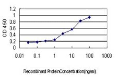
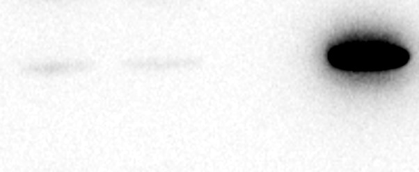
| Application Notes | This Ubiquilin 2 (6H9). antibody is useful for Immunocytochemistry/Immunofluorescence, Immunohistochemistry, and Western Blot, where a band can be seen at approx. 66-68 kDa. In Simple Western only 10 - 15 uL of the recommended dilution is used per data point. See Simple Western Antibody Database for Simple Western validation: Tested in HeLa lysate 0.5 mg/mL, separated by Size, antibody dilution of 1:100, apparent MW was 74 kDa. Use in Immunohistochemistry-Frozen reported in scientific literature (PMID:33028421). |
|
| Theoretical MW | 66-68 kDa. Disclaimer note: The observed molecular weight of the protein may vary from the listed predicted molecular weight due to post translational modifications, post translation cleavages, relative charges, and other experimental factors. |
|
| Reviewed Applications |
|
|
| Publications |
|
| Storage | Store at -20C. Avoid freeze-thaw cycles. |
| Buffer | 50% PBS, 50% glycerol |
| Preservative | 5mM Sodium Azide |
| Concentration | 1 mg/ml |
| Purity | Protein G purified |


 - BSA Free")
| Images | Ratings | Applications | Species | Date | Details | ||||||||||
|---|---|---|---|---|---|---|---|---|---|---|---|---|---|---|---|
Enlarge |
reviewed by:
Verified Customer |
IP | Human | 08/19/2022 |
Summary
Comments
|
||||||||||
Enlarge |
reviewed by:
Verified Customer |
ICC | Human | 02/09/2022 |
Summary
Comments
|
Secondary Antibodies |
Isotype Controls |
Research Areas for Ubiquilin 2 Antibody (NBP2-25164)Find related products by research area.
|
The concentration calculator allows you to quickly calculate the volume, mass or concentration of your vial. Simply enter your mass, volume, or concentration values for your reagent and the calculator will determine the rest.
5 | |
4 | |
3 | |
2 | |
1 |
| Verified Customer 08/19/2022 |
||
| Application: | IP | |
| Species: | Human |
| Verified Customer 02/09/2022 |
||
| Application: | ICC | |
| Species: | Human |


![Simple Western: Ubiquilin 2 Antibody (6H9) [NBP2-25164] - Simple Western lane view shows a specific band for Ubiquilin 2 in 0.5 mg/ml of HeLa lysate. This experiment was performed under reducing conditions using the 12-230 kDa separation system.](http://images.novusbio.com/fullsize/Ubiquilin-2-Antibody-6H9-Simple-Western-NBP2-25164-img0007.jpg "Simple Western: Ubiquilin 2 Antibody (6H9) [NBP2-25164] - Simple Western lane view shows a specific band for Ubiquilin 2 in 0.5 mg/ml of HeLa lysate. This experiment was performed under reducing conditions using the 12-230 kDa separation system.")
![Western Blot: Ubiquilin 2 Antibody (6H9) [NBP2-25164] - Analysis of different tissue and cell lysates using mouse mAb to ubiquilin 2, NBP2-25164, dilution 1:1,000 in green: [1] protein standard (red), [2] NIH-3T3, [3] C6, [4] HEK293, [5] HeLa, [6] SH-SY5Y, [7] rat whole brain, and [8] mouse whole brain. The band at 65-70kDa corresponds to ubiquilin 2 protein, which is known to differ between the human and rodent proteins.](http://images.novusbio.com/fullsize/Ubiquilin-2-Antibody-6H9-Western-Blot-NBP2-25164-img0009.jpg "Western Blot: Ubiquilin 2 Antibody (6H9) [NBP2-25164] - Analysis of different tissue and cell lysates using mouse mAb to ubiquilin 2, NBP2-25164, dilution 1:1,000 in green: [1] protein standard (red), [2] NIH-3T3, [3] C6, [4] HEK293, [5] HeLa, [6] SH-SY5Y, [7] rat whole brain, and [8] mouse whole brain. The band at 65-70kDa corresponds to ubiquilin 2 protein, which is known to differ between the human and rodent proteins.")
![Immunocytochemistry/Immunofluorescence: Ubiquilin 2 Antibody (6H9) [NBP2-25164] - Analysis of an NIH-3T3 cell culture stained with mouse mAb to ubiquilin 2, NBP2-25164, dilution 1:1,000 in green, and costained with chicken pAb to lamin A/C, dilution 1:5,000 in red. The blue is DAPI staining of nuclear DNA. The cells were treated with 50uM of chloroquine, an inhibitor of autophagy, for 16 hours prior to staining. NBP2-25164 antibody reveals vesicular staining of ubiquilin 2 protein accumulated in lysosomes in the cytoplasm, while the lamin A/C antibody stains the nuclear lamina.](http://images.novusbio.com/fullsize/Ubiquilin-2-Antibody-6H9-Immunocytochemistry-Immunofluorescence-NBP2-25164-img0008.jpg "Immunocytochemistry/Immunofluorescence: Ubiquilin 2 Antibody (6H9) [NBP2-25164] - Analysis of an NIH-3T3 cell culture stained with mouse mAb to ubiquilin 2, NBP2-25164, dilution 1:1,000 in green, and costained with chicken pAb to lamin A/C, dilution 1:5,000 in red. The blue is DAPI staining of nuclear DNA. The cells were treated with 50uM of chloroquine, an inhibitor of autophagy, for 16 hours prior to staining. NBP2-25164 antibody reveals vesicular staining of ubiquilin 2 protein accumulated in lysosomes in the cytoplasm, while the lamin A/C antibody stains the nuclear lamina.")
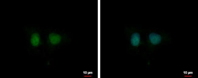
![Immunocytochemistry/Immunofluorescence: Ubiquilin 2 Antibody (6H9) [NBP2-25164] - Analysis of Human U2OS cells stained with Ubiquilin 2 antibody. Primary antibody dilution: 1:1000. Image from verified customer review.](http://images.novusbio.com/fullsize/Ubiquilin-2-Antibody-6H9-Immunocytochemistry-Immunofluorescence-NBP2-25164-img0011.jpg "Immunocytochemistry/Immunofluorescence: Ubiquilin 2 Antibody (6H9) [NBP2-25164] - Analysis of Human U2OS cells stained with Ubiquilin 2 antibody. Primary antibody dilution: 1:1000. Image from verified customer review.")
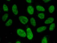
![Immunocytochemistry/Immunofluorescence: Ubiquilin 2 Antibody (6H9) [NBP2-25164] - HeLa cell cultures stained with NBP2-25164 (green) and chicken polyclonal antibody to Vimentin: NB300-223 (red). In most individual cells Ubiquilin 2 is present diffusely in the cytoplasm of cells, though some cells show enrichment of the protein in spherical autophagosome-like structure.](http://images.novusbio.com/fullsize/Ubiquilin-2-Antibody-6H9-Immunocytochemistry-Immunofluorescence-NBP2-25164-img0003.jpg "Immunocytochemistry/Immunofluorescence: Ubiquilin 2 Antibody (6H9) [NBP2-25164] - HeLa cell cultures stained with NBP2-25164 (green) and chicken polyclonal antibody to Vimentin: NB300-223 (red). In most individual cells Ubiquilin 2 is present diffusely in the cytoplasm of cells, though some cells show enrichment of the protein in spherical autophagosome-like structure.")
![Western Blot: Ubiquilin 2 Antibody (6H9) [NBP2-25164] - Lysates of HAP1 WT and Ubiquilin 2 KO were prepared, and 30 ug of protein were processed for immunoblot with NBP2-25164. The Ponceau stained transfer of the blot is shown. Antibody dilution used: 1/2000. Predicted band size: 65 kDa. Image, protocol and testing courtesy of YCharOS Inc. (ycharos.com).](http://images.novusbio.com/fullsize/Ubiquilin-2-Antibody-6H9-Knockout-Validated-NBP2-25164-img0012.jpg "Western Blot: Ubiquilin 2 Antibody (6H9) [NBP2-25164] - Lysates of HAP1 WT and Ubiquilin 2 KO were prepared, and 30 ug of protein were processed for immunoblot with NBP2-25164. The Ponceau stained transfer of the blot is shown. Antibody dilution used: 1/2000. Predicted band size: 65 kDa. Image, protocol and testing courtesy of YCharOS Inc. (ycharos.com).")
![Immunoprecipitation: Ubiquilin 2 Antibody (6H9) [NBP2-25164] - HAP1 lysates were prepared, and immunoprecipitation was performed using 2.0 ug of Ubiquilin 2 antibody (NBP2-25164) pre-coupled to Dynabeads protein G. Samples were washed and processed for immunoblot with NBP2-25164. For immunoblot, Recombinant Ubiquilin 2 (Rb x Ubiquilin 2) was used at 1/1000. The Ponceau stained transfer of the blot is shown. SM=4% starting material; UB=4% unbound fraction. Image, protocol and testing courtesy of YCharOS Inc. (ycharos.com).](http://images.novusbio.com/fullsize/Ubiquilin-2-Antibody-6H9-Immunoprecipitation-NBP2-25164-img0013.jpg "Immunoprecipitation: Ubiquilin 2 Antibody (6H9) [NBP2-25164] - HAP1 lysates were prepared, and immunoprecipitation was performed using 2.0 ug of Ubiquilin 2 antibody (NBP2-25164) pre-coupled to Dynabeads protein G. Samples were washed and processed for immunoblot with NBP2-25164. For immunoblot, Recombinant Ubiquilin 2 (Rb x Ubiquilin 2) was used at 1/1000. The Ponceau stained transfer of the blot is shown. SM=4% starting material; UB=4% unbound fraction. Image, protocol and testing courtesy of YCharOS Inc. (ycharos.com).")
![Immunocytochemistry/ Immunofluorescence: Ubiquilin 2 Antibody (6H9) [NBP2-25164] - HAP1 WT and Ubiquilin 2 KO cells were labeled with a green or a far-red fluorescent dye, respectively. WT and KO cells were mixed and plated to a 1:1 ratio in a 96-well plate with glass bottom. Cells were stained with Ubiquilin-2 antibody (NBP2-25164) and with the corresponding Alexa-fluor 555 coupled secondary antibody including DAPI. Acquisition of the blue (nucleus-DAPI), green (identification of WT cells), red (antibody staining) and far-red (identification of KO cells) channels was performed. Representative image of the blue and red (grayscale) channels is shown. WT and KO cells are outlined with green and magenta dashed line, respectively. Antibody dilution used: 1/1000. Bars = 10 um Image, protocol and testing courtesy of YCharOS Inc. (ycharos.com).](http://images.novusbio.com/fullsize/Ubiquilin-2-Antibody-6H9-Knockout-Validated-NBP2-25164-img0014.jpg "Immunocytochemistry/ Immunofluorescence: Ubiquilin 2 Antibody (6H9) [NBP2-25164] - HAP1 WT and Ubiquilin 2 KO cells were labeled with a green or a far-red fluorescent dye, respectively. WT and KO cells were mixed and plated to a 1:1 ratio in a 96-well plate with glass bottom. Cells were stained with Ubiquilin-2 antibody (NBP2-25164) and with the corresponding Alexa-fluor 555 coupled secondary antibody including DAPI. Acquisition of the blue (nucleus-DAPI), green (identification of WT cells), red (antibody staining) and far-red (identification of KO cells) channels was performed. Representative image of the blue and red (grayscale) channels is shown. WT and KO cells are outlined with green and magenta dashed line, respectively. Antibody dilution used: 1/1000. Bars = 10 um Image, protocol and testing courtesy of YCharOS Inc. (ycharos.com).")
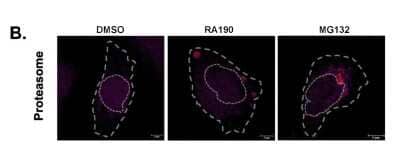
 UBQLN2 is not pelleted when cells are heat shocked post lysis. Cell lysates were incubated at 37 or 42C and then fractionated into soluble (S) and pellet (P) fraction. This indicates that UBQLN2 itself does not aggregate as a result of high temperature.(B) UBQLN2 levels are not upregulated in response to heat shock. HSP70 and GAPDH were used as a positive and negative controls, respectively.(C) Heat shock aggregates are insoluble in up to and including 1% SDS but are solubilized in 2% SDS. Blotting of soluble and pellet fractions with anti-ubiquitin and UQBLN2 antibodies confirmed dissolution of the aggregates in 2% SDS.(D) Proteasomes are active after heat shock. To confirm that proteasome activity was not affected by heat shock, we incubated U2OS and MEFs at the indicted temperatures for 2h. Cells were then harvested and cell lysates were incubated with the proteasome inhibitor MG132 or DMSO, followed by incubation with a fluorescent proteasome-activity probe, as indicated. The presence of fluorescently labeled beta-subunits at the same intensity under both heat stress and normal temperature, indicate that proteasome activity is not significantly affected by heat shock.(E) U2OS cells were treated with control or UBQLN2 siRNA and subjected to heat shock for the indicated times. Analysis of the pellet fraction revealed that insoluble ubiquitylated aggregates are generated within 5 min of heat shock, but that depletion of UBQLN2 does not noticeably alter the accumulation of these aggregates at any of the indicated time points. Image collected and cropped by CiteAb from the following open publication (//pubmed.ncbi.nlm.nih.gov/27477512), licensed under a CC-BY license. Not internally tested by Novus Biologicals.")
 Schematic representation on how the three fractions, total insoluble, nuclear soluble and total insoluble were generated.(B) Cells treated with puromycin were fractionated as indicated and treatment did not induce the nuclear localization of UBQLN2.(C) Puromycin treatment did not induce the upregulation of UBQLN2 protein.(D) Schematic showing the GFPu-NLS construct (Bennett et al., 2005).(E) HEK293 cells stably expressing GFPu-NLS were subject to heat shock for 2h at 42C and fractionated into soluble and insoluble fractions. GFPu-NLS recruited to the insoluble fraction after heat shock indicating its heat-induced aggregation.(F) GFPu-NLS cells were subject to heat shock for 2h at 42C and proteasome inhibition with 25 μM MG132 as indicated. UBQLN2 was immunoprecipitated and GFPu-NLS was found to co-immunoprecipitate only upon heat shock, consistent with UBQLN2 nuclear localization. Combined heat shock and proteasome inhibition increased the binding further.(G) GFPu-NLS cells were depleted of UBQLN2 or treated with a control non-targeting siRNA then treated with 50 μg/ml cycloheximide (CHX) for the indicated time to measure turnover. Turnover was quantified using data from three independent experiments.Error bars represent SE. Image collected and cropped by CiteAb from the following open publication (//pubmed.ncbi.nlm.nih.gov/27477512), licensed under a CC-BY license. Not internally tested by Novus Biologicals.")


![Western Blot Rad23 Antibody [Unconjugated]](https://images.novusbio.com/images/antibody/Rad23_AF4555_Western_Blot_5498.jpg)


 followed by 30 min incubation with Goat anti Mouse HRP conjugated secondary antibodies (Catalog # HAF007) at 1:20 dilution + DAB chromogen (brown). The tissue was counterstained with Hematoxylin (blue). Control was done by omitting primary antibody.")
![SDS-Page: Mouse IgG1 Isotype Control (MG1) [NBP1-97005] - Lane 1: , Non-reduced. M: Opal Pre-stained Ladder. Lane 2: , Reduced. Load: 1.0 ug per lane. Predicted/Observed: 120 kDa Non-reduced, 55 and 25 Reduced.](https://images.novusbio.com/images/Mouse-IgG1-Isotype-Control-MG1-SDS-Page-NBP1-97005-img0002.jpg "SDS-Page: Mouse IgG1 Isotype Control (MG1) [NBP1-97005] - Lane 1: , Non-reduced. M: Opal Pre-stained Ladder. Lane 2: , Reduced. Load: 1.0 ug per lane. Predicted/Observed: 120 kDa Non-reduced, 55 and 25 Reduced.")

