
| Reactivity | Hu, Mu, Rt, Gp, ZeSpecies Glossary |
| Applications | WB, ELISA, Flow, ICC/IF, IHC |
| Clonality | Polyclonal |
| Host | Rabbit |
| Conjugate | Unconjugated |
| Format | BSA Free |
| Concentration | 1.0 mg/ml |
| Immunogen | A synthetic peptide made to a region within the N-terminus (residues 1-100) of the human TRPA1 protein. [Swiss-Prot# O75762] |
| Localization | Membrane |
| Specificity | Additional modified form of TRPA1 can also be detected. |
| Isotype | IgG |
| Clonality | Polyclonal |
| Host | Rabbit |
| Gene | TRPA1 |
| Purity | Immunogen affinity purified |
| Innovator's Reward | Test in a species/application not listed above to receive a full credit towards a future purchase. |
| Dilutions |
|
|
| Theoretical MW | 127.5 kDa. Disclaimer note: The observed molecular weight of the protein may vary from the listed predicted molecular weight due to post translational modifications, post translation cleavages, relative charges, and other experimental factors. |
|
| Reviewed Applications |
|
|
| Publications |
|
| Storage | Store at 4C short term. Aliquot and store at -20C long term. Avoid freeze-thaw cycles. |
| Buffer | PBS |
| Preservative | 0.05% Sodium Azide |
| Concentration | 1.0 mg/ml |
| Purity | Immunogen affinity purified |


![Western Blot RFC1 Antibody [Unconjugated]](https://images.novusbio.com/images/antibody/RFC1_AF6457_Western_Blot_9874.jpg)


![Immunohistochemistry EN-RAGE/S100A12 Antibody [Unconjugated]](https://images.novusbio.com/images/antibody/EN-RAGE_AF1052_Immunohistochemistry_6671.jpg)
![Simple Western EN-RAGE/S100A12 Antibody [Unconjugated]](https://images.novusbio.com/images/antibody/af1052_human-en-rage-affinity-purified-polyclonal-ab-simple-western-2672021152820.jpg)
![Flow Cytometry EN-RAGE/S100A12 Antibody [Unconjugated]](https://images.novusbio.com/images/antibody/af1052_human-en-rage-affinity-purified-polyclonal-ab-flow-cytometry-572024104928.jpg)

| Images | Ratings | Applications | Species | Date | Details | ||||||||||
|---|---|---|---|---|---|---|---|---|---|---|---|---|---|---|---|

Enlarge |
reviewed by:
LEONARDO DE ASSIS |
Flow | Zebrafish | 10/28/2019 |
Summary
Comments
|
||||||||||

Enlarge |
reviewed by:
TinaMarie Lieu |
IF | Human | 08/21/2012 |
Summary
|
Secondary Antibodies |
Isotype Controls |
Research Areas for TRPA1 Antibody (NB110-40763)Find related products by research area.
|
|
Winter is coming, and TRPM8 welcomes the cold! TRPM8, or transient receptor potential melastatin 8, is a nonselective cation channel that is activated by cold environments and menthol-like cooling compounds. While TRPM8 is best known for its location in peripheral nerve endings, it has functio... Read full blog post. |
|
TRPA1: A contributor to itching and inflammation? Scratch that! Transient receptor potential A1 (TRPA1) is an ion channel found on the plasma membrane of many cell types that functions in diverse sensory processes such as pain and temperature. The TRPA1 ion channel is specifically expressed in nociceptive neurons,... Read full blog post. |
|
Touch Infographic: From Touch Receptors to the Brain The body contains thousands of receptors and nerves which allow us to experience the sense of touch, also referred to as tactile perception. The somatosensory system allows organisms to perceive and decode a wide range of tactile stimuli to allow for ... Read full blog post. |
The concentration calculator allows you to quickly calculate the volume, mass or concentration of your vial. Simply enter your mass, volume, or concentration values for your reagent and the calculator will determine the rest.
5 | |
4 | |
3 | |
2 | |
1 |
| LEONARDO DE ASSIS 10/28/2019 |
||
| Application: | Flow | |
| Species: | Zebrafish |
| TinaMarie Lieu 08/21/2012 |
||
| Application: | IF | |
| Species: | Human |


![Immunohistochemistry: TRPA1 Antibody [NB110-40763] - Staining TRPA1 in mouse intestine.](http://images.novusbio.com/fullsize/TRPA1-Antibody-Immunohistochemistry-NB110-40763-img0008.jpg "Immunohistochemistry: TRPA1 Antibody [NB110-40763] - Staining TRPA1 in mouse intestine.")

![Flow Cytometry: TRPA1 Antibody [NB110-40763] - An intracellular stain was performed on A549 cells with NB110-40763C (blue) and a matched isotype control (orange). Cells were fixed with 4% PFA and then permeabilized with 0.1% saponin. Cells were incubated in an antibody dilution of 2.5 ug/mL for 30 minutes at room temperature. Both antibodies were conjugated to DyLight 650.](http://images.novusbio.com/fullsize/TRPA1-Antibody-Flow-Cytometry-NB110-40763-img0010.jpg "Flow Cytometry: TRPA1 Antibody [NB110-40763] - An intracellular stain was performed on A549 cells with NB110-40763C (blue) and a matched isotype control (orange). Cells were fixed with 4% PFA and then permeabilized with 0.1% saponin. Cells were incubated in an antibody dilution of 2.5 ug/mL for 30 minutes at room temperature. Both antibodies were conjugated to DyLight 650.")
![Flow Cytometry: TRPA1 Antibody [NB110-40763] - An intracellular stain was performed on A549 cells with NB110-40763G (blue) and a matched isotype control (orange). Cells were fixed with 4% PFA and then permeabilized with 0.1% saponin. Cells were incubated in an antibody dilution of 5 ug/mL for 30 minutes at room temperature. Both antibodies were conjugated to DyLight 488](http://images.novusbio.com/fullsize/TRPA1-Antibody-Flow-Cytometry-NB110-40763-img0009.jpg "Flow Cytometry: TRPA1 Antibody [NB110-40763] - An intracellular stain was performed on A549 cells with NB110-40763G (blue) and a matched isotype control (orange). Cells were fixed with 4% PFA and then permeabilized with 0.1% saponin. Cells were incubated in an antibody dilution of 5 ug/mL for 30 minutes at room temperature. Both antibodies were conjugated to DyLight 488")

![Immunocytochemistry/Immunofluorescence: TRPA1 Antibody [NB110-40763] - Analysis of TRPA1 in HEK 293 cells (Flp In Trex system) cells.](http://images.novusbio.com/fullsize/TRPA1-Antibody-Immunocytochemistry-Immunofluorescence-NB110-40763-img0006.jpg "Immunocytochemistry/Immunofluorescence: TRPA1 Antibody [NB110-40763] - Analysis of TRPA1 in HEK 293 cells (Flp In Trex system) cells.")
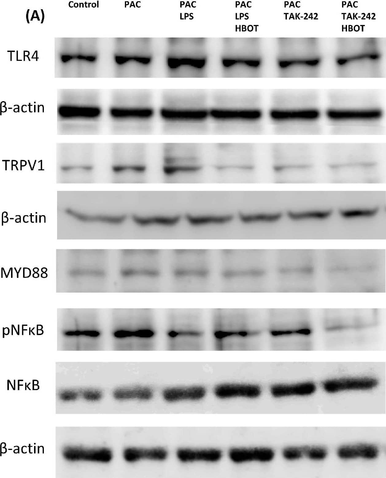
![Western Blot: TRPA1 Antibody - BSA Free [NB110-40763] - Effects of Lycium barbarum polysaccharides (LBP) & capsaicin (CAP) on colonic (A) Cyclooxygenase-2 (COX-2), (B) Transient receptor potential cation channel V1 (TRPV1), & (C) Transient receptor potential ankyrin 1 (TRPA1) protein expression. (D) Representatives of Western blot for COX-2, TRPV1, TRPA1, & beta -actin. N: control group, U: ulcerative colitis induced group, L: LBP treated group, C: CAP treated group, M: mixed LBP & CAP treated group. Data are presented as mean ± SEM & analyzed by one-way ANOVA & Fisher’s least significant difference test (n = 8). # p < 0.05 compared to the N group. * p < 0.05 compared to the U group. Image collected & cropped by CiteAb from the following publication (//pubmed.ncbi.nlm.nih.gov/35269566), licensed under a CC-BY license. Not internally tested by Novus Biologicals.](http://images.novusbio.com/fullsize/nb110-40763_rabbit-polyclonal-trpa1-antibody-310202415392558.jpg "Western Blot: TRPA1 Antibody - BSA Free [NB110-40763] - Effects of Lycium barbarum polysaccharides (LBP) & capsaicin (CAP) on colonic (A) Cyclooxygenase-2 (COX-2), (B) Transient receptor potential cation channel V1 (TRPV1), & (C) Transient receptor potential ankyrin 1 (TRPA1) protein expression. (D) Representatives of Western blot for COX-2, TRPV1, TRPA1, & beta -actin. N: control group, U: ulcerative colitis induced group, L: LBP treated group, C: CAP treated group, M: mixed LBP & CAP treated group. Data are presented as mean ± SEM & analyzed by one-way ANOVA & Fisher’s least significant difference test (n = 8). # p < 0.05 compared to the N group. * p < 0.05 compared to the U group. Image collected & cropped by CiteAb from the following publication (//pubmed.ncbi.nlm.nih.gov/35269566), licensed under a CC-BY license. Not internally tested by Novus Biologicals.")
![Immunocytochemistry/ Immunofluorescence: TRPA1 Antibody - BSA Free [NB110-40763] - Expression & localization of TRPA1 channels in wild-type (WT) & APP/PS1 Tg mice. (a) Brains were harvested from WT & APP/PS1 Tg mice at 8 months old. Western blot analysis of protein levels of TRPA1 & alpha -tubulin. Data are mean ± SEM from 6 mice in each group. *, P < 0.05 vs. WT mice. (b, c) Immunohistochemistry of specimens of cortex & hippocampus from 8-month-old WT & APP/PS1 Tg mice with the antibodies anti-TRPA1, anti-vWF (endothelial cell marker), anti-NeuN (neuron marker), anti-GFAP (astrocyte marker) & anti-IBA-1 (microglia marker), then FITC- or Texas red-conjugated secondary antibody. Bar = 50 μm. vWF-positive cells denoted endothelial cells, NeuN-positive cells denoted neurons, & GFAP-positive cells denoted astrocytes, as indicated by arrowheads, stars or arrows, respectively Image collected & cropped by CiteAb from the following publication (//jneuroinflammation.biomedcentral.com/articles/10.1186/s12974-016-0557-z), licensed under a CC-BY license. Not internally tested by Novus Biologicals.](http://images.novusbio.com/fullsize/nb110-40763_rabbit-polyclonal-trpa1-antibody-310202416235534.jpg "Immunocytochemistry/ Immunofluorescence: TRPA1 Antibody - BSA Free [NB110-40763] - Expression & localization of TRPA1 channels in wild-type (WT) & APP/PS1 Tg mice. (a) Brains were harvested from WT & APP/PS1 Tg mice at 8 months old. Western blot analysis of protein levels of TRPA1 & alpha -tubulin. Data are mean ± SEM from 6 mice in each group. *, P < 0.05 vs. WT mice. (b, c) Immunohistochemistry of specimens of cortex & hippocampus from 8-month-old WT & APP/PS1 Tg mice with the antibodies anti-TRPA1, anti-vWF (endothelial cell marker), anti-NeuN (neuron marker), anti-GFAP (astrocyte marker) & anti-IBA-1 (microglia marker), then FITC- or Texas red-conjugated secondary antibody. Bar = 50 μm. vWF-positive cells denoted endothelial cells, NeuN-positive cells denoted neurons, & GFAP-positive cells denoted astrocytes, as indicated by arrowheads, stars or arrows, respectively Image collected & cropped by CiteAb from the following publication (//jneuroinflammation.biomedcentral.com/articles/10.1186/s12974-016-0557-z), licensed under a CC-BY license. Not internally tested by Novus Biologicals.")
![Immunocytochemistry/ Immunofluorescence: TRPA1 Antibody - BSA Free [NB110-40763] - Evaluation of anti-TRPA1 antibody specificity by western blotting & immunofluorescence. (a) Lysates derived from HEK293T cells transiently transfected with the TRPA1 & GFP expression construct (TRPA1 OE) or TRPM2 & GFP negative control (TRPM2 OE) were probed with the antibodies indicated. Additional untransfected & GFP only controls are presented in the first blot (6G8). Uncropped full length blots are displayed, gaps between the blots are present to indicate the use of a different antibody. Mouse mAbs C-5 & 6G8, & NB110-40763 can detect TRPA1 only in the membrane fraction of lysates at the expected molecular weight (127.5 kDa). Several TRPA1-specific bands are observed above this weight. NB110-40763 also detects several other antigens. Ab58844 & ACC- 037 only appear to detect antigens other than TRPA1 in the conditions used. (b) The performance of the same antibodies was evaluated by immunofluorescence (red) in comparison to the expression of the GFP reporter (green), or control TRPM2 & GFP overexpressing cells as for western blotting. Mouse mAb C-5 again shows high specificity with a strong correlation between the expression of the GFP reporter & antibody staining, & no staining in TRPM2 & GFP overexpressing controls. NB110-40763 also demonstrates some sensitivity but has high background that likely does not correspond to Fc binding of the antibody, as the other two rabbit polyclonal antibodies or isotype control did show different staining patterns. Ab58844 & ACC- 037 only appear to detect antigens other than TRPA1 in the conditions used. Image collected & cropped by CiteAb from the following publication (//pubmed.ncbi.nlm.nih.gov/31811235), licensed under a CC-BY license. Not internally tested by Novus Biologicals.](http://images.novusbio.com/fullsize/nb110-40763_rabbit-polyclonal-trpa1-antibody-31020241623556.jpg "Immunocytochemistry/ Immunofluorescence: TRPA1 Antibody - BSA Free [NB110-40763] - Evaluation of anti-TRPA1 antibody specificity by western blotting & immunofluorescence. (a) Lysates derived from HEK293T cells transiently transfected with the TRPA1 & GFP expression construct (TRPA1 OE) or TRPM2 & GFP negative control (TRPM2 OE) were probed with the antibodies indicated. Additional untransfected & GFP only controls are presented in the first blot (6G8). Uncropped full length blots are displayed, gaps between the blots are present to indicate the use of a different antibody. Mouse mAbs C-5 & 6G8, & NB110-40763 can detect TRPA1 only in the membrane fraction of lysates at the expected molecular weight (127.5 kDa). Several TRPA1-specific bands are observed above this weight. NB110-40763 also detects several other antigens. Ab58844 & ACC- 037 only appear to detect antigens other than TRPA1 in the conditions used. (b) The performance of the same antibodies was evaluated by immunofluorescence (red) in comparison to the expression of the GFP reporter (green), or control TRPM2 & GFP overexpressing cells as for western blotting. Mouse mAb C-5 again shows high specificity with a strong correlation between the expression of the GFP reporter & antibody staining, & no staining in TRPM2 & GFP overexpressing controls. NB110-40763 also demonstrates some sensitivity but has high background that likely does not correspond to Fc binding of the antibody, as the other two rabbit polyclonal antibodies or isotype control did show different staining patterns. Ab58844 & ACC- 037 only appear to detect antigens other than TRPA1 in the conditions used. Image collected & cropped by CiteAb from the following publication (//pubmed.ncbi.nlm.nih.gov/31811235), licensed under a CC-BY license. Not internally tested by Novus Biologicals.")
![Western Blot: TRPA1 Antibody - BSA Free [NB110-40763] - Expression & localization of TRPA1 channels in wild-type (WT) & APP/PS1 Tg mice. (a) Brains were harvested from WT & APP/PS1 Tg mice at 8 months old. Western blot analysis of protein levels of TRPA1 & alpha -tubulin. Data are mean ± SEM from 6 mice in each group. *, P < 0.05 vs. WT mice. (b, c) Immunohistochemistry of specimens of cortex & hippocampus from 8-month-old WT & APP/PS1 Tg mice with the antibodies anti-TRPA1, anti-vWF (endothelial cell marker), anti-NeuN (neuron marker), anti-GFAP (astrocyte marker) & anti-IBA-1 (microglia marker), then FITC- or Texas red-conjugated secondary antibody. Bar = 50 μm. vWF-positive cells denoted endothelial cells, NeuN-positive cells denoted neurons, & GFAP-positive cells denoted astrocytes, as indicated by arrowheads, stars or arrows, respectively Image collected & cropped by CiteAb from the following publication (//jneuroinflammation.biomedcentral.com/articles/10.1186/s12974-016-0557-z), licensed under a CC-BY license. Not internally tested by Novus Biologicals.](http://images.novusbio.com/fullsize/nb110-40763_rabbit-polyclonal-trpa1-antibody-310202416235536.jpg "Western Blot: TRPA1 Antibody - BSA Free [NB110-40763] - Expression & localization of TRPA1 channels in wild-type (WT) & APP/PS1 Tg mice. (a) Brains were harvested from WT & APP/PS1 Tg mice at 8 months old. Western blot analysis of protein levels of TRPA1 & alpha -tubulin. Data are mean ± SEM from 6 mice in each group. *, P < 0.05 vs. WT mice. (b, c) Immunohistochemistry of specimens of cortex & hippocampus from 8-month-old WT & APP/PS1 Tg mice with the antibodies anti-TRPA1, anti-vWF (endothelial cell marker), anti-NeuN (neuron marker), anti-GFAP (astrocyte marker) & anti-IBA-1 (microglia marker), then FITC- or Texas red-conjugated secondary antibody. Bar = 50 μm. vWF-positive cells denoted endothelial cells, NeuN-positive cells denoted neurons, & GFAP-positive cells denoted astrocytes, as indicated by arrowheads, stars or arrows, respectively Image collected & cropped by CiteAb from the following publication (//jneuroinflammation.biomedcentral.com/articles/10.1186/s12974-016-0557-z), licensed under a CC-BY license. Not internally tested by Novus Biologicals.")


![Western Blot: Goat anti-Rabbit IgG (H+L) Secondary Antibody [HRP] [NB7160] - Western blot showing vemurafenib treatment in BRAFV600E CRC cells inhibits fission mediator DRP1 with no significant effect on fusion proteins (Mfn1 & 2) using MFN-1 antibody (NBP1-51841) and corresponding secondary antibody, goat anti-rabbit IgG-HRP (NB7160). Image collected and cropped by CiteAb from the following publication (https://pubmed.ncbi.nlm.nih.gov/33738242).](https://images.novusbio.com/images/Goat-anti-Rabbit-IgG-H+L-Secondary-Antibody-HRP-Western-Blot-NB7160-img0001.jpg "Western Blot: Goat anti-Rabbit IgG (H+L) Secondary Antibody [HRP] [NB7160] - Western blot showing vemurafenib treatment in BRAFV600E CRC cells inhibits fission mediator DRP1 with no significant effect on fusion proteins (Mfn1 & 2) using MFN-1 antibody (NBP1-51841) and corresponding secondary antibody, goat anti-rabbit IgG-HRP (NB7160). Image collected and cropped by CiteAb from the following publication (https://pubmed.ncbi.nlm.nih.gov/33738242).")
 followed by 30 min incubation with Goat anti Rabbit HRP conjugated secondary antibodies (Catalog # HAF008) at 1:20 dilution + DAB chromogen (brown). The tissue was counterstained with Hematoxylin (blue). Control was done by omitting primary antibody.")
![Flow Cytometry: Rabbit IgG Isotype Control [NBP2-24891] - An intracellular stain was performed on Raji cells with Adiponectin antibody NB100-65810 (blue) and a matched isotype control NBP2-24893 (orange). Cells were fixed with 4% PFA and then permeablized with 0.1% saponin. Cells were incubated in an antibody dilution of 1 ug/mL for 30 minutes at room temperature, followed by Dylight488-conjugated anti-rabbit secondary antibody. Image using the Azide Free form of this antibody.](https://images.novusbio.com/images/Rabbit--Mouse-IgG-Isotype-Control-Flow-Cytometry-NBP2-24891-img0006.jpg "Flow Cytometry: Rabbit IgG Isotype Control [NBP2-24891] - An intracellular stain was performed on Raji cells with Adiponectin antibody NB100-65810 (blue) and a matched isotype control NBP2-24893 (orange). Cells were fixed with 4% PFA and then permeablized with 0.1% saponin. Cells were incubated in an antibody dilution of 1 ug/mL for 30 minutes at room temperature, followed by Dylight488-conjugated anti-rabbit secondary antibody. Image using the Azide Free form of this antibody.")
