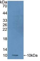
 HAF007). A specific band was detected for S100A8 at approximately 11 kDa (as indicated). This experiment was conducted under reducing conditions and using Immunoblot Buffer Group 1." title="Western blot shows lysates of HL-60 human acute promyelocytic leukemia cell line. PVDF membrane was probed with 2 µg/mL of Mouse Anti-Human S100A8 Monoclonal Antibody (Catalog # MAB4570) followed by HRP-conjugated Anti-Mouse IgG Secondary Antibody (Catalog # HAF007). A specific band was detected for S100A8 at approximately 11 kDa (as indicated). This experiment was conducted under reducing conditions and using Immunoblot Buffer Group 1." />
| Reactivity | HuSpecies Glossary |
| Applications | WB, IHC |
| Clone | 749916 |
| Clonality | Monoclonal |
| Host | Mouse |
| Conjugate | Unconjugated |
| Concentration | LYOPH |
| Immunogen | E. coli-derived recombinant human S100A8 Met1-Glu93 Accession # P05109 |
| Specificity | Detects human S100A8 in direct ELISAs.
In direct ELISAs, 100% cross-reactivity
with recombinant human (rh) S100A8/A9 is observed and no cross-reactivity with
rhS100A9, recombinant mouse (rm) S100A8, rmS100A9, or rmS100A8/A9 is observed. |
| Source | N/A |
| Isotype | IgG1 |
| Clonality | Monoclonal |
| Host | Mouse |
| Gene | S100A8 |
| Purity Statement | Protein A or G purified from hybridoma culture supernatant |
| Innovator's Reward | Test in a species/application not listed above to receive a full credit towards a future purchase. |
| Dilutions |
|
|
| Publications |
|
| Storage | Use a manual defrost freezer and avoid repeated freeze-thaw cycles.
|
| Buffer | Lyophilized from a 0.2 μm filtered solution in PBS with Trehalose. *Small pack size (SP) is supplied either lyophilized or as a 0.2 µm filtered solution in PBS. |
| Preservative | No Preservative |
| Concentration | LYOPH |
| Reconstitution Instructions | Sterile PBS to a final concentration of 0.5 mg/mL. |
S100A8 (also known as MRP8 and calgranulin A) is a 10 kDa member of the S100 family, EF-hand superfamily of Ca-binding proteins. It is produced by neutrophils and monocytes, and forms Ca‑dependent heterodimer/heterotetramer complexes (termed calprotectin) with S100A9. It functions both intracellularly and extracellularly, where it binds to RAGE and CD36. Human S100A8 is 93 amino acids (aa) in length. It contains two EF-hand motifs (aa 12-47 and 46-81) and one high-affinity Ca‑binding site (aa 59-70). There may be one splice form that shows a 15 aa substitution for the C-terminal 14 amino acids. Although mouse S100A8 is cleaved by MMP‑2 after Asn21, it is unclear if human S100A8 is susceptible. Full-length human S100A8 is 57% and 74% aa identical to mouse and canine S100A8, respectively.
![Immunohistochemistry EN-RAGE/S100A12 Antibody [Unconjugated]](https://images.novusbio.com/images/antibody/EN-RAGE_AF1052_Immunohistochemistry_6671.jpg)
![Simple Western EN-RAGE/S100A12 Antibody [Unconjugated]](https://images.novusbio.com/images/antibody/af1052_human-en-rage-affinity-purified-polyclonal-ab-simple-western-2672021152820.jpg)
![Flow Cytometry EN-RAGE/S100A12 Antibody [Unconjugated]](https://images.novusbio.com/images/antibody/af1052_human-en-rage-affinity-purified-polyclonal-ab-flow-cytometry-572024104928.jpg)


")
Secondary Antibodies |
Isotype Controls |
The concentration calculator allows you to quickly calculate the volume, mass or concentration of your vial. Simply enter your mass, volume, or concentration values for your reagent and the calculator will determine the rest.


 CTS013). Tissue was stained using the Anti-Mouse HRP-DAB Cell & Tissue Staining Kit (brown; Catalog #
  Reverse transcriptase and real-time PCR analysis of mRNAs for BMP8A, CCR6, HOXA9, NANOG and S100A8 in LCL and DLBCL (HBL-2, HT, SU-DHL-5, U2932 and U2940) cell lines. Data were normalized by the amount of GAPDH mRNA, expressed relative to the corresponding value for LCL, and are shown as means ± SD from triplicate data. (B) After quantification of Western blot intensity, the relative expression levels of each protein are shown with normalization of GAPDH. U2940 cells have the highest expression of the stem cell genes, HOXA9 and NANOG. Molecular weight listed in parenthesis. Error bars represent the standard error of the mean of three independent experiments. *p < 0.05, **p < 0.01, ***p < 0.001, Student t-test. Image collected and cropped by CiteAb from the following open publication (//pubmed.ncbi.nlm.nih.gov/33288848), licensed under a CC-BY license. Not internally tested by R&D Systems.")
 Reverse transcriptase and real-time PCR analysis of mRNAs for BMP8A, CCR6, HOXA9, NANOG and S100A8 in LCL and DLBCL (HBL-2, HT, SU-DHL-5, U2932 and U2940) cell lines. Data were normalized by the amount of GAPDH mRNA, expressed relative to the corresponding value for LCL, and are shown as means ± SD from triplicate data. (B) After quantification of Western blot intensity, the relative expression levels of each protein are shown with normalization of GAPDH. U2940 cells have the highest expression of the stem cell genes, HOXA9 and NANOG. Molecular weight listed in parenthesis. Error bars represent the standard error of the mean of three independent experiments. *p < 0.05, **p < 0.01, ***p < 0.001, Student t-test. Image collected and cropped by CiteAb from the following open publication (//pubmed.ncbi.nlm.nih.gov/33288848), licensed under a CC-BY license. Not internally tested by R&D Systems.")
![Immunocytochemistry S100A9 Antibody [Unconjugated]](https://images.novusbio.com/images/antibody/S100A9_AF2065_Immunocytochemistry__Immunofluorescence_22492.jpg)
![Immunohistochemistry S100A9 Antibody [Unconjugated]](https://images.novusbio.com/images/af2065_goat-s100a9-pab-alexa-fluor-594-immunohistochemistry-12122025852812.jpg)
![SDS-Page TNF-alpha [Unconjugated]](https://images.novusbio.com/images/protein/TNF-alpha_210-TA_256.jpg)
![Bioactivity TNF-alpha [Unconjugated]](https://images.novusbio.com/images/protein/TNFalpha_210TA_1658.jpg)
![SEC-MALS TNF-alpha [Unconjugated]](https://images.novusbio.com/images/210-ta_recombinant-human-tnf-alpha-protein-sec-mals-35202312244..jpg)
![N/A IL-6 [HRP]](https://images.novusbio.com/images/elisa/DATA_IL6_M6000_ELISA_936.jpg)
![N/A IL-6 [HRP]](https://images.novusbio.com/images/elisa/IL-6_M6000_ELISA_415.jpg)
![N/A IL-6 [HRP]](https://images.novusbio.com/images/m6000b_mouse-il-6-quantikine-elisa-kit-1752025024034.jpg)


![N/A IL-10 [Biotin]](https://images.novusbio.com/images/elisa/DATA_IL10_DY417_ELISA_2014.jpg)
![N/A C-Reactive Protein/CRP [Biotin]](https://images.novusbio.com/images/elisa/DATA_CReactive_Protein_DY1707_ELISA_1701.jpg)


![N/A CCL2/JE/MCP-1 [HRP]](https://images.novusbio.com/images/elisa/CCL2_DCP00_ELISA_67.jpg)
![N/A CCL2/JE/MCP-1 [HRP]](https://images.novusbio.com/images/elisa/DATA_CCL2_DCP00_ELISA_633.jpg)
![N/A CCL2/JE/MCP-1 [HRP]](https://images.novusbio.com/images/elisa/DATA_CCL2_DCP00_ELISA_634.jpg)


 followed by 30 min incubation with Goat anti Mouse HRP conjugated secondary antibodies (Catalog # HAF007) at 1:20 dilution + DAB chromogen (brown). The tissue was counterstained with Hematoxylin (blue). Control was done by omitting primary antibody.")
![SDS-Page: Mouse IgG1 Isotype Control (MG1) [NBP1-97005] - Lane 1: , Non-reduced. M: Opal Pre-stained Ladder. Lane 2: , Reduced. Load: 1.0 ug per lane. Predicted/Observed: 120 kDa Non-reduced, 55 and 25 Reduced.](https://images.novusbio.com/images/Mouse-IgG1-Isotype-Control-MG1-SDS-Page-NBP1-97005-img0002.jpg "SDS-Page: Mouse IgG1 Isotype Control (MG1) [NBP1-97005] - Lane 1: , Non-reduced. M: Opal Pre-stained Ladder. Lane 2: , Reduced. Load: 1.0 ug per lane. Predicted/Observed: 120 kDa Non-reduced, 55 and 25 Reduced.")