| Reactivity | MuSpecies Glossary |
| Applications | Bioactivity |
| Format | Carrier-Free |
| Details of Functionality | Measured by its binding ability in a functional ELISA. When Recombinant Mouse Glypican 3 is coated at 5 μg/mL (100 μL/well), the concentration of recombinant human FGF basic that produces 50% of the optimal binding response is found to be approximately 0.75-3.75 ng/mL. |
| Source | Mouse myeloma cell line, NS0-derived mouse Glypican 3 protein Gln25-Met557 & Ser358-Met557, both with a C-terminal 6-His tag |
| Accession # | |
| N-terminal Sequence | Gln25 predicted (No result obtained, sequencing might be blocked) & Ser358 |
| Protein/Peptide Type | Recombinant Proteins |
| Gene | Gpc3 |
| Purity | >90%, by SDS-PAGE under reducing conditions and visualized by silver stain |
| Endotoxin Note | <0.10 EU per 1 μg of the protein by the LAL method. |
| Dilutions |
|
|
| Theoretical MW | 61.3 kDa. Disclaimer note: The observed molecular weight of the protein may vary from the listed predicted molecular weight due to post translational modifications, post translation cleavages, relative charges, and other experimental factors. |
|
| SDS-PAGE | 66-98 kDa & 30-40 kDa, reducing conditions |
|
| Publications |
|
| Storage | Use a manual defrost freezer and avoid repeated freeze-thaw cycles.
|
| Buffer | Lyophilized from a 0.2 μm filtered solution in PBS. |
| Purity | >90%, by SDS-PAGE under reducing conditions and visualized by silver stain |
| Reconstitution Instructions | Reconstitute at 100 μg/mL in PBS. |
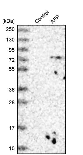
![Western Blot alpha-Fetoprotein/AFP Antibody (189502) [Unconjugated]](https://images.novusbio.com/images/antibody/alphaFetoprotein_MAB1368_Western_Blot_20276.jpg)
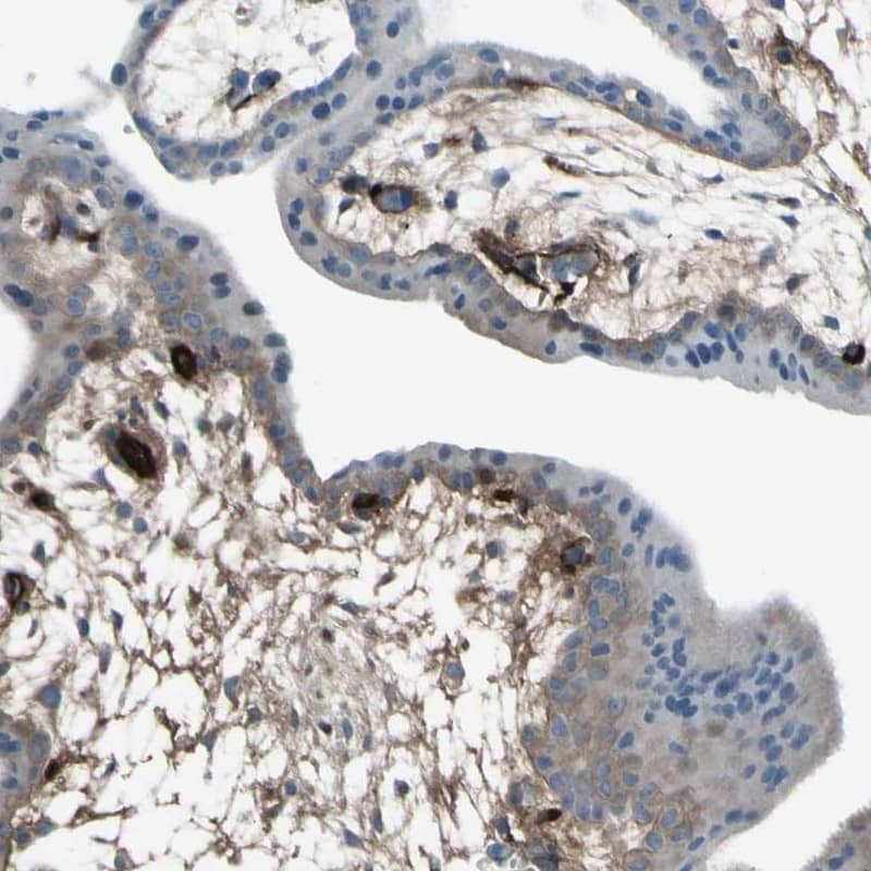
![Immunocytochemistry/ Immunofluorescence alpha-Fetoprotein/AFP Antibody (189502) [Unconjugated]](https://images.novusbio.com/images/mab1368_human-mouse-alpha-fetoprotein-mab-clone-189502-41202410325519.jpg)
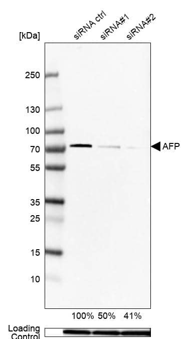
![Knockout Validated alpha-Fetoprotein/AFP Antibody (189502) [Unconjugated]](https://images.novusbio.com/images/antibody/alphaFetoprotein_MAB1368_Knockout_Validated_21503.jpg)


![Immunoprecipitation CD44 Antibody (2C5) [Unconjugated] - s Pan Specific](https://images.novusbio.com/images/antibody/bba10_human-cd44s-pan-specific-mab-clone-2c5-immunoprecipitation-2852025105258..png)
![Knockout Validated CD44 Antibody (2C5) [Unconjugated] - s Pan Specific](https://images.novusbio.com/images/antibody/bba10_human-cd44s-pan-specific-mab-clone-2c5-knockout-validated-285202510529..png)
![Western Blot CD44 Antibody (2C5) [Unconjugated] - s Pan Specific](https://images.novusbio.com/images/antibody/CD44_BBA10_Western_Blot_21459.jpg)


![Western Blot Decorin Antibody [Unconjugated]](https://images.novusbio.com/images/antibody/Decorin_AF1060_Western_Blot_19006.jpg)

|
Glypican 3 as a biomarker for gastro-esophageal adenocarcinoma By Jamshed Arslan, Pharm. D., PhD. Gastroesophageal adenocarcinoma originates from the glandular epithelium of the esophagus, gastroesophageal junction and stomach. The incidence of gastroesophageal adenocarcinoma is ... Read full blog post. |
The concentration calculator allows you to quickly calculate the volume, mass or concentration of your vial. Simply enter your mass, volume, or concentration values for your reagent and the calculator will determine the rest.


![Flow Cytometry Syndecan-2/CD362 Antibody (305515) [Unconjugated]](https://images.novusbio.com/images/antibody/Syndecan2_MAB2965_Flow_Cytometry_20268.jpg)
![Flow Cytometry Glypican 1 Antibody [Unconjugated]](https://images.novusbio.com/images/antibody/Glypican_1_AF4519_Flow_Cytometry_8254.jpg)
![Immunocytochemistry/ Immunofluorescence Glypican 1 Antibody [Unconjugated]](https://images.novusbio.com/images/af4519_human-glypican-1-affinity-purified-polyclonal-ab-immunocytochemistry-immunofluorescence-1212202582239.jpg)
![Immunocytochemistry Glypican 1 Antibody [Unconjugated]](https://images.novusbio.com/images/antibody/Glypican_1_AF4519_Flow_Cytometry_17434.jpg)


![SDS-Page IGF-II/IGF2 [Unconjugated]](https://images.novusbio.com/images/protein/IGF-II_292-G2_668.jpg)
![Bioactivity IGF-II/IGF2 [Unconjugated]](https://images.novusbio.com/images/protein/IGF-II_292-G2_669.jpg)


![Immunohistochemistry Insulin Antibody (182410) [Unconjugated]](https://images.novusbio.com/images/antibody/mab1417_human-bovine-mouse-insulin-mab-clone-182410-immunohistochemistry-308202115145.jpg)
![Immunocytochemistry Insulin Antibody (182410) [Unconjugated]](https://images.novusbio.com/images/antibody/Insulin_MAB1417_Immunocytochemistry_9376.jpg)
![Immunocytochemistry/Immunofluorescence: Glypican 3 Antibody (1G12) [NBP2-44484] - ICC/IF analysis of MeOH-fixed HepG2 cells labeling Glypican-3 with followed by Goat anti-Mouse IgG-CF488 (Green). The nuclear counterstain is Red Dot (Red)](https://images.novusbio.com/images/Glypican-3-Antibody-1G12-Immunofluorescence-NBP2-44484-img0003.jpg "Immunocytochemistry/Immunofluorescence: Glypican 3 Antibody (1G12) [NBP2-44484] - ICC/IF analysis of MeOH-fixed HepG2 cells labeling Glypican-3 with followed by Goat anti-Mouse IgG-CF488 (Green). The nuclear counterstain is Red Dot (Red)")