has a molecular weight (MW) of 37-40 kDa as analyzed by SEC-MALS, suggesting that this protein is a monomer. MW may differ from predicted MW due to post-translational modifications (PTMs) present (i.e. Glycosylation).")
| Reactivity | HuSpecies Glossary |
| Applications | Enzyme Activity |
| Format | Carrier-Free |
| Additional Information | Analyzed by SEC-MALS |
| Details of Functionality | Measured by its ability to transfer phosphate from adenosine triphosphate (ATP) to a synthetic peptide substrate. The specific activity is >35 pmol/min/μg, as measured under the described conditions. |
| Source | Spodoptera frugiperda, Sf 21 (baculovirus)-derived human EGFR protein Gly696-Gly1022, with an N-terminal Met and 6-His tag |
| Accession # | |
| N-terminal Sequence | Met |
| Protein/Peptide Type | Recombinant Enzymes |
| Purity | >95%, by SDS-PAGE visualized with Silver Staining and quantitative densitometry by Coomassie® Blue Staining |
| Endotoxin Note | <0.10 EU per 1 μg of the protein by the LAL method. |
| Dilutions |
|
| Theoretical MW | 38 kDa. Disclaimer note: The observed molecular weight of the protein may vary from the listed predicted molecular weight due to post translational modifications, post translation cleavages, relative charges, and other experimental factors. |
| SDS-PAGE | 39 kDa, under reducing conditions |
| Storage | Use a manual defrost freezer and avoid repeated freeze-thaw cycles.
|
|||
| Buffer | Supplied as a 0.2 μm filtered solution in Tris, NaCl and DTT. |
|||
| Purity | >95%, by SDS-PAGE visualized with Silver Staining and quantitative densitometry by Coomassie® Blue Staining |
|||
| Assay Procedure |
*Adjusted for Substrate Control **Derived using ADP-GloTM Kinase Assay Kit protocol (Promega) Per Well:
|
![Intracellular Staining by Flow Cytometry AKT [p Ser473] Antibody [Unconjugated] - Pan Specific](https://images.novusbio.com/images/antibody/Akt3_AF887_Flow_Cytometry_8283.jpg)
![Western Blot AKT [p Ser473] Antibody [Unconjugated] - Pan Specific](https://images.novusbio.com/images/af887_phospho-akt-s473-pan-specific-affinity-purified-pab-41202410485440.jpg)
![Western Blot AKT [p Ser473] Antibody [Unconjugated] - Pan Specific](https://images.novusbio.com/images/af887_phospho-akt-s473-pan-specific-affinity-purified-pab-8120255552843.jpg)


![Western Blot EphB2 Antibody [Unconjugated]](https://images.novusbio.com/images/af467_human-mouse-ephb2-affinity-purified-polyclonal-ab-41202410485412.jpg)
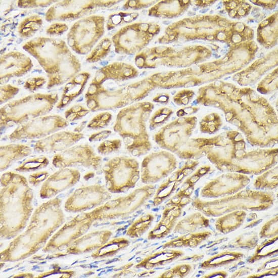
![Immunohistochemistry EphB2 Antibody [Unconjugated]](https://images.novusbio.com/images/antibody/EphB2_AF467_Immunohistochemistry_20369.jpg)
![Western Blot EphB2 Antibody [Unconjugated]](https://images.novusbio.com/images/antibody/EphB2_AF467_Western_Blot_21511.jpg)
![Neutralization ErbB3/Her3 Antibody (66223) [Unconjugated]](https://images.novusbio.com/images/antibody/ErbB3_MAB3481_Block_Neutralize_9158.jpg)
![ELISA ErbB3/Her3 Antibody (66223) [Unconjugated]](https://images.novusbio.com/images/mab3481_human-erbb3-her3-mab-clone-66223-elisa-639201777920228845.jpg)


|
Hypoxia-Dependent CAR Stabilizing Construct in T cells Improves Solid Tumor Targeting and Efficacy By Victoria Osinski, PhDDespite advances in the development of cancer immunotherapies, those specifically targeting tumors still remains limited. Currently, there is great interest in utilizing chimeric antigen rece... Read full blog post. |
|
Dual applications of a c-Myc antibody in mitochondrial research c-Myc, a proto-oncogene, has documented involvement in cellular differentiation, cell growth, cell death and tumor formation. Target genes of the Myc family include those that participate in cell survival, translation, transcription, metabolism and... Read full blog post. |
|
The dynamic use of a PCNA antibody in fish, porcine and primate species Proliferating cell nuclear antigen (PCNA) plays a crucial role in nucleic acid metabolism as it pertains to DNA replication and repair. Most noted for its activation of subunits of DNA polymerase, it has also been found to interact with cell-cycl... Read full blog post. |
|
Using a STAT3 antibody in chromatin immunoprecipitation (ChIP) Signal transducer and activator of transcription 3 (STAT3) is an important oncogenic transcriptional factor that mediates tumor induced immune suppression. Specifically, STAT3 transmits signals from cytokines and growth factor receptors in the pla... Read full blog post. |
|
Beta Tubulin III and neurogenesis Beta tubulin III, also known as Tuj-1, is a class III member of the beta tubulin protein family. Beta tubulins are one of two structural components that form our microtubule network. While general tubulins play a role in a wide range of cellular pr... Read full blog post. |
|
The relationship between Ki67 and HIF-1 in cancer Ki67, also known as MKI67, is best known as the leading marker of cellular proliferation. Ki67 is regulated by a balance between synthesis and degradation, and often carries a very short half-life. First discovered to be located to dividing cells,... Read full blog post. |
|
Niemann Pick-C1 and cholesterol dynamics Niemann-Pick type C1 (NPC1) mediates low-density cholesterol transport from late endosomes and lysosomes to other areas of the cell via receptor mediation endocytosis. Although cholesterol moves freely inside the cell, it cannot independently expo... Read full blog post. |
|
MAPK3/ERK1 - A signal transduction pathway with roles in development and disease Mitogen-activated protein kinases (MAPKs) are important signaling proteins needed to transmit and relay extracellular stimuli and to illicit intracellular responses (1). The MAPK family of proteins are serine/threonine kinases that are able to phos... Read full blog post. |
|
Using EGF Protein from Novus Biologicals EGF (epidermal growth factor) stimulates differentiation, proliferation and cell growth by binding to its receptor, EGFR. EGF was first discovered in the mouse submandibular gland in 1986 by Stanley Cohen of Vanderbilt University, leading to a Nobel P... Read full blog post. |
|
It's a Wiz: Merlin Antibodies Advance Hepatic Tumor Research The NF2 gene, also known as “Merlin”, was discovered through studies into Neurofibromatosis Type II, a rare genetic disease which causes formation of non-malignant, but life-limiting, brain tumors. NF2 encodes a cytoskeletal protein involved in extrac... Read full blog post. |
The concentration calculator allows you to quickly calculate the volume, mass or concentration of your vial. Simply enter your mass, volume, or concentration values for your reagent and the calculator will determine the rest.
| Uniprot |
|


 was resolved with SDS-PAGE under reducing (R) and non-reducing (NR) conditions and visualized by Coomassie® Blue staining, showing bands at 39 kDa.")
![Bioactivity EGF [Unconjugated]](https://images.novusbio.com/images/protein/EGF_236EG_1570.jpg)
![Cell Culture EGF [Unconjugated]](https://images.novusbio.com/images/236-eg_recombinant-human-egf-protein-cf-25202394526.jpg)
![Cell Culture EGF [Unconjugated]](https://images.novusbio.com/images/236-eg_recombinant-human-egf-protein-cf-bioactivity-25202394053.jpg)
![Bioactivity ErbB2/Her2 [Unconjugated]](https://images.novusbio.com/images/protein/1129-er_recombinant-human-erbb2-her2-fc-chimera-protein-cf-bioactivity-7122020142841.jpg)
![SDS-Page ErbB2/Her2 [Unconjugated]](https://images.novusbio.com/images/protein/ErbB2_1129-ER_70.jpg)
![Western Blot ERK2 Antibody [Unconjugated]](https://images.novusbio.com/images/antibody/ERK2_AF1230_Western_Blot_5097.jpg)
![Knockout Validated ERK2 Antibody [Unconjugated]](https://images.novusbio.com/images/antibody/ERK2_AF1230_Knockout_Validated_22864.jpg)
![Immunohistochemistry ERK2 Antibody [Unconjugated]](https://images.novusbio.com/images/antibody/ERK2_AF1230_Immunohistochemistry_20696.jpg)


![N/A VEGF [HRP]](https://images.novusbio.com/images/elisa/VEGF_DVE00_ELISA_208.jpg)
![N/A VEGF [HRP]](https://images.novusbio.com/images/elisa/DATA_VEGF_DVE00_ELISA_871.jpg)
![N/A VEGF [HRP]](https://images.novusbio.com/images/elisa/DATA_VEGF_DVE00_ELISA_872.jpg)
![Immunocytochemistry ErbB4/Her4 Antibody (182803) [Unconjugated]](https://images.novusbio.com/images/antibody/ErbB4_MAB1131_Immunocytochemistry__Immunofluorescence_21145.jpg)
![Immunohistochemistry ErbB4/Her4 Antibody (182803) [Unconjugated]](https://images.novusbio.com/images/antibody/ErbB4_MAB1131_Immunohistochemistry_12968.jpg)
![Western Blot Src [p Tyr419] Antibody [Unconjugated]](https://images.novusbio.com/images/antibody/Src_AF2685_Western_Blot_5209.jpg)
![Immunohistochemistry Src [p Tyr419] Antibody [Unconjugated]](https://images.novusbio.com/images/af2685_human-phospho-src-y419-affinity-purified-polyclonal-ab-412024124022.jpg)
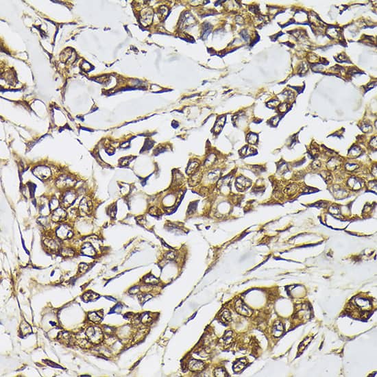
![Immunohistochemistry Src [p Tyr419] Antibody [Unconjugated]](https://images.novusbio.com/images/antibody/Src_AF2685_Immunohistochemistry_9943.jpg)


![SEC-MALS TGF-alpha [Unconjugated]](https://images.novusbio.com/images/239-a_recombinant-human-tgf-alpha-protein-cf-sec-mals-2810202585510.jpg)