 and Recombinant Mouse EphB2 Fc Chimera (Catalog # 467-B2). PVDF Membrane was probed with 0.1 µg/mL of Goat Anti-Human/Mouse EphB2 Antigen Affinity-purified Polyclonal Antibody (Catalog # AF467) followed by HRP-conjugated Anti-Goat IgG Secondary Antibody (Catalog # HAF109). A specific band was detected for EphB2 at approximately 120 kDa (as indicated). This experiment was conducted under reducing conditions and using Immunoblot Buffer Group 3.")
| Reactivity | Hu, MuSpecies Glossary |
| Applications | WB, Simple Western, Flow, IHC, CyTOF-ready, ICC/IF |
| Clonality | Polyclonal |
| Host | Goat |
| Conjugate | Unconjugated |
| Concentration | LYOPH |
| Immunogen | Mouse myeloma cell line NS0-derived recombinant mouse EphB2 Val27-Lys548 Accession # P54763 |
| Specificity | Detects mouse and human EphB2 in direct ELISAs and Western blots. In Western blots, approximately 5% cross-reactivity with recombinant rat (rr) EphB1, recombinant mouse (rm) EphA8, rmEphA6, rmEphB6, rmEphA3, rmEphA4, rmEphA7, rrEphA5, recombinant human EphA1, rmEphA2 and rmEphB3 is observed. |
| Source | N/A |
| Isotype | IgG |
| Clonality | Polyclonal |
| Host | Goat |
| Gene | EPHB2 |
| Purity Statement | Antigen Affinity-purified |
| Innovator's Reward | Test in a species/application not listed above to receive a full credit towards a future purchase. |
| Dilutions |
|
|
| Reviewed Applications |
|
|
| Publications |
|
| Storage | Use a manual defrost freezer and avoid repeated freeze-thaw cycles.
|
| Buffer | Lyophilized from a 0.2 μm filtered solution in PBS with Trehalose. See Certificate of Analysis for details. *Small pack size (-SP) is supplied either lyophilized or as a 0.2 µm filtered solution in PBS. |
| Preservative | No Preservative |
| Concentration | LYOPH |
| Reconstitution Instructions | Reconstitute at 0.2 mg/mL in sterile PBS. For liquid material, refer to CoA for concentration. |
EphB2, also known as Cek5, Nuk, Erk, Qek2, Tyro5, Sek3, Hek5, and Drt (1), is a member of the Eph receptor family which binds members of the ephrin ligand family. There are two classes of receptors, designated A and B. Both the A and B class receptors have an extracellular region consisting of a globular domain, a cysteine-rich domain, and two fibronectin type III domains. This is followed by the transmembrane region and the cytoplasmic region. The cytoplasmic region contains a juxtamembrane motif with two tyrosine residues which are the major autophosphorylation sites, a kinase domain, and a conserved sterile alpha motif (SAM) in the carboxy tail which contains one conserved tyrosine residue. Activation of kinase activity occurs after ligand recognition and binding. EphB2 has been shown to bind ephrin-B1, ephrin-B2, and ephrin-B3 (2, 3). The extracellular domains of human and mouse EphB2 share 99% amino acid identity. Only membrane-bound or
Fc‑clustered ligands are capable of activating the receptor in vitro. Soluble monomeric ligands bind the receptor but do not induce receptor autophosphorylation and activation (2). In vivo, the ligands and receptors display reciprocal expression (3). It has been found that nearly all the receptors and ligands are expressed in developing and adult neural tissue (3). The ephrin/Eph families also appear to play a role in angiogenesis (3).
![Intracellular Staining by Flow Cytometry AKT [p Ser473] Antibody [Unconjugated] - Pan Specific](https://images.novusbio.com/images/antibody/Akt3_AF887_Flow_Cytometry_8283.jpg)
![Western Blot AKT [p Ser473] Antibody [Unconjugated] - Pan Specific](https://images.novusbio.com/images/af887_phospho-akt-s473-pan-specific-affinity-purified-pab-41202410485440.jpg)
![Western Blot AKT [p Ser473] Antibody [Unconjugated] - Pan Specific](https://images.novusbio.com/images/af887_phospho-akt-s473-pan-specific-affinity-purified-pab-8120255552843.jpg)


![Immunocytochemistry EGFR Antibody [Unconjugated]](https://images.novusbio.com/images/antibody/EGF_R_AF231_Immunocytochemistry__Immunofluorescence_21143.jpg)
![Flow Cytometry EGFR Antibody [Unconjugated]](https://images.novusbio.com/images/antibody/EGF_R_AF231_Flow_Cytometry_20401.jpg)
![Western Blot EGFR Antibody [Unconjugated]](https://images.novusbio.com/images/antibody/EGF_R_AF231_Western_Blot_19925.jpg)
![EphB2 Antibody [Unconjugated]](/sites/all/modules/enterprise-tech/et_datasheets/images/novus_guarantee.png "EphB2 Antibody [Unconjugated]")
| Images | Ratings | Applications | Species | Date | Details | ||||||||||
|---|---|---|---|---|---|---|---|---|---|---|---|---|---|---|---|
Enlarge |
reviewed by:
STAVROULA THEOFANOUS |
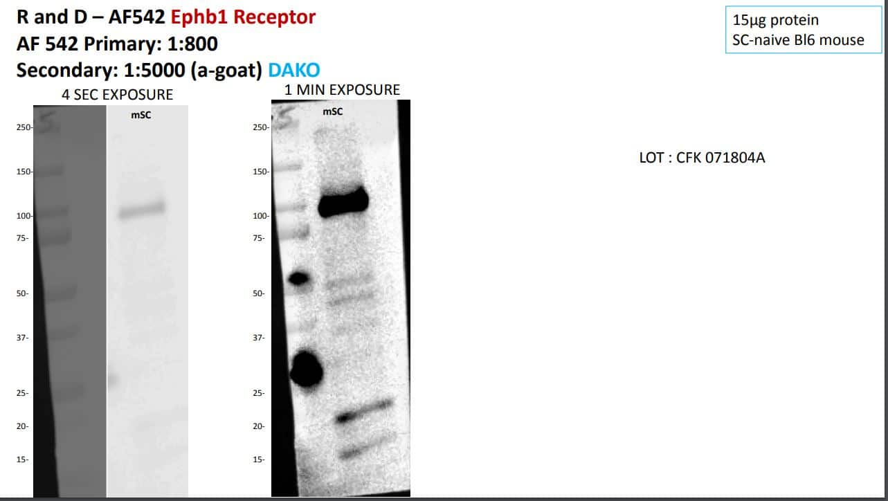
WB | Mouse | 07/05/2018 |
Summary
Comments
|
||||||||||
Enlarge |
reviewed by:
STAVROULA THEOFANOUS |
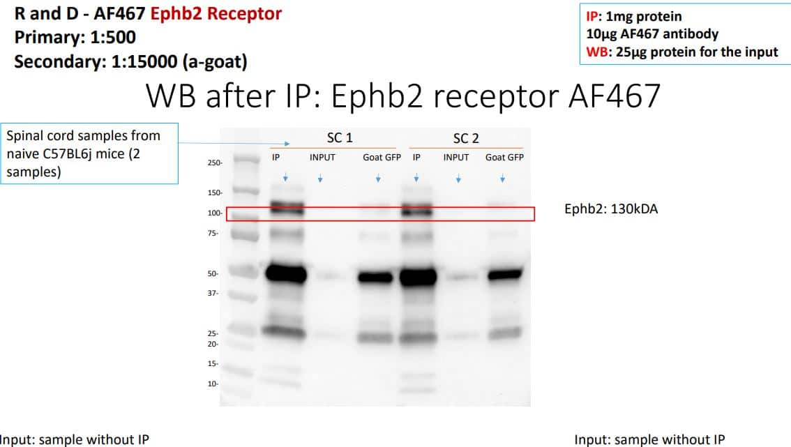
IP | Mouse | 04/16/2018 |
Summary
Comments
|
||||||||||
Enlarge |
reviewed by:
Verified Customer |
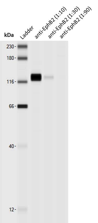
Simple Western | Mouse | 07/12/2017 |
Summary
Comments
|
||||||||||
|
reviewed by:
Verified Customer |
WB | Mouse | 01/08/2015 |
Summary
|
|||||||||||
|
reviewed by:
Verified Customer |
WB | Other | 01/08/2015 |
Summary
|
Secondary Antibodies |
Isotype Controls |
|
Stemness is responsible for onset and metastasis of colorectal cancer By Jamshed Arslan, Pharm. D., PhD. Colorectal cancer stem cells are a rare subpopulation of colorectal cancer cells that can self-renew and initiate and sustain tumor growth when transplanted into an animal host.1,2 C... Read full blog post. |
The concentration calculator allows you to quickly calculate the volume, mass or concentration of your vial. Simply enter your mass, volume, or concentration values for your reagent and the calculator will determine the rest.
5 | |
4 | |
3 | |
2 | |
1 |
| STAVROULA THEOFANOUS 07/05/2018 |
||
| Application: | WB | |
| Species: | Mouse |
| STAVROULA THEOFANOUS 04/16/2018 |
||
| Application: | IP | |
| Species: | Mouse |
| Verified Customer 07/12/2017 |
||
| Application: | Simple Western | |
| Species: | Mouse |


 or isotype control antibody (Catalog # AB-108-C, open histogram), followed by Phycoerythrin-conjugated Anti-Goat IgG Secondary Antibody (Catalog # F0107). View our protocol for Staining Membrane-associated Proteins.")
 or isotype control antibody (Catalog # AB-108-C, open histogram), followed by Phycoerythrin-conjugated Anti-Goat IgG Secondary Antibody (Catalog # F0107). View our protocol for Staining Membrane-associated Proteins.")
 at 5 µg/mL for 3 hours at room temperature. Cells were stained using the NorthernLights™ 557-conjugated Anti-Goat IgG Secondary Antibody (red; Catalog # NL001) and counterstained with DAPI (blue). Specific staining was localized to cytoplasm. View our protocol for Fluorescent ICC Staining of Cells on Coverslips.")
 using 15 µg/mL Goat Anti-Mouse EphB2 Antigen Affinity-purified Polyclonal Antibody (Catalog # AF467) overnight at 4 °C. Tissue was stained with the NorthernLights™ 557-conjugated Anti-Goat IgG Secondary Antibody (red; Catalog # NL001) and counterstained (green). View our protocol for Fluorescent IHC Staining of Frozen Tissue Sections.")
 at 3 µg/mL for 1 hour at room temperature followed by incubation with the Anti-Goat IgG VisUCyte™ HRP Polymer Antibody (Catalog # VC004). Tissue was stained using DAB (brown) and counterstained with hematoxylin (blue). Specific staining was localized to cytoplasm in cancer cells. View our protocol for IHC Staining with VisUCyte HRP Polymer Detection Reagents.")
 using Goat Anti-Human/Mouse EphB2 Antigen Affinity-purified Polyclonal Antibody (Catalog # AF467) at 1.7 µg/mL for 1 hour at room temperature followed by incubation with the Anti-Goat IgG VisUCyte™ HRP Polymer Antibody (Catalog # VC004). Tissue was stained using DAB (brown) and counterstained with hematoxylin (blue). Specific staining was localized to developing brain. View our protocol for IHC Staining with VisUCyte HRP Polymer Detection Reagents.")
 (NBP2-49845) and COLO 205 human colorectal adenocarcinoma cell line, loaded at 0.5 mg/ml. A specific band was detected for EphB2 at approximately 135 kDa (as indicated) using 1 µg/ml of Goat Anti-Human/Mouse EphB2 Antigen Affinity-purified Polyclonal Antibody (Catalog # AF467) followed by HRP-conjugated Donkey Anti-Goat Secondary Antibody (Catalog # 042-206). This experiment was conducted under reducing conditions and using the 12-230kDa separation system.")
 using 20 µg/mL of Goat Anti-Human/Mouse EphB2 Antigen Affinity-purified Polyclonal Antibody (Catalog # AF467) followed by 1:50 dilution of HRP-conjugated Anti-Goat IgG Secondary Antibody (Catalog # HAF109). This experiment was conducted under reducing conditions and using the 12-230 kDa separation system.")
 and ERBB3 (red, A and B) by co-immunofluorescence in normal colon (A) and colorectal cancer (B) (DAPI, blue). Scale bar, 50μm. Image collected and cropped by CiteAb from the following publication (//pubmed.ncbi.nlm.nih.gov/26367378), licensed under a CC-BY license. Not internally tested by R&D Systems.")
 Isolated cell fractions from livers of mice subjected to chronic CCl4 injections were analyzed for EphB2, Ephrin-B1, Ephrin-B2 and Ephrin-B3 mRNA levels using RT-qPCR. Results are shown as fold change compared to liver cell fractions obtained from vehicle-treated controls. Error bars represent mean ± SEM.; n = 6 animals; CD11b = macrophages, LSEC = Liver sinusoidal endothelial cells, HEP = Hepatocytes and HSCs = Hepatic stellate cells. (b) OCT liver sections from C57BL/6 J mice chronically injected with CCl4 or vehicle (oil) controls were stained with EphB2 (red), alpha SMA (green) and DAPI/DNA (blue) and analyzed using confocal microscopy. Scale bar = 100 µm, “C” denotes the central vein. All images are representative of 5 mice per group. (c) OCT liver sections from C57BL/6 J mice chronically injected with CCl4 or vehicle controls were stained with phospho-EphB1/EphB2-Y594 (red), PDGFR beta (green) and DAPI/DNA (blue) and analyzed using confocal microscopy. Scale bar = 50 µm. All images are representative of 5 mice per group. Image collected and cropped by CiteAb from the following publication (//pubmed.ncbi.nlm.nih.gov/29416088), licensed under a CC-BY license. Not internally tested by R&D Systems.")
 Simulation of cell-cell segregation using the same adhesion term in both cell types (Aeph = Aephrin = 100, left panel) vs. increased adhesion only in the green cell population (Aeph = 110, Aephrin = 100, right panel). Both simulations started with the same number of Eph (green) and ephrin (black) expressing cells. In the “Equal adhesion” case, an ‘Islands-in-a-sea’ pattern is less apparent. B) Representative images from segregation assays of unlabelled ephrin-B1 cells co-cultured with Cell Tracker-green labelled (green staining) EphB2 cells, without (left) or with E-cadherin-cherry expression (red staining, right); scale bar, 75 µm. C) Quantitation of cell densities in the cell clusters shown in B (n = 10). D) Western blot analysis of lysates from parental and E-cadherin-cherry-transduced cells, using the indicated antibodies. Image collected and cropped by CiteAb from the following publication (//dx.plos.org/10.1371/journal.pone.0111803), licensed under a CC-BY license. Not internally tested by R&D Systems.")


![Western Blot ERK2 Antibody [Unconjugated]](https://images.novusbio.com/images/antibody/ERK2_AF1230_Western_Blot_5097.jpg)
![Knockout Validated ERK2 Antibody [Unconjugated]](https://images.novusbio.com/images/antibody/ERK2_AF1230_Knockout_Validated_22864.jpg)
![Immunohistochemistry ERK2 Antibody [Unconjugated]](https://images.novusbio.com/images/antibody/ERK2_AF1230_Immunohistochemistry_20696.jpg)
![Western Blot JNK1 Antibody (228601) [Unconjugated]](https://images.novusbio.com/images/antibody/JNK1_MAB17761_Western_Blot_5991.jpg)
![Western Blot JNK1 Antibody (228601) [Unconjugated]](https://images.novusbio.com/images/antibody/JNK1_MAB17761_Western_Blot_6327.jpg)
![Immunocytochemistry JNK1 Antibody (228601) [Unconjugated]](https://images.novusbio.com/images/antibody/JNK1_MAB17761_Immunocytochemistry__Immunofluorescence_20184.jpg)
![Western Blot p38 alpha Antibody [Unconjugated]](https://images.novusbio.com/images/antibody/p38_alpha_AF8691_Western_Blot_7402.jpg)
![Immunohistochemistry p38 alpha Antibody [Unconjugated]](https://images.novusbio.com/images/antibody/p38_alpha_AF8691_Immunohistochemistry_7087.jpg)
![Western Blot p38 alpha Antibody [Unconjugated]](https://images.novusbio.com/images/antibody/p38_alpha_AF8691_Western_Blot_7401.jpg)
![Western Blot p38 gamma/SAPK3 Antibody [Unconjugated]](https://images.novusbio.com/images/antibody/p38_gamma_AF1347_Western_Blot_12768.jpg)
![Simple Western p38 gamma/SAPK3 Antibody [Unconjugated]](https://images.novusbio.com/images/antibody/p38_gamma_AF1347_Simple_Western_16621.jpg)
![Western Blot p38 gamma/SAPK3 Antibody [Unconjugated]](https://images.novusbio.com/images/af1347_human-mouse-rat-p38-gamma-affinity-purified-pab-44202415425098.jpg)


![Immunocytochemistry/ Immunofluorescence c-jun [p Ser63] Antibody (SY0297)](https://images.novusbio.com/images/c-jun-p-Ser63-Antibody-SY0297-Immunocytochemistry-Immunofluorescence-NBP2-67471-img0006.jpg)
![Western Blot c-jun [p Ser63] Antibody (SY0297)](https://images.novusbio.com/images/c-jun-p-Ser63-Antibody-SY0297-Western-Blot-NBP2-67471-img0008.jpg)
![Western Blot c-jun [p Ser63] Antibody (SY0297)](https://images.novusbio.com/images/c-jun-p-Ser63-Antibody-SY0297-Western-Blot-NBP2-67471-img0009.jpg)


![SDS-Page TNF-alpha [Unconjugated]](https://images.novusbio.com/images/protein/TNF-alpha_210-TA_256.jpg)
![Bioactivity TNF-alpha [Unconjugated]](https://images.novusbio.com/images/protein/TNFalpha_210TA_1658.jpg)
![SEC-MALS TNF-alpha [Unconjugated]](https://images.novusbio.com/images/210-ta_recombinant-human-tnf-alpha-protein-sec-mals-35202312244..jpg)
![Immunohistochemistry Raf-1 Antibody (563002) [Unconjugated]](https://images.novusbio.com/images/antibody/Raf-1_MAB4540_Immunohistochemistry_9891.jpg)
![Western Blot Raf-1 Antibody (563002) [Unconjugated]](https://images.novusbio.com/images/antibody/Raf-1_MAB4540_Western_Blot_9426.jpg)
![Simple Western Raf-1 Antibody (563002) [Unconjugated]](https://images.novusbio.com/images/mab4540_human-mouse-rat-raf-1-mab-clone-563002-simple-western-158202212919.jpg)
 or Normal Goat IgG Isotype Control Antibody (Catalog # AB-108-C, open histogram), followed by Phycoerythrin-conjugated Anti-Goat IgG Secondary Antibody (Catalog # F0107).")


