![Western Blot: PGAM1/2/4 Antibody [NB100-774] - staining of Mouse (A), Rat (B) and Pig (C) Liver lysate (35ug protein in RIPA buffer). Primary incubation was 1 hour. Detected by chemiluminescence.](http://images.novusbio.com/fullsize/PGAM1-2-4-Antibody-Western-Blot-NB100-774-img0002.jpg "Western Blot: PGAM1/2/4 Antibody [NB100-774] - staining of Mouse (A), Rat (B) and Pig (C) Liver lysate (35ug protein in RIPA buffer). Primary incubation was 1 hour. Detected by chemiluminescence.")
| Reactivity | Hu, Mu, Rt, Po, Bv, CaSpecies Glossary |
| Applications | WB, ELISA, ICC/IF |
| Clonality | Polyclonal |
| Host | Goat |
| Conjugate | Unconjugated |
| Concentration | 0.5 mg/ml |
| Immunogen | Peptide with sequence C-KAMEAVAAQGKAKK, from the C Terminus of the protein sequence according to NP_002620.1; NP_000281.2; NP_001025062.1. |
| Specificity | Please note this antibody is expected to recognize the products of 3 highly similar genes. |
| Predicted Species | Canine (100%), Bovine (100%). Backed by our 100% Guarantee. |
| Isotype | IgG |
| Clonality | Polyclonal |
| Host | Goat |
| Gene | PGAM1 |
| Purity | Immunogen affinity purified |
| Innovator's Reward | Test in a species/application not listed above to receive a full credit towards a future purchase. |
| Dilutions |
|
||||||
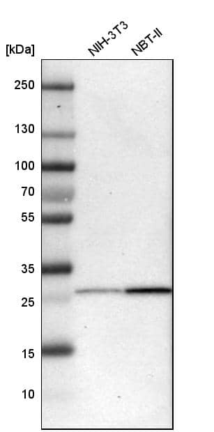
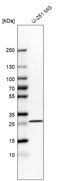
| Application Notes | WB: Approx 27kDa band observed in Human Cerebellum and in Human and Mouse Liver lysates, and approx. 28kDa band observed in Rat and Pig Liver lysates (calculated MW of 28.8kDa according to Human NP_002620.1, NP_000281.2, and NP_001025062.1, Mouse NP_075907.2, Rat NP_445742.2 and Pig XP_003483583.1 ). |
||||||
| Theoretical MW | 29 kDa. Disclaimer note: The observed molecular weight of the protein may vary from the listed predicted molecular weight due to post translational modifications, post translation cleavages, relative charges, and other experimental factors. |
||||||
| Control |
|
||||||
| Publications |
|
| Storage | Store at -20C. Avoid freeze-thaw cycles. |
| Buffer | Tris saline (20 mM Tris pH 7.3, 150 mM NaCl), 0.5% BSA |
| Preservative | 0.02% Sodium Azide |
| Concentration | 0.5 mg/ml |
| Purity | Immunogen affinity purified |


![Western Blot Galectin-1 Antibody [Unconjugated]](https://images.novusbio.com/images/antibody/Galectin-1_AF1152_Western_Blot_12374.jpg)
![Simple Western Galectin-1 Antibody [Unconjugated]](https://images.novusbio.com/images/antibody/af1152_human-galectin-1-affinity-purified-polyclonal-ab-simple-western-236202593440..jpg)
![Simple Western Galectin-1 Antibody [Unconjugated]](https://images.novusbio.com/images/antibody/Galectin1_AF1152_Simple_Western_16673.jpg)
 - Azide and BSA Free")


| Human Liver Whole Tissue Lysate (Adult Whole Normal) | |
| PGAM1 Overexpression Lysate | |
| Human Brain Whole Tissue Lysate (Adult Whole Normal) |
Secondary Antibodies |
Isotype Controls |
Research Areas for PGAM1/2/4 Antibody (NB100-774)Find related products by research area.
|
The concentration calculator allows you to quickly calculate the volume, mass or concentration of your vial. Simply enter your mass, volume, or concentration values for your reagent and the calculator will determine the rest.


 mRNA and protein levels of R-Ras2 in Sirt6 WT and KO MEFs. (B) In-gel fluorescence (with NH2OH treatment) showing that R-Ras2 has higher lysine fatty acylation level in Sirt6 KO MEFs than in Sirt6 WT MEFs. Right histogram shows the quantification of bands on the fluorescence gel. Values with error bars indicate mean ± s.d. of three biological replicates. * indicates p<0.05. The full fluorescence gel is shown in Figure 2—figure supplement 2A. (C) Detection of R-Ras2 lysine fatty acylation levels in control and SIRT6 knockown HEK 293 T cells by in-gel fluorescence. Right histogram shows the quantification of bands on the fluorescence gel. Values with error bars indicate mean ± s.d. of three biological replicates. * indicates p<0.05. (D) Lysine fatty acylation levels of endogenous R-Ras2 in Sirt6 WT and KO MEFs. (E, F) SIRT6 defatty-acylated R-Ras2 in a NAD+-dependent manner in vitro. In-gel fluorescence was used to detect R-Ras2 lysine fatty acylation (E). A 32P-NAD+ assay was used to detect fatty acyl ADPR product from defatty-acylation reaction. (F). (G) In-gel fluorescence (with NH2OH treatment) showing that mutation of four lysine residues at the C-terminus of R-Ras2 significantly decreased lysine fatty acylation in Sirt6 KO MEFs. Right histogram shows the quantification of bands on the fluorescence gel. Values with error bars indicate mean ± s.d. of three biological replicates. * indicates p<0.05. The full fluorescence gel including R-Ras2 total fatty acylation levels (without NH2OH treatment) is shown in Figure 2—figure supplement 2C. (H) Tandem mass (MS/MS) spectrum of doubly charged Alk14 modified (on K194) R-Ras2 peptide. The b- and y- ions are shown along with the peptide sequence. (I) In-gel fluorescence (with NH2OH treatment) showing that single mutation of four lysine residues at the C-terminus of R-Ras2 did not affect R-Ras2 lysine fatty acylation. (J) Confocal imaging showed that R-Ras2 W")
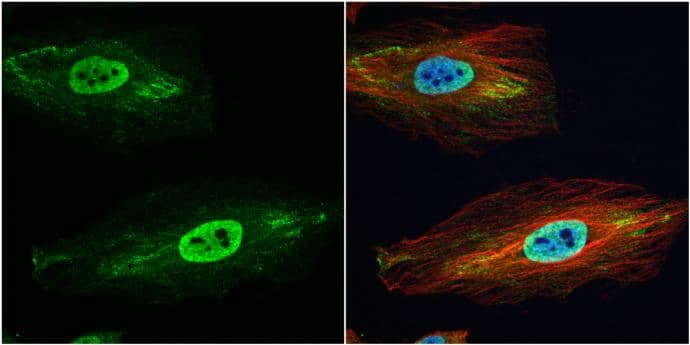
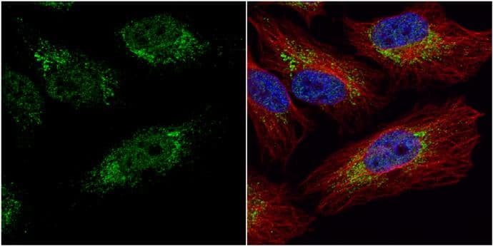
![Immunocytochemistry/ Immunofluorescence: PGAM1/2/4 Antibody [NB100-774] - Detection of PGAM in KLN-205 cells with antibodies directed to whole PGAM protein or to C-terminal peptide of PGAMA. control conditions (scan parameters in red channel were set to emphasize nucleolar staining with propidium iodide – PI) B. RNase-treated cells. Bar=15 μm. Image collected & cropped by CiteAb from the following publication (//www.oncotarget.com/lookup/doi/10.18632/oncotarget.4044), licensed under a CC-BY license. Not internally tested by Novus Biologicals.](http://images.novusbio.com/fullsize/nb100-774_goat-polyclonal-pgam1-2-4-antibody-310202415371946.jpg "Immunocytochemistry/ Immunofluorescence: PGAM1/2/4 Antibody [NB100-774] - Detection of PGAM in KLN-205 cells with antibodies directed to whole PGAM protein or to C-terminal peptide of PGAMA. control conditions (scan parameters in red channel were set to emphasize nucleolar staining with propidium iodide – PI) B. RNase-treated cells. Bar=15 μm. Image collected & cropped by CiteAb from the following publication (//www.oncotarget.com/lookup/doi/10.18632/oncotarget.4044), licensed under a CC-BY license. Not internally tested by Novus Biologicals.")


![Western Blot Aldo-keto Reductase 1C1/AKR1C1 Antibody (859026) [Unconjugated]](https://images.novusbio.com/images/antibody/Aldoketo_Reductase_1C1_MAB6529_Western_Blot_13068.jpg)
![Simple Western Aldo-keto Reductase 1C1/AKR1C1 Antibody (859026) [Unconjugated]](https://images.novusbio.com/images/antibody/Aldoketo_Reductase_1C1_MAB6529_Simple_Western_17147.jpg)
![Western Blot Annexin A4 Antibody [Unconjugated]](https://images.novusbio.com/images/antibody/Annexin_A4_AF4146_Western_Blot_5405.jpg)
![Immunohistochemistry Annexin A4 Antibody [Unconjugated]](https://images.novusbio.com/images/antibody/Annexin_A4_AF4146_Immunohistochemistry_10406.jpg)
![Simple Western Annexin A4 Antibody [Unconjugated]](https://images.novusbio.com/images/antibody/af4146_human-mouse-rat-annexin-a4-affinity-purified-polyclonal-ab-simple-western-1712025133556..jpg)


![Western Blot: Human Brain Whole Tissue Lysate (Adult Normal) [NB820-59177] - Beta Actin expression on human brain whole tissue lysate using anti-Beta Actin antibody (Cat. # NBP1-47423). Image from verified customer review.](https://images.novusbio.com/images/Human-Brain-Whole-Tissue-Lysate-Adult-Normal-Western-Blot-NB820-59177-img0001.jpg "Western Blot: Human Brain Whole Tissue Lysate (Adult Normal) [NB820-59177] - Beta Actin expression on human brain whole tissue lysate using anti-Beta Actin antibody (Cat. # NBP1-47423). Image from verified customer review.")
 and a low dose of doxorubicin (0.006 μmol/L) for 2 hours. Whole-cell lysates were harvested, and Simple Western was used to show the inhibition of pDNA-PK upon combination treatment of ZL-2201 with doxorubicin. Image collected and cropped by CiteAb from the following open publication (https://pubmed.ncbi.nlm.nih.gov/37663435), licensed under a CC-BY license. Not internally tested by R&D Systems.")