HAF109). A specific band was detected for Park7/DJ-1 at approximately 22 kDa (as indicated). This experiment was conducted under reducing conditions and using Immunoblot Buffer Group 1." title="Western blot shows lysates of HeLa human cervical epithelial carcinoma cell line and A172 human glioblastoma cell line. PVDF membrane was probed with 0.2 µg/mL of Goat Anti-Human Park7/DJ-1 Antigen Affinity-purified Polyclonal Antibody (Catalog # AF3995) followed by HRP-conjugated Anti-Goat IgG Secondary Antibody (HAF109). A specific band was detected for Park7/DJ-1 at approximately 22 kDa (as indicated). This experiment was conducted under reducing conditions and using Immunoblot Buffer Group 1." />
| Reactivity | HuSpecies Glossary |
| Applications | WB, Simple Western, IHC, ICC/IF, KO |
| Clonality | Polyclonal |
| Host | Goat |
| Conjugate | Unconjugated |
| Concentration | LYOPH |
| Immunogen | E. coli-derived recombinant human Park7/DJ-1 Met1-Asp189 Accession # Q99497 |
| Specificity | Detects human Park7/DJ-1 in Western blots. |
| Source | N/A |
| Isotype | IgG |
| Clonality | Polyclonal |
| Host | Goat |
| Gene | PARK7 |
| Purity Statement | Antigen Affinity-purified |
| Innovator's Reward | Test in a species/application not listed above to receive a full credit towards a future purchase. |
| Dilutions |
|
|
| Application Notes | In Simple Western only 10-15 uL of the recommended dilution is used per data point. |
|
| Publications |
|
| Storage | Use a manual defrost freezer and avoid repeated freeze-thaw cycles.
|
| Buffer | Lyophilized from a 0.2 μm filtered solution in PBS with Trehalose. *Small pack size (SP) is supplied either lyophilized or as a 0.2 µm filtered solution in PBS. |
| Preservative | No Preservative |
| Concentration | LYOPH |
| Reconstitution Instructions | Reconstitute at 0.2 mg/mL in sterile PBS. |
Park7, also known as DJ-1, is a cytoplasmic protein that belongs to the ThiJ/Pfp1/DJ-1 superfamily of highly conserved proteins that function as protein chaperones, catalases, proteases and kinases. Park7 is widely expressed in the brain as well as in peripheral tissues. It exists as a homodimer that can be localized in the cytoplasm, nucleus and mitochondria. Park7 is a redox-sensitive protein that has been ascribed various functions including that as a redox sensor and antioxidant protein. Mutations in Park7 are associated with a small percentage of hereditary early onset Parkinson’s disease.




![Intracellular Staining by Flow Cytometry AKT [p Ser473] Antibody [Unconjugated] - Pan Specific](https://images.novusbio.com/images/antibody/Akt3_AF887_Flow_Cytometry_8283.jpg)
![Western Blot AKT [p Ser473] Antibody [Unconjugated] - Pan Specific](https://images.novusbio.com/images/af887_phospho-akt-s473-pan-specific-affinity-purified-pab-41202410485440.jpg)
![Western Blot AKT [p Ser473] Antibody [Unconjugated] - Pan Specific](https://images.novusbio.com/images/af887_phospho-akt-s473-pan-specific-affinity-purified-pab-8120255552843.jpg)


![Immunohistochemistry-Paraffin Tau [p Thr181] Antibody - BSA Free](https://images.novusbio.com/images/Tau-[p-Thr181]-Antibody-Immunohistochemistry-Paraffin-NB100-82245-img0005.jpg)
![Simple Western Tau [p Thr181] Antibody - BSA Free](https://images.novusbio.com/images/Tau-[p-Thr181]-Antibody-Simple-Western-NB100-82245-img0004.jpg)
![Western Blot Tau [p Thr181] Antibody - BSA Free](https://images.novusbio.com/images/Tau-[p-Thr181]-Antibody-Western-Blot-NB100-82245-img0001.jpg)
![Western Blot PTEN Antibody [Unconjugated]](https://images.novusbio.com/images/antibody/PTEN_AF847_Western_Blot_5922.jpg)
![Intracellular Staining by Flow Cytometry PTEN Antibody [Unconjugated]](https://images.novusbio.com/images/antibody/PTEN_AF847_Flow_Cytometry_8281.jpg)
![Western Blot PTEN Antibody [Unconjugated]](https://images.novusbio.com/images/af847_human-mouse-rat-pten-affinity-purified-polyclonal-ab-822024951286.jpg)



Secondary Antibodies |
Isotype Controls |
|
Park7/DJ-1: A Reliable Biomarker for Parkinson's Disease? The product of the Parkinson's disease 7 (Park7/DJ-1) gene belongs to the peptidase C56 family of proteins and appears to have two transcriptional variants. It is a positive regulator of androgen receptor-dependent transcription, and some evidence sug... Read full blog post. |
The concentration calculator allows you to quickly calculate the volume, mass or concentration of your vial. Simply enter your mass, volume, or concentration values for your reagent and the calculator will determine the rest.


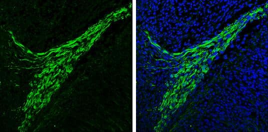
 NL001) and counterstained with DAPI (blue). Specific staining was localized to cytoplasm. View our protocol for
  HAF109). This experiment was conducted under reducing conditions and using the 12-230 kDa separation system." title="Simple Western lane view shows lysates of HeLa human cervical epithelial carcinoma cell line and A172 human glioblastoma cell line, loaded at 0.2 mg/mL. A specific band was detected for Park7/DJ-1 at approximately 28 kDa (as indicated) using 2 µg/mL of Goat Anti-Human Park7/DJ-1 Antigen Affinity-purified Polyclonal Antibody (Catalog # AF3995) followed by 1:50 dilution of HRP-conjugated Anti-Goat IgG Secondary Antibody (
 HAF017). A specific band was detected for Park7/DJ-1 at approximately 23 kDa (as indicated) in the parental HEK293T cell line, but is not detectable in knockout HEK293T cell line. GAPDH (
 VC004). Before incubation with the primary antibody, tissue was subjected to heat-induced epitope retrieval using VisUCyte Antigen Retrieval Reagent-Basic (Catalog #



![Western Blot Androgen R/NR3C4 [p Ser213, p Ser210] Antibody (156C135.2) - BSA Free](https://images.novusbio.com/images/Androgen-R-NR3C4-p-Ser213-p-Ser210-Antibody-156C135-2-Western-Blot-NB100-56603-img0004.jpg)