 tissue and mouse brain (cerebellum) tissue. PVDF membrane was probed with 0.5 µg/mL of Goat Anti-Human/Mouse NCAM-1/CD56 Antigen Affinity-purified Polyclonal Antibody (Catalog # AF2408) followed by HRP-conjugated Anti-Goat IgG Secondary Antibody (Catalog # HAF017). Specific bands were detected for NCAM-1/CD56 at approximately 100-150 kDa (as indicated). This experiment was conducted under reducing conditions and using Immunoblot Buffer Group 1.")
| Reactivity | Hu, MuSpecies Glossary |
| Applications | WB, Simple Western, Flow, CyTOF-ready, ICC/IF, KO |
| Clonality | Polyclonal |
| Host | Goat |
| Conjugate | Unconjugated |
| Concentration | LYOPH |
| Immunogen | Mouse myeloma cell line NS0-derived recombinant human NCAM1/CD56 Leu20-Pro603 Accession # NP_001070150 |
| Specificity | Detects human NCAM-1/CD56 in direct ELISAs and Western blots. In direct ELISAs and Western blots, less than 1% cross‑reactivity with recombinant human (rh) ALCAM, rhBCAM and rhEpCAM is observed. |
| Source | N/A |
| Isotype | IgG |
| Clonality | Polyclonal |
| Host | Goat |
| Gene | NCAM1 |
| Purity Statement | Antigen Affinity-purified |
| Innovator's Reward | Test in a species/application not listed above to receive a full credit towards a future purchase. |
| Dilutions |
|
|
| Publications |
|
| Storage | Use a manual defrost freezer and avoid repeated freeze-thaw cycles.
|
| Buffer | Lyophilized from a 0.2 μm filtered solution in PBS with Trehalose. *Small pack size (SP) is supplied either lyophilized or as a 0.2 µm filtered solution in PBS. |
| Preservative | No Preservative |
| Concentration | LYOPH |
| Reconstitution Instructions | Reconstitute at 0.2 mg/mL in sterile PBS. |
Neural cell adhesion molecule 1 (NCAM-1) is a multifunctional member of the Ig superfamily. It belongs to a family of membrane-bound glycoproteins that are involved in Ca++ independent cell matrix and homophilic or heterophilic cell-cell interactions. NCAM-1 specifically binds to heparan sulfate proteoglycans (1), the extracellular matrix protein agrin (2), and several chondroitin sulfate proteoglycans that include neurocan and phosphocan (3). There are three main forms of human NCAM-1 that arise by alternate splicing. These are designated NCAM-120/NCAM-1 (761 amino acids [aa]), NCAM-140 (848 aa), and NCAM-180 (1120 aa). NCAM-120 is GPI-linked, while NCAM-140 and NCAM-180 are type I transmembrane glycoproteins (4‑6). Additional alternate splicing adds considerable diversity to all three forms, and extracellular proteolytic processing is possible for NCAM-180 (7‑8). NCAM-1 is synthesized as a 761 aa preproprecursor that contains a 19 aa signal sequence, a 722 aa GPI-linked mature region, and a 20 aa C-terminal prosegment (4). The molecule contains five C-2 type Ig-like domains and two fibronectin type-III domains. Human to mouse, NCAM-1 is 93% aa identical. NCAM-1 appears to be highly sialylated. The polysialyation of NCAM-1 reduces its adhesive property and increases its neurite outgrowth promoting features (9). NCAM-1 in the adult brain shows a decline of sialylation relative to earlier developmental periods. In regions that retain a high degree of neuronal plasticity, however, the adult brain continues to express polysialylation-NCAM-1, suggesting sialylation of NCAM-1 is involved in regenerative processes and synaptic plasticity (10‑13).


![NCAM-1/CD56 Antibody [Unconjugated]](/sites/all/modules/enterprise-tech/et_datasheets/images/novus_guarantee.png "NCAM-1/CD56 Antibody [Unconjugated]")
Secondary Antibodies |
Isotype Controls |
The concentration calculator allows you to quickly calculate the volume, mass or concentration of your vial. Simply enter your mass, volume, or concentration values for your reagent and the calculator will determine the rest.
| Gene Symbol | NCAM1 |
| Uniprot |


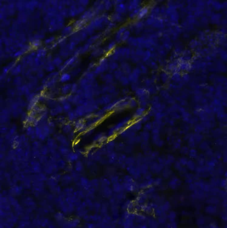
 prior to immersion fixation. Neural Cell Adhesion Molecule 1 (NCAM-1)/CD56 was detected using a Goat Anti-Human/Mouse NCAM-1/CD56 Antigen Affinity-purified Polyclonal Antibody (Catalog # AF2408). The cells were stained with the NorthernLights 557-conjugated Donkey Anti-Goat IgG Affinity-purified Secondary Antibody (red; Catalog # NL001). Actin filaments were stained with FITC-conjugated Phalloidin (green) and cell nuclei were counter-stained with DAPI (blue). NCAM-1/CD56 immuno-reactivity was localized to the plasma membrane. View our protocol for Fluorescent ICC Staining of Cells on Coverslips.")
 at 10 µg/mL for 3 hours at room temperature. Cells were stained using the NorthernLights™ 557-conjugated Anti-Goat IgG Secondary Antibody (red; Catalog # NL001) and counterstained with DAPI (blue). Specific staining was localized to cytoplasm. View our protocol for Fluorescent ICC Staining of Stem Cells on Coverslips.")
 tissue, loaded at 0.2 mg/mL. A specific band was detected for NCAM-1/CD56 at approximately 143 kDa (as indicated) using 5 µg/mL for human lysates and 25 µg/mL for mouse lysates of Goat Anti-Human/Mouse NCAM-1/ CD56 Antigen Affinity-purified Polyclonal Antibody (Catalog # AF2408) followed by 1:50 dilution of HRP-conjugated Anti-Goat IgG Secondary Antibody (Catalog # HAF109). This experiment was conducted under reducing conditions and using the 12-230 kDa separation system.")
. PVDF membrane was probed with 0.25 µg/mL of Goat Anti-Human/Mouse NCAM‑1/CD56 Antigen Affinity-purified Polyclonal Antibody (Catalog # AF2408) followed by HRP-conjugated Anti-Goat IgG Secondary Antibody (HAF017). A specific band was detected for NCAM‑1/CD56 at approximately 160 kDa (as indicated) in the parental U937 human histiocytic lymphoma cell line, but is not detectable in knockout U937 human histiocytic lymphoma cell line. GAPDH (AF5718) is shown as a loading control. This experiment was conducted under reducing conditions and using Western Blot Buffer Group 1.")
![Bioactivity CTLA-4 [Unconjugated]](https://images.novusbio.com/images/protein/CTLA4_7268CT_2293.jpg)
![Bioactivity IL-2 [Unconjugated]](https://images.novusbio.com/images/202-il_recombinant-human-il-2-protein-bioactivity-174202314946.jpg)
![Binding Activity Fc gamma RIIIB/CD16b [Unconjugated]](https://images.novusbio.com/images/protein/Fc_gamma_RIIIB_1597-FC_CF_776.jpg)
![SDS-Page Fc gamma RIIIB/CD16b [Unconjugated]](https://images.novusbio.com/images/protein/Fc_gamma_RIIIB_1597-FC_CF_756.jpg)
![SEC-MALS Fc gamma RIIIA/CD16a [Unconjugated]](https://images.novusbio.com/images/4325-fc_recombinant-human-fc-gamma-riiia-cd16a-protein-cf-sec-mals-2842023172947.jpg)
![Binding Activity Fc gamma RIIIA/CD16a [Unconjugated]](https://images.novusbio.com/images/protein/Fc_gamma_RIIIA_4325-FC_491.jpg)
![SDS-Page Fc gamma RIIIA/CD16a [Unconjugated]](https://images.novusbio.com/images/protein/Fc_gamma_RIIIA_4325-FC_411.jpg)
![Immunohistochemistry CD45 Antibody [Unconjugated]](https://images.novusbio.com/images/antibody/CD45_AF114_Immunohistochemistry_23525.jpg)
![Immunocytochemistry CD45 Antibody [Unconjugated]](https://images.novusbio.com/images/antibody/af114_mouse-cd45-affinity-purified-polyclonal-ab-immunocytochemistry-6122021145449.jpg)
![SDS-Page TNF-alpha [Unconjugated]](https://images.novusbio.com/images/protein/TNF-alpha_210-TA_256.jpg)
![Bioactivity TNF-alpha [Unconjugated]](https://images.novusbio.com/images/protein/TNFalpha_210TA_1658.jpg)
![SEC-MALS TNF-alpha [Unconjugated]](https://images.novusbio.com/images/210-ta_recombinant-human-tnf-alpha-protein-sec-mals-35202312244..jpg)
![SEC-MALS IFN-gamma [Unconjugated]](https://images.novusbio.com/images/485-mi_recombinant-mouse-ifn-gamma-protein-sec-mals-1612202583245.jpg)
![Bioactivity IFN-gamma [Unconjugated]](https://images.novusbio.com/images/protein/IFN-gamma_485-MI_455.jpg)
![SDS-Page IFN-gamma [Unconjugated]](https://images.novusbio.com/images/protein/IFN-gamma_485-MI_407.jpg)


![N/A CD25/IL-2R alpha [HRP]](https://images.novusbio.com/images/elisa/IL-2_R_alpha_DR2A00_ELISA_167.jpg)
![N/A CD25/IL-2R alpha [HRP]](https://images.novusbio.com/images/elisa/DATA_CD25_DR2A00_ELISA_800.jpg)
 or Normal Goat IgG Isotype Control Antibody (Catalog # AB-108-C, open histogram), followed by Phycoerythrin-conjugated Anti-Goat IgG Secondary Antibody (Catalog # F0107).")