 and CoraFluor™ 2, amine reactive (Catalog # 7950) are terbium-based probes that have been developed for use as TR-FRET donors. They emit wavelengths compatible with commonly used fluorescent acceptor dyes such as BODIPY® (or BDY) and Janelia Fluor® dyes, FITC (Catalog # 5440), TMR and Cyanine 5 (Catalog # 5436). CoraFluor™ fluorescence is brighter and more stable in biological media than existing TR-FRET donors, leading to enhanced sensitivity and improved data generation. CoraFluor™ 1 exhibits excitation upon exposure to a 337 nm UV laser.")
| Reactivity | Hu, MuSpecies Glossary |
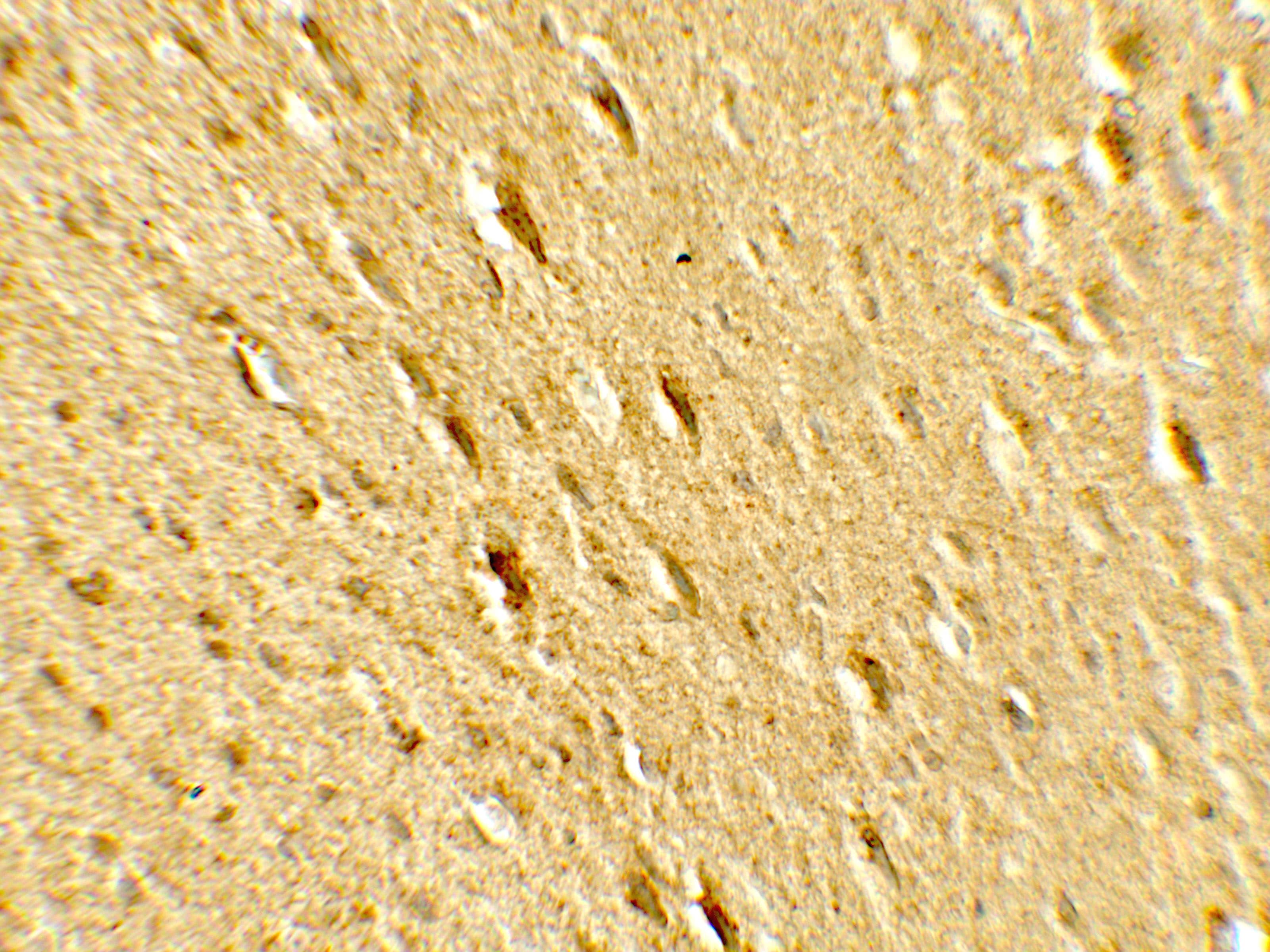
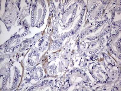
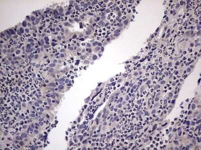
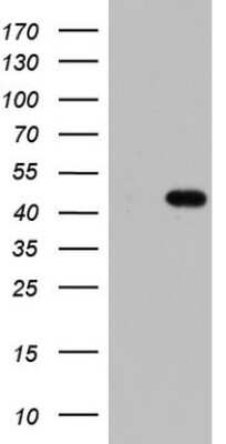
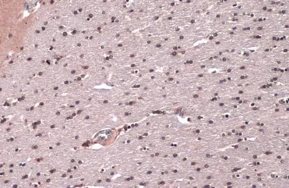
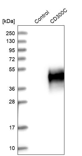
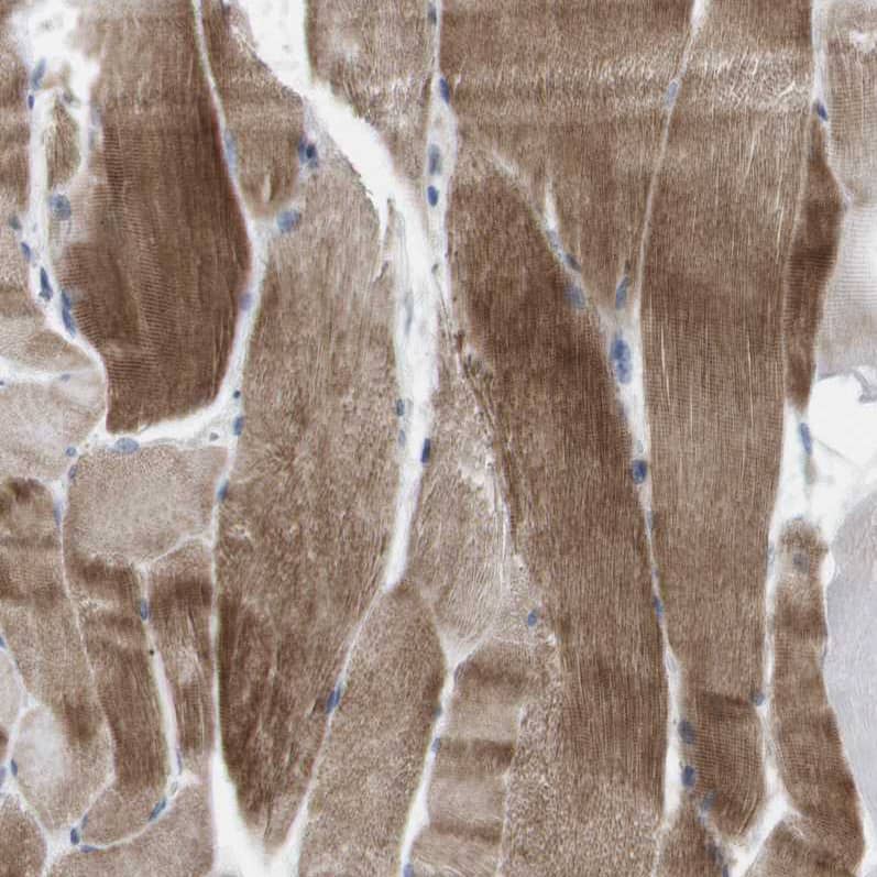
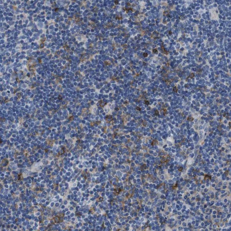
| Applications | WB, ELISA, ICC/IF, IHC, ELISA |
| Clonality | Polyclonal |
| Host | Rabbit |
| Conjugate | CoraFluor 1 |
| Description | CoraFluor(TM) 1 is a high performance terbium-based TR-FRET (Time-Resolved Fluorescence Resonance Energy Transfer) or TRF (Time-Resolved Fluorescence) donor for high throughput assay development. CoraFluor(TM) 1 absorbs UV light at approximately 340 nm, and emits at approximately 490 nm, 545 nm, 585 nm and 620 nm. It is compatible with common acceptor dyes that absorb at the emission wavelengths of CoraFluor(TM) 1. CoraFluor(TM) 1 can be used for the development of robust and scalable TR-FRET binding assays such as target engagement, ternary complex, protein-protein interaction and protein quantification assays.
CoraFluor(TM) 1, amine reactive CoraFluor(TM) 1, thiol reactive For more information, please see our CoraFluor(TM) TR-FRET technology flyer. |
| Immunogen | A genomic peptide made to an internal region of the human NBR1 protein (within residues 50-250). [Swiss-Prot Q14596] |
| Localization | Cytoplasm. Cytoplasmic vesicle - autophagosome. Lysosome. Cytoplasm - myofibril - sarcomere - M line. Note: In cardiac muscles localizes to the sarcomeric M line. Is targeted to lysosomes for degradation. |
| Isotype | IgG |
| Clonality | Polyclonal |
| Host | Rabbit |
| Gene | NBR1 |
| Purity | Immunogen affinity purified |
| Innovator's Reward | Test in a species/application not listed above to receive a full credit towards a future purchase. |
| Dilutions |
|
| Application Notes | Optimal dilution of this antibody should be experimentally determined. |
| Storage | Store at 4C in the dark. Do not freeze. |
| Buffer | PBS |
| Preservative | No Preservative |
| Purity | Immunogen affinity purified |


![NBR1 Antibody [CoraFluor™ 1]](/sites/all/modules/enterprise-tech/et_datasheets/images/novus_guarantee.png "NBR1 Antibody [CoraFluor™ 1]")
Secondary Antibodies |
Isotype Controls |
|
Understanding Mitophagy Mechanisms: Canonical PINK1/Parkin, LC3-Dependent Piecemeal, and LC3-Independent Mitochondrial Derived Vesicles By Christina Towers, PhD What is Mitophagy?The selective degradation of mitochondria via double membrane autophagosome vesicles is called mitophagy. Damaged mitochondria can generate harmful amounts of reactive ox... Read full blog post. |
|
Monitoring Autophagy in Neurons By Christina Towers, PhD. Autophagy is a critical cellular process used by most cells in the body to recycle nutrients and prevent harmful buildup of damaged proteins. It is particularly important in the brain, where ... Read full blog post. |
|
Why LC3B Antibodies Make Ideal Autophagosomes Membrane Markers The human form of microtubule-associated protein light chain 3 (LC3) is expressed as 3 splice variants LC3A, LC3B, and LC3C.1 LC3B is a subunit of the MAP1A and MAP1B microtubule-binding proteins and plays a central role in autophagosome membrane stru... Read full blog post. |
The concentration calculator allows you to quickly calculate the volume, mass or concentration of your vial. Simply enter your mass, volume, or concentration values for your reagent and the calculator will determine the rest.
| Gene Symbol | NBR1 |


![Simple Western MuRF1/TRIM63 Antibody [Unconjugated]](https://images.novusbio.com/images/af5366_human-mouse-rat-murf1-trim63-affinity-purified-polyclonal-ab-simple-western-639195798985228037.jpg)
![Western Blot MuRF1/TRIM63 Antibody [Unconjugated]](https://images.novusbio.com/images/af5366_human-mouse-rat-murf1-trim63-affinity-purified-polyclonal-ab-41202412403588.jpg)


![Western Blot: Goat anti-Rabbit IgG (H+L) Secondary Antibody [HRP] [NB7160] - Western blot showing vemurafenib treatment in BRAFV600E CRC cells inhibits fission mediator DRP1 with no significant effect on fusion proteins (Mfn1 & 2) using MFN-1 antibody (NBP1-51841) and corresponding secondary antibody, goat anti-rabbit IgG-HRP (NB7160). Image collected and cropped by CiteAb from the following publication (https://pubmed.ncbi.nlm.nih.gov/33738242).](https://images.novusbio.com/images/Goat-anti-Rabbit-IgG-H+L-Secondary-Antibody-HRP-Western-Blot-NB7160-img0001.jpg "Western Blot: Goat anti-Rabbit IgG (H+L) Secondary Antibody [HRP] [NB7160] - Western blot showing vemurafenib treatment in BRAFV600E CRC cells inhibits fission mediator DRP1 with no significant effect on fusion proteins (Mfn1 & 2) using MFN-1 antibody (NBP1-51841) and corresponding secondary antibody, goat anti-rabbit IgG-HRP (NB7160). Image collected and cropped by CiteAb from the following publication (https://pubmed.ncbi.nlm.nih.gov/33738242).")
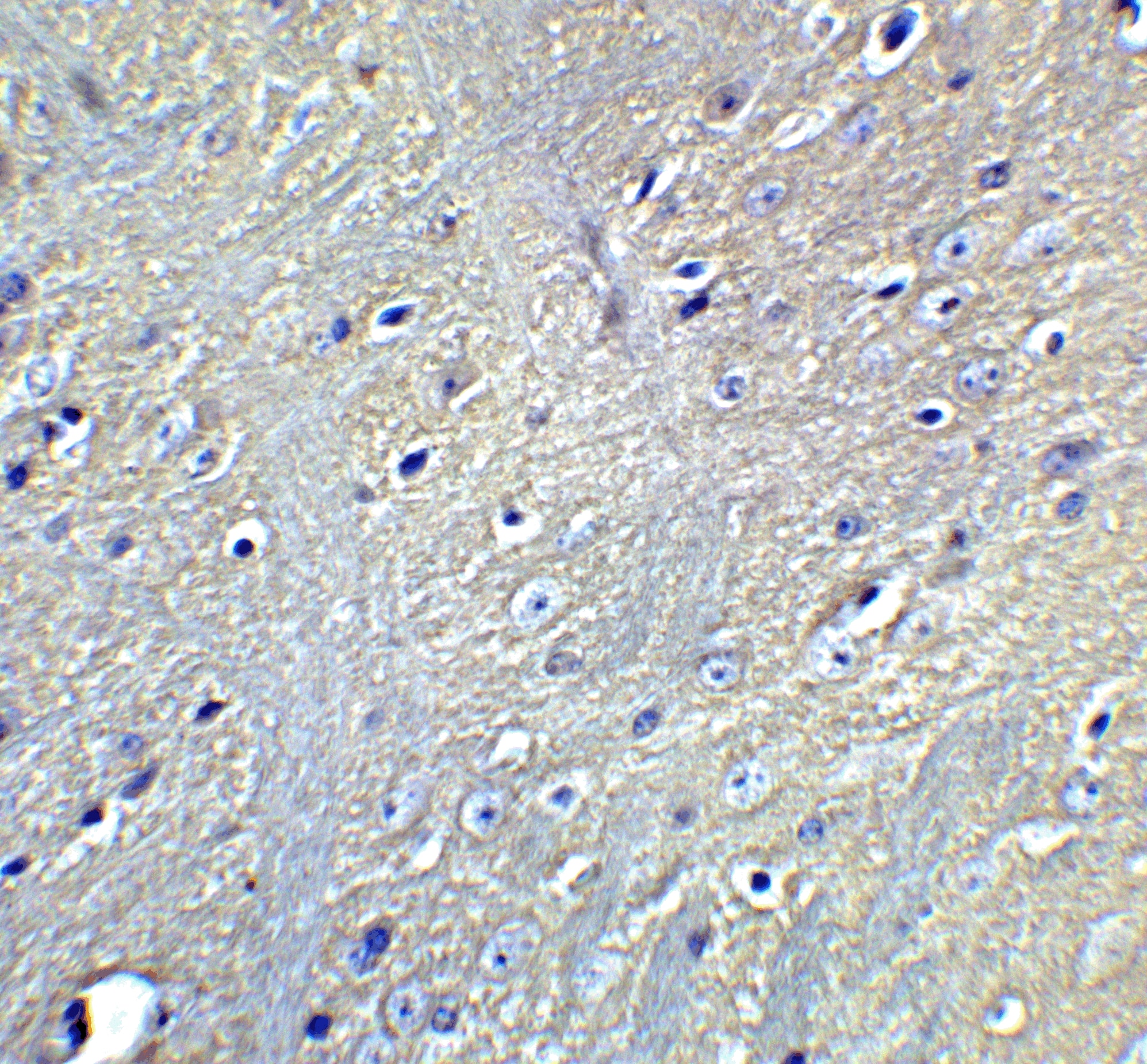
 followed by 30 min incubation with Goat anti Rabbit HRP conjugated secondary antibodies (Catalog # HAF008) at 1:20 dilution + DAB chromogen (brown). The tissue was counterstained with Hematoxylin (blue). Control was done by omitting primary antibody.")
![Flow Cytometry: Rabbit IgG Isotype Control [NBP2-24891] - An intracellular stain was performed on Raji cells with Adiponectin antibody NB100-65810 (blue) and a matched isotype control NBP2-24893 (orange). Cells were fixed with 4% PFA and then permeablized with 0.1% saponin. Cells were incubated in an antibody dilution of 1 ug/mL for 30 minutes at room temperature, followed by Dylight488-conjugated anti-rabbit secondary antibody. Image using the Azide Free form of this antibody.](https://images.novusbio.com/images/Rabbit--Mouse-IgG-Isotype-Control-Flow-Cytometry-NBP2-24891-img0006.jpg "Flow Cytometry: Rabbit IgG Isotype Control [NBP2-24891] - An intracellular stain was performed on Raji cells with Adiponectin antibody NB100-65810 (blue) and a matched isotype control NBP2-24893 (orange). Cells were fixed with 4% PFA and then permeablized with 0.1% saponin. Cells were incubated in an antibody dilution of 1 ug/mL for 30 minutes at room temperature, followed by Dylight488-conjugated anti-rabbit secondary antibody. Image using the Azide Free form of this antibody.")