| Reactivity | HuSpecies Glossary |
| Applications | AC |
| Description | A recombinant protein antigen with a N-terminal His6-ABP tag corresponding to human LC3 Source: E.coli Amino Acid Sequence: TKFLVPDHVNMSELIKIIRRRLQLN Fusion Tag: N-terminal His6ABP (ABP = Albumin Binding Protein derived from Streptococcal Protein G) This product is intended to be used as a blocking antigen for antibody competition assays. Any other use of this antigen is done at the risk of the user. The use of this product for commercial production is strictly prohibited. Please contact technical support if you have any questions. |
| Source | E. coli |
| Protein/Peptide Type | Recombinant Protein Antigen |
| Gene | MAP1LC3B |
| Purity | >80% by SDS-PAGE and Coomassie blue staining |
| Dilutions |
|
| Application Notes | This recombinant antigen is only intended to be used as a blocking agent to confirm antibody specificity with the corresponding antibody, catalog number NBP3-21266. It is purified by IMAC chromatography, and the expected concentration is greater than 0.5 mg/ml.For current lot information, including availability, please contact our technical support team click nb-technical@bio-techne.com |
| Theoretical MW | 21 kDa. Disclaimer note: The observed molecular weight of the protein may vary from the listed predicted molecular weight due to post translational modifications, post translation cleavages, relative charges, and other experimental factors. |
| Storage | Store at -20C. Avoid freeze-thaw cycles. |
| Buffer | PBS and 1M Urea, pH 7.4. |
| Preservative | No Preservative |
| Purity | >80% by SDS-PAGE and Coomassie blue staining |


![Immunohistochemistry ATG7 Antibody (683906) [Unconjugated]](https://images.novusbio.com/images/antibody/ATG7_MAB6608_Immunohistochemistry_10631.jpg)
![Simple Western ATG7 Antibody (683906) [Unconjugated]](https://images.novusbio.com/images/antibody/ATG7_MAB6608_Simple_Western_16409.jpg)
![Intracellular Staining by Flow Cytometry AKT [p Ser473] Antibody [Unconjugated] - Pan Specific](https://images.novusbio.com/images/antibody/Akt3_AF887_Flow_Cytometry_8283.jpg)
![Western Blot AKT [p Ser473] Antibody [Unconjugated] - Pan Specific](https://images.novusbio.com/images/af887_phospho-akt-s473-pan-specific-affinity-purified-pab-41202410485440.jpg)
![Western Blot AKT [p Ser473] Antibody [Unconjugated] - Pan Specific](https://images.novusbio.com/images/af887_phospho-akt-s473-pan-specific-affinity-purified-pab-8120255552843.jpg)


Research Areas for LC3 Recombinant Protein Antigen (NBP3-21266PEP)Find related products by research area.
|
|
Autophagy and Metastasis By Christina Towers, PhD The majority of cancer patients die from metastatic disease at secondary sites. The threshold to undergo metastasis is high. Only a minority of cancer cells acquire invasive phenotypes... Read full blog post. |
|
Optogenetic Control of Mitophagy: AMBRA1 based mitophagy switch By Christina Towers, PhD Mitophagy in the BrainSelective autophagic degradation of damaged mitochondria, known as mitophagy, has been described as a cyto-protective process. Accordingly, defects in mitophagy h... Read full blog post. |
|
Read full blog post. |
|
Read full blog post. |
|
Read full blog post. |
|
How to visualize autophagy by microscopy By Christina Towers, PhD Autophagy is a recycling process that relies on the formation of a unique organelle termed an autophagosome. An elegant way to monitor autophagy is through various microscopy techniques to... Read full blog post. |
|
Microglia: pruning shears for homeostatic maintenance in the brain By Jennifer Sokolowski, MD, PhD.Microglia play a critical role in pruning neurons and synapses during homeostatic maintenance in the adult brain.1 A recent study by Ayata et al. (2018) identified regional differe... Read full blog post. |
|
Read full blog post. |
|
Read full blog post. |
|
Read full blog post. |
The concentration calculator allows you to quickly calculate the volume, mass or concentration of your vial. Simply enter your mass, volume, or concentration values for your reagent and the calculator will determine the rest.
| Gene Symbol | MAP1LC3B |


![Immunohistochemistry TOR/mTOR [p Ser2448] Antibody - BSA Free](https://images.novusbio.com/images/nb600-607_rabbit-polyclonal-tor-mtor-p-ser2448-antibody-235202318143142.jpg)
![Western Blot TOR/mTOR [p Ser2448] Antibody - BSA Free](https://images.novusbio.com/images/nb600-607_rabbit-polyclonal-tor-mtor-p-ser2448-antibody-31020241535647.jpg)
![Data TOR/mTOR [p Ser2448] Antibody - BSA Free](https://images.novusbio.com/images/TOR-mTOR-[p-Ser2448]-Antibody-N-A-NB600-607-img0008.jpg)
![Immunohistochemistry Caspase-3 Antibody [Unconjugated] - Active](https://images.novusbio.com/images/af835_human-mouse-active-caspase-3-affinity-purified-pab-41202410331943.jpg)
![Western Blot Caspase-3 Antibody [Unconjugated] - Active](https://images.novusbio.com/images/af835_human-mouse-active-caspase-3-affinity-purified-pab-812025554170.jpg)
![Western Blot Caspase-3 Antibody [Unconjugated] - Active](https://images.novusbio.com/images/af835_human-mouse-active-caspase-3-affinity-purified-pab-8120255534731.jpg)


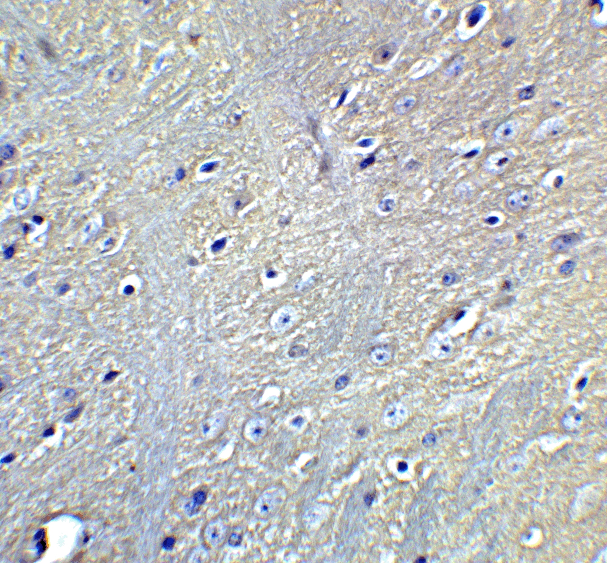
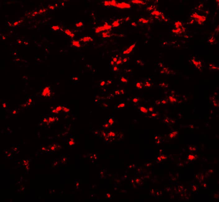
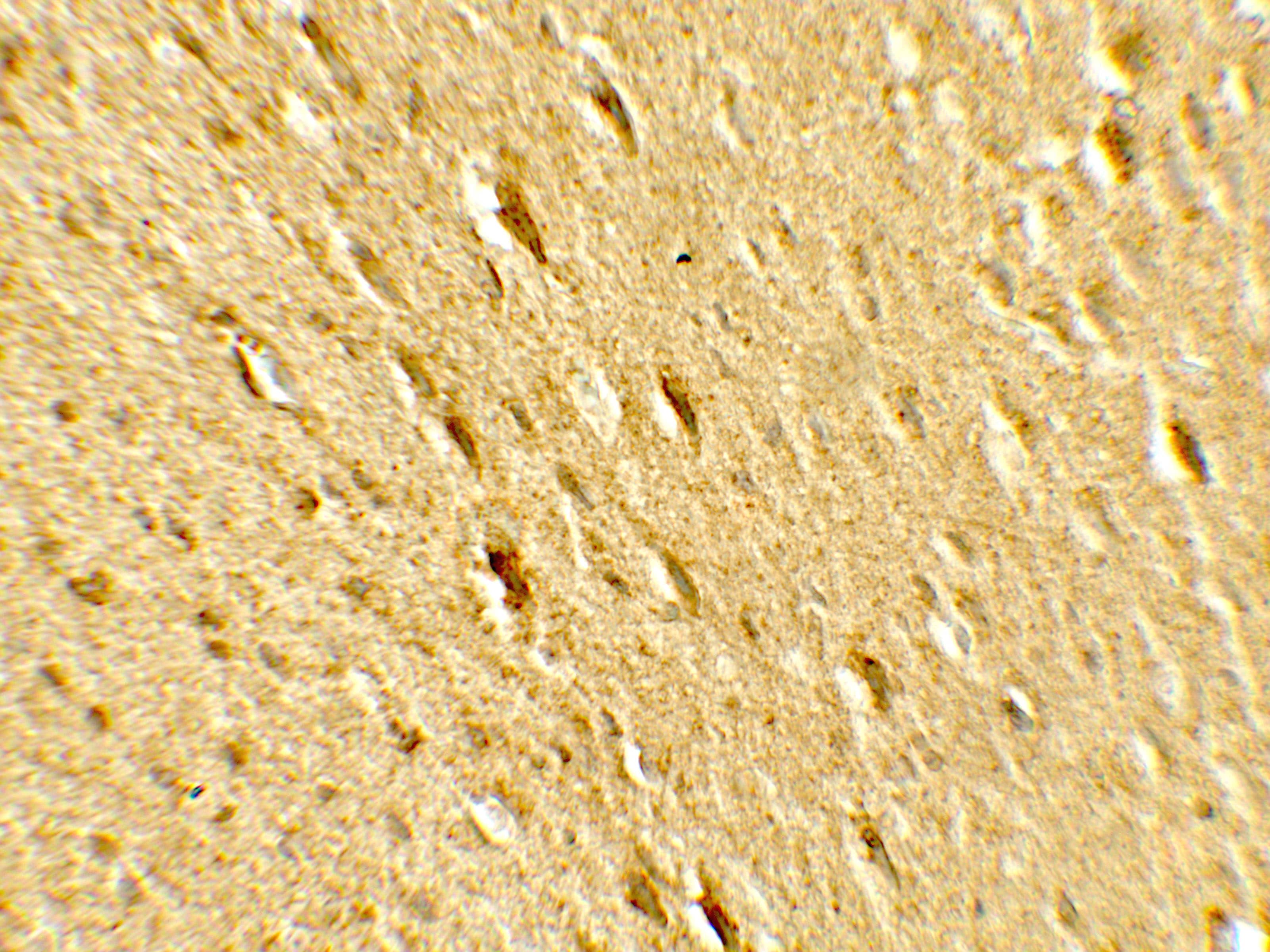
![Immunocytochemistry/ Immunofluorescence: LC3 Antibody Pack [NB910-40435] - The autophagy level was increased in degenerated mouse SGNs. (a-c) Western blot results revealed that the levels of the autophagy-related proteins LC3 & BECN1 were increased in the degenerated SGNs on the 5th, 15th & 30th day after ototoxic drug administration & were significantly different from those in the normal mice. *, P < 0.05. (d) Immunofluorescence staining of LC3 puncta (red) also demonstrated that the LC3 level in the degenerated SGNs (green) was significantly increased on the 30th day after drug administration. Con, normal mice without drug treatment; 30D, 30 days after drug administration. Images of immunofluorescence staining were taken from the middle turn of cochlea. Scale bar: 10 µm. Image collected & cropped by CiteAb from the following publication (https://pubmed.ncbi.nlm.nih.gov/30706760), licensed under a CC-BY license. Not internally tested by Novus Biologicals.](https://images.novusbio.com/images/nb910-40435_lc3-antibody-pack-31020241538249.jpg "Immunocytochemistry/ Immunofluorescence: LC3 Antibody Pack [NB910-40435] - The autophagy level was increased in degenerated mouse SGNs. (a-c) Western blot results revealed that the levels of the autophagy-related proteins LC3 & BECN1 were increased in the degenerated SGNs on the 5th, 15th & 30th day after ototoxic drug administration & were significantly different from those in the normal mice. *, P < 0.05. (d) Immunofluorescence staining of LC3 puncta (red) also demonstrated that the LC3 level in the degenerated SGNs (green) was significantly increased on the 30th day after drug administration. Con, normal mice without drug treatment; 30D, 30 days after drug administration. Images of immunofluorescence staining were taken from the middle turn of cochlea. Scale bar: 10 µm. Image collected & cropped by CiteAb from the following publication (https://pubmed.ncbi.nlm.nih.gov/30706760), licensed under a CC-BY license. Not internally tested by Novus Biologicals.")