![Immunocytochemistry/Immunofluorescence: Ki67/MKI67 Antibody [NB500-170] - HeLa cells were fixed in 4% paraformaldehyde for 10 minutes and permeabilized in 0.5% Triton X-100 in PBS for 5 minutes. The cells were incubated with anti- NB500-170 at 2 ug/ml overnight at 4C and detected with an anti-rabbit Dylight 488 (Green) at a 1:1000 dilution for 60 minutes. Alpha tubulin (DM1A) NB100-690 was used as a co-stain at a 1:1000 dilution and detected with an anti-mouse Dylight 550 (Red) at a 1:1000 dilution. Nuclei were counterstained with DAPI (Blue). Cells were imaged using a 100X objective and digitally deconvolved.](http://images.novusbio.com/fullsize/Ki67-MKI67-Antibody-Immunocytochemistry-Immunofluorescence-NB500-170-img0025.jpg "Immunocytochemistry/Immunofluorescence: Ki67/MKI67 Antibody [NB500-170] - HeLa cells were fixed in 4% paraformaldehyde for 10 minutes and permeabilized in 0.5% Triton X-100 in PBS for 5 minutes. The cells were incubated with anti- NB500-170 at 2 ug/ml overnight at 4C and detected with an anti-rabbit Dylight 488 (Green) at a 1:1000 dilution for 60 minutes. Alpha tubulin (DM1A) NB100-690 was used as a co-stain at a 1:1000 dilution and detected with an anti-mouse Dylight 550 (Red) at a 1:1000 dilution. Nuclei were counterstained with DAPI (Blue). Cells were imaged using a 100X objective and digitally deconvolved.")
| Reactivity | Hu, Mu, Rt, Po, AvSpecies Glossary |
| Applications | WB, Flow, ICC/IF, IHC, IP, WB, KO |
| Clonality | Polyclonal |
| Host | Rabbit |
| Conjugate | Unconjugated |
| Format | BSA Free |
| Concentration | 1.0 mg/ml |
| Immunogen | The immunogen for this KI67/MKI67 Antibody was made using a synthetic peptide from the internal region of Human KI67/MKI67, between amino acids: 1550-1600 [Uniprot: P46013]. |
| Localization | Nuclear |
| Marker | Proliferation Marker |
| Isotype | IgG |
| Clonality | Polyclonal |
| Host | Rabbit |
| Gene | MKI67 |
| Purity | Immunogen affinity purified |
| Innovator's Reward | Test in a species/application not listed above to receive a full credit towards a future purchase. |
| Dilutions |
|
|
| Application Notes | Formalin fixed paraffin embedded tissue sections require high temperature antigen unmasking with 10 mM citrate buffer (pH 6.0) prior to immunostaining. This antibody will not work without optimal antigen retrieval, and is a critical step. NOTE: We suggest an incubation period of 30 minutes at room temperature and to use DAB to stain the protein. |
|
| Theoretical MW | 359 kDa. Disclaimer note: The observed molecular weight of the protein may vary from the listed predicted molecular weight due to post translational modifications, post translation cleavages, relative charges, and other experimental factors. |
|
| Reviewed Applications |
|
|
| Publications |
|
| Storage | Store at 4C short term. Aliquot and store at -20C long term. Avoid freeze-thaw cycles. |
| Buffer | PBS |
| Preservative | 0.05% Sodium Azide |
| Concentration | 1.0 mg/ml |
| Purity | Immunogen affinity purified |


![Immunocytochemistry EGFR Antibody [Unconjugated]](https://images.novusbio.com/images/antibody/EGF_R_AF231_Immunocytochemistry__Immunofluorescence_21143.jpg)
![Flow Cytometry EGFR Antibody [Unconjugated]](https://images.novusbio.com/images/antibody/EGF_R_AF231_Flow_Cytometry_20401.jpg)
![Western Blot EGFR Antibody [Unconjugated]](https://images.novusbio.com/images/antibody/EGF_R_AF231_Western_Blot_19925.jpg)


![Immunohistochemistry PAK3 Antibody [Unconjugated]](https://images.novusbio.com/images/antibody/PAK3_AF6897_Immunohistochemistry_11144.jpg)
![Simple Western PAK3 Antibody [Unconjugated]](https://images.novusbio.com/images/antibody/af6897_human-mouse-pak3-affinity-purified-polyclonal-ab-simple-western-245202494437..jpg)
![Immunohistochemistry PAK3 Antibody [Unconjugated]](https://images.novusbio.com/images/antibody/PAK3_AF6897_Immunohistochemistry_11480.jpg)


| Images | Ratings | Applications | Species | Date | Details | ||||||||||
|---|---|---|---|---|---|---|---|---|---|---|---|---|---|---|---|

Enlarge |
reviewed by:
Shinford Sun |
WB | Human | 08/10/2023 |
Summary
|
||||||||||
Enlarge |
reviewed by:
Nick Dordai |
WB | Human | 12/01/2022 |
Summary
|
||||||||||

Enlarge |
reviewed by:
Verified Customer |
IP | Human | 04/27/2021 |
Summary
Comments
|
||||||||||

Enlarge |
reviewed by:
Verified Customer |
WB | Human | 04/21/2021 |
Summary
Comments
|
||||||||||

Enlarge |
reviewed by:
Lang Zhao |
IHC-P | Mouse | 03/17/2018 |
Summary
|
||||||||||

Enlarge |
reviewed by:
Alexandra Ambrico |
Immunocytochemistry/Immunofluorescence | Mouse | 10/11/2016 |
Summary
|
||||||||||

Enlarge |
reviewed by:
Natalie Erdmann |
ICC | Human | 08/24/2015 |
Summary
|
||||||||||
|
reviewed by:
Verified Customer |
IHC-P | Human | 12/29/2014 |
Summary
|
|||||||||||
-(01-ml)_NB500-170_8061.bmp)
Enlarge |
reviewed by:
Bryan Tinsley |
ICC | Human | 06/09/2014 |
Summary
|
||||||||||
|
reviewed by:
Jian Zong |
WB | Mouse | 09/30/2010 |
Summary
|
Secondary Antibodies |
Isotype Controls |
Research Areas for Ki67/MKI67 Antibody (NB500-170)Find related products by research area.
|
|
The relationship between Ki67 and HIF-1 in cancer Ki67, also known as MKI67, is best known as the leading marker of cellular proliferation. Ki67 is regulated by a balance between synthesis and degradation, and often carries a very short half-life. First discovered to be located to dividing cells,... Read full blog post. |
|
Ki67 - an established marker for labelling proliferating cells Ki-67/MKI67 is an antigen which is expressed during G1, S, G2, and M phases of the cell cycle (mitotically active cells), but not during G0 phase (resting cells). It is a large protein with expected molecular weight of about 395 kDa, and it has a v... Read full blog post. |
|
Ki67 - A Crucial Cellular Proliferation Marker The Ki67 antigen is a prototypic cell cycle-related protein expressed by proliferating cells in all phases of the active cell cycle (G1, S, G2 and M). It is a non-histone nuclear protein originally identified in a Hodgkin's lymphoma-derived cell line.... Read full blog post. |
|
The Ki67 Antibody in Cell Marker Studies The MK167, or Ki67 antibody recognizes a nuclear protein encoded by the MK167 gene. Ki167 is involved with RNA transcription and essential to cellular proliferation, being expressed by proliferating cells at all stages of the active cell cycle; it is ... Read full blog post. |
The concentration calculator allows you to quickly calculate the volume, mass or concentration of your vial. Simply enter your mass, volume, or concentration values for your reagent and the calculator will determine the rest.
5 | |
4 | |
3 | |
2 | |
1 |
| Shinford Sun 08/10/2023 |
||
| Application: | WB | |
| Species: | Human |
| Nick Dordai 12/01/2022 |
||
| Application: | WB | |
| Species: | Human |
| Verified Customer 04/27/2021 |
||
| Application: | IP | |
| Species: | Human |


![Simple Western: Ki67/MKI67 Antibody [NB500-170] - Detection of Ki67/MKI67 by Simple WesternTM. Simple Western lane view shows lysates of HeLa parental cell line and Ki67 knockout (KO) HeLa cell line. A specific band was detected for Ki67/MKI67 at approximately 320 kDa (as indicated) in the parental cell line, but is not detectable in the knockout HeLa cell line using 20 ug/mL of Rabbit Anti-Ki67/MKI67 Polyclonal Antibody (Catalog # NB500-170). GAPDH is shown as a loading control. This experiment was conducted under reducing conditions and using the 12-230 kDa separation system.](http://images.novusbio.com/fullsize/Ki67-MKI67-Antibody-Knockout-Validated-NB500-170-img0023.jpg "Simple Western: Ki67/MKI67 Antibody [NB500-170] - Detection of Ki67/MKI67 by Simple WesternTM. Simple Western lane view shows lysates of HeLa parental cell line and Ki67 knockout (KO) HeLa cell line. A specific band was detected for Ki67/MKI67 at approximately 320 kDa (as indicated) in the parental cell line, but is not detectable in the knockout HeLa cell line using 20 ug/mL of Rabbit Anti-Ki67/MKI67 Polyclonal Antibody (Catalog # NB500-170). GAPDH is shown as a loading control. This experiment was conducted under reducing conditions and using the 12-230 kDa separation system.")

![Immunohistochemistry-Paraffin: Ki67/MKI67 Antibody [NB500-170] - Ki-67/MKI67 Antibody [NB500-170] - Tissue section of human tonsil using 1:50 dilution of rabbit anti-KI67 antibody. The staining was developed with HRP labeled anti-rabbit IgG secondary antibody and DAB reagent, and nuclei of cells were counter-stained with hematoxylin. This Ki67 antibody generated a specific nuclear staining in the cells in germinal centers of the tested tonsil tissue.](http://images.novusbio.com/fullsize/Ki-67-MKI67-Antibody-Immunohistochemistry-Paraffin-NB500-170-img0019.jpg "Immunohistochemistry-Paraffin: Ki67/MKI67 Antibody [NB500-170] - Ki-67/MKI67 Antibody [NB500-170] - Tissue section of human tonsil using 1:50 dilution of rabbit anti-KI67 antibody. The staining was developed with HRP labeled anti-rabbit IgG secondary antibody and DAB reagent, and nuclei of cells were counter-stained with hematoxylin. This Ki67 antibody generated a specific nuclear staining in the cells in germinal centers of the tested tonsil tissue.")
![Immunohistochemistry-Paraffin: Ki67/MKI67 Antibody [NB500-170] - Ki-67/MKI67 Antibody [NB500-170] - Human tonsil using 1:200 dilution of rabbit anti-KI67 antibody. The staining was developed with HRP labeled anti-rabbit IgG secondary antibody and DAB reagent, and nuclei of cells were counter-stained with hematoxylin. This Ki67 antibody generated a specific nuclear staining in the cells in germinal centers of the tested tonsil tissue.](http://images.novusbio.com/fullsize/Ki-67-MKI67-Antibody-Immunohistochemistry-Paraffin-NB500-170-img0020.jpg "Immunohistochemistry-Paraffin: Ki67/MKI67 Antibody [NB500-170] - Ki-67/MKI67 Antibody [NB500-170] - Human tonsil using 1:200 dilution of rabbit anti-KI67 antibody. The staining was developed with HRP labeled anti-rabbit IgG secondary antibody and DAB reagent, and nuclei of cells were counter-stained with hematoxylin. This Ki67 antibody generated a specific nuclear staining in the cells in germinal centers of the tested tonsil tissue.")
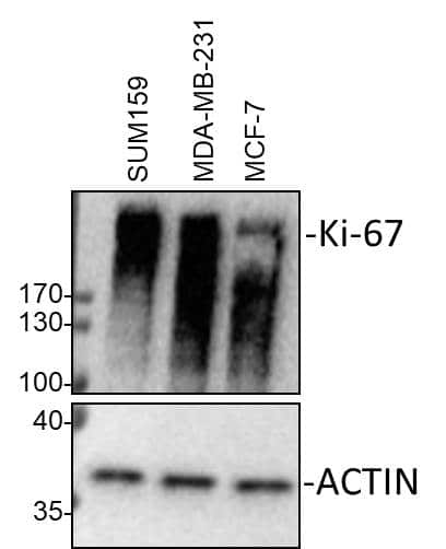
![Western Blot: Ki67/MKI67 Antibody - BSA Free [NB500-170] - Whole cell lysates from SUM159, MDA-MB-231 and MCF-7 cells were loaded with 30 ug/lane. 10% SDS-PAGE. Ki67/MKI67 antibody (NB500-170) was used for primary antibody: 1:2000, 4C, overnight. Image from verified customer review.](http://images.novusbio.com/fullsize/Ki67-MKI67-Antibody---BSA-Free-Western-Blot-NB500-170-img0028.jpg "Western Blot: Ki67/MKI67 Antibody - BSA Free [NB500-170] - Whole cell lysates from SUM159, MDA-MB-231 and MCF-7 cells were loaded with 30 ug/lane. 10% SDS-PAGE. Ki67/MKI67 antibody (NB500-170) was used for primary antibody: 1:2000, 4C, overnight. Image from verified customer review.")
![Immunocytochemistry/Immunofluorescence: Ki67/MKI67 Antibody [NB500-170] - NIH3T3 cells were fixed in 4% paraformaldehyde for 10 minutes and permeabilized in 0.5% Triton X-100 in PBS for 5 minutes. The cells were incubated with anti-Ki67/MKI67 Antibody NB500-170 at 2 ug/ml overnight at 4C and detected with an anti-rabbit Dylight 488 (Green) at a 1:1000 dilution for 60 minutes. Nuclei were counterstained with DAPI (Blue). Cells were imaged using a 100X objective and digitally deconvolved.](http://images.novusbio.com/fullsize/Ki67-MKI67-Antibody-Immunocytochemistry-Immunofluorescence-NB500-170-img0026.jpg "Immunocytochemistry/Immunofluorescence: Ki67/MKI67 Antibody [NB500-170] - NIH3T3 cells were fixed in 4% paraformaldehyde for 10 minutes and permeabilized in 0.5% Triton X-100 in PBS for 5 minutes. The cells were incubated with anti-Ki67/MKI67 Antibody NB500-170 at 2 ug/ml overnight at 4C and detected with an anti-rabbit Dylight 488 (Green) at a 1:1000 dilution for 60 minutes. Nuclei were counterstained with DAPI (Blue). Cells were imaged using a 100X objective and digitally deconvolved.")
![Flow Cytometry: Ki67/MKI67 Antibody - BSA Free [NB500-170] - An intracellular stain was performed on U-251 MG cells with Ki67/MKI67 Antibody NB500-170AF594 (blue) and a matched isotype control NBP2-24891 (orange). Cells were fixed with 4% PFA and then permeabilized with 0.1% saponin. Cells were incubated in an antibody dilution of 2.5 ug/mL for 30 minutes at room temperature. Both antibodies were conjugated to Alexa Fluor 594.](http://images.novusbio.com/fullsize/Ki67-MKI67-Antibody---BSA-Free-Flow-Cytometry-NB500-170-img0027.jpg "Flow Cytometry: Ki67/MKI67 Antibody - BSA Free [NB500-170] - An intracellular stain was performed on U-251 MG cells with Ki67/MKI67 Antibody NB500-170AF594 (blue) and a matched isotype control NBP2-24891 (orange). Cells were fixed with 4% PFA and then permeabilized with 0.1% saponin. Cells were incubated in an antibody dilution of 2.5 ug/mL for 30 minutes at room temperature. Both antibodies were conjugated to Alexa Fluor 594.")
![Western Blot: Ki67/MKI67 Antibody [NB500-170] - Ki-67/MKI67 Antibody [NB500-170] - Analysis of A431 (A) and Hek293 (B) cell lysate using Ki67 antibody (NB500-170) at 2 ug/ml.](http://images.novusbio.com/fullsize/Ki-67-MKI67-Antibody-Western-Blot-NB500-170-img0017.jpg "Western Blot: Ki67/MKI67 Antibody [NB500-170] - Ki-67/MKI67 Antibody [NB500-170] - Analysis of A431 (A) and Hek293 (B) cell lysate using Ki67 antibody (NB500-170) at 2 ug/ml.")
![Immunocytochemistry/Immunofluorescence: Ki67/MKI67 Antibody [NB500-170] - Ki67 antibody was tested at 1:25 in HeLa cells with FITC (green). Nuclei and actin were counterstained with Dapi (blue) and Phalloidin (red).](http://images.novusbio.com/fullsize/Ki-67-MKI67-Antibody-Immunocytochemistry-Immunofluorescence-NB500-170-img0014.jpg "Immunocytochemistry/Immunofluorescence: Ki67/MKI67 Antibody [NB500-170] - Ki67 antibody was tested at 1:25 in HeLa cells with FITC (green). Nuclei and actin were counterstained with Dapi (blue) and Phalloidin (red).")
![Immunocytochemistry/Immunofluorescence: Ki67/MKI67 Antibody [NB500-170] - Confocal immunofluorescent analysis of MCF7 cells using Ki67 antibody (NB500-170, 1:5). An Alexa Fluor 488-conjugated Goat to rabbit IgG was used as secondary antibody (green). Actin filaments were labeled with Alexa Fluor 568 phalloidin (red). DAPI was used to stain the cell nuclei (blue).](http://images.novusbio.com/fullsize/Ki-67-MKI67-Antibody-Immunocytochemistry-Immunofluorescence-NB500-170-img0016.jpg "Immunocytochemistry/Immunofluorescence: Ki67/MKI67 Antibody [NB500-170] - Confocal immunofluorescent analysis of MCF7 cells using Ki67 antibody (NB500-170, 1:5). An Alexa Fluor 488-conjugated Goat to rabbit IgG was used as secondary antibody (green). Actin filaments were labeled with Alexa Fluor 568 phalloidin (red). DAPI was used to stain the cell nuclei (blue).")
![Immunocytochemistry/Immunofluorescence: Ki67/MKI67 Antibody [NB500-170] - A431 cells were fixed in 4% paraformaldehyde for 10 minutes and permeabilized in 0.5% Triton X-100 in PBS for 5 minutes. The cells were incubated with anti- NB500-170 at 2 ug/ml overnight at 4C and detected with an anti-rabbit Dylight 488 (Green) at a 1:1000 dilution for 60 minutes. Alpha tubulin (DM1A) NB100-690 was used as a co-stain at a 1:1000 dilution and detected with an anti-mouse Dylight 550 (Red) at a 1:1000 dilution. Nuclei were counterstained with DAPI (Blue). Cells were imaged using a 100X objective and digitally deconvolved.](http://images.novusbio.com/fullsize/Ki67-MKI67-Antibody-Immunocytochemistry-Immunofluorescence-NB500-170-img0024.jpg "Immunocytochemistry/Immunofluorescence: Ki67/MKI67 Antibody [NB500-170] - A431 cells were fixed in 4% paraformaldehyde for 10 minutes and permeabilized in 0.5% Triton X-100 in PBS for 5 minutes. The cells were incubated with anti- NB500-170 at 2 ug/ml overnight at 4C and detected with an anti-rabbit Dylight 488 (Green) at a 1:1000 dilution for 60 minutes. Alpha tubulin (DM1A) NB100-690 was used as a co-stain at a 1:1000 dilution and detected with an anti-mouse Dylight 550 (Red) at a 1:1000 dilution. Nuclei were counterstained with DAPI (Blue). Cells were imaged using a 100X objective and digitally deconvolved.")
![Immunohistochemistry-Paraffin: Ki67/MKI67 Antibody [NB500-170] - Ki-67/MKI67 Antibody [NB500-170] - Human tonsil.](http://images.novusbio.com/fullsize/Ki-67-MKI67-Antibody-Immunohistochemistry-Paraffin-NB500-170-img0015.jpg "Immunohistochemistry-Paraffin: Ki67/MKI67 Antibody [NB500-170] - Ki-67/MKI67 Antibody [NB500-170] - Human tonsil.")
 at 5 µg/mL overnight at 4C. Cells were stained using Streptavidin conjugated to DyLight 550 (red) and counterstained with DAPI (blue). Cells were imaged using a 100X objective and digitally deconvolved.")
 (green) at 2 µg/mL overnight at 4C. Cells were counterstained with DAPI (blue). Cells were imaged using a 100X objective and digitally deconvolved.")
 or matched control antibody (orange histogram) at 2.5 µg/mL for 30 minutes at RT.")
 (green) at 5 µg/mL overnight at 4C. Cells were counterstained with DAPI (blue). Cells were imaged using a 100X objective and digitally deconvolved.")
 (light blue) at 5 µg/mL overnight at 4C. Cells were counterstained with DAPI (blue). Cells were imaged using a 100X objective and digitally deconvolved.")
![Immunohistochemistry: Ki67/MKI67 Antibody - BSA Free [NB500-170] - PKD2 enzymatic deficiency had no effect on the proliferation or apoptosis of intestinal epithelial cells in mice exposed to DSS.(a) Representative 200× immunohistochemical images (left) & quantification of Ki-67+ proliferating intestinal epithelial cells (right) in wild-type & PKD2SSAA/SSAA mice. Bar represents 5 μm. (b) Representative 200× immunohistochemical images (left) & quantification of cleaved Caspase-3+ apoptotic intestinal epithelial cells (right) in wild-type & PKD2SSAA/SSAA mice. Bar represents 5 μm. Results are representative of three separate experiments with three mice per group. All p > 0.05 as measured by one way-ANOVA among four groups. Image collected & cropped by CiteAb from the following publication (//www.nature.com/articles/srep34079), licensed under a CC-BY license. Not internally tested by Novus Biologicals.](http://images.novusbio.com/fullsize/nb500-170_rabbit-polyclonal-ki67-mki67-antibody-3102024153437.jpg "Immunohistochemistry: Ki67/MKI67 Antibody - BSA Free [NB500-170] - PKD2 enzymatic deficiency had no effect on the proliferation or apoptosis of intestinal epithelial cells in mice exposed to DSS.(a) Representative 200× immunohistochemical images (left) & quantification of Ki-67+ proliferating intestinal epithelial cells (right) in wild-type & PKD2SSAA/SSAA mice. Bar represents 5 μm. (b) Representative 200× immunohistochemical images (left) & quantification of cleaved Caspase-3+ apoptotic intestinal epithelial cells (right) in wild-type & PKD2SSAA/SSAA mice. Bar represents 5 μm. Results are representative of three separate experiments with three mice per group. All p > 0.05 as measured by one way-ANOVA among four groups. Image collected & cropped by CiteAb from the following publication (//www.nature.com/articles/srep34079), licensed under a CC-BY license. Not internally tested by Novus Biologicals.")
![Immunohistochemistry: Ki67/MKI67 Antibody - BSA Free [NB500-170] - Deletion of Vangl2, but not Vangl1, in the mammary gland results in narrow ducts & low cell turnover. (A) RT-qPCR analysis of Vangl1 & Vangl2 mRNA levels in the mammary glands of MMTV-Cre, MMTV-Cre;Vangl2flox/+ & MMTV-Cre;Vangl2flox/flox at 10 weeks of age. (B) Representative images of 10 week old, carmine stained mammary glands & quantification of total branch number from MMTV-Cre, MMTV-Cre;Vangl2flox/+ & MMTV-Cre;Vangl2flox/flox mice (n = 3 mice per genotype). (C) Histological analysis by H&E staining show normal duct formation in 10 week old MMTV-Cre & MMTV-Cre;Vangl2flox/+ glands, whereas ducts from MMTV-Cre;Vangl2flox/flox glands are narrow & have significantly diminished lumens (n = 3 mice/genotype). (D) Immunostaining in mammary ducts show normal distribution of luminal (Cytokeratin 8 (K8), green), & basal (K5), red; SMA magenta) cell populations. (E) Immunofluorescence of adhesion junctional protein E-Cadherin (CDH1, green) & apical membrane marker pERM (red). (F,G) Immunostaining in 10 week old MMTV-Cre;Vangl2+/+ & MMTV-Cre;Vangl2flox/flox glands & quantitation of (F) Ki67 & (G) Cleaved Caspase 3 (n = 3–5 mice per genotype). Lu indicates lumen. * indicates non-magnified duct. Data are represented as mean +/− SEM. Scale bar represents 100 μm (C,F,G) 50 μm (D) & 10 μm (E). Student’s t-test *p < 0.05 & ****p < 0.0001. Image collected & cropped by CiteAb from the following publication (//pubmed.ncbi.nlm.nih.gov/31068622), licensed under a CC-BY license. Not internally tested by Novus Biologicals.](http://images.novusbio.com/fullsize/nb500-170_rabbit-polyclonal-ki67-mki67-antibody-310202416171470.jpg "Immunohistochemistry: Ki67/MKI67 Antibody - BSA Free [NB500-170] - Deletion of Vangl2, but not Vangl1, in the mammary gland results in narrow ducts & low cell turnover. (A) RT-qPCR analysis of Vangl1 & Vangl2 mRNA levels in the mammary glands of MMTV-Cre, MMTV-Cre;Vangl2flox/+ & MMTV-Cre;Vangl2flox/flox at 10 weeks of age. (B) Representative images of 10 week old, carmine stained mammary glands & quantification of total branch number from MMTV-Cre, MMTV-Cre;Vangl2flox/+ & MMTV-Cre;Vangl2flox/flox mice (n = 3 mice per genotype). (C) Histological analysis by H&E staining show normal duct formation in 10 week old MMTV-Cre & MMTV-Cre;Vangl2flox/+ glands, whereas ducts from MMTV-Cre;Vangl2flox/flox glands are narrow & have significantly diminished lumens (n = 3 mice/genotype). (D) Immunostaining in mammary ducts show normal distribution of luminal (Cytokeratin 8 (K8), green), & basal (K5), red; SMA magenta) cell populations. (E) Immunofluorescence of adhesion junctional protein E-Cadherin (CDH1, green) & apical membrane marker pERM (red). (F,G) Immunostaining in 10 week old MMTV-Cre;Vangl2+/+ & MMTV-Cre;Vangl2flox/flox glands & quantitation of (F) Ki67 & (G) Cleaved Caspase 3 (n = 3–5 mice per genotype). Lu indicates lumen. * indicates non-magnified duct. Data are represented as mean +/− SEM. Scale bar represents 100 μm (C,F,G) 50 μm (D) & 10 μm (E). Student’s t-test *p < 0.05 & ****p < 0.0001. Image collected & cropped by CiteAb from the following publication (//pubmed.ncbi.nlm.nih.gov/31068622), licensed under a CC-BY license. Not internally tested by Novus Biologicals.")
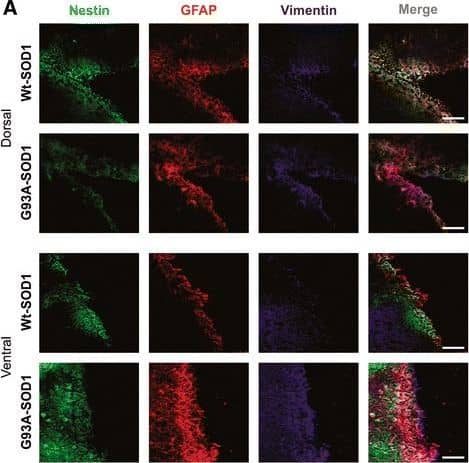
![Immunocytochemistry/ Immunofluorescence: Ki67/MKI67 Antibody - BSA Free [NB500-170] - Senescent MSPCs are characterized by loss of nestin expression. a, b Representative images of GFP immunofluorescence staining (green) & quantitative analysis of GFP+ cells in femoral primary spongiosa from 4, 6, 8, & 12-week-old male Nestin-GFP mice. Images in upper panels in a are lower power w/ boxes outlining area of higher power in lower panels. Numbers of GFP+ cells per mm2 tissue area in primary spongiosa (N. GFP+ cells/PS.Ar) b. 4W, 6W, 8W, & 12W represent 4-, 6-, 8-, & 12-week-old mice, respectively. DAPI stains nuclei blue. c, d Representative images of flow cytometry analysis (c) & % of CD45−GFP+ cells (d) in femoral metaphysis from 4-, 6-, 8-, & 12-week-old male Nestin-GFP mice. e, f Double-immunofluorescence images of femoral metaphysis sections from 4-, 6-, 8-, & 12-week-old male Nestin-GFP mice using antibodies against GFP (green) & Ki67 (red) e. DAPI stains nuclei blue. GP growth plate. PS primary spongiosa. Quantification of % of GFP+ cells that express Ki67 f. Five mice per group. Data are represented as mean ± s.e.m. *P < 0.05 as determined by ANOVA. g Diagram showing isolation of Nestin-GFP+ (red) & Nestin-GFP− (purple) mesenchymal stem/progenitor cells (MSPCs) by fluorescence-activated cell sorting. Detailed information on isolation of MSPCs from femoral metaphysis from Nestin-GFP mice are described in Supplementary Fig. 3 & Methods section. h–j The sorted cells cultured, & SA-beta Gal staining (h), BrdU incorporation (i), & p16INK4a immunostaining (j) performed, & representative images shown. k–m Quantification of % of cells that express SA-beta Gal (k), BrdU (l), & p16INK4a (m). n = 5. Data are represented as mean ± s.e.m. *P < 0.01 as determined by Student’s t-tests Image collected & cropped by CiteAb from following publication (//pubmed.ncbi.nlm.nih.gov/29101351), licensed under a CC-BY license. Not internally tested by Novus Biologicals.](http://images.novusbio.com/fullsize/nb500-170_rabbit-polyclonal-ki67-mki67-antibody-310202416175292.jpg "Immunocytochemistry/ Immunofluorescence: Ki67/MKI67 Antibody - BSA Free [NB500-170] - Senescent MSPCs are characterized by loss of nestin expression. a, b Representative images of GFP immunofluorescence staining (green) & quantitative analysis of GFP+ cells in femoral primary spongiosa from 4, 6, 8, & 12-week-old male Nestin-GFP mice. Images in upper panels in a are lower power w/ boxes outlining area of higher power in lower panels. Numbers of GFP+ cells per mm2 tissue area in primary spongiosa (N. GFP+ cells/PS.Ar) b. 4W, 6W, 8W, & 12W represent 4-, 6-, 8-, & 12-week-old mice, respectively. DAPI stains nuclei blue. c, d Representative images of flow cytometry analysis (c) & % of CD45−GFP+ cells (d) in femoral metaphysis from 4-, 6-, 8-, & 12-week-old male Nestin-GFP mice. e, f Double-immunofluorescence images of femoral metaphysis sections from 4-, 6-, 8-, & 12-week-old male Nestin-GFP mice using antibodies against GFP (green) & Ki67 (red) e. DAPI stains nuclei blue. GP growth plate. PS primary spongiosa. Quantification of % of GFP+ cells that express Ki67 f. Five mice per group. Data are represented as mean ± s.e.m. *P < 0.05 as determined by ANOVA. g Diagram showing isolation of Nestin-GFP+ (red) & Nestin-GFP− (purple) mesenchymal stem/progenitor cells (MSPCs) by fluorescence-activated cell sorting. Detailed information on isolation of MSPCs from femoral metaphysis from Nestin-GFP mice are described in Supplementary Fig. 3 & Methods section. h–j The sorted cells cultured, & SA-beta Gal staining (h), BrdU incorporation (i), & p16INK4a immunostaining (j) performed, & representative images shown. k–m Quantification of % of cells that express SA-beta Gal (k), BrdU (l), & p16INK4a (m). n = 5. Data are represented as mean ± s.e.m. *P < 0.01 as determined by Student’s t-tests Image collected & cropped by CiteAb from following publication (//pubmed.ncbi.nlm.nih.gov/29101351), licensed under a CC-BY license. Not internally tested by Novus Biologicals.")
![Immunocytochemistry/ Immunofluorescence: Ki67/MKI67 Antibody - BSA Free [NB500-170] - Cellular senescence occurs in primary spongiosa of long bone during late puberty. a–e Representative senescence-associated beta -galactosidase (SA-beta Gal) staining (blue) & quantitative analysis of SA-beta Gal+ cells in femoral metaphysis (a–c) & diaphysis (d, e) sections from increasing ages of male mice. 4, 6, 8, & 12W represent 4-, 6-, 8-, & 12-week-old mice, respectively. Images in a are lower power with boxes outlining the area of higher power in b. Numbers of SA-beta Gal+ cells per mm2 tissue area in primary spongiosa (N. SA-beta Gal+ cells/PS.Ar) (c) & diaphysis (N. SA-beta Gal+ cells/DP.Ar) (e). Counterstained with eosin (pink). f, g Representative images of immunofluorescence staining (f) & quantitative analysis of ki67+ (g) cells (red) in femoral primary spongiosa from 4, 6, 8, & 12-week-old male mice. DAPI stains nuclei blue. Images in upper panels in f are lower power with boxes outlining the area of higher power in lower panels. Five mice per group. Data are represented as mean ± s.e.m. Ar tissue area, DP diaphysis, GP growth plate, N number, PS primary spongiosa. *P < 0.01 as determined by ANOVA Image collected & cropped by CiteAb from the following publication (//pubmed.ncbi.nlm.nih.gov/29101351), licensed under a CC-BY license. Not internally tested by Novus Biologicals.](http://images.novusbio.com/fullsize/nb500-170_rabbit-polyclonal-ki67-mki67-antibody-310202416175289.jpg "Immunocytochemistry/ Immunofluorescence: Ki67/MKI67 Antibody - BSA Free [NB500-170] - Cellular senescence occurs in primary spongiosa of long bone during late puberty. a–e Representative senescence-associated beta -galactosidase (SA-beta Gal) staining (blue) & quantitative analysis of SA-beta Gal+ cells in femoral metaphysis (a–c) & diaphysis (d, e) sections from increasing ages of male mice. 4, 6, 8, & 12W represent 4-, 6-, 8-, & 12-week-old mice, respectively. Images in a are lower power with boxes outlining the area of higher power in b. Numbers of SA-beta Gal+ cells per mm2 tissue area in primary spongiosa (N. SA-beta Gal+ cells/PS.Ar) (c) & diaphysis (N. SA-beta Gal+ cells/DP.Ar) (e). Counterstained with eosin (pink). f, g Representative images of immunofluorescence staining (f) & quantitative analysis of ki67+ (g) cells (red) in femoral primary spongiosa from 4, 6, 8, & 12-week-old male mice. DAPI stains nuclei blue. Images in upper panels in f are lower power with boxes outlining the area of higher power in lower panels. Five mice per group. Data are represented as mean ± s.e.m. Ar tissue area, DP diaphysis, GP growth plate, N number, PS primary spongiosa. *P < 0.01 as determined by ANOVA Image collected & cropped by CiteAb from the following publication (//pubmed.ncbi.nlm.nih.gov/29101351), licensed under a CC-BY license. Not internally tested by Novus Biologicals.")
 at 2 µg/mL overnight at 4C. Cells were stained using Streptavidin conjugated to DyLight 550 (red) and counterstained with DAPI (blue). Cells were imaged using a 100X objective and digitally deconvolved.")


![Bioactivity ErbB2/Her2 [Unconjugated]](https://images.novusbio.com/images/protein/1129-er_recombinant-human-erbb2-her2-fc-chimera-protein-cf-bioactivity-7122020142841.jpg)
![SDS-Page ErbB2/Her2 [Unconjugated]](https://images.novusbio.com/images/protein/ErbB2_1129-ER_70.jpg)


![N/A MMP-9 [HRP]](https://images.novusbio.com/images/elisa/DATA_MMP9_DMP900_ELISA_770.jpg)
![N/A MMP-9 [HRP]](https://images.novusbio.com/images/elisa/MMP-9_DMP900_ELISA_148.jpg)


![SDS-Page TNF-alpha [Unconjugated]](https://images.novusbio.com/images/protein/TNF-alpha_210-TA_256.jpg)
![Bioactivity TNF-alpha [Unconjugated]](https://images.novusbio.com/images/protein/TNFalpha_210TA_1658.jpg)
![SEC-MALS TNF-alpha [Unconjugated]](https://images.novusbio.com/images/210-ta_recombinant-human-tnf-alpha-protein-sec-mals-35202312244..jpg)
![Western Blot: Goat anti-Rabbit IgG (H+L) Secondary Antibody [HRP] [NB7160] - Western blot showing vemurafenib treatment in BRAFV600E CRC cells inhibits fission mediator DRP1 with no significant effect on fusion proteins (Mfn1 & 2) using MFN-1 antibody (NBP1-51841) and corresponding secondary antibody, goat anti-rabbit IgG-HRP (NB7160). Image collected and cropped by CiteAb from the following publication (https://pubmed.ncbi.nlm.nih.gov/33738242).](https://images.novusbio.com/images/Goat-anti-Rabbit-IgG-H+L-Secondary-Antibody-HRP-Western-Blot-NB7160-img0001.jpg "Western Blot: Goat anti-Rabbit IgG (H+L) Secondary Antibody [HRP] [NB7160] - Western blot showing vemurafenib treatment in BRAFV600E CRC cells inhibits fission mediator DRP1 with no significant effect on fusion proteins (Mfn1 & 2) using MFN-1 antibody (NBP1-51841) and corresponding secondary antibody, goat anti-rabbit IgG-HRP (NB7160). Image collected and cropped by CiteAb from the following publication (https://pubmed.ncbi.nlm.nih.gov/33738242).")
 followed by 30 min incubation with Goat anti Rabbit HRP conjugated secondary antibodies (Catalog # HAF008) at 1:20 dilution + DAB chromogen (brown). The tissue was counterstained with Hematoxylin (blue). Control was done by omitting primary antibody.")
![Flow Cytometry: Rabbit IgG Isotype Control [NBP2-24891] - An intracellular stain was performed on Raji cells with Adiponectin antibody NB100-65810 (blue) and a matched isotype control NBP2-24893 (orange). Cells were fixed with 4% PFA and then permeablized with 0.1% saponin. Cells were incubated in an antibody dilution of 1 ug/mL for 30 minutes at room temperature, followed by Dylight488-conjugated anti-rabbit secondary antibody. Image using the Azide Free form of this antibody.](https://images.novusbio.com/images/Rabbit--Mouse-IgG-Isotype-Control-Flow-Cytometry-NBP2-24891-img0006.jpg "Flow Cytometry: Rabbit IgG Isotype Control [NBP2-24891] - An intracellular stain was performed on Raji cells with Adiponectin antibody NB100-65810 (blue) and a matched isotype control NBP2-24893 (orange). Cells were fixed with 4% PFA and then permeablized with 0.1% saponin. Cells were incubated in an antibody dilution of 1 ug/mL for 30 minutes at room temperature, followed by Dylight488-conjugated anti-rabbit secondary antibody. Image using the Azide Free form of this antibody.")


