| Luo, J;Liang, Q;Gong, M;Liu, Y;D'Ursi, P;Zheng, Y;Yang, XD; Oxyphenbutazone suppresses non-canonical inflammasome activation and protects against LPS-induced sepsis International immunopharmacology 2026-05-18 [PMID: 42150290] (Western Blot, Mouse) |
Western Blot |
Mouse |
| Herr, SM;Stalkopf, D;Padaszus, S;Herbst, LA;Dörrie, A;Niedenthal, R;Ronkina, N;Yakovleva, T;Kotlyarov, A;Gaestel, M; MK2/p38/p53 Suppress Basal IL-1? and Non-Canonical NF-?B Signaling in Macrophages International journal of molecular sciences 2026-04-02 [PMID: 41977413] (Western Blot, Mouse, Transgenic Mouse) |
Western Blot |
Mouse, Transgenic Mouse |
| Park, YH;Batu, ED;Matthiasardottir, B;Kim, Y;Akkaya-Ulum, YZ;Kastner, DL;Chae, JJ; Cofilin-1 is a redox-sensitive guard of the NLRP3 inflammasome Nature immunology 2026-03-18 [PMID: 41851524] (Western Blot, Mouse) |
Western Blot |
Mouse |
| Zhang, ZH;Wang, XY;Quan, SL;Zeng, ZY;Zhang, KJ;Wu, YL;Zhan, ZY;Nan, JX;Lian, LH; Entadamide A-?-D-glucopyranoside Attenuates Psoriatic Dermatitis by Restoring Keratinocyte Homeostasis and Suppressing Neutrophil Extracellular Traps Journal of ethnopharmacology 2026-03-13 [PMID: 41833762] (Western Blot, Mouse) |
Western Blot |
Mouse |
| Chen, L;Li, J;Wang, S;Ren, R;Xiong, W;Gan, X;Kang, H;Wen, Y;Li, J;He, L; Daucosterol alleviates osteoarthritis by targeting chondrocyte senescence via inhibiting of the JNK pathway International immunopharmacology 2026-03-02 [PMID: 41775106] (Bioassay, Mouse) |
Bioassay |
Mouse |
| Xiong, J;Cheng, X;Xiong, X;Zhang, H;An, Q;Li, Z;Fan, H;Li, G;Li, W;Tian, M;Lv, J; MYSM1-mediated epigenetic modification dysregulation leads to immunosuppression and secondary infections in sepsis PLoS pathogens 2026-02-01 [PMID: 41678561] (Westen Blot, Immunohistochemistry, Mouse) |
Westen Blot, Immunohistochemistry |
Mouse |
| Montero-Jodra, A;Estebán-Amo, MJ;Fernández-Martínez, SP;García Martínez, C;de la Fuente García, MÁ;García-Concejo, A;Martín-Fernández, M;Tamayo, E;Simarro, M; The KEAP1/NRF2 axis controls LPS-induced oxidative stress, inflammasome activation and caspase-1 activity in human endothelial cells PloS one 2026-02-04 [PMID: 41637413] (Western Blot, Human) |
Western Blot |
Human |
| Zeng, M;Xing, W;Liang, G;Yuan, F;Wang, W;Liu, J;He, Z; ?-Methylene-?-butyrolactone 3 inhibits the activation of NLRP3 inflammasome in sepsis by blocking both the priming and inflammasome assembly International immunopharmacology 2026-03-15 [PMID: 41628514] (Western Blot, Mouse) |
Western Blot |
Mouse |
| Yang, H;Liu, W;Zhang, D;Xu, Q;Lee, J;Sun, J;Xue, S;Sun, X;Che, C; IL-24 regulates corneal inflammation and fibrosis in fungal keratitis via the caspase-11/gasdermin D pathway The ocular surface 2026-01-21 [PMID: 41577293] (Western Blot, Mouse) |
Western Blot |
Mouse |
| Huang, Y;He, Y;Wang, Z;Zhang, F;Han, M;Guo, X;Xie, Y;Wang, J;Ma, X;Dan, J;Deng, Y;Wu, H;Hu, H;Wang, J;Yu, L;Jiang, BC;Yan, M;Ma, L; A positive feedback loop between FGR and p65 sustains satellite glial cell activation and chronic neuropathic pain Cell reports 2026-02-24 [PMID: 41575848] (Neutralization, Mouse) |
Neutralization |
Mouse |
| Herrmann, BI;Zamba-Campero, M;Garcia-Sillas, R;Yost, WW;Peterson, LW;Roncaioili, JL;Rankin, SC;Schifferli, DM;Ravichandran, K;Brodsky, IE; Innate detection of Salmonella replication triggers caspase-8-dependent apoptosis via TLR-driven TNF signaling and NLRC4-mediated sensing of the SPI-2 Type III secretion system bioRxiv : the preprint server for biology 2025-10-11 [PMID: 40964383] (Western Blot, Mouse) |
Western Blot |
Mouse |
| Miao, F;Cai, H;Chen, Y;Yan, Z;Jin, R;Dai, Y;Li, L;Wang, H;Xu, Y;Sun, W; NLRP3 inflammasome impairs fracture repair in Rheumatoid arthritis through RhoA/Rac1-IL1? axis-mediated suppression of osteoblast differentiation Journal of orthopaedic translation 2025-09-01 [PMID: 40822518] (Immunohistochemistry, Western Blot, Immunocytochemistry, Mouse) |
Immunohistochemistry, Western Blot, Immunocytochemistry |
Mouse |
| Sato, T;Ishikawa, M;Izumi, Y;Shibata, N;Sato, K;Ohno-Oishi, M;Tawarayama, H;Kunikata, H;Zorumski, CF;Nakazawa, T; Involvement of Microglia in Retinal Ganglion Cell Injury Induced by IOP Elevation in a Rat Ex Vivo Acute Glaucoma Model Biomedicines 2025-07-08 [PMID: 40722741] (Western Blot, Rat) |
Western Blot |
Rat |
| Chen, X;Zheng, Y;Zhang, X;Zheng, A;Huang, J;Deng, G;Wu, X;Peng, Y;Zhang, X;Chen, R;Xiao, Q;Ye, W; Harnessing FDA-approved dipyridamole to inhibit NLRP3 inflammasome and improve outcomes of acute lung injury in sepsis Toxicology and applied pharmacology 2025-07-01 [PMID: 40360054] (Western Blot, Mouse) |
Western Blot |
Mouse |
| Kharitonova, A;Patel, RS;Osborne, B;Krause-Hauch, M;Lui, A;Vidyarthi, G;Li, S;Cai, J;Patel, NA; NPC86 Increases LncRNA Gas5 In Vivo to Improve Insulin Sensitivity and Metabolic Function in Diet-Induced Obese Diabetic Mouse Model International journal of molecular sciences 2025-04-14 [PMID: 40332318] (Western Blot, Mouse) |
Western Blot |
Mouse |
| Son, S;Xu, C;Jang, J;Dinh, M;Skorobogatko, Y;Fu, H;Valentine, JM;An, G;Ying, W;Yu, RT;Downes, M;Evans, RM;Saltiel, AR; Sympathetic activation of white adipose tissue recruits neutrophils to limit energy expenditure Research square 2025-04-16 [PMID: 40321773] (Western Blot, Mouse) |
Western Blot |
Mouse |
| Xu, H;Xu, H;Li, W;Liang, Z;Luo, W;Sheng, S;Liang, G;Zhang, Z; Modulating the NLRP3 Inflammasome: Acitretin as a potential treatment for Sepsis-induced acute lung injury International immunopharmacology 2025-04-24 [PMID: 40187888] (Western Blot, Mouse) |
Western Blot |
Mouse |
| Pushpakumar, S;Juin, SK;Almarshood, H;Gondim, DD;Ouseph, R;Sen, U; Diallyl Trisulfide Attenuates Ischemia-Reperfusion-Induced ER Stress and Kidney Dysfunction in Aged Female Mice Cells 2025-03-12 [PMID: 40136669] (Western Blot, Mouse) |
Western Blot |
Mouse |
| Zhang, SQ;Deng, Q;Tian, C;Zhao, HH;Yang, LY;Cheng, XW;Wang, GP;Liu, D; Costunolide normalizes neuroinflammation and improves neurogenesis deficits in a mouse model of depression through inhibiting microglial Akt/mTOR/NF-?B pathway Acta pharmacologica Sinica 2025-02-26 [PMID: 40011631] (Western Blots, Mouse) |
Western Blots |
Mouse |
| Chan, FHM;Yeap, HW;Liu, Z;Rosli, SN;Low, KE;Bonne, I;Wu, Y;Chong, SZ;Chen, KW; Plasticity of cell death pathways ensures GSDMD activation during Yersinia pseudotuberculosis infection Cell reports 2025-01-16 [PMID: 39823227] (Western Blot, Mouse) |
Western Blot |
Mouse |
| Wei, S;Guan, G;Luan, X;Yu, C;Miao, L;Yuan, X;Chen, P;Di, G; NLRP3 inflammasome constrains liver regeneration through impairing MerTK-mediated macrophage efferocytosis Science advances 2025-01-03 [PMID: 39742469] (In vivo assay, Mouse) |
In vivo assay |
Mouse |
| Zhong, X;Wu, H;Zhang, W;Shi, Y;Gwack, Y;Xue, HH;Sun, Z; Distinct ROR?t-dependent Th17 immune responses are required for autoimmune pathogenesis and protection against bacterial infection Cell reports 2024-11-05 [PMID: 39504243] (Neutralization, Mouse) |
Neutralization |
Mouse |
| Li, D;Yang, Q;Luo, J;Xu, Y;Li, J;Tao, L; Bacterial toxins induce non-canonical migracytosis to aggravate acute inflammation Cell discovery 2024-11-05 [PMID: 39500876] (Immunohistochemistry, Mouse) |
Immunohistochemistry |
Mouse |
| Huynh, P;Hoffmann, JD;Gerhardt, T;Kiss, MG;Zuraikat, FM;Cohen, O;Wolfram, C;Yates, AG;Leunig, A;Heiser, M;Gaebel, L;Gianeselli, M;Goswami, S;Khamhoung, A;Downey, J;Yoon, S;Chen, Z;Roudko, V;Dawson, T;Ferreira da Silva, J;Ameral, NJ;Morgenroth-Rebin, J;D'Souza, D;Koekkoek, LL;Jacob, W;Munitz, J;Lee, D;Fullard, JF;van Leent, MMT;Roussos, P;Kim-Schulze, S;Shah, N;Kleinstiver, BP;Swirski, FK;Leistner, D;St-Onge, MP;McAlpine, CS; Myocardial infarction augments sleep to limit cardiac inflammation and damage Nature 2024-10-30 [PMID: 39478215] (Neutralization, Transgenic Mouse) |
Neutralization |
Transgenic Mouse |
| Zhang, ZH;Zhan, ZY;Jiang, M;Wang, XY;Quan, SL;Wu, YL;Nan, JX;Lian, LH; Casting NETs on Psoriasis: The modulation of inflammatory feedback targeting IL-36/IL-36R axis International immunopharmacology 2024-09-21 [PMID: 39306890] (Western Blot, Immunocytochemistry, Mouse) |
Western Blot, Immunocytochemistry |
Mouse |
| Nabukhotna, K;Kordus, SL;Shupe, JA;Cano Rodríguez, R;Smith, A;Bohannon, JK;Washington, MK;Lacy, DB; Purified CDT toxins and a clean deletion within the CDT locus provide novel insights into the contribution of binary toxin in cellular inflammation and Clostridioides difficile infection PLoS pathogens 2024-09-19 [PMID: 39298531] (Western Blot, Mouse) |
Western Blot |
Mouse |
| Liu, W;Yang, H;Xu, Q;Lee, J;Sun, J;Xue, S;Yang, X;Sun, X;Che, C; Role of MYO1F in neutrophil and macrophage recruitment and pro-inflammatory cytokine production in Aspergillus fumigatus keratitis International immunopharmacology 2024-09-13 [PMID: 39276460] (Western Blot, Mouse) |
Western Blot |
Mouse |
| Song, M;Deng, M;Peng, Z;Dai, F;Wang, Y;Shu, W;Zhou, X;Zhang, J;Hou, Y;Yu, B; Granulocyte colony-stimulating factor mediates bone loss via the activation of IL-1?/JNK signaling pathway in murine Staphylococcus aureus-induced osteomyelitis International immunopharmacology 2024-08-19 [PMID: 39163688] (In vivo assay, Mouse) |
In vivo assay |
Mouse |
| Yao, Q;Wen, J;Chen, S;Wang, Y;Wen, X;Wang, X;Li, C;Zheng, C;Li, J;Ma, Z;Zhan, X;Xiao, X;Bai, Z; Shuangdan Jiedu Decoction improved LPS-induced acute lung injury by regulating both cGAS-STING pathway and inflammasome Journal of ethnopharmacology 2024-08-17 [PMID: 39159837] (Western Blot, Mouse) |
Western Blot |
Mouse |
| Wang, M;Flaswinkel, H;Joshi, A;Napoli, M;Masgrau-Alsina, S;Kamper, JM;Henne, A;Heinz, A;Berouti, M;Schmacke, NA;Hiller, K;Kremmer, E;Wefers, B;Wurst, W;Sperandio, M;Ruland, J;Fröhlich, T;Hornung, V; Phosphorylation of PFKL regulates metabolic reprogramming in macrophages following pattern recognition receptor activation Nature communications 2024-07-31 [PMID: 39085210] (Western Blot, Human) |
Western Blot |
Human |
| Sun, Y;Lu, Y;Liu, L;Saaoud, F;Shao, Y;Xu, K;Drummer Iv, C;Cueto, R;Shan, H;Jiang, X;Zhao, H;Wang, H;Yang, X; Caspase-4/11 promotes hyperlipidemia and chronic kidney disease-accelerated vascular inflammation by enhancing trained immunity JCI insight 2024-07-18 [PMID: 39024553] (Western Blot, Human, Transgenic Mouse) |
Western Blot |
Human, Transgenic Mouse |
| Yow, SJ;Rosli, SN;Hutchinson, PE;Chen, KW; Differential signalling requirements for RIPK1-dependent pyroptosis in neutrophils and macrophages Cell death & disease 2024-07-04 [PMID: 38965211] (Western Blot, Mouse, Transgenic Mouse) |
Western Blot |
Mouse, Transgenic Mouse |
| Kang, H;Choi, SW;Kim, JY;Oh, SJ;Kim, SJ;Lee, MS; ER-to-lysosome Ca2+ refilling followed by K+ efflux-coupled store-operated Ca2+ entry in inflammasome activation and metabolic inflammation eLife 2024-07-02 [PMID: 38953285] (Western Blot, Mouse) |
Western Blot |
Mouse |
| Redmer, T;Raigel, M;Sternberg, C;Ziegler, R;Probst, C;Lindner, D;Aufinger, A;Limberger, T;Trachtova, K;Kodajova, P;Högler, S;Schlederer, M;Stoiber, S;Oberhuber, M;Bolis, M;Neubauer, HA;Miranda, S;Tomberger, M;Harbusch, NS;Garces de Los Fayos Alonso, I;Sternberg, F;Moriggl, R;Theurillat, JP;Tichy, B;Bystry, V;Persson, JL;Mathas, S;Aberger, F;Strobl, B;Pospisilova, S;Merkel, O;Egger, G;Lagger, S;Kenner, L; JUN mediates the senescence associated secretory phenotype and immune cell recruitment to prevent prostate cancer progression Molecular cancer 2024-05-29 [PMID: 38811984] (Immunohistochemistry, Human) |
Immunohistochemistry |
Human |
| Ge, X;Han, H;Desert, R;Das, S;Song, Z;Komakula, SSB;Chen, W;Athavale, D;Lantvit, D;Nieto, N; A PROTEIN COMPLEX OF LIVER ORIGIN ACTIVATES A PRO-INFLAMMATORY PROGRAM THAT DRIVES HEPATIC AND INTESTINAL INJURY IN ALCOHOL-ASSOCIATED LIVER DISEASE Cellular and molecular gastroenterology and hepatology 2024-05-22 [PMID: 38788899] (Western Blot, Transgenic Mouse) |
Western Blot |
Transgenic Mouse |
| Wang, Z;Liu, J;Mou, Y;Liao, W;Li, Y;Liu, J;Tang, J; Anti-inflammatory and uric acid lowering effects of Euodiae fructus on hyperuricemia and gout mice Frontiers in pharmacology 2024-04-19 [PMID: 38708084] (Western Blot, Mouse) |
Western Blot |
Mouse |
| Liu, A;Hayashi, M;Ohsugi, Y;Katagiri, S;Akira, S;Iwata, T;Nakashima, T; The IL-33/ST2 axis is protective against acute inflammation during the course of periodontitis Nature communications 2024-03-28 [PMID: 38548743] (Immunohistochemistry, Mouse) |
Immunohistochemistry |
Mouse |
| Tran, HT;Kratina, T;Coutansais, A;Michalek, D;Hogan, BM;Lawlor, KE;Vince, JE;Silke, J;Lalaoui, N; RIPK3 cleavage is dispensable for necroptosis inhibition but restricts NLRP3 inflammasome activation Cell death and differentiation 2024-03-21 [PMID: 38514849] (Western Blot, Transgenic Mouse) |
Western Blot |
Transgenic Mouse |
| Li, Y;Wu, C;Lee, J;Ning, Q;Lim, J;Eoh, H;Wang, S;Hurrell, BP;Akbari, O;Ou, JJ; Hepatitis B virus e antigen induces atypical metabolism and differentially regulates programmed cell deaths of macrophages PLoS pathogens 2024-03-11 [PMID: 38466743] (Human) |
|
Human |
| Gleeson, TA;Kaiser, C;Lawrence, CB;Brough, D;Allan, SM;Green, JP; The NLRP3 inflammasome is essential for IL-18 production in a murine model of macrophage activation syndrome bioRxiv : the preprint server for biology 2024-03-01 [PMID: 38464243] (Western Blot, Mouse) |
Western Blot |
Mouse |
| Zhan, ZY;Zhang, ZH;Sun, RH;Wu, YL;Nan, JX;Lian, LH; A therapeutic strategy of parthenolide in improving imiquimod-induced psoriasis-like skin inflammation targeting IL-36/NETs through skin transdermal therapeutic system International immunopharmacology 2024-03-09 [PMID: 38461633] (Western Blot, Mouse) |
Western Blot |
Mouse |
| Cao, F;Chen, G;Xu, Y;Wang, X;Tang, X;Zhang, W;Song, X;Yang, X;Zeng, W;Xie, J; METTL14 contributes to acute lung injury by stabilizing NLRP3 expression in an IGF2BP2-dependent manner Cell death & disease 2024-01-13 [PMID: 38218935] (Western Blot, Mouse) |
Western Blot |
Mouse |
| Lee, S;Yun, S;Yang, H;Lee, N;Kim, Y;Lee, S;Zamora, NA;Montero, SS;Yi, DK;Kim, SY;Choi, S;Choi, T;Kim, MS;Lee, Y;Park, YH; Guarea microcarpa C. DC. extract inhibits NLRP3 inflammasome by suppressing its ATPase activity Journal of ethnopharmacology 2024-01-03 [PMID: 38176663] (Western Blot, Mouse) |
Western Blot |
Mouse |
| Exconde, PM;Hernandez-Chavez, C;Bourne, CM;Richards, RM;Bray, MB;Lopez, JL;Srivastava, T;Egan, MS;Zhang, J;Yoo, W;Shin, S;Discher, BM;Taabazuing, CY; The tetrapeptide sequence of IL-18 and IL-1? regulates their recruitment and activation by inflammatory caspases Cell reports 2023-12-15 [PMID: 38103201] (Western Blot, Bioassay, Human) |
Western Blot, Bioassay |
Human |
| Cho, E;Mun, SJ;Kim, HK;Ham, YS;Gil, WJ;Yang, CS; Colon-targeted S100A8/A9-specific peptide systems ameliorate colitis and colitis-associated colorectal cancer in mouse models Acta pharmacologica Sinica 2023-12-01 [PMID: 38040838] (Western Blot, Mouse) |
Western Blot |
Mouse |
| McCrory, C;Verma, J;Tucey, TM;Turner, R;Weerasinghe, H;Beilharz, TH;Traven, A; The short-chain fatty acid crotonate reduces invasive growth and immune escape of Candida albicans by regulating hyphal gene expression mBio 2023-11-06 [PMID: 37929941] (Western Blot, Mouse) |
Western Blot |
Mouse |
| Caronni, N;La Terza, F;Vittoria, FM;Barbiera, G;Mezzanzanica, L;Cuzzola, V;Barresi, S;Pellegatta, M;Canevazzi, P;Dunsmore, G;Leonardi, C;Montaldo, E;Lusito, E;Dugnani, E;Citro, A;Ng, MSF;Schiavo Lena, M;Drago, D;Andolfo, A;Brugiapaglia, S;Scagliotti, A;Mortellaro, A;Corbo, V;Liu, Z;Mondino, A;Dellabona, P;Piemonti, L;Taveggia, C;Doglioni, C;Cappello, P;Novelli, F;Iannacone, M;Ng, LG;Ginhoux, F;Crippa, S;Falconi, M;Bonini, C;Naldini, L;Genua, M;Ostuni, R; IL-1?+ macrophages fuel pathogenic inflammation in pancreatic cancer Nature 2023-11-01 [PMID: 37914939] (IHC, Mouse) |
IHC |
Mouse |
| Meibers, HE;Warrick, KA;VonHandorf, A;Vallez, CN;Kawarizadeh, K;Saha, I;Donmez, O;Jain, VG;Kottyan, LC;Weirauch, MT;Pasare, C; Effector memory T cells induce innate inflammation by triggering DNA damage and a non-canonical STING pathway in dendritic cells Cell reports 2023-09-27 [PMID: 37794597] (Flow Cytometry, Western Blot, Mouse) |
Flow Cytometry, Western Blot |
Mouse |
| Wong, MP;Juan, EYW;Chelluri, SS;Wang, P;Pahmeier, F;Castillo-Rojas, B;Blanc, SF;Biering, SB;Vance, RE;Harris, E; The Inflammasome Pathway is Activated by Dengue Virus Non-structural Protein 1 and is Protective During Dengue Virus Infection bioRxiv : the preprint server for biology 2023-09-21 [PMID: 37790301] (Western Blot, Mouse) |
Western Blot |
Mouse |
| Shosha, E;Shahror, RA;Morris, CA;Xu, Z;Lucas, R;McGee-Lawrence, ME;Rusch, NJ;Caldwell, RB;Fouda, AY; The arginase 1/ornithine decarboxylase pathway suppresses HDAC3 to ameliorate the myeloid cell inflammatory response: implications for retinal ischemic injury Cell death & disease 2023-09-21 [PMID: 37735154] (IHC, Western Blot, Mouse) |
IHC, Western Blot |
Mouse |
| Didi Liu, Yi Huang Protocol for in vivo and in vitro activation of NLRP3 inflammasome in mice using monosodium urate STAR Protoc 9/7/2023 [PMID: 37682717] |
|
|
| Liu H, Zhang L, Liu Z et al. Galectin-3 as TREM2 upstream factor contributes to lung ischemia-reperfusion injury by regulating macrophage polarization iScience 2023-09-15 [PMID: 37636061] |
|
|
| Liu H, Zhang L, Liu Z et al. Galectin-3 as TREM2 upstream factor contributes to lung ischemia-reperfusion injury by regulating macrophage polarization iScience 9/15/2023 [PMID: 37636061] |
|
|
| Sin, R;Sotogaku, N;Ohnishi, YN;Shuto, T;Kuroiwa, M;Kawahara, Y;Sugiyama, K;Murakami, Y;Kanai, M;Funakoshi, H;Chakraborti, A;Bibb, JA;Nishi, A; Inhibition of STAT-mediated cytokine responses to chemically-induced colitis prevents inflammation-associated neurobehavioral impairments Brain, behavior, and immunity 2023-08-23 [PMID: 37625556] (Western Blot, Mouse) |
Western Blot |
Mouse |
| Ciana Diskin, Emily A. Day, Órlaith C. Henry, Juliana E. Toller-Kawahisa, Luke A. J. O’Neill 4-Octyl Itaconate and Dimethyl Fumarate Induce Secretion of the Anti-Inflammatory Protein Annexin A1 via NRF2 The Journal of Immunology 9/15/2023 [PMID: 37578391] |
|
|
| Rondeaux, J;Groussard, D;Renet, S;Tardif, V;Dumesnil, A;Chu, A;Di Maria, L;Lemarcis, T;Valet, M;Henry, JP;Badji, Z;Vézier, C;Béziau-Gasnier, D;Neele, AE;de Winther, MPJ;Guerrot, D;Brand, M;Richard, V;Durand, E;Brakenhielm, E;Fraineau, S; Ezh2 emerges as an epigenetic checkpoint regulator during monocyte differentiation limiting cardiac dysfunction post-MI Nature communications 2023-07-25 [PMID: 37491334] (IHC, Mouse) |
IHC |
Mouse |
| Bai, Y;Min, R;Chen, P;Mei, S;Deng, F;Zheng, Z;Jiang, C;Miao, R;Wu, Z;Zhang, P;Pan, Y;Lieberman, J;Liu, X; Disulfiram blocks inflammatory TLR4 signaling by targeting MD-2 Proceedings of the National Academy of Sciences of the United States of America 2023-08-01 [PMID: 37487070] (Western Blot, Mouse) |
Western Blot |
Mouse |
| Veronica Mutti, Giulia Carini, Alice Filippini, Stefania Castrezzati, Lorena Giugno, Massimo Gennarelli et al. LRRK2 Kinase Inhibition Attenuates Neuroinflammation and Cytotoxicity in Animal Models of Alzheimer’s and Parkinson’s Disease-Related Neuroinflammation Cells 7/6/2023 [PMID: 37443833] |
|
|
| Bingwei Wang, Yan Ma, Sheng Li, Hang Yao, Mingna Gu, Ying Liu et al. GSDMD in peripheral myeloid cells regulates microglial immune training and neuroinflammation in Parkinson's disease Acta Pharmaceutica Sinica B 6/1/2023 [PMID: 37425058] |
|
|
| Germana Lentini, Agata Famà, Giuseppe Valerio De Gaetano, Francesco Coppolino, Ahlem Khachroub Mahjoub, Liv Ryan et al. Caspase-8 inhibition improves the outcome of bacterial infections in mice by promoting neutrophil activation Cell Reports Medicine 7/18/2023 [PMID: 37390829] |
|
|
| Yingting Hou, Hongbin He, Ming Ma, Rongbin Zhou Apilimod activates the NLRP3 inflammasome through lysosome-mediated mitochondrial damage Frontiers in Immunology 6/8/2023 [PMID: 37359517] |
|
|
| Leem YH, Kim DY, Park JE, Kim HS Necrosulfonamide exerts neuroprotective effect by inhibiting necroptosis, neuroinflammation, and ?-synuclein oligomerization in a subacute MPTP mouse model of Parkinson's disease Scientific reports 5/31/2023 [PMID: 37258791] |
|
|
| Fang L, Ohashi K, Hayakawa S et al. Adipolin protects against renal injury via PPAR?-dependent reduction of inflammasome activation iScience 2023-05-01 [PMID: 37250342] |
|
|
| Fang L, Ohashi K, Hayakawa S et al. Adipolin protects against renal injury via PPAR?-dependent reduction of inflammasome activation iScience 5/1/2023 [PMID: 37250342] |
|
|
| Zhang, Z;Song, J;Kwon, SH;Wang, Z;Park, SG;Piao, X;Ryu, JH;Kim, N;Kim, OS;Kim, SH;Koh, JT; Pirfenidone Inhibits Alveolar Bone Loss in Ligature-Induced Periodontitis by Suppressing the NF-?B Signaling Pathway in Mice International journal of molecular sciences 2023-05-12 [PMID: 37240020] (IHC, Mouse) |
IHC |
Mouse |
| Jack P. Green, Lina Y. El-Sharkawy, Stefan Roth, Jie Zhu, Jiayu Cao, Andrew G. Leach et al. Discovery of an inhibitor of DNA-driven inflammation that preferentially targets the AIM2 inflammasome iScience 5/19/2023 [PMID: 37216118] |
|
|
| Jack P. Green, Lina Y. El-Sharkawy, Stefan Roth, Jie Zhu, Jiayu Cao, Andrew G. Leach et al. Discovery of an inhibitor of DNA-driven inflammation that preferentially targets the AIM2 inflammasome iScience 2023-05-19 [PMID: 37216118] (Western Blot, Mouse) |
Western Blot |
Mouse |
| Weerasinghe, H;Simm, C;Djajawi, TM;Tedja, I;Lo, TL;Simpson, DS;Shasha, D;Mizrahi, N;Olivier, FAB;Speir, M;Lawlor, KE;Ben-Ami, R;Traven, A; Candida auris uses metabolic strategies to escape and kill macrophages while avoiding robust activation of the NLRP3 inflammasome response Cell reports 2023-05-18 [PMID: 37204928] (Western Blot, Mouse) |
Western Blot |
Mouse |
| Foster, SL;Dutton, AJ;Yerzhan, A;March, LB;Barry, K;Seehus, CR;Huang, X;Talbot, S;Woolf, CJ; A Preliminary Study of Mild Heat Stress on Inflammasome Activation in Murine Macrophages Cells 4/19/2023 [PMID: 37190098] |
|
|
| Ren, M;Chen, J;Xu, H;Li, W;Wang, T;Chi, Z;Lin, Y;Zhang, A;Chen, G;Wang, X;Sun, X;Liang, G;Wang, J;Luo, W; Ergolide covalently binds NLRP3 and inhibits NLRP3 inflammasome-mediated pyroptosis International immunopharmacology 2023-05-12 [PMID: 37182452] (Western Blot, Transgenic Mouse, Mouse) |
Western Blot |
Transgenic Mouse, Mouse |
| Liu, L;Feng, L;Gao, J;Hu, J;Li, A;Zhu, Y;Zhang, C;Qiu, B;Shen, Z; Parthenolide targets NLRP3 to treat inflammasome-related diseases International immunopharmacology 2023-06-01 [PMID: 37167640] (Bioassay, Mouse) |
Bioassay |
Mouse |
| Huang, LS;Anas, M;Xu, J;Zhou, B;Toth, PT;Krishnan, Y;Di, A;Malik, AB; Endosomal trafficking of two-pore K+ efflux channel TWIK2 to plasmalemma mediates NLRP3 inflammasome activation and inflammatory injury eLife 2023-05-09 [PMID: 37158595] (Mouse) |
|
Mouse |
| Siopi, E;Galerne, M;Rivagorda, M;Saha, S;Moigneu, C;Moriceau, S;Bigot, M;Oury, F;Lledo, PM; Gut microbiota changes require vagus nerve integrity to promote depressive-like behaviors in mice Molecular psychiatry 2023-05-02 [PMID: 37131071] (Western Blot, Mouse) |
Western Blot |
Mouse |
| JH Kang, SB Lee, J Seok, DH Kim, G Ma, J Park, AJ Jeong, SK Ye, TB Kang Novel Activity of ODZ10117, a STAT3 Inhibitor, for Regulation of NLRP3 Inflammasome Activation International Journal of Molecular Sciences, 2023-03-23;24(7):. 2023-03-23 [PMID: 37047051] (Western Blot, Mouse) |
Western Blot |
Mouse |
| A Ta, R Ricci-Azev, SO Vasudevan, SS Wright, P Kumari, MS Havira, M Surendran, VA Rathinam, SK Vanaja A bacterial autotransporter impairs innate immune responses by targeting the transcription factor TFE3 Nature Communications, 2023-04-11;14(1):2035. 2023-04-11 [PMID: 37041208] (Western Blot, Mouse) |
Western Blot |
Mouse |
| SB Lee, JH Kang, EJ Sim, YR Jung, JH Kim, PF Hillman, SJ Nam, TB Kang Cornus officinalis Seed Extract Inhibits AIM2-Inflammasome Activation and Attenuates Imiquimod-Induced Psoriasis-like Skin Inflammation International Journal of Molecular Sciences, 2023-03-15;24(6):. 3/15/2023 [PMID: 36982727] |
|
|
| Jeon Y, Chow SH, Stuart I et al. FBXO11 governs macrophage cell death and inflammation in response to bacterial toxins Life science alliance 6/1/2023 [PMID: 36977592] |
|
|
| S Bollenbeck, K Heitman, B Czaya, M Easter, MJ Hirsch, S Vang, E Harris, ES Helton, JW Barnes, C Faul, S Krick Phosphate induces inflammation and exacerbates injury from cigarette smoke in the bronchial epithelium Scientific Reports, 2023-03-25;13(1):4898. 2023-03-25 [PMID: 36966182] (IHC, Mouse) |
IHC |
Mouse |
| J Li, Q Lin, X Shao, S Li, X Zhu, J Wu, S Mou, L Gu, Q Wang, M Zhang, K Zhang, J Lu, Z Ni HIF1alpha-BNIP3-mediated mitophagy protects against renal fibrosis by decreasing ROS and inhibiting activation of the NLRP3 inflammasome Cell Death & Disease, 2023-03-17;14(3):200. 2023-03-17 [PMID: 36928344] (ICC, IHC, Western Blot, Mouse) |
ICC, IHC, Western Blot |
Mouse |
| TT Lin, CY Jiang, L Sheng, L Wan, W Fan, JC Li, XD Sun, CJ Xu, L Hu, XF Wu, Y Han, WT Liu, YB Pan Suppressing high mobility group box-1 release alleviates morphine tolerance via the adenosine 5'-monophosphate-activated protein kinase/heme oxygenase-1 pathway Neural regeneration research, 2023-09-01;18(9):2067-2074. 2023-09-01 [PMID: 36926733] (Western Blot, Mouse) |
Western Blot |
Mouse |
| Y Chen, J Pu, X Li, L Lian, C Ge, Z Liu, W Wang, L Hou, W Chen, J Li Aim2 Deficiency Ameliorates Lacrimal Gland Destruction and Corneal Epithelium Defects in an Experimental Dry Eye Model Investigative Ophthalmology & Visual Science, 2023-03-01;64(3):26. 2023-03-01 [PMID: 36920364] (Western Blot, Mouse) |
Western Blot |
Mouse |
| A Hooftman, CG Peace, DG Ryan, EA Day, M Yang, AF McGettrick, M Yin, EN Montano, L Huo, JE Toller-Kaw, V Zecchini, TAJ Ryan, A Bolado-Car, AM Casey, HA Prag, ASH Costa, G De Los San, M Ishimori, DJ Wallace, S Venuturupa, E Nikitopoul, N Frizzell, C Johansson, A Von Kriegs, MP Murphy, C Jefferies, C Frezza, LAJ O'Neill Macrophage fumarate hydratase restrains mtRNA-mediated interferon production Nature, 2023-03-08;615(7952):490-498. 2023-03-08 [PMID: 36890227] (Western Blot, Mouse) |
Western Blot |
Mouse |
| WJ Zhong, J Zhang, JX Duan, CY Zhang, SC Ma, YS Li, NS Yang, HH Yang, JB Xiong, CX Guan, ZX Jiang, ZJ You, Y Zhou TREM-1 triggers necroptosis of macrophages through mTOR-dependent mitochondrial fission during acute lung injury Journal of Translational Medicine, 2023-03-06;21(1):179. 2023-03-06 [PMID: 36879273] (Western Blot, Mouse) |
Western Blot |
Mouse |
| Haowen Xu, Jiahao Chen, Pan Chen, Weifeng Li, Jingjing Shao, Shanshan Hong et al. Costunolide covalently targets NACHT domain of NLRP3 to inhibit inflammasome activation and alleviate NLRP3-driven inflammatory diseases Acta Pharmaceutica Sinica B 2/1/2023 [PMID: 36873170] |
|
|
| Ah-Ra Jang, Ha-Nul Lee, Jung Joo Hong, Young-Min Kim, Jong-Hwan Park Ethanol extract of Chrysanthemum zawadskii inhibits the NLRP3 inflammasome by suppressing ASC oligomerization in macrophages Experimental and Therapeutic Medicine 2/7/2023 [PMID: 36845948] |
|
|
| Yi Huang, Yun Guo, Yan Zhou, Qian Huang, Yi Ru, Yingli Luo et al. Tivantinib alleviates inflammatory diseases by directly targeting NLRP3 iScience 3/17/2023 [PMID: 36843841] |
|
|
| A Filippini, V Salvi, V Dattilo, C Magri, S Castrezzat, R Veerhuis, D Bosisio, M Gennarelli, I Russo LRRK2 Kinase Inhibition Attenuates Astrocytic Activation in Response to Amyloid beta1-42 Fibrils Biomolecules, 2023-02-06;13(2):. 2023-02-06 [PMID: 36830676] (Western Blot, Mouse) |
Western Blot |
Mouse |
| PM Exconde, C Hernandez-, MB Bray, JL Lopez, T Srivastava, MS Egan, J Zhang, S Shin, BM Discher, CY Taabazuing The tetrapeptide sequence of IL-1beta regulates its recruitment and activation by inflammatory caspases bioRxiv : the preprint server for biology, 2023-02-16;0(0):. 2/16/2023 [PMID: 36824844] |
|
|
| Y Wang, D Sadike, B Huang, P Li, Q Wu, N Jiang, Y Fang, G Song, L Xu, W Wang, M Xie Regulatory T cells alleviate myelin loss and cognitive dysfunction by regulating neuroinflammation and microglial pyroptosis via TLR4/MyD88/NF-kappaB pathway in LPC-induced demyelination Journal of Neuroinflammation, 2023-02-18;20(1):41. 2023-02-18 [PMID: 36803990] (IHC, Mouse) |
IHC |
Mouse |
| X Shu, C Wei, WY Tu, K Zhong, S Qi, A Wang, L Bai, SX Zhang, B Luo, ZZ Xu, K Zhang, C Shen Negative regulation of TREM2-mediated C9orf72 poly-GA clearance by the NLRP3 inflammasome Cell Reports, 2023-02-16;42(2):112133. 2023-02-16 [PMID: 36800288] (Western Blot, Mouse) |
Western Blot |
Mouse |
| Fangfang Wang, Meng Zhang, Meng Yuan, Zixuan Xia, Fengge Yang, Sihao Zhang et al. A novel sorbicillinoid compound as a potent anti‐inflammation agent through inducing NLRP3 protein degradation British Journal of Pharmacology 8/1/2023 [PMID: 36788033] |
|
|
| Fangfang Wang, Meng Zhang, Meng Yuan, Zixuan Xia, Fengge Yang, Sihao Zhang et al. A novel sorbicillinoid compound as a potent anti‐inflammation agent through inducing NLRP3 protein degradation British Journal of Pharmacology 2023-08-01 [PMID: 36788033] (Western Blot, Mouse) |
Western Blot |
Mouse |
| Nevien Hendawy, Tala H. Salaheldin, Sally A. Abuelezz PCSK9 Inhibition Reduces Depressive like Behavior in CUMS-Exposed Rats: Highlights on HMGB1/RAGE/TLR4 Pathway, NLRP3 Inflammasome Complex and IDO-1 Journal of Neuroimmune Pharmacology 2/13/2023 [PMID: 36781714] |
|
|
| Wenhua Zhao, Yanan Cao, Yue Chen, Yuyi Chen, Tianxiao Wang, Lu Li et al. NLRP3 Regulates Mandibular Healing through Interaction with UCHL5 in MSCs International Journal of Biological Sciences 1/16/2023 [PMID: 36778113] |
|
|
| A Ramadan, Z Cao, M Gadjeva, TS Zaidi, VA Rathinam, N Panjwani The NLRP3 Inflammasome Is Required for Protection Against Pseudomonas Keratitis Investigative Ophthalmology & Visual Science, 2023-02-01;64(2):11. 2023-02-01 [PMID: 36749596] (Western Blot, Mouse) |
Western Blot |
Mouse |
| Junedh M. Lavine, Junedh Amrute, Vinay Penna, Andrea Bredemeyer, Andrea Bredemeyer, Steven Yamawaki et al. Targeting Immune-Fibroblast Crosstalk in Myocardial Infarction and Cardiac Fibrosis Research Square 8/25/2023 [PMID: 36747878] |
|
|
| G Lin, N Li, D Li, L Chen, H Deng, S Wang, J Tang, W Ouyang Carnosic acid inhibits NLRP3 inflammasome activation by targeting both priming and assembly steps International immunopharmacology, 2023-02-02;116(0):109819. 2023-02-02 [PMID: 36738671] (Western Blot, Mouse) |
Western Blot |
Mouse |
| MB Reynolds, HS Hong, BC Michmerhui, AE Lawrence, L Zhang, JS Knight, CA Lyssiotis, BH Abuaita, MX O'Riordan Cardiolipin coordinates inflammatory metabolic reprogramming through regulation of Complex II disassembly and degradation Science Advances, 2023-02-03;9(5):eade8701. 2023-02-03 [PMID: 36735777] (Western Blot, Mouse) |
Western Blot |
Mouse |
| Zhouyangfan Peng, Xiqing Tan, Liangpeng Xie, Ze Li, Sufang Zhou, Yapei Li PKR deficiency delays vascular aging via inhibiting GSDMD-mediated endothelial cell hyperactivation iScience 1/20/2023 [PMID: 36691613] |
|
|
| L González-C, J P Green, I Cuadrado, Á Amesty, S Oramas-Roy, David Brou, A Estévez-Br, S Hortelano, B de Las Her Phenolic and quinone methide nor-triterpenes as selective NLRP3 inflammasome inhibitors Bioorganic chemistry, 2023-01-14;132(0):106362. 2023-01-14 [PMID: 36657273] (Western Blot, Mouse) |
Western Blot |
Mouse |
| Meyers AK, Wang Z, Han W et al. Pyruvate dehydrogenase kinase supports macrophage NLRP3 inflammasome activation during acute inflammation Cell Reports 2023-01-31 [PMID: 36640341] |
|
|
| Meyers AK, Wang Z, Han W et al. Pyruvate dehydrogenase kinase supports macrophage NLRP3 inflammasome activation during acute inflammation Cell Reports 1/31/2023 [PMID: 36640341] |
|
|
| HW Xu, WF Li, SS Hong, JJ Shao, JH Chen, N Chattipako, D Wu, W Luo, G Liang Tabersonine, a natural NLRP3 inhibitor, suppresses inflammasome activation in macrophages and attenuate NLRP3-driven diseases in mice Acta pharmacologica Sinica, 2023-01-10;0(0):. 2023-01-10 [PMID: 36627344] (Western Blot, Mouse) |
Western Blot |
Mouse |
| G Lee, H Ahn, JH Yun, J Park, E Lee, S Oh, GS Lee JC2-11, a benzylideneacetophenone derivative, attenuates inflammasome activation Scientific Reports, 2022-12-28;12(1):22484. 2022-12-28 [PMID: 36577816] (Western Blot, Mouse) |
Western Blot |
Mouse |
| Jae-Sung Kim, Hyo Keun Kim, Minsoo Kim, Sein Jang, Euni Cho, Seok-Jun Mun et al. Colon-Targeted eNAMPT-Specific Peptide Systems for Treatment of DSS-Induced Acute and Chronic Colitis in Mouse Antioxidants (Basel) 11/30/2022 [PMID: 36552583] |
|
|
| SK Juin, S Pushpakuma, SC Tyagi, U Sen Glucosidase inhibitor, Nimbidiol ameliorates renal fibrosis and dysfunction in type-1 diabetes Scientific Reports, 2022-12-15;12(1):21707. 2022-12-15 [PMID: 36522378] (Western Blot, Mouse) |
Western Blot |
Mouse |
| LM Branco, MP Amaral, H Boekhoff, ABF de Lima, IS Farias, SL Lage, GJS Pereira, BS Franklin, KR Bortoluci Lysosomal cathepsins act in concert with Gasdermin-D during NAIP/NLRC4-dependent IL-1beta secretion Cell Death & Disease, 2022-12-08;13(12):1029. 2022-12-08 [PMID: 36481780] (Mouse) |
|
Mouse |
| C Oh, L Li, A Verma, AD Reuven, EA Miao, JB Bliska, Y Aachoui Neutrophil inflammasomes sense the subcellular delivery route of translocated bacterial effectors and toxins Cell Reports, 2022-11-22;41(8):111688. 2022-11-22 [PMID: 36417874] (Western Blot, Mouse, Transgenic Mouse) |
Western Blot |
Mouse, Transgenic Mouse |
| Ilia D. Vainchtein, Astrid M. Alsema, Marissa L. Dubbelaar, Corien Grit, Jonathan Vinet, Hilmar R. J. van Weering et al. Characterizing microglial gene expression in a model of secondary progressive multiple sclerosis Glia 3/1/2023 [PMID: 36377669] |
|
|
| YH Wu, ST Mo, IT Chen, FY Hsieh, SL Hsieh, J Zhang, MZ Lai Caspase-8 inactivation drives autophagy-dependent inflammasome activation in myeloid cells Science Advances, 2022-11-11;8(45):eabn9912. 2022-11-11 [PMID: 36367942] (Western Blot, Transgenic Mouse) |
Western Blot |
Transgenic Mouse |
| MSJ Mangan, F Gorki, K Krause, A Heinz, A Pankow, T Ebert, D Jahn, K Hiller, V Hornung, M Maurer, FI Schmidt, R Gerhard, E Latz Transcriptional licensing is required for Pyrin inflammasome activation in human macrophages and bypassed by mutations causing familial Mediterranean fever PloS Biology, 2022-11-07;20(11):e3001351. 2022-11-07 [PMID: 36342970] (Western Blot, Human) |
Western Blot |
Human |
| Tianyin Sun, Ruiqian Xie, Hongbin He, Qianqian Xie, Xueqin Zhao, Guijie Kang et al. Kynurenic acid ameliorates NLRP3 inflammasome activation by blocking calcium mobilization via GPR35 Frontiers in Immunology 10/13/2022 [PMID: 36311752] |
|
|
| Huijeong Ahn, Gilyoung Lee, Byung-Cheol Han, Seung-Ho Lee, Geun-Shik Lee Maltol, a Natural Flavor Enhancer, Inhibits NLRP3 and Non-Canonical Inflammasome Activation Antioxidants (Basel) 9/28/2022 [PMID: 36290645] |
|
|
| Huijeong Ahn, Gilyoung Lee, Byung-Cheol Han, Seung-Ho Lee, Geun-Shik Lee Maltol, a Natural Flavor Enhancer, Inhibits NLRP3 and Non-Canonical Inflammasome Activation Antioxidants (Basel) 2022-09-28 [PMID: 36290645] (Western Blot, Mouse) |
Western Blot |
Mouse |
| SL Kim, MW Shin, SW Kim Lipocalin 2 activates the NLRP3 inflammasome via LPS?induced NF?kappaB signaling and plays a role as a pro?inflammatory regulator in murine macrophages Molecular Medicine Reports, 2022-10-20;26(6):. 2022-10-20 [PMID: 36263611] (Western Blot, Mouse) |
Western Blot |
Mouse |
| Hiroyasu Tsutsuki, Tianli Zhang, Kinnosuke Yahiro, Touya Toyomoto, Tomohiro Sawa Non-canonical inflammasome activation analysis in a mouse model of Citrobacter rodentium infection STAR Protocols 12/16/2022 [PMID: 36201318] |
|
|
| Q Lin, D Zhang, J Zhang, W Luo, Z Xu, J Yao, L Wang Identification of lncRNA DLEU2 as a potential diagnostic biomarker and anti-inflammatory target for ulcerative colitis Frontiers in Pharmacology, 2022-09-14;13(0):991448. 2022-09-14 [PMID: 36188540] (Western Blot, Human) |
Western Blot |
Human |
| P Sharma, K Karnam, A Mahale, K Sedmaki, V Krishna Ve, OP Kulkarni HDAC5 RNA interference ameliorates acute renal injury by upregulating KLF2 and inhibiting NALP3 expression in a mouse model of oxalate nephropathy International immunopharmacology, 2022-09-29;112(0):109264. 2022-09-29 [PMID: 36183679] (Western Blot, Mouse) |
Western Blot |
Mouse |
| Hadar Cohen, Noam Baram, Chaya Mushka Fridman, Liat Edry-Botzer, Dor Salomon, Motti Gerlic Post-phagocytosis activation of NLRP3 inflammasome by two novel T6SS effectors eLife 9/26/2022 [PMID: 36155655] |
|
|
| AA Abdelrahma, KL Bunch, PV Sandow, PN Cheng, RB Caldwell, RW Caldwell Systemic Administration of Pegylated Arginase-1 Attenuates the Progression of Diabetic Retinopathy Cells, 2022-09-16;11(18):. 2022-09-16 [PMID: 36139465] (Western Blot, Mouse) |
Western Blot |
Mouse |
| M Duhalde Ve, D Olivera, G Gastão Dav, M Bertullo, V Noya, G Fabiano de, S Primon Mur, I Castro, AP Arévalo, M Crispo, G Galliussi, S Russo, D Charbonnie, F Rammauro, M Jeldres, C Alamón, V Varela, C Batthyany, M Bollati-Fo, P Oppezzo, O Pritsch, JL Proença-Mó, HI Nakaya, E Trias, L Barbeito, I Anegon, MC Cuturi, P Moraes-Vie, M Segovia, M Hill PD-1/PD-L1 blockade abrogates a dysfunctional innate-adaptive immune axis in critical beta-coronavirus disease Science Advances, 2022-09-21;8(38):eabn6545. 2022-09-21 [PMID: 36129987] (Neutralization, Mouse) |
Neutralization |
Mouse |
| I de Castro, A Bianchi, NU Deshpande, P Sharma, S Mehra, VT Garrido, SJ Saigh, J England, PJ Hosein, D Kwon, NB Merchant, J Datta Neutrophil-mediated fibroblast-tumor cell il-6/stat-3 signaling underlies the association between neutrophil-to-lymphocyte ratio dynamics and chemotherapy response in localized pancreatic cancer: A hybrid clinical-preclinical study Elife, 2022-09-15;11(0):. 2022-09-15 [PMID: 36107485] (Neutralization, Mouse) |
Neutralization |
Mouse |
| Y Zhu, H Zhang, H Mao, S Zhong, Y Huang, S Chen, K Yan, Z Zhao, X Hao, Y Zhang, H Yao, X Huang, M Wang, W Zhang, J Li, G Meng, X Qin, Z Ye, J Shen, Y Song, Y Xu, Z Yang, L Wang, Y Zhang, L Wen FAAH served a key membrane-anchoring and stabilizing role for NLRP3 protein independently of the endocannabinoid system Cell Death and Differentiation, 2022-09-14;0(0):. 2022-09-14 [PMID: 36104448] (Western Blot, Mouse) |
Western Blot |
Mouse |
| SM Ruwanpura, L McLeod, LF Dousha, HJ Seow, AC West, AJ West, T Weng, M Alanazi, M MacDonald, PT King, PG Bardin, C Gabay, DM Klinman, S Bozinovski, R Vlahos, GP Anderson, S Rose-John, MI Saad, BJ Jenkins Cross-talk between IL-6 trans-signaling and AIM2 inflammasome/IL-1beta axes bridge innate immunity and epithelial apoptosis to promote emphysema Proceedings of the National Academy of Sciences of the United States of America, 2022-08-29;119(36):e2201494119. 2022-08-29 [PMID: 36037355] (Western Blot, Mouse) |
Western Blot |
Mouse |
Lee J, Kim SJ, Choi GE et al. Sweet taste receptor agonists attenuate macrophage IL-1 beta expression and eosinophilic inflammation linked to autophagy deficiency in myeloid cells Clinical and translational medicine 8/1/2022 [PMID: 35988262]
Details:
Dilution used 1:400 |
|
|
| C Yuan, K Ai, M Xiang, C Xie, M Zhao, M Wu, H Li, Y Wu, Y Cao, C Li, Y Zhong, X Pei, HKW Law, L Gao, Q Xiao, X Yang Novel 1-hydroxy phenothiazinium-based derivative protects against bacterial sepsis by inhibiting AAK1-mediated LPS internalization and caspase-11 signaling Cell Death & Disease, 2022-08-18;13(8):722. 8/18/2022 [PMID: 35982051] |
|
|
Feriotti C, SA-Pessoa J, CalderOn-GonzAlez R et al. Klebsiella pneumoniae hijacks the Toll-IL-1R protein SARM1 in a type I IFN-dependent manner to antagonize host immunity Cell reports 8/9/2022 [PMID: 35947948]
Details:
Dilutions: 1:1000 |
|
|
| H Shi, Y Zhu, C Xing, S Li, Z Bao, L Lei, D Lin, Y Wang, H Chen, X Xu An injectable thermosensitive hydrogel for dual delivery of diclofenac and Avastin� to effectively suppress inflammatory corneal neovascularization International journal of pharmaceutics, 2022-08-05;0(0):122081. 2022-08-05 [PMID: 35934166] (IHC, Rabbit) |
IHC |
Rabbit |
| AQ Dong, YP Yang, SM Jiang, XY Yao, D Qi, CJ Mao, XY Cheng, F Wang, LF Hu, CF Liu Pramipexole inhibits astrocytic NLRP3 inflammasome activation via Drd3-dependent autophagy in a mouse model of Parkinson's disease Acta pharmacologica Sinica, 2022-07-27;0(0):. 2022-07-27 [PMID: 35896696] (Western Blot, Mouse) |
Western Blot |
Mouse |
| AB Lopez-Rodr, C Decouty-Pe, V Farré-Alin, A Palomino-A, P Narros-Fer, J Egea Activation of NLRP3 Is Required for a Functional and Beneficial Microglia Response after Brain Trauma Pharmaceutics, 2022-07-26;14(8):. 2022-07-26 [PMID: 35893807] (Western Blot, Mouse) |
Western Blot |
Mouse |
| Jae Sang Han, Ye Lin Kim, Hyo Jeong Yu, Jung Mee Park, Yeonji Kim, So Young Park et al. Safety and Efficacy of Intratympanic Alpha-Lipoic Acid Injection in a Mouse Model of Noise-Induced Hearing Loss Antioxidants (Basel) 7/22/2022 [PMID: 35892625] |
|
|
| Xintong Dong, Nathachit Limjunyawong, Elizabeth I. Sypek, Gaofeng Wang, Roger V. Ortines, Christine Youn et al. Keratinocyte-derived defensins activate neutrophil-specific receptors Mrgpra2a/b to prevent skin dysbiosis and bacterial infection Immunity 9/1/2022 [PMID: 35882236] |
|
|
| Xintong Dong, Nathachit Limjunyawong, Elizabeth I. Sypek, Gaofeng Wang, Roger V. Ortines, Christine Youn et al. Keratinocyte-derived defensins activate neutrophil-specific receptors Mrgpra2a/b to prevent skin dysbiosis and bacterial infection Immunity 2022-09-01 [PMID: 35882236] (Western Blot, Human) |
Western Blot |
Human |
| JH Ma, E Lee, SH Yoon, H Min, JH Oh, I Hwang, Y Sung, JH Ryu, J Bok, JW Yu Therapeutic effect of NLRP3 inhibition on hearing loss induced by systemic inflammation in a CAPS-associated mouse model EBioMedicine, 2022-07-20;82(0):104184. 2022-07-20 [PMID: 35870427] (Western Blot, Mouse) |
Western Blot |
Mouse |
| Hao Zhou, Xun Shen, Chen Yan, Wu Xiong, Zemeng Ma, Zhenggang Tan et al. Extracellular vesicles derived from human umbilical cord mesenchymal stem cells alleviate osteoarthritis of the knee in mice model by interacting with METTL3 to reduce m6A of NLRP3 in macrophage Stem Cell Research & Therapy 12/1/2022 [PMID: 35842714] |
|
|
| Zhouyangfan Peng, Mingwu Duan, Yiting Tang, Jianfeng Wu, Kai Zhao, Yanjun Zhong et al. Impaired interferon-gamma signaling promotes the development of silicosis iScience 2022-07-15 [PMID: 35800765] (Western Blot, Mouse) |
Western Blot |
Mouse |
| Zhouyangfan Peng, Mingwu Duan, Yiting Tang, Jianfeng Wu, Kai Zhao, Yanjun Zhong et al. Impaired interferon-gamma signaling promotes the development of silicosis iScience 7/15/2022 [PMID: 35800765] |
|
|
| W Chen, S Chen, C Yan, Y Zhang, R Zhang, M Chen, S Zhong, W Fan, S Zhu, D Zhang, X Lu, J Zhang, Y Huang, L Zhu, X Li, D Lv, Y Fu, H Iv, Z Ling, L Ma, H Jiang, G Long, J Zhu, D Wu, B Wu, B Sun Allergen protease-activated stress granule assembly and gasdermin D fragmentation control interleukin-33 secretion Nature Immunology, 2022-07-06;23(7):1021-1030. 2022-07-06 [PMID: 35794369] (Western Blot, Mouse) |
Western Blot |
Mouse |
| H Dong, B Zhao, J Chen, Z Liu, X Li, L Li, H Wen Mitochondrial calcium uniporter promotes phagocytosis-dependent activation of the NLRP3 inflammasome Proceedings of the National Academy of Sciences of the United States of America, 2022-06-22;119(26):e2123247119. 6/22/2022 [PMID: 35733245] |
|
|
| W Yu, Z Wang, X Yu, Y Zhao, Z Xie, K Zhang, Z Chi, S Chen, T Xu, D Jiang, X Guo, M Li, J Zhang, H Fang, D Yang, Y Guo, X Yang, X Zhang, Y Wu, W Yang, D Wang Kir2.1-mediated membrane potential promotes nutrient acquisition and inflammation through regulation of nutrient transporters Nature Communications, 2022-06-21;13(1):3544. 2022-06-21 [PMID: 35729093] (Western Blot, Mouse) |
Western Blot |
Mouse |
| Shuhan Liu, Shengxiang Wang, Runze Gu, Na Che, Jing Wang, Jinbo Cheng et al. The XPO1 Inhibitor KPT-8602 Ameliorates Parkinson’s Disease by Inhibiting the NF-kappa B/NLRP3 Pathway Frontiers in Pharmacology 6/1/2022 [PMID: 35721113] |
|
|
| Shuhan Liu, Shengxiang Wang, Runze Gu, Na Che, Jing Wang, Jinbo Cheng et al. The XPO1 Inhibitor KPT-8602 Ameliorates Parkinson’s Disease by Inhibiting the NF-kappa B/NLRP3 Pathway Frontiers in Pharmacology 2022-06-01 [PMID: 35721113] (Western Blot, Mouse) |
Western Blot |
Mouse |
| Di Carlo S, H�cker G, Gentle IE. GM-CSF suppresses antioxidant signaling and drives IL-1? secretion through NRF2 downregulation EMBO reports 8/3/2022 [PMID: 35695080] |
|
|
| Di Carlo S, H�cker G, Gentle IE. GM-CSF suppresses antioxidant signaling and drives IL-1? secretion through NRF2 downregulation EMBO reports 2022-08-03 [PMID: 35695080] |
|
|
| Y Jin, Y Liu, L Xu, J Xu, Y Xiong, Y Peng, K Ding, S Zheng, N Yang, Z Zhang, L Li, L Tan, H Song, J Fu Novel role for caspase 1 inhibitor VX765 in suppressing NLRP3 inflammasome assembly and atherosclerosis via promoting mitophagy and efferocytosis Cell Death & Disease, 2022-05-31;13(5):512. 2022-05-31 [PMID: 35641492] (Western Blot, Mouse) |
Western Blot |
Mouse |
| Isak W. Tengesdal, Suzhao Li, Nicholas E. Powers, Makenna May, Charles P. Neff, Leo A. B. Joosten et al. Activation of Host-NLRP3 Inflammasome in Myeloid Cells Dictates Response to Anti-PD-1 Therapy in Metastatic Breast Cancers Pharmaceuticals (Basel) 5/4/2022 [PMID: 35631400] |
|
|
| Isak W. Tengesdal, Suzhao Li, Nicholas E. Powers, Makenna May, Charles P. Neff, Leo A. B. Joosten et al. Activation of Host-NLRP3 Inflammasome in Myeloid Cells Dictates Response to Anti-PD-1 Therapy in Metastatic Breast Cancers Pharmaceuticals (Basel) 2022-05-04 [PMID: 35631400] (Western Blot, Mouse) |
Western Blot |
Mouse |
| Q Zhang, W Liu, H Wang, H Zhou, K Bulek, X Chen, CJ Zhang, J Zhao, R Zhang, C Liu, Z Kang, RA Bermel, G Dubyak, DW Abbott, TS Xiao, LE Nagy, X Li TH17 cells promote CNS inflammation by sensing danger signals via Mincle Nature Communications, 2022-05-03;13(1):2406. 2022-05-03 [PMID: 35504893] (Western Blot, Mouse) |
Western Blot |
Mouse |
| L Zhou, S Yang, X Zou Farrerol Alleviates Myocardial Ischemia/Reperfusion Injury by Targeting Macrophages and NLRP3 Frontiers in Pharmacology, 2022-04-13;13(0):879232. 2022-04-13 [PMID: 35496295] (Western Blot, Mouse) |
Western Blot |
Mouse |
| LK Billingham, JS Stoolman, K Vasan, AE Rodriguez, TA Poor, M Szibor, HT Jacobs, CR Reczek, A Rashidi, P Zhang, J Miska, NS Chandel Mitochondrial electron transport chain is necessary for NLRP3 inflammasome activation Nature Immunology, 2022-04-28;0(0):. 2022-04-28 [PMID: 35484407] (Simple Western, Mouse) |
Simple Western |
Mouse |
| ES Jung, K Suh, J Han, H Kim, HS Kang, WS Choi, I Mook-Jung Amyloid-beta activates NLRP3 inflammasomes by affecting microglial immunometabolism through the Syk-AMPK pathway Aging Cell, 2022-04-27;0(0):e13623. 2022-04-27 [PMID: 35474599] (Western Blot, Mouse) |
Western Blot |
Mouse |
| F Li, L Lin, Y He, G Sun, D Dong, B Wu BMAL1 regulates Propionibacterium acnes-induced skin inflammation via REV-ERBalpha in mice International journal of biological sciences, 2022-03-21;18(6):2597-2608. 2022-03-21 [PMID: 35414779] (Western Blot, Mouse) |
Western Blot |
Mouse |
| Peili Li, Yasutaka Kurata, Fikri Taufiq, Masanari Kuwabara, Haruaki Ninomiya, Katsumi Higaki et al. Kv1.5 channel mediates monosodium urate-induced activation of NLRP3 inflammasome in macrophages and arrhythmogenic effects of urate on cardiomyocytes Molecular Biology Reports 7/1/2022 [PMID: 35368226] |
|
|
| Hiroyasu Tsutsuki, Tianli Zhang, Kinnosuke Yahiro, Katsuhiko Ono, Yukio Fujiwara, Sunao Iyoda et al. Subtilase cytotoxin from Shiga-toxigenic Escherichia coli impairs the inflammasome and exacerbates enteropathogenic bacterial infection iScience 4/15/2022 [PMID: 35345462] |
|
|
| K Molnár, B Nógrádi, R Kristóf, Á Mészáros, K Pajer, L Siklós, A Nógrádi, I Wilhelm, IA Krizbai Motoneuronal inflammasome activation triggers excessive neuroinflammation and impedes regeneration after sciatic nerve injury Journal of Neuroinflammation, 2022-03-19;19(1):68. 2022-03-19 [PMID: 35305649] (Western Blot, Mouse) |
Western Blot |
Mouse |
| CMA Rego, AF Francisco, CN Boeno, MV Paloschi, JA Lopes, MDS Silva, HM Santana, SN Serrath, JE Rodrigues, CTL Lemos, RSS Dutra, JN da Cruz, CBR Dos Santos, S da S Setúb, MRM Fontes, AM Soares, WL Pires, JP Zuliani Inflammasome NLRP3 activation induced by Convulxin, a C-type lectin-like isolated from Crotalus durissus terrificus snake venom Scientific Reports, 2022-03-18;12(1):4706. 2022-03-18 [PMID: 35304541] (ELISA, Human) |
ELISA |
Human |
| B Czaya, K Heitman, I Campos, C Yanucil, D Kentrup, D Westbrook, O Gutierrez, JL Babitt, G Jung, IB Salusky, M Hanudel, C Faul Hyperphosphatemia increases inflammation to exacerbate anemia and skeletal muscle wasting independently of FGF23-FGFR4 signaling Elife, 2022-03-18;11(0):. 2022-03-18 [PMID: 35302487] (Neutralization, Mouse) |
Neutralization |
Mouse |
| MF Mohamed, K Gupta, JW Goldufsky, R Roy, LT Callaghan, DM Wetzel, TM Kuzel, J Reiser, SH Shafikhani CrkII/Abl phosphorylation cascade is critical for NLRC4 inflammasome activity and is blocked by Pseudomonas aeruginosa ExoT Nature Communications, 2022-03-11;13(1):1295. 2022-03-11 [PMID: 35277504] (Western Blot, Mouse) |
Western Blot |
Mouse |
| Cong Li, Hualong Lin, Hongbin He, Ming Ma, Wei Jiang, Rongbin Zhou Inhibition of the NLRP3 Inflammasome Activation by Manoalide Ameliorates Experimental Autoimmune Encephalomyelitis Pathogenesis Frontiers in Cell and Developmental Biology 2/16/2022 [PMID: 35252186] |
|
|
| Hualong Lin, Mingyang Yang, Cong Li, Bolong Lin, Xianming Deng, Hongbin He et al. An RRx-001 Analogue With Potent Anti-NLRP3 Inflammasome Activity but Without High-Energy Nitro Functional Groups Frontiers in Pharmacology 2/17/2022 [PMID: 35250572] |
|
|
| K Eislmayr, A Bestehorn, L Morelli, M Borroni, LV Walle, M Lamkanfi, P Kovarik Nonredundancy of IL-1alpha and IL-1beta is defined by distinct regulation of tissues orchestrating resistance versus tolerance to infection Science Advances, 2022-03-02;8(9):eabj7293. 2022-03-02 [PMID: 35235356] (Western Blot, Mouse) |
Western Blot |
Mouse |
| Tan Zhang, Jingyao Zhao, Tiemin Liu, Wei Cheng, Yibing Wang, Shuzhe Ding et al. A novel mechanism for NLRP3 inflammasome activation Metabolism Open 3/1/2022 [PMID: 35198946] |
|
|
| YT Wang, TY Liu, CH Shen, SY Lin, CC Hung, LC Hsu, GC Chen K48/K63-linked polyubiquitination of ATG9A by TRAF6 E3 ligase regulates oxidative stress-induced autophagy Cell Reports, 2022-02-22;38(8):110354. 2022-02-22 [PMID: 35196483] (Western Blot, Mouse) |
Western Blot |
Mouse |
| Y Jiao, J Nan, B Mu, Y Zhang, N Zhou, S Yang, S Zhang, W Lin, F Wang, A Xia, Z Cao, P Chen, Z Pan, G Lin, S Pan, H Bin, L Li, S Yang Discovery of a novel and potent inhibitor with differential species-specific effects against NLRP3 and AIM2 inflammasome-dependent pyroptosis European Journal of Medicinal Chemistry, 2022-02-11;232(0):114194. 2022-02-11 [PMID: 35183871] (Western Blot, Mouse) |
Western Blot |
Mouse |
| Daniel S. Simpson, Jiyi Pang, Ashley Weir, Isabella Y. Kong, Melanie Fritsch, Maryam Rashidi et al. Interferon-gamma primes macrophages for pathogen ligand-induced killing via a caspase-8 and mitochondrial cell death pathway Immunity 3/1/2022 [PMID: 35139355] |
|
|
| Christopher Hoyle, Jack P. Green, Stuart M. Allan, David Brough, Eloise Lemarchand Itaconate and fumarate derivatives inhibit priming and activation of the canonical NLRP3 inflammasome in macrophages Immunology 4/1/2022 [PMID: 35137954] |
|
|
| Alejandra Romero, Pilar Dongil, Inés Valencia, Susana Vallejo, Álvaro San Hipólito-Luengo, Guillermo Díaz-Araya et al. Pharmacological Blockade of NLRP3 Inflammasome/IL-1 beta -Positive Loop Mitigates Endothelial Cell Senescence and Dysfunction Aging and disease 2/1/2022 [PMID: 35111374] |
|
|
| T Xu, W Yu, H Fang, Z Wang, Z Chi, X Guo, D Jiang, K Zhang, S Chen, M Li, Y Guo, J Zhang, D Yang, Q Yu, D Wang, X Zhang Ubiquitination of NLRP3 by gp78/Insig-1 restrains NLRP3 inflammasome activation Cell Death and Differentiation, 2022-02-02;0(0):. 2022-02-02 [PMID: 35110683] (Western Blot, Human) |
Western Blot |
Human |
| K Tóth, N Lénárt, P Berki, R Fekete, E Szabadits, B Pósfai, C Cserép, A Alatshan, S Benk?, D Kiss, CA Hübner, A Gulyás, K Kaila, Z Környei, Á Dénes The NKCC1 ion transporter modulates microglial phenotype and inflammatory response to brain injury in a cell-autonomous manner PloS Biology, 2022-01-27;20(1):e3001526. 2022-01-27 [PMID: 35085235] (IHC, Mouse) |
IHC |
Mouse |
| S Wang, K Jing, H Wu, X Li, C Yang, T Li, H Tang, T Zou, Y She, HF Liu Activation of Transcription Factor EB Alleviates Tubular Epithelial Cell Injury via Restoring Lysosomal Homeostasis in Diabetic Nephropathy Oxidative Medicine and Cellular Longevity, 2022-01-12;2022(0):2812493. 2022-01-12 [PMID: 35082964] (Western Blot, Mouse) |
Western Blot |
Mouse |
| Y Li, Y Zhu, S Feng, Y Ishida, TP Chiu, T Saito, S Wang, DK Ann, JJ Ou Macrophages activated by hepatitis B virus have distinct metabolic profiles and suppress the virus via IL-1beta to downregulate PPARalpha and FOXO3 Cell Reports, 2022-01-25;38(4):110284. 1/25/2022 [PMID: 35081341] |
|
|
| S Murthy, S Baruah, JL Bowen, K Keck, BA Wagner, GR Buettner, DB Sykes, J Klesney-Ta TREM-1 is required for enhanced OpZ-induced superoxide generation following priming Journal of leukocyte biology, 2022-01-24;0(0):. 2022-01-24 [PMID: 35075692] (Western Blot, Mouse) |
Western Blot |
Mouse |
| D Szabó, P Tod, F Gölöncsér, V Román, B Lendvai, L Otrokocsi, B Sperlágh Maternal P2X7 receptor inhibition prevents autism-like phenotype in male mouse offspring through the NLRP3-IL-1beta pathway Brain, Behavior, and Immunity, 2022-01-20;0(0):. 2022-01-20 [PMID: 35065198] (In Vivo, Mouse) |
In Vivo |
Mouse |
| Jakob A. Østergaard, Jay C. Jha, Arpeeta Sharma, Aozhi Dai, Judy S.Y. Choi, Judy B. de Haan et al. Adverse renal effects of NLRP3 inflammasome inhibition by MCC950 in an interventional model of diabetic kidney disease Clinical Science 1/28/2022 [PMID: 35048962] |
|
|
| MA Munoz, EK Fletcher, OP Skinner, J Jurczyluk, E Kristianto, MP Hodson, S Sun, FH Ebetino, DR Croucher, PM Hansbro, JR Center, MJ Rogers Bisphosphonate drugs have actions in the lung and inhibit the mevalonate pathway in alveolar macrophages Elife, 2021-12-30;10(0):. 2021-12-30 [PMID: 34967731] (Western Blot, Mouse) |
Western Blot |
Mouse |
| Yuzi Tian, Patrick Li, Zhenyu Wu, Qiufang Deng, Baihong Pan, Kathleen A. Stringer et al. Citrullinated Histone H3 Mediates Sepsis-Induced Lung Injury Through Activating Caspase-1 Dependent Inflammasome Pathway Frontiers in Immunology 12/7/2021 [PMID: 34950139] |
|
|
| NC Williams, DG Ryan, ASH Costa, EL Mills, MP Jedrychows, SM Cloonan, C Frezza, LA O'Neill Signalling metabolite L-2-hydroxyglutarate activates the transcription factor HIF-1alpha in lipopolysaccharide-activated macrophages The Journal of Biological Chemistry, 2021-12-17;0(0):101501. 2021-12-17 [PMID: 34929172] (Western Blot, Mouse) |
Western Blot |
Mouse |
| Jun Zhang, Chun-Lin Mai, Ying Xiong, Zhen-Jia Lin, Ying-Tao Jie, Jie-Zhen Mai et al. The Causal Role of Magnesium Deficiency in the Neuroinflammation, Pain Hypersensitivity and Memory/Emotional Deficits in Ovariectomized and Aged Female Mice Journal of Inflammation Research 12/7/2021 [PMID: 34908863] |
|
|
| Jun Zhang, Chun-Lin Mai, Ying Xiong, Zhen-Jia Lin, Ying-Tao Jie, Jie-Zhen Mai et al. The Causal Role of Magnesium Deficiency in the Neuroinflammation, Pain Hypersensitivity and Memory/Emotional Deficits in Ovariectomized and Aged Female Mice Journal of Inflammation Research 2021-12-07 [PMID: 34908863] (Immunohistochemistry, Mouse) |
Immunohistochemistry |
Mouse |
| K Yuan, J Zheng, X Shen, Y Wu, Y Han, X Jin, X Huang Sensory nerves promote corneal inflammation resolution via CGRP mediated transformation of macrophages to the M2 phenotype through the PI3K/AKT signaling pathway International immunopharmacology, 2021-12-11;102(0):108426. 2021-12-11 [PMID: 34906854] (IHC, Western Blot, Mouse) |
IHC, Western Blot |
Mouse |
| Ao Duan, Zemeng Ma, Wanshun Liu, Kai Shen, Hao Zhou, Shunbing Wang et al. 1,25-Dihydroxyvitamin D Inhibits Osteoarthritis by Modulating Interaction Between Vitamin D Receptor and NLRP3 in Macrophages Journal of Inflammation Research 12/3/2021 [PMID: 34887675] |
|
|
| Debabrata Chowdhury, Jason C. Gardner, Abhijit Satpati, Suba Nookala, Santhosh Mukundan, Aleksey Porollo et al. Metallothionein 3-Zinc Axis Suppresses Caspase-11 Inflammasome Activation and Impairs Antibacterial Immunity Frontiers in Immunology 11/12/2021 [PMID: 34867993] |
|
|
| Debabrata Chowdhury, Jason C. Gardner, Abhijit Satpati, Suba Nookala, Santhosh Mukundan, Aleksey Porollo et al. Metallothionein 3-Zinc Axis Suppresses Caspase-11 Inflammasome Activation and Impairs Antibacterial Immunity Frontiers in Immunology 2021-11-12 [PMID: 34867993] (Western Blot, Mouse) |
Western Blot |
Mouse |
| Xenia Kodji, Zizheng Kee, Robyn McKenna, Joao de Sousa Valente, Harriet Ravenscroft, Hayley McMillan et al. Evidence That a TRPA1-Mediated Murine Model of Temporomandibular Joint Pain Involves NLRP3 Inflammasome Activation Pharmaceuticals (Basel) 10/23/2021 [PMID: 34832855] |
|
|
| Xingchen Dong, Lin-Feng Chen Protocol for measuring NLRC4 inflammasome activation and pyroptosis in murine bone-marrow-derived macrophages STAR Protocols 2021-12-01 [PMID: 34723211] (Western Blot, Mouse) |
Western Blot |
Mouse |
| Xingchen Dong, Lin-Feng Chen Protocol for measuring NLRC4 inflammasome activation and pyroptosis in murine bone-marrow-derived macrophages STAR Protocols 12/1/2021 [PMID: 34723211] |
|
|
| Z Wei, X Zhan, K Ding, G Xu, W Shi, L Ren, Z Fang, T Liu, X Hou, J Zhao, H Li, J Li, Z Li, Q Li, L Lin, Y Yang, X Xiao, Z Bai, J Cao Dihydrotanshinone I Specifically Inhibits NLRP3 Inflammasome Activation and Protects Against Septic Shock In Vivo Frontiers in Pharmacology, 2021-10-14;12(0):750815. 2021-10-14 [PMID: 34721038] (Western Blot, Mouse) |
Western Blot |
Mouse |
| B Liu, X Li, H Yu, X Shi, Y Zhou, S Alvarez, MJ Naldrett, SD Kachman, SH Ro, X Sun, S Chung, L Jing, J Yu Therapeutic potential of garlic chive-derived vesicle-like nanoparticles in NLRP3 inflammasome-mediated inflammatory diseases Theranostics, 2021-09-07;11(19):9311-9330. 9/7/2021 [PMID: 34646372] |
|
|
| P Chen, Q Bai, Y Wu, Q Zeng, X Song, Y Guo, P Zhou, Y Wang, X Liao, Q Wang, Z Ren, Y Wang The Essential Oil of Artemisia argyi H.L�v. and Vaniot Attenuates NLRP3 Inflammasome Activation in THP-1 Cells Frontiers in Pharmacology, 2021-09-16;12(0):712907. 2021-09-16 [PMID: 34603026] (Western Blot, Mouse) |
Western Blot |
Mouse |
| H Ahn, H Lee, G Lee, J Park, HW Sung, E Lee, GS Lee Parabens disrupt non-canonical inflammasome activation International immunopharmacology, 2021-09-30;101(0):108196. 2021-09-30 [PMID: 34601332] (Western Blot, Mouse) |
Western Blot |
Mouse |
| B Wen, YY Dang, SH Wu, YM Huang, KY Ma, YM Xu, XL Zheng, XY Dai Antiatherosclerotic effect of dehydrocorydaline on ApoE-/- mice: inhibition of macrophage inflammation Acta pharmacologica Sinica, 2021-09-22;0(0):. 2021-09-22 [PMID: 34552216] (Western Blot, Mouse) |
Western Blot |
Mouse |
| S Jiang, NM Maphis, J Binder, D Chisholm, L Weston, W Duran, C Peterson, A Zimmerman, MA Mandell, SD Jett, E Bigio, C Geula, N Mellios, JP Weick, GA Rosenberg, E Latz, MT Heneka, K Bhaskar Proteopathic tau primes and activates interleukin-1beta via myeloid-cell-specific MyD88- and NLRP3-ASC-inflammasome pathway Cell Reports, 2021-09-21;36(12):109720. 2021-09-21 [PMID: 34551296] (Western Blot, Mouse) |
Western Blot |
Mouse |
| Y Tang, KT Kline, XS Zhong, Y Xiao, H Lian, J Peng, X Liu, DW Powell, G Tang, Q Li Chronic colitis upregulates microRNAs suppressing brain-derived neurotrophic factor in the adult heart PLoS ONE, 2021-09-20;16(9):e0257280. 2021-09-20 [PMID: 34543287] (Neutralization, Rat) |
Neutralization |
Rat |
| Xiong S, Zhang L, Richner JM et al. Interleukin-1RA Mitigates SARS-CoV-2-Induced Inflammatory Lung Vascular Leakage and Mortality in Humanized K18-hACE-2 Mice Arteriosclerosis, thrombosis, and vascular biology 9/9/2021 [PMID: 34496633] |
|
|
| Julien Stackowicz, Nicolas Gaudenzio, Nadine Serhan, Eva Conde, Ophélie Godon, Thomas Marichal et al. Neutrophil-specific gain-of-function mutations in Nlrp3 promote development of cryopyrin-associated periodic syndrome Journal of Experimental Medicine 10/4/2021 [PMID: 34477811] |
|
|
| Julien Stackowicz, Nicolas Gaudenzio, Nadine Serhan, Eva Conde, Ophélie Godon, Thomas Marichal et al. Neutrophil-specific gain-of-function mutations in Nlrp3 promote development of cryopyrin-associated periodic syndrome Journal of Experimental Medicine 2021-10-04 [PMID: 34477811] (Immunohistochemistry, Mouse) |
Immunohistochemistry |
Mouse |
| D Reimers, M Vallejo-Mu, MJ Casarejos, A Jimenez-Es, R Gonzalo-Go, E Bazan Immunohistochemical Study of ASC Expression and Distribution in the Hippocampus of an Aged Murine Model of Alzheimer's Disease International Journal of Molecular Sciences, 2021-08-13;22(16):. 2021-08-13 [PMID: 34445402] (Western Blot, Mouse) |
Western Blot |
Mouse |
| X Li, X Zhang, J Xia, L Zhang, B Chen, G Lian, C Yun, J Yang, Y Yan, P Wang, X Wang, B Liu, H Liu, H Liang, Y Pang, X Wang, C Jiang Macrophage HIF-2alpha suppresses NLRP3 inflammasome activation and alleviates insulin resistance Cell Reports, 2021-08-24;36(8):109607. 2021-08-24 [PMID: 34433035] (ChIP, ICC, Western Blot, Mouse) |
ChIP, ICC, Western Blot |
Mouse |
| P Ortiz-Rome, A González-S, G Egea, LA Pérez-Jura, V Campuzano Co-Treatment With Verapamil and Curcumin Attenuates the Behavioral Alterations Observed in Williams-Beuren Syndrome Mice by Regulation of MAPK Pathway and Microglia Overexpression Frontiers in Pharmacology, 2021-08-03;12(0):670785. 2021-08-03 [PMID: 34413771] (IHC, Mouse) |
IHC |
Mouse |
| Jian Liu, Min Jiang, Quan Jin, Yan-Ling Wu, Zhen-Yu Cui, Ben-Wen Cui et al. Modulation of HMGB1 Release in APAP-Induced Liver Injury: A Possible Strategy of Chikusetsusaponin V Targeting NETs Formation Frontiers in Pharmacology 7/21/2021 [PMID: 34366873] |
|
|
| H Ahn, G Lee, J Kim, J Park, SG Kang, SI Yoon, E Lee, GS Lee NLRP3 Triggers Attenuate Lipocalin-2 Expression Independent with Inflammasome Activation Cells, 2021-07-02;10(7):. 2021-07-02 [PMID: 34359830] (Western Blot, Mouse) |
Western Blot |
Mouse |
| XY Wu, JY Lv, SQ Zhang, X Yi, ZW Xu, YX Zhi, BX Zhao, JX Pang, KKL Yung, SW Liu, PZ Zhou ML365 inhibits TWIK2 channel to block ATP-induced NLRP3 inflammasome Acta pharmacologica Sinica, 2021-08-02;0(0):. 2021-08-02 [PMID: 34341510] (Western Blot, Mouse) |
Western Blot |
Mouse |
| Zou Y, Bhat Om, Yuan X Et Al. Release and Actions of Inflammatory Exosomes in Pulmonary Emphysema: Potential Therapeutic Target of Acupuncture Journal of Inflammation Research 7/1/2021 [PMID: 34335040] |
|
|
| S Rastogi, S Ellinwood, J Augenstrei, KD Mayer-Barb, V Briken Mycobacterium tuberculosis inhibits the NLRP3 inflammasome activation via its phosphokinase PknF PloS Pathogens, 2021-07-29;17(7):e1009712. 2021-07-29 [PMID: 34324582] (Western Blot, Mouse) |
Western Blot |
Mouse |
| S Nizami, V Millar, K Arunasalam, T Zarganes-T, D Brough, G Tresadern, PE Brennan, JB Davis, D Ebner, E Di Daniel A phenotypic high-content, high-throughput screen identifies inhibitors of NLRP3 inflammasome activation Scientific Reports, 2021-07-28;11(1):15319. 2021-07-28 [PMID: 34321581] (Western Blot, Mouse) |
Western Blot |
Mouse |
| Mehboob Ali, Mehak Gupta, Abubakar Wani, Ankita Sharma, Mohd Abdullaha, Dilpreet Kour et al. IIIM-941, a Stilbene Derivative Inhibits NLRP3 Inflammasome Activation by Inducing Autophagy Frontiers in Pharmacology 6/25/2021 [PMID: 34248643] |
|
|
| Mihály Kozma, Ádám Mészáros, Ádám Nyúl-Tóth, Kinga Molnár, Laura Costea, Zsófia Hernádi et al. Cerebral Pericytes and Endothelial Cells Communicate through Inflammasome-Dependent Signals International Journal of Molecular Sciences 6/6/2021 [PMID: 34204159] |
|
|
| CC Lin, YR Shen, CC Chang, XY Guo, YY Young, TY Lai, IS Yu, CY Lee, TH Chuang, HY Tsai, LC Hsu Terminal uridyltransferase 7 regulates TLR4-triggered inflammation by controlling Regnase-1 mRNA uridylation and degradation Nature Communications, 2021-06-29;12(1):3878. 2021-06-29 [PMID: 34188032] (Western Blot, Mouse) |
Western Blot |
Mouse |
| Wang X, Shi J, Li Z et al. An 8-Hydroxy-Quinoline Derivative Protects Against Lipopolysaccharide-Induced Lethality in Endotoxemia by Inhibiting HMGB1-Mediated Caspase-11 Signaling Frontiers in Pharmacology 2021-05-21 [PMID: 34093202] |
|
|
| Wang X, Shi J, Li Z et al. An 8-Hydroxy-Quinoline Derivative Protects Against Lipopolysaccharide-Induced Lethality in Endotoxemia by Inhibiting HMGB1-Mediated Caspase-11 Signaling Frontiers in Pharmacology 5/21/2021 [PMID: 34093202] |
|
|
| Greg A. Timblin, Kevin M. Tharp, Breanna Ford, Janet M. Winchester, Jerome Wang, Stella Zhu et al. Mitohormesis reprogrammes macrophage metabolism to enforce tolerance Nature Metabolism 5/20/2021 [PMID: 34031590] |
|
|
| B Ratitong, M Marshall, E Pearlman &beta-Glucan-stimulated neutrophil secretion of IL-1&alpha is independent of GSDMD and mediated through extracellular vesicles Cell Reports, 2021-05-18;35(7):109139. 2021-05-18 [PMID: 34010648] (Neutralization, Human) |
Neutralization |
Human |
| Tadakimi Tomita, Debanjan Mukhopadhyay, Bing Han, Rama Yakubu, Vincent Tu, Joshua Mayoral et al. Toxoplasma gondii Matrix Antigen 1 Is a Secreted Immunomodulatory Effector mBio 6/29/2021 [PMID: 34006649] |
|
|
| SL Vijayaraj, R Feltham, M Rashidi, D Frank, Z Liu, DS Simpson, G Ebert, A Vince, MJ Herold, A Kueh, JS Pearson, LF Dagley, JM Murphy, AI Webb, KE Lawlor, JE Vince The ubiquitylation of IL-1&beta limits its cleavage by caspase-1 and targets it for proteasomal degradation Nature Communications, 2021-05-11;12(1):2713. 2021-05-11 [PMID: 33976225] (Western Blot, Mouse) |
Western Blot |
Mouse |
| Ewelina Flis, Gillian Barber, Ciara Nulty, Brian Keogh, Peter McGuirk, Akanksha Anand et al. Identification of TLR2 Signalling Mechanisms Which Contribute to Barrett’s and Oesophageal Adenocarcinoma Disease Progression Cancers (Basel) 4/25/2021 [PMID: 33922955] |
|
|
| S Xia, Z Zhang, VG Magupalli, JL Pablo, Y Dong, SM Vora, L Wang, TM Fu, MP Jacobson, A Greka, J Lieberman, J Ruan, H Wu Gasdermin D pore structure reveals preferential release of mature interleukin-1 Nature, 2021-04-21;0(0):. 2021-04-21 [PMID: 33883744] (Western Blot, Mouse) |
Western Blot |
Mouse |
| X Wang, Z Li, Y Bai, R Zhang, R Meng, F Chen, H Wang, TR Billiar, X Xiao, B Lu, Y Tang A small molecule binding HMGB1 inhibits caspase-11-mediated lethality in sepsis Cell Death & Disease, 2021-04-14;12(4):402. 2021-04-14 [PMID: 33854044] (Flow Cytometry, Proximity Ligation Assay, Western Blot, Mouse) |
Flow Cytometry, Proximity Ligation Assay, Western Blot |
Mouse |
| B Zhou, DW Abbott Gasdermin E permits interleukin-1 beta release in distinct sublytic and pyroptotic phases Cell Reports, 2021-04-13;35(2):108998. 2021-04-13 [PMID: 33852854] (Western Blot, Mouse) |
Western Blot |
Mouse |
| C Zhang, H Yue, P Sun, L Hua, S Liang, Y Ou, D Wu, X Wu, H Chen, Y Hao, W Hu, Z Yang Discovery of chalcone analogues as novel NLRP3 inflammasome inhibitors with potent anti-inflammation activities European Journal of Medicinal Chemistry, 2021-04-01;219(0):113417. 2021-04-01 [PMID: 33845232] (Western Blot, Mouse) |
Western Blot |
Mouse |
| Jūratė Skerniškytė, Emilija Karazijaitė, Asta Lučiūnaitė, Edita Sužiedėlienė OmpA Protein-Deficient Acinetobacter baumannii Outer Membrane Vesicles Trigger Reduced Inflammatory Response Pathogens 3/31/2021 [PMID: 33807410] |
|
|
| A Shimada, S Hasegawa-I Increased cytokine expression in the choroid plexus stroma and epithelium in response to endotoxin-induced systemic inflammation in mice Toxicology reports, 2021-03-10;8(0):520-528. 2021-03-10 [PMID: 33747797] (IHC, Mouse) |
IHC |
Mouse |
| MN Lee, Y Lee, D Wu, M Pae Luteolin inhibits NLRP3 inflammasome activation via blocking ASC oligomerization The Journal of nutritional biochemistry, 2021-03-08;0(0):108614. 2021-03-08 [PMID: 33705947] (Western Blot, Mouse) |
Western Blot |
Mouse |
| Z Li, Y Cao, Y Li, Y Zhao, X Chen Vaccine delivery alerts innate immune systems for more immunogenic vaccination JCI Insight, 2021-04-08;0(0):. 2021-04-08 [PMID: 33690222] (Western Blot, Mouse) |
Western Blot |
Mouse |
Martini H, Lefevre L et al. Selective Cardiomyocyte Oxidative Stress Leads to Bystander Senescence of Cardiac Stromal Cells. Int J Mol Sci 2024-02-24 [PMID: 33668142] (Simple Western, Mouse)
Details:
1:250 |
Simple Western |
Mouse |
| Hana Starobova, Mercedes Monteleone, Christelle Adolphe, Lena Batoon, Cheyenne J. Sandrock, Bryan Tay et al. Vincristine-induced peripheral neuropathy is driven by canonical NLRP3 activation and IL-1 beta release Journal of Experimental Medicine 5/3/2021 [PMID: 33656514] |
|
|
| Wu M, Ma Y, Chen X et al. Hyperuricemia causes kidney damage by promoting autophagy and NLRP3-mediated inflammation in rats with urate oxidase deficiency Disease models & mechanisms 3/24/2021 [PMID: 33648977] |
|
|
| J Li, H Zhang, Y Dong, X Wang, G Wang Omega-3FAs Can Inhibit the Inflammation and Insulin Resistance of Adipose Tissue Caused by HHcy Induced Lipids Profile Changing in Mice Frontiers in Physiology, 2021-02-12;12(0):628122. 2021-02-12 [PMID: 33643070] (Western Blot, Mouse) |
Western Blot |
Mouse |
| Y Mishima, T Osaki, A Shimada, S Kamiya, S Hasegawa-I Sex-dependent differences in the gut microbiota following chronic nasal inflammation in adult mice Scientific Reports, 2021-02-25;11(1):4640. 2021-02-25 [PMID: 33633180] (IHC, Mouse) |
IHC |
Mouse |
| Li Y, Chen X, Jin R et al. Injectable hydrogel with MSNs/microRNA-21-5p delivery enables both immunomodification and enhanced angiogenesis for myocardial infarction therapy in pigs Science advances 2/5/2021 [PMID: 33627421] |
|
|
| Xingyi Chen, Baolong Liu, Xingzhi Li, Thuy T. An, You Zhou, Gang Li et al. Identification of anti‐inflammatory vesicle‐like nanoparticles in honey Journal of Extracellular Vesicles 2/12/2021 [PMID: 33613874] |
|
|
| Xingchen Dong, Xiangming Hu, Yan Bao, Guo Li, Xiao-dong Yang, James M. Slauch et al. Brd4 regulates NLRC4 inflammasome activation by facilitating IRF8-mediated transcription of Naips Journal of Cell Biology 3/1/2021 [PMID: 33535228] |
|
|
| Y Shi, Q Lv, M Zheng, H Sun, F Shi NLRP3 inflammasome inhibitor INF39 attenuated NLRP3 assembly in macrophages International immunopharmacology, 2021-01-25;92(0):107358. 2021-01-25 [PMID: 33508701] (ICC, Western Blot, Mouse) |
ICC, Western Blot |
Mouse |
| S Estfanous, K Krause, MNK Anne, M Eltobgy, K Caution, A Abu Khweek, K Hamilton, A Badr, K Daily, C Carafice, D Baetzhold, X Zhang, T Li, H Wen, MA Gavrilin, H Haffez, S Soror, AO Amer Gasdermin D restricts Burkholderia cenocepacia infection in vitro and in vivo Scientific Reports, 2021-01-13;11(1):855. 2021-01-13 [PMID: 33441602] (Western Blot, Mouse) |
Western Blot |
Mouse |
| Brian V Tsu, Christopher Beierschmitt, Andrew P Ryan, Rimjhim Agarwal, Patrick S Mitchell, Matthew D Daugherty Diverse viral proteases activate the NLRP1 inflammasome eLife 1/7/2021 [PMID: 33410748] |
|
|
| Yuyi Chen, Qiudong Yang, Chunhua Lv, Yue Chen, Wenhua Zhao, Wenlei Li et al. NLRP3 regulates alveolar bone loss in ligature‐induced periodontitis by promoting osteoclastic differentiation Cell Proliferation 2/1/2021 [PMID: 33382502] |
|
|
| Víctor Farré-Alins, Paloma Narros-Fernández, Alejandra Palomino-Antolín, Céline Decouty-Pérez, Ana Belen Lopez-Rodriguez, Esther Parada et al. Melatonin Reduces NLRP3 Inflammasome Activation by Increasing alpha 7 nAChR-Mediated Autophagic Flux Antioxidants (Basel) 12/18/2020 [PMID: 33353046] |
|
|
| Guang Xu, Shubin Fu, Xiaoyan Zhan, Zhilei Wang, Ping Zhang, Wei Shi et al. Echinatin effectively protects against NLRP3 inflammasome–driven diseases by targeting HSP90 JCI Insight 1/25/2021 [PMID: 33350984] |
|
|
| Q Lin, S Li, N Jiang, H Jin, X Shao, X Zhu, J Wu, M Zhang, Z Zhang, J Shen, W Zhou, L Gu, R Lu, Z Ni Inhibiting NLRP3 inflammasome attenuates apoptosis in contrast-induced acute kidney injury through the upregulation of HIF1A and BNIP3-mediated mitophagy Autophagy, 2020-12-19;0(0):1-16. 2020-12-19 [PMID: 33345685] (Western Blot, Mouse) |
Western Blot |
Mouse |
| B Nógrádi, Á Nyúl-Tóth, M Kozma, K Molnár, R Patai, L Siklós, I Wilhelm, IA Krizbai Upregulation of Nucleotide-Binding Oligomerization Domain-, LRR- and Pyrin Domain-Containing Protein 3 in Motoneurons Following Peripheral Nerve Injury in Mice Frontiers in Pharmacology, 2020-11-26;11(0):584184. 2020-11-26 [PMID: 33328988] (Western Blot, Mouse) |
Western Blot |
Mouse |
| Lise Lefèvre, Jason S. Iacovoni, Hélène Martini, Julie Bellière, Damien Maggiorani, Marianne Dutaur et al. Kidney inflammaging is promoted by CCR2+ macrophages and tissue-derived micro-environmental factors Cellular and Molecular Life Sciences 4/1/2021 [PMID: 33313981] |
|
|
| J Nilsson, M Hörnberg, A Schmidt-Ch, K Linde, M Nilsson, M Carlus, SF Erttmann, S Mayans, D Holmberg NKT cells promote both type 1 and type 2 inflammatory responses in a mouse model of liver fibrosis Scientific Reports, 2020-12-11;10(1):21778. 2020-12-11 [PMID: 33311540] (Western Blot, Mouse) |
Western Blot |
Mouse |
| Nader Morshed, William T Ralvenius, Alexi Nott, L Ashley Watson, Felicia H Rodriguez, Leyla A Akay et al. Phosphoproteomics identifies microglial Siglec‐F inflammatory response during neurodegeneration Molecular Systems Biology 12/8/2020 [PMID: 33289969] |
|
|
| Morena S. Havira, Atri Ta, Puja Kumari, Chengliang Wang, Ashley J. Russo, Jianbin Ruan et al. Shiga toxin suppresses noncanonical inflammasome responses to cytosolic LPS Science Immunology 11/19/2020 [PMID: 33246946] |
|
|
| Morena S. Havira, Atri Ta, Puja Kumari, Chengliang Wang, Ashley J. Russo, Jianbin Ruan et al. Shiga toxin suppresses noncanonical inflammasome responses to cytosolic LPS Science Immunology 2020-11-19 [PMID: 33246946] (Western Blot, Mouse) |
Western Blot |
Mouse |
| W Wang, D Hu, Y Feng, C Wu, Y Song, W Liu, A Li, Y Wang, K Chen, M Tian, F Xiao, Q Zhang, W Chen, P Pan, P Wan, Y Liu, H Lan, K Wu, J Wu Paxillin mediates ATP-induced activation of P2X7 receptor and NLRP3 inflammasome BMC Biol, 2020-11-26;18(1):182. 2020-11-26 [PMID: 33243234] (Western Blot, Human) |
Western Blot |
Human |
| Jack P Green, Tessa Swanton, Lucy V Morris, Lina Y El-Sharkawy, James Cook, Shi Yu et al. LRRC8A is essential for hypotonicity-, but not for DAMP-induced NLRP3 inflammasome activation eLife 11/20/2020 [PMID: 33216713] |
|
|
| DR Sweet, NT Vasudevan, L Fan, CE Booth, KS Keerthy, X Liao, V Vinayachan, Y Takami, D Tugal, N Sharma, ER Chan, L Zhang, Y Qing, SL Gerson, C Fu, A Wynshaw-Bo, P Sangwung, L Nayak, P Holvoet, K Matoba, Y Lu, G Zhou, MK Jain Myeloid Kr�ppel-like factor 2 is a critical regulator of metabolic inflammation Nat Commun, 2020-11-18;11(1):5872. 2020-11-18 [PMID: 33208733] (Western Blot, Mouse) |
Western Blot |
Mouse |
| Y Duan, L Zhang, D Angosto-Ba, P Pelegrín, G Núñez, Y He RACK1 Mediates NLRP3 Inflammasome Activation by Promoting NLRP3 Active Conformation and Inflammasome Assembly Cell Rep, 2020-11-17;33(7):108405. 11/17/2020 [PMID: 33207200] |
|
|
| JS Kim, SJ Mun, E Cho, D Kim, W Son, HI Jeon, HK Kim, K Jang, CS Yang Toxoplasma gondii GRA9 Regulates the Activation of NLRP3 Inflammasome to Exert Anti-Septic Effects in Mice Int J Mol Sci, 2020-11-10;21(22):. 11/10/2020 [PMID: 33182702] |
|
|
| R Kajino-Sak, T Fujishita, MM Taketo, M Aoki Synthetic lethality between MyD88 loss and mutations in Wnt/&beta-catenin pathway in intestinal tumor epithelial cells Oncogene, 2020-11-12;0(0):. 2020-11-12 [PMID: 33177648] (In Vivo, Mouse) |
In Vivo |
Mouse |
| H Ahn, GS Lee Riboflavin, vitamin B2, attenuates NLRP3, NLRC4, AIM2, and non-canonical inflammasomes by the inhibition of caspase-1 activity Sci Rep, 2020-11-05;10(1):19091. 2020-11-05 [PMID: 33154451] (Western Blot, Human) |
Western Blot |
Human |
| T Matsuoka, G Yoshimatsu, N Sakata, R Kawakami, T Tanaka, T Yamada, Y Yoshida, S Hasegawa, S Kodama Inhibition of NLRP3 inflammasome by MCC950 improves the metabolic outcome of islet transplantation by suppressing IL-1&beta and islet cellular death Sci Rep, 2020-10-21;10(1):17920. 2020-10-21 [PMID: 33087823] (IHC, Mouse) |
IHC |
Mouse |
| Sheng-Li Ming, Lei Zeng, Yu-Kun Guo, Shuang Zhang, Guo-Li Li, Ying-Xian Ma et al. The Human-Specific STING Agonist G10 Activates Type I Interferon and the NLRP3 Inflammasome in Porcine Cells Frontiers in Immunology 9/24/2020 [PMID: 33072119] |
|
|
| Baptiste Guey, Mélanie Bodnar-Wachtel, Annabelle Drouillard, Anaïs Eberhardt, Manon Pratviel, Nadège Goutagny et al. Inflammasome Deletion Promotes Anti-tumor NK Cell Function in an IL-1/IL-18 Independent Way in Murine Invasive Breast Cancer Frontiers in Oncology 9/16/2020 [PMID: 33042810] |
|
|
| Xiangxi Kong, Yajin Liao, Lujun Zhou, Ying Zhang, Jinbo Cheng, Zengqiang Yuan et al. Hematopoietic Cell Kinase (HCK) Is Essential for NLRP3 Inflammasome Activation and Lipopolysaccharide-Induced Inflammatory Response In Vivo Frontiers in Pharmacology 9/15/2020 [PMID: 33041826] |
|
|
| YH Kim, MS Kwak, B Lee, JM Shin, S Aum, IH Park, MG Lee, JS Shin Secretory autophagy machinery and vesicular trafficking are involved in HMGB1 secretion Autophagy, 2020-10-05;0(0):1-18. 2020-10-05 [PMID: 33017561] (Western Blot, Mouse) |
Western Blot |
Mouse |
| Haitao Guo, Jenny P.‐Y. Ting Inflammasome Assays In Vitro and in Mouse Models Current Protocols in Immunology 12/15/2020 [PMID: 33017103] |
|
|
| Haitao Guo, Jenny P.‐Y. Ting Inflammasome Assays In Vitro and in Mouse Models Current Protocols in Immunology 2020-12-15 [PMID: 33017103] (Western Blot, Mouse) |
Western Blot |
Mouse |
| Q Zhao, Y Bi, J Guo, YX Liu, J Zhong, LR Pan, Y Tan, XJ Yu Pristimerin protects against inflammation and metabolic disorder in mice through inhibition of NLRP3 inflammasome activation Acta Pharmacol Sin, 2020-09-28;0(0):. 2020-09-28 [PMID: 32989235] (Immunoprecipitation, Western Blot, Mouse) |
Immunoprecipitation, Western Blot |
Mouse |
| Magupalli VG, Negro R, Tian Y et al. HDAC6 mediates an aggresome-like mechanism for NLRP3 and pyrin inflammasome activation Science 9/18/2020 [PMID: 32943500] |
|
|
| Magupalli VG, Negro R, Tian Y et al. HDAC6 mediates an aggresome-like mechanism for NLRP3 and pyrin inflammasome activation Science 2020-09-18 [PMID: 32943500] |
|
|
| A Volchuk, A Ye, L Chi, BE Steinberg, NM Goldenberg Indirect regulation of HMGB1 release by gasdermin D Nat Commun, 2020-09-11;11(1):4561. 2020-09-11 [PMID: 32917873] (Western Blot, Mouse) |
Western Blot |
Mouse |
| Wenjuan Rui, Sheng Li, Hong Xiao, Ming Xiao, Jingping Shi Baicalein Attenuates Neuroinflammation by Inhibiting NLRP3/Caspase-1/GSDMD Pathway in MPTP-Induced Mice Model of Parkinson’s Disease International Journal of Neuropsychopharmacology 12/17/2020 [PMID: 32761175] |
|
|
| TM Tucey, J Verma, FAB Olivier, TL Lo, AAB Robertson, T Naderer, A Traven Metabolic competition between host and pathogen dictates inflammasome responses to fungal infection PLoS Pathog., 2020-08-04;16(8):e1008695. 2020-08-04 [PMID: 32750090] (Western Blot, Human, Mouse) |
Western Blot |
Human, Mouse |
| Lucy Sneezum, Kevin Eislmayr, Helene Dworak, Vitaly Sedlyarov, Anita Le Heron, Florian Ebner et al. Context-Dependent IL-1 mRNA-Destabilization by TTP Prevents Dysregulation of Immune Homeostasis Under Steady State Conditions Frontiers in Immunology 7/7/2020 [PMID: 32733464] |
|
|
| Lucy Sneezum, Kevin Eislmayr, Helene Dworak, Vitaly Sedlyarov, Anita Le Heron, Florian Ebner et al. Context-Dependent IL-1 mRNA-Destabilization by TTP Prevents Dysregulation of Immune Homeostasis Under Steady State Conditions Frontiers in Immunology 2020-07-07 [PMID: 32733464] (Western Blot, Mouse) |
Western Blot |
Mouse |
| Andrew T. Templin, Mahnaz Mellati, Daniel T. Meier, Nathalie Esser, Meghan F. Hogan, Joseph J. Castillo et al. Low concentration IL-1 beta promotes islet amyloid formation by increasing hIAPP release from humanised mouse islets in vitro Diabetologia 11/1/2020 [PMID: 32728889] |
|
|
| KM von Herrma, FL Anderson, EM Martinez, AL Young, MC Havrda Slc6a3-dependent expression of a CAPS-associated Nlrp3 allele results in progressive behavioral abnormalities and neuroinflammation in aging mice J Neuroinflammation, 2020-07-17;17(1):213. 2020-07-17 [PMID: 32680528] (IHC, Mouse) |
IHC |
Mouse |
| YH Park, EF Remmers, W Lee, AK Ombrello, LK Chung, Z Shilei, DL Stone, MI Ivanov, NA Loeven, KS Barron, P Hoffmann, M Nehrebecky, YZ Akkaya-Ulu, E Sag, B Balci-Peyn, I Aksentijev, A Gül, CN Rotimi, H Chen, JB Bliska, S Ozen, DL Kastner, D Shriner, JJ Chae Ancient familial Mediterranean fever mutations in human pyrin and resistance to Yersinia pestis Nat. Immunol., 2020-06-29;0(0):. 2020-06-29 [PMID: 32601469] (Western Blot, Mouse) |
Western Blot |
Mouse |
| K Bulek, J Zhao, Y Liao, N Rana, D Corridoni, A Antanavici, X Chen, H Wang, W Qian, WA Miller-Lit, S Swaidani, F Tang, BB Willard, K McCrae, Z Kang, GR Dubyak, F Cominelli, A Simmons, TT Pizarro, X Li Epithelial-derived gasdermin D mediates nonlytic IL-1&beta release during experimental colitis J. Clin. Invest., 2020-08-03;0(0):. 2020-08-03 [PMID: 32597834] (IP, Human) |
IP |
Human |
| Ah-Ra Jang, Min-Jung Kang, Jeong-Ih Shin, Soon-Wook Kwon, Ji-Yeon Park, Jae-Hun Ahn et al. Unveiling the Crucial Role of Type IV Secretion System and Motility of Helicobacter pylori in IL-1 beta Production via NLRP3 Inflammasome Activation in Neutrophils Frontiers in Immunology 6/9/2020 [PMID: 32582201] |
|
|
| Mailhot B, Christin M, Tessandier N et al. Neuronal interleukin-1 receptors mediate pain in chronic inflammatory diseases J. Exp. Med. 9/7/2020 [PMID: 32573694] |
|
|
| Natacha Ipseiz, Robert J Pickering, Marcela Rosas, Victoria J Tyrrell, Luke C Davies, Selinda J Orr et al. Tissue‐resident macrophages actively suppress IL‐1beta release via a reactive prostanoid/IL‐10 pathway The EMBO Journal 7/15/2020 [PMID: 32484988] |
|
|
| Christopher Hoyle, Elena Redondo‐Castro, James Cook, Te‐Chen Tzeng, Stuart M. Allan, David Brough et al. Hallmarks of NLRP3 inflammasome activation are observed in organotypic hippocampal slice culture Immunology 9/1/2020 [PMID: 32445196] |
|
|
| Y Huang, H Wang, Y Hao, H Lin, M Dong, J Ye, L Song, Y Wang, Q Li, B Shan, Y Jiang, H Li, Z Shao, G Kroemer, H Zhang, L Bai, T Jin, C Wang, Y Ma, Y Cai, C Ding, S Liu, Y Pan, W Jiang, R Zhou Myeloid PTEN promotes chemotherapy-induced NLRP3-inflammasome activation and antitumour immunity Nat. Cell Biol., 2020-05-04;0(0):. 2020-05-04 [PMID: 32367047] (Bioassay, Mouse) |
Bioassay |
Mouse |
| Rosalie Heilig, Marisa Dilucca, Dave Boucher, Kaiwen W Chen, Dora Hancz, Benjamin Demarco et al. Caspase-1 cleaves Bid to release mitochondrial SMAC and drive secondary necrosis in the absence of GSDMD Life Science Alliance 6/1/2020 [PMID: 32345661] |
|
|
| S Xiong, Z Hong, LS Huang, Y Tsukasaki, S Nepal, A Di, M Zhong, W Wu, Z Ye, X Gao, GN Rao, D Mehta, J Rehman, AB Malik IL-1beta suppression of VE-cadherin transcription underlies sepsis-induced inflammatory lung injury J. Clin. Invest., 2020-07-01;130(7):3684-3698. 2020-07-01 [PMID: 32298238] (Immunoprecipitation, Western Blot, Mouse) |
Immunoprecipitation, Western Blot |
Mouse |
| Y Wang, H Li, F Li ELMO2 association with G&alphai2 regulates pancreatic cancer cell chemotaxis and metastasis PeerJ, 2020-04-06;8(0):e8910. 2020-04-06 [PMID: 32292657] (Western Blot, Mouse) |
Western Blot |
Mouse |
| CA Donado, AB Cao, DP Simmons, BA Croker, PJ Brennan, MB Brenner A Two-Cell Model for IL-1&beta Release Mediated by Death-Receptor Signaling Cell Rep, 2020-04-07;31(1):107466. 2020-04-07 [PMID: 32268091] (Western Blot, Mouse) |
Western Blot |
Mouse |
| F Cao, X Tian, Z Li, Y Lv, J Han, R Zhuang, B Cheng, Y Gong, B Ying, S Jin, Y Gao Suppression of NLRP3 Inflammasome by Erythropoietin via the EPOR/JAK2/STAT3 Pathway Contributes to Attenuation of Acute Lung Injury in Mice Front Pharmacol, 2020-03-19;11(0):306. 2020-03-19 [PMID: 32265704] (Western Blot, Mouse) |
Western Blot |
Mouse |
| H Tozaki-Sai, I Sasaki, T Yamashita, M Hosoi, TA Kato, M Tsuda Involvement of exchange protein directly activated by cAMP and tumor progression locus 2 in IL-1&beta production in microglial cells following activation of &beta-adrenergic receptors J. Pharmacol. Sci., 2020-03-21;0(0):. 2020-03-21 [PMID: 32253104] (Western Blot, Mouse) |
Western Blot |
Mouse |
| S Hamarsheh, L Osswald, BS Saller, S Unger, D De Feo, JM Vinnakota, M Konantz, FM Uhl, H Becker, M Lübbert, K Shoumariye, C Schürch, G Andrieux, N Venhoff, A Schmitt-Gr, S Duquesne, D Pfeifer, MA Cooper, C Lengerke, M Boerries, J Duyster, CM Niemeyer, M Erlacher, BR Blazar, B Becher, O Gro beta , T Brummer, R Zeiser Oncogenic KrasG12D causes myeloproliferation via NLRP3 inflammasome activation Nat Commun, 2020-04-03;11(1):1659. 2020-04-03 [PMID: 32246016] (Western Blot, Mouse) |
Western Blot |
Mouse |
| Q Li, Y Cao, C Dang, B Han, R Han, H Ma, J Hao, L Wang Inhibition of double-strand DNA-sensing cGAS ameliorates brain injury after ischemic stroke EMBO Mol Med, 2020-04-01;12(4):e11002. 2020-04-01 [PMID: 32239625] (IHC, Mouse) |
IHC |
Mouse |
| BR Kim, BJ Kim, YH Kook, BJ Kim Mycobacterium abscessus infection leads to enhanced production of type 1 interferon and NLRP3 inflammasome activation in murine macrophages via mitochondrial oxidative stress PLoS Pathog., 2020-03-25;16(3):e1008294. 2020-03-25 [PMID: 32210476] (Neutralization, Mouse) |
Neutralization |
Mouse |
| R Nakata, Y Nakamura, S Hosomi, H Okuda, Y Nishida, N Sugita, S Itani, Y Nadatani, K Otani, F Tanaka, N Kamata, K Taira, Y Nagami, T Tanigawa, T Watanabe, H Yamagami, T Nakanishi, Y Fujiwara Slco2a1 deficiency exacerbates experimental colitis via inflammasome activation in macrophages: a possible mechanism of chronic enteropathy associated with SLCO2A1 gene Sci Rep, 2020-03-17;10(1):4883. 2020-03-17 [PMID: 32184453] (Western Blot, Mouse) |
Western Blot |
Mouse |
| S Subbarao, J Sanchez-Ga, N Krishnan, AR Shenoy, BD Robertson Genetic and pharmacological inhibition of inflammasomes reduces the survival of Mycobacterium tuberculosis strains in macrophages Sci Rep, 2020-02-28;10(1):3709. 2020-02-28 [PMID: 32111888] (Western Blot, Mouse) |
Western Blot |
Mouse |
| Z Zas?ona, E Flis, MM Wilk, RG Carroll, EM Palsson-Mc, MM Hughes, C Diskin, K Banahan, DG Ryan, A Hooftman, A Misiak, J Kearney, G Lochnit, W Bertrams, T Greulich, B Schmeck, OJ McElvaney, KHG Mills, EC Lavelle, M Wygrecka, EM Creagh, LAJ O'Neill Caspase-11 promotes allergic airway inflammation Nat Commun, 2020-02-26;11(1):1055. 2020-02-26 [PMID: 32103022] (Western Blot, Human) |
Western Blot |
Human |
| K Das Gupta, MR Shakespear, JEB Curson, AMV Murthy, A Iyer, MP Hodson, D Ramnath, VA Tillu, JB von Pein, RC Reid, K Tunny, DM Hohenhaus, SV Moradi, GM Kelly, T Kobayashi, JH Gunter, AJ Stevenson, W Xu, L Luo, A Jones, WA Johnston, A Blumenthal, K Alexandrov, BM Collins, JL Stow, DP Fairlie, MJ Sweet Class IIa Histone Deacetylases Drive Toll-like Receptor-Inducible Glycolysis and Macrophage Inflammatory Responses via Pyruvate Kinase M2 Cell Rep, 2020-02-25;30(8):2712-2728.e8. 2020-02-25 [PMID: 32101747] (Western Blot, Mouse) |
Western Blot |
Mouse |
| Jonathan Muri, Helen Thut, Qian Feng, Manfred Kopf Thioredoxin-1 distinctly promotes NF-kappa B target DNA binding and NLRP3 inflammasome activation independently of Txnip eLife 2/25/2020 [PMID: 32096759] |
|
|
| B Liu, Y Lu, X Chen, PG Muthuraj, X Li, M Pattabiram, J Zempleni, SD Kachman, SK Natarajan, J Yu Protective Role of Shiitake Mushroom-Derived Exosome-Like Nanoparticles in D-Galactosamine and Lipopolysaccharide-Induced Acute Liver Injury in Mice Nutrients, 2020-02-13;12(2):. 2020-02-13 [PMID: 32069862] (Western Blot, Mouse) |
Western Blot |
Mouse |
| Y Wooff, N Fernando, JHC Wong, C Dietrich, R Aggio-Bruc, JA Chu-Tan, AAB Robertson, SL Doyle, SM Man, R Natoli Caspase-1-dependent inflammasomes mediate photoreceptor cell death in photo-oxidative damage-induced retinal degeneration Sci Rep, 2020-02-10;10(1):2263. 2020-02-10 [PMID: 32041990] (IHC, Western Blot, Mouse) |
IHC, Western Blot |
Mouse |
| Ming He, Hou-Hsien Chiang, Hanzhi Luo, Zhifang Zheng, Qi Qiao, Li Wang et al. An Acetylation Switch of the NLRP3 Inflammasome Regulates Aging-Associated Chronic Inflammation and Insulin Resistance Cell Metabolism 3/1/2020 [PMID: 32032542] |
|
|
| D Fox, A Mathur, Y Xue, Y Liu, WH Tan, S Feng, A Pandey, C Ngo, JA Hayward, II Atmosukart, JD Price, MD Johnson, N Jessberger, AAB Robertson, G Burgio, DC Tscharke, EM Fox, DL Leyton, NO Kaakoush, E Märtlbauer, SH Leppla, SM Man Bacillus cereus non-haemolytic enterotoxin activates the NLRP3 inflammasome Nat Commun, 2020-02-06;11(1):760. 2020-02-06 [PMID: 32029733] (Western Blot, Mouse) |
Western Blot |
Mouse |
| Hadar Cohen, Noam Baram, Liat Edry-Botzer, Ariel Munitz, Dor Salomon, Motti Gerlic Vibrio pore-forming leukocidin activates pyroptotic cell death via the NLRP3 inflammasome Emerging Microbes & Infections 2/5/2020 [PMID: 32013758] |
|
|
| J Wang, H Liu, G Xie, W Cai, J Xu Identification of hub genes and key pathways of dietary advanced glycation end products?induced non?alcoholic fatty liver disease by bioinformatics analysis and animal experiments Mol Med Rep, 2019-12-09;21(2):685-694. 2019-12-09 [PMID: 31974594] (IHC, Mouse) |
IHC |
Mouse |
| Tripathi H, Peng H, Donahue R et al. Isolation Methods for Human CD34 Subsets Using Fluorescent and Magnetic Activated Cell Sorting: an In Vivo Comparative Study Stem Cell Rev Rep 1/17/2020 [PMID: 31953639] |
|
|
| J Kim, H Ahn, S Yu, JH Ahn, HJ Ko, MN Kweon, EJ Hong, BS An, E Lee, GS Lee I&kappaB? controls NLRP3 inflammasome activation via upregulation of the Nlrp3 gene Cytokine, 2020-01-07;127(0):154983. 2020-01-07 [PMID: 31918161] (Western Blot, Mouse) |
Western Blot |
Mouse |
| Zebing Rao, Xin Chen, Junxian Wu, Mengjun Xiao, Jing Zhang, Binghao Wang et al. Vitamin D Receptor Inhibits NLRP3 Activation by Impeding Its BRCC3-Mediated Deubiquitination Frontiers in Immunology 12/4/2019 [PMID: 31866999] |
|
|
| A Bhatta, M Atianand, Z Jiang, J Crabtree, J Blin, KA Fitzgerald A Mitochondrial Micropeptide Is Required for Activation of the Nlrp3 Inflammasome J. Immunol., 2019-12-13;0(0):. 2019-12-13 [PMID: 31836654] (Western Blot, Mouse) |
Western Blot |
Mouse |
| C Quach, Y Song, H Guo, S Li, H Maazi, M Fung, N Sands, D O'Connell, S Restrepo-V, B Chai, D Nemecio, V Punj, O Akbari, GE Idos, SM Mumenthale, N Wu, SE Martin, A Hagiya, J Hicks, H Cui, C Liang A truncating mutation in the autophagy gene UVRAG drives inflammation and tumorigenesis in mice Nat Commun, 2019-12-12;10(1):5681. 12/12/2019 [PMID: 31831743] |
|
|
| M Speir, CJ Nowell, AA Chen, JA O'Donnell, IS Shamie, PR Lakin, AA D'Cruz, RO Braun, JJ Babon, RS Lewis, M Bliss-More, I Shlomovitz, S Wang, LH Cengia, AI Stoica, R Hakem, MA Kelliher, LA O'Reilly, H Patsiouras, KE Lawlor, E Weller, NE Lewis, AW Roberts, M Gerlic, BA Croker Ptpn6 inhibits caspase-8- and Ripk3/Mlkl-dependent inflammation Nat. Immunol., 2019-12-09;21(1):54-64. 2019-12-09 [PMID: 31819256] (Western Blot, Mouse) |
Western Blot |
Mouse |
| M Di Gioia, R Spreafico, JR Springstea, MM Mendelson, R Joehanes, D Levy, I Zanoni Endogenous oxidized phospholipids reprogram cellular metabolism and boost hyperinflammation Nat. Immunol., 2019-11-25;0(0):. 2019-11-25 [PMID: 31768073] (Western Blot, Mouse) |
Western Blot |
Mouse |
| Yuqin Wu, Zhicai Feng, Shengnan Jiang, Jing Chen, Yuefu Zhan, Jianqiang Chen Secreting-lux/pT-ClyA engineered bacteria suppresses tumor growth via interleukin-1 beta in two pathways AMB Express 12/1/2019 [PMID: 31754923] |
|
|
| Cheryl F. Harding, Carolyn L. Pytte, Kimberly G. Page, Kelly J. Ryberg, Edna Normand, Gregory J. Remigio et al. Mold inhalation causes innate immune activation, neural, cognitive and emotional dysfunction Brain, Behavior, and Immunity 7/1/2020 [PMID: 31751617] |
|
|
| Inês Dinis Aires, Maria Helena Madeira, Raquel Boia, Ana Catarina Rodrigues-Neves, Joana Margarida Martins, António Francisco Ambrósio et al. Intravitreal injection of adenosine A2A receptor antagonist reduces neuroinflammation, vascular leakage and cell death in the retina of diabetic mice Scientific Reports 11/20/2019 [PMID: 31748653] |
|
|
| T Yoshizaki, S Itoh, S Yamaguchi, T Numata, A Nambu, N Kimura, H Suto, K Okumura, K Sudo, A Yamaguchi, S Nakae IL-25 exacerbates autoimmune aortitis in IL-1 receptor antagonist-deficient mice Sci Rep, 2019-11-19;9(1):17067. 2019-11-19 [PMID: 31745167] (IHC, Mouse) |
IHC |
Mouse |
| Seung Il Jo, Jung Hwan Bae, Seong Jin Kim, Jong Min Lee, Ji Hun Jeong, Jong-Seok Moon PF-04620110, a Potent Antidiabetic Agent, Suppresses Fatty Acid-Induced NLRP3 Inflammasome Activation in Macrophages Diabetes & Metabolism Journal 10/24/2019 [PMID: 31694081] |
|
|
| Israel C. Nnah, Chih‐Hao Lee, Marianne Wessling-Resnick Iron potentiates microglial interleukin‐1 beta secretion induced by amyloid‐ beta Journal of Neurochemistry 7/1/2020 [PMID: 31693761] |
|
|
| Jeeyoung Kim, Woo Jin Kim, Kwon-Soo Ha, Eun-Taek Han, Won Sun Park, Se-Ran Yang et al. Perivascular Stem Cells Suppress Inflammasome Activation during Inflammatory Responses in Macrophages International Journal of Stem Cells 11/30/2019 [PMID: 31658511] |
|
|
| Kathrin Krause, Kylene Daily, Shady Estfanous, Kaitlin Hamilton, Asmaa Badr, Arwa Abu Khweek et al. Caspase‐11 counteracts mitochondrial ROS‐mediated clearance of Staphylococcus aureus in macrophages EMBO reports 12/5/2019 [PMID: 31637841] |
|
|
| Y Chen, H Wang, J Shen, R Deng, X Yao, Q Guo, A Lu, B Sun, Y Zhang, G Meng Gasdermin D Drives the Nonexosomal Secretion of Galectin-3, an Insulin Signal Antagonist J. Immunol., 2019-10-09;0(0):. 2019-10-09 [PMID: 31597705] (Western Blot, Mouse) |
Western Blot |
Mouse |
| MM Hughes, A Hooftman, S Angiari, P Tummala, Z Zaslona, MC Runtsch, AF McGettrick, CE Sutton, C Diskin, M Rooke, S Takahashi, S Sundararaj, MG Casarotto, JE Dahlstrom, EM Palsson-Mc, SC Corr, KHG Mills, RJS Preston, N Neamati, Y Xie, JB Baell, PG Board, LAJ O'Neill Glutathione Transferase Omega-1 Regulates NLRP3 Inflammasome Activation through NEK7 Deglutathionylation Cell Rep, 2019-10-01;29(1):151-161.e5. 2019-10-01 [PMID: 31577945] (Western Blot, Mouse) |
Western Blot |
Mouse |
Bonnardel J, T'Jonck W, Gaublomme D et al.. Stellate Cells, Hepatocytes, and Endothelial Cells Imprint the Kupffer Cell Identity on Monocytes Colonizing the Liver Macrophage Niche Immunity. 10/15/2019 [PMID: 31561945]
Details:
Citation used the Alexa Fluor 647 format of this antibody. |
|
|
| Meng Deng, Haitao Guo, Jason W. Tam, Brandon M. Johnson, W. June Brickey, James S. New et al. Platelet-activating factor (PAF) mediates NLRP3-NEK7 inflammasome induction independently of PAFR Journal of Experimental Medicine 2019-12-02 [PMID: 31558613] (Western Blot, Human) |
Western Blot |
Human |
| Meng Deng, Haitao Guo, Jason W. Tam, Brandon M. Johnson, W. June Brickey, James S. New et al. Platelet-activating factor (PAF) mediates NLRP3-NEK7 inflammasome induction independently of PAFR Journal of Experimental Medicine 12/2/2019 [PMID: 31558613] |
|
|
| P Wang, X Qi, G Xu, J Liu, J Guo, X Li, X Ma, H Sun CCL28 promotes locomotor recovery after spinal cord injury via recruiting regulatory T cells Aging (Albany NY), 2019-09-26;11(18):7402-7415. 2019-09-26 [PMID: 31557129] (Neutralization, Mouse) |
Neutralization |
Mouse |
| Simone Lipinski, Steffen Pfeuffer, Philipp Arnold, Christian Treitz, Konrad Aden, Henriette Ebsen et al. Prdx4 limits caspase‐1 activation and restricts inflammasome‐mediated signaling by extracellular vesicles The EMBO Journal 10/15/2019 [PMID: 31544965] |
|
|
| P Samir, S Kesavardha, DM Patmore, S Gingras, RKS Malireddi, R Karki, CS Guy, B Briard, DE Place, A Bhattachar, BR Sharma, A Nourse, SV King, A Pitre, AR Burton, S Pelletier, RJ Gilbertson, TD Kanneganti DDX3X acts as a live-or-die checkpoint in stressed cells by regulating NLRP3 inflammasome Nature, 2019-09-11;0(0):. 2019-09-11 [PMID: 31511697] (Western Blot, Mouse) |
Western Blot |
Mouse |
| MK Akula, MX Ibrahim, EG Ivarsson, OM Khan, IT Kumar, M Erlandsson, C Karlsson, X Xu, M Brisslert, C Brakebusch, D Wang, M Bokarewa, VI Sayin, MO Bergo Protein prenylation restrains innate immunity by inhibiting Rac1 effector interactions Nat Commun, 2019-09-04;10(1):3975. 2019-09-04 [PMID: 31484924] (Western Blot, Mouse) |
Western Blot |
Mouse |
| Ruochan Chen, Shan Zhu, Ling Zeng, Qingde Wang, Yi Sheng, Borong Zhou et al. AGER-Mediated Lipid Peroxidation Drives Caspase-11 Inflammasome Activation in Sepsis Frontiers in Immunology 8/8/2019 [PMID: 31440260] |
|
|
| Soo Jung Cho, Kyoung Sook Hong, Ji Hun Jeong, Mihye Lee, Augustine M. K. Choi, Heather W. Stout-Delgado et al. DROSHA-Dependent AIM2 Inflammasome Activation Contributes to Lung Inflammation during Idiopathic Pulmonary Fibrosis Cells 2019-08-20 [PMID: 31434287] (Western Blot, Mouse) |
Western Blot |
Mouse |
| Soo Jung Cho, Kyoung Sook Hong, Ji Hun Jeong, Mihye Lee, Augustine M. K. Choi, Heather W. Stout-Delgado et al. DROSHA-Dependent AIM2 Inflammasome Activation Contributes to Lung Inflammation during Idiopathic Pulmonary Fibrosis Cells 8/20/2019 [PMID: 31434287] |
|
|
| Tian Feng, Toru Yamashita, Jingwei Shang, Xiaowen Shi, Yumiko Nakano, Ryuta Morihara et al. Clinical and Pathological Benefits of Edaravone for Alzheimer’s Disease with Chronic Cerebral Hypoperfusion in a Novel Mouse Model Journal of Alzheimer's Disease 9/3/2019 [PMID: 31403949] |
|
|
| Andrew S. Kimball, Frank M. Davis, Aaron denDekker, Amrita D. Joshi, Matthew A. Schaller, Jennifer Bermick et al. The Histone Methyltransferase Setdb2 Modulates Macrophage Phenotype and Uric Acid Production in Diabetic Wound Repair Immunity 8/1/2019 [PMID: 31350176] |
|
|
| Mian Wu, M Zhang, Y Ma, F Liu, S Chen, J Lu, H Chen Chaetocin attenuates gout in mice through inhibiting HIF-1? and NLRP3 inflammasome-dependent IL-1? secretion in macrophages Arch. Biochem. Biophys., 2019-06-28;0(0):. 2019-06-28 [PMID: 31255694] (Western Blot, Mouse) |
Western Blot |
Mouse |
| May-Kristin Torp, Kuan Yang, Trine Ranheim, Knut Husø Lauritzen, Katrine Alfsnes, Leif E. Vinge et al. Mammalian Target of Rapamycin (mTOR) and the Proteasome Attenuates IL-1 beta Expression in Primary Mouse Cardiac Fibroblasts Frontiers in Immunology 6/6/2019 [PMID: 31244838] |
|
|
| Q Lin, S Li, N Jiang, X Shao, M Zhang, H Jin, Z Zhang, J Shen, Y Zhou, W Zhou, L Gu, R Lu, Z Ni PINK1-parkin pathway of mitophagy protects against contrast-induced acute kidney injury via decreasing mitochondrial ROS and NLRP3 inflammasome activation Redox Biol, 2019-06-11;26(0):101254. 2019-06-11 [PMID: 31229841] (IHC, Mouse) |
IHC |
Mouse |
| S Shikano, Y Gon, S Maruoka, T Shimizu, Y Kozu, Y Iida, M Hikichi, M Takahashi, S Okamoto, K Tsuya, A Fukuda, K Mizumura, S Hashimoto Increased extracellular vesicle miRNA-466 family in the bronchoalveolar lavage fluid as a precipitating factor of ARDS BMC Pulm Med, 2019-06-20;19(1):110. 6/20/2019 [PMID: 31221118] |
|
|
| Maryam Rashidi, Daniel S. Simpson, Anne Hempel, Daniel Frank, Emma Petrie, Angelina Vince et al. The Pyroptotic Cell Death Effector Gasdermin D Is Activated by Gout-Associated Uric Acid Crystals but Is Dispensable for Cell Death and IL-1 beta Release The Journal of Immunology 2019-08-01 [PMID: 31209100] (Western Blot, Mouse) |
Western Blot |
Mouse |
| Maryam Rashidi, Daniel S. Simpson, Anne Hempel, Daniel Frank, Emma Petrie, Angelina Vince et al. The Pyroptotic Cell Death Effector Gasdermin D Is Activated by Gout-Associated Uric Acid Crystals but Is Dispensable for Cell Death and IL-1 beta Release The Journal of Immunology 8/1/2019 [PMID: 31209100] |
|
|
| MR Spalinger, TS Schmidt, M Schwarzfis, L Hering, K Atrott, S Lang, C Gottier, A Geirnaert, C Lacroix, X Dai, DJ Rawlings, AC Chan, C von Mering, G Rogler, M Scharl Protein tyrosine phosphatase non-receptor type 22 modulates colitis in a microbiota-dependent manner J. Clin. Invest., 2019-05-20;130(0):2527-2541. 2019-05-20 [PMID: 31107248] (Western Blot, Mouse) |
Western Blot |
Mouse |
| Samuel J. Carpentier, Minjian Ni, Jeffrey M. Duggan, Richard G. James, Brad T. Cookson, Jessica A. Hamerman The signaling adaptor BCAP inhibits NLRP3 and NLRC4 inflammasome activation in macrophages through interactions with Flightless-1 Science Signaling 5/16/2019 [PMID: 31088976] |
|
|
| Mercedes Segovia, Sofia Russo, Mathias Jeldres, Yamil D. Mahmoud, Valentina Perez, Maite Duhalde et al. Targeting TMEM176B Enhances Antitumor Immunity and Augments the Efficacy of Immune Checkpoint Blockers by Unleashing Inflammasome Activation Cancer Cell 5/13/2019 [PMID: 31085177] |
|
|
| Evan H. Phillips, Adam H. Lorch, Abigail C. Durkes, Craig J. Goergen Early pathological characterization of murine dissecting abdominal aortic aneurysms APL Bioengineering 2018-12-20 [PMID: 31069328] (Immunohistochemistry, Mouse) |
Immunohistochemistry |
Mouse |
| Evan H. Phillips, Adam H. Lorch, Abigail C. Durkes, Craig J. Goergen Early pathological characterization of murine dissecting abdominal aortic aneurysms APL Bioengineering 12/20/2018 [PMID: 31069328] |
|
|
| LS da Costa, A Outlioua, A Anginot, K Akarid, D Arnoult RNA viruses promote activation of the NLRP3 inflammasome through cytopathogenic effect-induced potassium efflux Cell Death Dis, 2019-04-25;10(5):346. 2019-04-25 [PMID: 31024004] (Western Blot, Mouse) |
Western Blot |
Mouse |
| DM Ojcius, A Jafari, L Yeruva, CW Schindler, AA Abdul-Sate Dicer regulates activation of the NLRP3 inflammasome PLoS ONE, 2019-04-23;14(4):e0215689. 2019-04-23 [PMID: 31013297] (Western Blot, Mouse) |
Western Blot |
Mouse |
| R Meng, L Gu, Y Lu, K Zhao, J Wu, H Wang, J Han, Y Tang, B Lu High mobility group box 1 enables bacterial lipids to trigger receptor-interacting protein kinase 3 (RIPK3)-mediated necroptosis and apoptosis in mice J. Biol. Chem., 2019-04-18;0(0):. 2019-04-18 [PMID: 31000631] (Western Blot, Mouse) |
Western Blot |
Mouse |
| H Ahn, BC Han, J Kim, SG Kang, PH Kim, KH Jang, SH So, SH Lee, GS Lee Nonsaponin fraction of Korean Red Ginseng attenuates cytokine production via inhibition of TLR4 expression J Ginseng Res, 2018-04-04;43(2):291-299. 2018-04-04 [PMID: 30976167] (Western Blot, Mouse) |
Western Blot |
Mouse |
| JH Bae, SI Jo, SJ Kim, JM Lee, JH Jeong, JS Kang, NJ Cho, SS Kim, EY Lee, JS Moon Circulating Cell-Free mtDNA Contributes to AIM2 Inflammasome-Mediated Chronic Inflammation in Patients with Type 2 Diabetes Cells, 2019-04-08;8(4):. 4/8/2019 [PMID: 30965677] |
|
|
| Ghulam Ilyas, Francesca Cingolani, Enpeng Zhao, Kathryn Tanaka, Mark J. Czaja Decreased Macrophage Autophagy Promotes Liver Injury and Inflammation from Alcohol Alcoholism: Clinical and Experimental Research 7/1/2019 [PMID: 30964198] |
|
|
| P Zhang, L Cao, R Zhou, X Yang, M Wu The lncRNA Neat1 promotes activation of inflammasomes in macrophages Nat Commun, 2019-04-02;10(1):1495. 2019-04-02 [PMID: 30940803] (Western Blot, Mouse) |
Western Blot |
Mouse |
| VS Tapia, MJD Daniels, P Palazón-Ri, M Dewhurst, NM Luheshi, J Rivers-Aut, J Green, E Redondo-Ca, P Kaldis, G Lopez-Cast, D Brough The three cytokines IL-1?, IL-18, and IL-1? share related but distinct secretory routes J. Biol. Chem., 2019-04-02;294(21):8325-8335. 2019-04-02 [PMID: 30940725] (Western Blot, Mouse) |
Western Blot |
Mouse |
| Xin Li, Meng Deng, Alex S. Petrucelli, Cheng Zhu, Jinyao Mo, Lu Zhang et al. Viral DNA Binding to NLRC3, an Inhibitory Nucleic Acid Sensor, Unleashes STING, a Cyclic Dinucleotide Receptor that Activates Type I Interferon Immunity 3/19/2019 [PMID: 30893587] |
|
|
| Yajing Chen, Tingting Wu, Heng Li, Xuan Li, Qing Li, Xiaoying Zhu et al. Dl-3-n-Butylphthalide Exerts Dopaminergic Neuroprotection Through Inhibition of Neuroinflammation Frontiers in Aging Neuroscience 2/28/2019 [PMID: 30873019] |
|
|
| Yi Liao, Tariq Hussain, Chunfa Liu, Yongyong Cui, Jie Wang, Jiao Yao et al. Endoplasmic Reticulum Stress Induces Macrophages to Produce IL-1 beta During Mycobacterium bovis Infection via a Positive Feedback Loop Between Mitochondrial Damage and Inflammasome Activation Frontiers in Immunology 2/21/2019 [PMID: 30846986] |
|
|
| I-Che Chung, Chun-Nan OuYang, Sheng-Ning Yuan, Hsin-Chung Lin, Kuo-Yang Huang, Pao-Shu Wu et al. Pretreatment with a Heat-Killed Probiotic Modulates the NLRP3 Inflammasome and Attenuates Colitis-Associated Colorectal Cancer in Mice Nutrients 2/28/2019 [PMID: 30823406] |
|
|
| J Lee, E Jung, M Heur Injury induces endothelial to mesenchymal transition in the mouse corneal endothelium in vivo via FGF2 Mol. Vis., 2019-01-20;25(0):22-34. 2019-01-20 [PMID: 30804659] (Western Blot, Mouse) |
Western Blot |
Mouse |
| Guangming Ren, Xuanyi Zhang, Yang Xiao, Wen Zhang, Yu Wang, Wenbing Ma et al. ABRO1 promotes NLRP3 inflammasome activation through regulation of NLRP3 deubiquitination The EMBO Journal 3/15/2019 [PMID: 30787184] |
|
|
| Tomohiko Murakami, Lerdluck Ruengsinpinya, Eriko Nakamura, Yoshifumi Takahata, Kenji Hata, Hiroaki Okae et al. Cutting Edge: G Protein Subunit beta 1 Negatively Regulates NLRP3 Inflammasome Activation The Journal of Immunology 4/1/2019 [PMID: 30777924] |
|
|
| S Morita-Tak, K Nakahara, S Hasegawa-I, A Isonishi, K Tatsumi, H Okuda, T Tanaka, M Kitabatake, T Ito, A Wanaka Responses of perivascular macrophages to circulating lipopolysaccharides in the subfornical organ with special reference to endotoxin tolerance J Neuroinflammation, 2019-02-14;16(1):39. 2019-02-14 [PMID: 30764851] (IHC-Fr, Mouse) |
IHC-Fr |
Mouse |
| Z Erlich, I Shlomovitz, L Edry-Botze, H Cohen, D Frank, H Wang, AM Lew, KE Lawlor, Y Zhan, JE Vince, M Gerlic Macrophages, rather than DCs, are responsible for inflammasome activity in the GM-CSF BMDC model Nat. Immunol., 2019-02-11;0(0):. 2019-02-11 [PMID: 30742078] (Western Blot, Mouse) |
Western Blot |
Mouse |
| WL Hwang, HY Lan, WC Cheng, SC Huang, MH Yang Tumor stem-like cell-derived exosomal RNAs prime neutrophils for facilitating tumorigenesis of colon cancer J Hematol Oncol, 2019-01-25;12(1):10. 2019-01-25 [PMID: 30683126] (Neutralization, Mouse) |
Neutralization |
Mouse |
| H Cui, S Banerjee, S Guo, N Xie, J Ge, D Jiang, M Zörnig, VJ Thannickal, G Liu Long noncoding RNA Malat1 regulates differential activation of macrophage and response to lung injury JCI Insight, 2019-02-21;0(0):. 2019-02-21 [PMID: 30676324] (Western Blot, Mouse) |
Western Blot |
Mouse |
| RT Gunaratna, A Santos, L Luo, C Nagi, I Lambertz, M Spier, CJ Conti, RS Fuchs-Youn Dynamic role of the codon 72 p53 single-nucleotide polymorphism in mammary tumorigenesis in a humanized mouse model Oncogene, 2019-01-16;0(0):. 2019-01-16 [PMID: 30651598] (IHC-P, Mouse) |
IHC-P |
Mouse |
| D Kurundkar, AR Kurundkar, NB Bone, EJ Becker, W Liu, B Chacko, V Darley-Usm, JW Zmijewski, VJ Thannickal SIRT3 diminishes inflammation and mitigates endotoxin-induced acute lung injury JCI Insight, 2019-01-10;4(1):. 2019-01-10 [PMID: 30626741] (Western Blot, Mouse) |
Western Blot |
Mouse |
| Zhimin Xu, Abdelrahman Y. Fouda, Tahira Lemtalsi, Esraa Shosha, Modesto Rojas, Fang Liu et al. Retinal Neuroprotection From Optic Nerve Trauma by Deletion of Arginase 2 Frontiers in Neuroscience 12/20/2018 [PMID: 30618589] |
|
|
| Zhimin Xu, Abdelrahman Y. Fouda, Tahira Lemtalsi, Esraa Shosha, Modesto Rojas, Fang Liu et al. Retinal Neuroprotection From Optic Nerve Trauma by Deletion of Arginase 2 Frontiers in Neuroscience 2018-12-20 [PMID: 30618589] (Western Blot, Mouse) |
Western Blot |
Mouse |
| C Widmann, C Gandin, A Petit-Pait, M Lazdunski, C Heurteaux The Traditional Chinese Medicine MLC901 inhibits inflammation processes after focal cerebral ischemia Sci Rep, 2018-12-24;8(1):18062. 2018-12-24 [PMID: 30584250] (IHC, Mouse) |
IHC |
Mouse |
| Irena Kaplanov, Yaron Carmi, Rachel Kornetsky, Avishai Shemesh, Galina V. Shurin, Michael R. Shurin et al. Blocking IL-1 beta reverses the immunosuppression in mouse breast cancer and synergizes with anti–PD-1 for tumor abrogation Proceedings of the National Academy of Sciences 1/22/2019 [PMID: 30545915] |
|
|
| A Mathur, S Feng, JA Hayward, C Ngo, D Fox, II Atmosukart, JD Price, K Schauer, E Märtlbauer, AAB Robertson, G Burgio, EM Fox, SH Leppla, NO Kaakoush, SM Man A multicomponent toxin from Bacillus cereus incites inflammation and shapes host outcome via the NLRP3 inflammasome Nat Microbiol, 2018-12-10;0(0):. 2018-12-10 [PMID: 30531979] (Western Blot, Mouse) |
Western Blot |
Mouse |
| JE Vince, D De Nardo, W Gao, AJ Vince, C Hall, K McArthur, D Simpson, S Vijayaraj, LM Lindqvist, P Bouillet, MA Rizzacasa, SM Man, J Silke, SL Masters, G Lessene, DCS Huang, DHD Gray, BT Kile, F Shao, KE Lawlor The Mitochondrial Apoptotic Effectors BAX/BAK Activate Caspase-3 and -7 to Trigger NLRP3 Inflammasome and Caspase-8 Driven IL-1? Activation Cell Rep, 2018-11-27;25(9):2339-2353.e4. 2018-11-27 [PMID: 30485804] (Western Blot, Mouse) |
Western Blot |
Mouse |
| B Koch, AA Barugahare, TL Lo, C Huang, RB Schittenhe, DR Powell, TH Beilharz, A Traven A Metabolic Checkpoint for the Yeast-to-Hyphae Developmental Switch Regulated by Endogenous Nitric Oxide Signaling Cell Rep, 2018-11-20;25(8):2244-2258.e7. 2018-11-20 [PMID: 30463019] (Western Blot, Mouse) |
Western Blot |
Mouse |
| D Sharma, A Malik, C Guy, P Vogel, TD Kanneganti TNF/TNFR axis promotes pyrin inflammasome activation and distinctly modulates pyrin inflammasomopathy J. Clin. Invest., 2018-11-19;0(0):. 2018-11-19 [PMID: 30457980] (Western Blot, Mouse) |
Western Blot |
Mouse |
| Isabella Russo, Giulietta Di Benedetto, Alice Kaganovich, Jinhui Ding, Daniela Mercatelli, Michele Morari et al. Leucine-rich repeat kinase 2 controls protein kinase A activation state through phosphodiesterase 4 Journal of Neuroinflammation 2018-12-01 [PMID: 30368241] (Western Blot, Mouse) |
Western Blot |
Mouse |
| Isabella Russo, Giulietta Di Benedetto, Alice Kaganovich, Jinhui Ding, Daniela Mercatelli, Michele Morari et al. Leucine-rich repeat kinase 2 controls protein kinase A activation state through phosphodiesterase 4 Journal of Neuroinflammation 12/1/2018 [PMID: 30368241] |
|
|
| L Kasper, A König, PA Koenig, MS Gresnigt, J Westman, RA Drummond, MS Lionakis, O Gro beta , J Ruland, JR Naglik, B Hube The fungal peptide toxin Candidalysin activates the NLRP3 inflammasome and causes cytolysis in mononuclear phagocytes Nat Commun, 2018-10-15;9(1):4260. 2018-10-15 [PMID: 30323213] |
|
|
| S Wang, Y Lin, X Yuan, F Li, L Guo, B Wu REV-ERB? integrates colon clock with experimental colitis through regulation of NF-?B/NLRP3 axis Nat Commun, 2018-10-12;9(1):4246. 2018-10-12 [PMID: 30315268] (Western Blot, Mouse) |
Western Blot |
Mouse |
| Deng M, Tang Y, Li W et al. The Endotoxin Delivery Protein HMGB1 Mediates Caspase-11-Dependent Lethality in Sepsis. Immunity. 10/16/2018 [PMID: 30314759] |
|
|
| JT Benjamin, DJ Moore, C Bennett, R van der Me, A Royce, R Loveland, JL Wynn Cutting Edge: IL-1? and Not IL-1? Drives IL-1R1-Dependent Neonatal Murine Sepsis Lethality J. Immunol., 2018-10-10;0(0):. 2018-10-10 [PMID: 30305325] (Western Blot, Mouse) |
Western Blot |
Mouse |
| Su‑Ping Xu, Yong‑Sheng Li Fisetin inhibits pristine-induced systemic lupus erythematosus in a murine model through CXCLs regulation International Journal of Molecular Medicine 10/1/2018 [PMID: 30272314] |
|
|
| Z Zhang, X Shao, N Jiang, S Mou, L Gu, S Li, Q Lin, Y He, M Zhang, W Zhou, Z Ni Caspase-11-mediated tubular epithelial pyroptosis underlies contrast-induced acute kidney injury Cell Death Dis, 2018-09-24;9(10):983. 2018-09-24 [PMID: 30250284] (IHC-P, Mouse) |
IHC-P |
Mouse |
| Lee S, Kim GL, Kim NY et al. ATF3 Stimulates IL-17A by Regulating Intracellular Ca2+/ROS-Dependent IL-1B Activation During Streptococcus pneumoniae Infection. Front Immunol 8/30/2018 [PMID: 30214444] |
|
|
| Agnieszka D. Truax, Liang Chen, Jason W. Tam, Ning Cheng, Hao Guo, A. Alicia Koblansky et al. The Inhibitory Innate Immune Sensor NLRP12 Maintains a Threshold against Obesity by Regulating Gut Microbiota Homeostasis Cell Host & Microbe 9/12/2018 [PMID: 30212649] |
|
|
| Pablo Palazón‐Riquelme, Jonathan D Worboys, Jack Green, Ana Valera, Fatima Martín‐Sánchez, Carolina Pellegrini et al. USP7 and USP47 deubiquitinases regulate NLRP3 inflammasome activation EMBO reports 10/1/2018 [PMID: 30206189] |
|
|
| Wenjun Zhao, Xin Feng, Baolin Liu, Jiechen Xian, Ning Zhang Er-Miao-Fang Extracts Inhibits Adipose Lipolysis and Reduces Hepatic Gluconeogenesis via Suppression of Inflammation Frontiers in Physiology 8/14/2018 [PMID: 30154727] |
|
|
| Y Chen, X He, X Yuan, J Hong, O Bhat, G Li, PL Li, J Guo NLRP3 Inflammasome Formation and Activation in Nonalcoholic Steatohepatitis: Therapeutic Target for Antimetabolic Syndrome Remedy FTZ Oxid Med Cell Longev, 2018-07-22;2018(0):2901871. 2018-07-22 [PMID: 30140364] (IHC, Mouse) |
IHC |
Mouse |
| M Monteleone, AC Stanley, KW Chen, DL Brown, JS Bezbradica, JB von Pein, CL Holley, D Boucher, MR Shakespear, R Kapetanovi, V Rolfes, MJ Sweet, JL Stow, K Schroder Interleukin-1? Maturation Triggers Its Relocation to the Plasma Membrane for Gasdermin-D-Dependent and -Independent Secretion Cell Rep, 2018-08-07;24(6):1425-1433. 2018-08-07 [PMID: 30089254] (ICC, Mouse) |
ICC |
Mouse |
| C Marchetti, B Swartzwelt, MI Koenders, T Azam, IW Tengesdal, N Powers, DM de Graaf, CA Dinarello, LAB Joosten NLRP3 inflammasome inhibitor OLT1177 suppresses joint inflammation in murine models of acute arthritis Arthritis Res. Ther., 2018-08-03;20(1):169. 2018-08-03 [PMID: 30075804] (Western Blot, Mouse) |
Western Blot |
Mouse |
| SJ Thygesen, KE Takizawa, AAB Robertson, DP Sester, KJ Stacey Compromised NLRP3 and AIM2 inflammasome function in autoimmune NZB/W F1 mouse macrophages Immunol. Cell Biol., 2018-08-19;0(0):. 8/19/2018 [PMID: 30052286] |
|
|
| M Shahid, MY Lee, A Yeon, E Cho, V Sairam, L Valdiviez, S You, J Kim Menthol, a unique urinary volatile compound, is associated with chronic inflammation in interstitial cystitis Sci Rep, 2018-07-18;8(1):10859. 2018-07-18 [PMID: 30022124] (Western Blot, Mouse) |
Western Blot |
Mouse |
| H He, H Jiang, Y Chen, J Ye, A Wang, C Wang, Q Liu, G Liang, X Deng, W Jiang, R Zhou Oridonin is a covalent NLRP3 inhibitor with strong anti-inflammasome activity Nat Commun, 2018-06-29;9(1):2550. 2018-06-29 [PMID: 29959312] (Western Blot, Mouse) |
Western Blot |
Mouse |
| Gantsetseg Tumurkhuu, Jargalsaikhan Dagvadorj, Rebecca A. Porritt, Timothy R. Crother, Kenichi Shimada, Elizabeth J. Tarling et al. Chlamydia pneumoniae hijacks a host auto-regulatory IL-1 beta loop to drive foam cell formation and accelerate atherosclerosis Cell Metabolism 9/1/2018 [PMID: 29937375] |
|
|
| Rui Kang, Ling Zeng, Shan Zhu, Yangchun Xie, Jiao Liu, Qirong Wen et al. Lipid Peroxidation Drives Gasdermin D-Mediated Pyroptosis in Lethal Polymicrobial Sepsis Cell Host & Microbe 7/1/2018 [PMID: 29937272] |
|
|
| E Lee, I Hwang, S Park, S Hong, B Hwang, Y Cho, J Son, JW Yu MPTP-driven NLRP3 inflammasome activation in microglia plays a central role in dopaminergic neurodegeneration Cell Death Differ., 2018-05-21;0(0):. 2018-05-21 [PMID: 29786072] (Western Blot, Mouse) |
Western Blot |
Mouse |
| SY Zhang, YQ Dong, P Wang, X Zhang, Y Yan, L Sun, B Liu, D Zhang, H Zhang, H Liu, W Kong, G Hu, YM Shah, FJ Gonzalez, X Wang, C Jiang Adipocyte-derived Lysophosphatidylcholine Activates Adipocyte and Adipose Tissue Macrophage Nod-Like Receptor Protein 3 Inflammasomes Mediating Homocysteine-Induced Insulin Resistance EBioMedicine, 2018-04-27;31(0):202-216. 4/27/2018 [PMID: 29735414] |
|
|
| Timothy M. Tucey, Jiyoti Verma, Paul F. Harrison, Sarah L. Snelgrove, Tricia L. Lo, Allison K. Scherer et al. Glucose Homeostasis Is Important for Immune Cell Viability during Candida Challenge and Host Survival of Systemic Fungal Infection Cell Metabolism 2018-05-01 [PMID: 29719235] (Western Blot, Mouse) |
Western Blot |
Mouse |
| Timothy M. Tucey, Jiyoti Verma, Paul F. Harrison, Sarah L. Snelgrove, Tricia L. Lo, Allison K. Scherer et al. Glucose Homeostasis Is Important for Immune Cell Viability during Candida Challenge and Host Survival of Systemic Fungal Infection Cell Metabolism 5/1/2018 [PMID: 29719235] |
|
|
| SB Kwak, S Koppula, EJ In, X Sun, YK Kim, MK Kim, KH Lee, TB Kang Artemisia Extract Suppresses NLRP3 and AIM2 Inflammasome Activation by Inhibition of ASC Phosphorylation Mediators Inflamm., 2018-03-04;2018(0):6054069. 2018-03-04 [PMID: 29686531] (Western Blot, Mouse) |
Western Blot |
Mouse |
| Ya-Hui Liu, Yung-Chi Chang, Liang-Kuei Chen, Po-An Su, Wen-Chien Ko, Yau-Sheng Tsai et al. The ATP-P2X7 Signaling Axis Is an Essential Sentinel for Intracellular Clostridium difficile Pathogen-Induced Inflammasome Activation Frontiers in Cellular and Infection Microbiology 3/16/2018 [PMID: 29616195] |
|
|
| LG Coleman, R Maile, SW Jones, BA Cairns, FT Crews HMGB1/IL-1? complexes in plasma microvesicles modulate immune responses to burn injury PLoS ONE, 2018-03-30;13(3):e0195335. 2018-03-30 [PMID: 29601597] (Western Blot, Mouse) |
Western Blot |
Mouse |
| YK Kim, S Koppula, DW Shim, EJ In, SB Kwak, MK Kim, SH Yu, KH Lee, TB Kang Inhibitory Effect and Mechanism ofArctium lappaExtracton NLRP3Inflammasome Activation Evid Based Complement Alternat Med, 2018-01-18;2018(0):6346734. 2018-01-18 [PMID: 29576797] (Western Blot, Mouse) |
Western Blot |
Mouse |
| C Cochain, E Vafadarnej, P Arampatzi, P Jaroslav, H Winkels, K Ley, D Wolf, AE Saliba, A Zernecke Single-Cell RNA-Seq Reveals the Transcriptional Landscape and Heterogeneity of Aortic Macrophages in Murine Atherosclerosis Circ. Res., 2018-03-15;0(0):. 2018-03-15 [PMID: 29545365] (IHC, Mouse) |
IHC |
Mouse |
| Y Huang, H Jiang, Y Chen, X Wang, Y Yang, J Tao, X Deng, G Liang, H Zhang, W Jiang, R Zhou Tranilast directly targets NLRP3 to treat inflammasome-driven diseases EMBO Mol Med, 2018-04-01;0(0):. 2018-04-01 [PMID: 29531021] (Western Blot, Mouse) |
Western Blot |
Mouse |
| LH Chu, M Indramohan, RA Ratsimandr, A Gangopadhy, EP Morris, DM Monack, A Dorfleutne, C Stehlik The oxidized phospholipid oxPAPC protects from septic shock by targeting the non-canonical inflammasome in macrophages Nat Commun, 2018-03-08;9(1):996. 2018-03-08 [PMID: 29520027] (Western Blot, Mouse) |
Western Blot |
Mouse |
| W Zhuang, C Wang, X Shi, S Qiu, S Zhang, B Xu, M Chen, W Jiang, H Dong, Y Qiao MCMV triggers ROS/NLRP3-associated inflammasome activation in the inner ear of mice and cultured spiral ganglion neurons, contributing to sensorineural hearing loss Int. J. Mol. Med., 2018-03-06;0(0):. 2018-03-06 [PMID: 29512778] (Western Blot, Mouse) |
Western Blot |
Mouse |
| Mary-Anne Hartley, Remzi O. Eren, Matteo Rossi, Florence Prevel, Patrik Castiglioni, Nathalie Isorce et al. Leishmania guyanensis parasites block the activation of the inflammasome by inhibiting maturation of IL-1 beta Microbial Cell 3/5/2018 [PMID: 29487860] |
|
|
| Dhivya Thiyagarajan, Hege L. Pedersen, Natalya Seredkina, Kjersti D. Horvei, Lorena Arranz, Ramon Sonneveld et al. IL-1 beta Promotes a New Function of DNase I as a Transcription Factor for the Fas Receptor Gene Frontiers in Cell and Developmental Biology 2/6/2018 [PMID: 29468159] |
|
|
| RG Carroll, Z Zas?ona, S Galván-Peñ, EL Koppe, DC Sévin, S Angiari, M Triantafil, K Triantafil, LK Modis, LA O'Neill An unexpected link between fatty acid synthase and cholesterol synthesis in proinflammatory macrophage activation J. Biol. Chem., 2018-02-20;293(15):5509-5521. 2018-02-20 [PMID: 29463677] (Western Blot, Mouse) |
Western Blot |
Mouse |
| JC Santos, MS Dick, B Lagrange, D Degrandi, K Pfeffer, M Yamamoto, E Meunier, P Pelczar, T Henry, P Broz LPS targets host guanylate-binding proteins to the bacterial outer membrane for non-canonical inflammasome activation EMBO J., 2018-02-19;0(0):. 2018-02-19 [PMID: 29459437] (Western Blot, Mouse) |
Western Blot |
Mouse |
| D Boucher, M Monteleone, RC Coll, KW Chen, CM Ross, JL Teo, GA Gomez, CL Holley, D Bierschenk, KJ Stacey, AS Yap, JS Bezbradica, K Schroder Caspase-1 self-cleavage is an intrinsic mechanism to terminate inflammasome activity J. Exp. Med., 2018-02-06;0(0):. 2018-02-06 [PMID: 29432122] (Western Blot, Mouse) |
Western Blot |
Mouse |
| Subo Yuan, Yuqiang Shi, Kaiwen Guo, Shao-Jun Tang Nucleoside Reverse Transcriptase Inhibitors (NRTIs) Induce Pathological Pain through Wnt5a-Mediated Neuroinflammation in Aging Mice Journal of Neuroimmune Pharmacology 2018-06-01 [PMID: 29429030] (Western Blot, Mouse) |
Western Blot |
Mouse |
| Subo Yuan, Yuqiang Shi, Kaiwen Guo, Shao-Jun Tang Nucleoside Reverse Transcriptase Inhibitors (NRTIs) Induce Pathological Pain through Wnt5a-Mediated Neuroinflammation in Aging Mice Journal of Neuroimmune Pharmacology 6/1/2018 [PMID: 29429030] |
|
|
| Z Czimmerer, B Daniel, A Horvath, D Rückerl, G Nagy, M Kiss, M Peloquin, MM Budai, I Cuaranta-M, Z Simandi, L Steiner, B Nagy, S Poliska, C Banko, Z Bacso, IG Schulman, S Sauer, JF Deleuze, JE Allen, S Benko, L Nagy The Transcription Factor STAT6 Mediates Direct Repression of Inflammatory Enhancers and Limits Activation of Alternatively Polarized Macrophages Immunity, 2018-01-16;48(1):75-90.e6. 1/16/2018 [PMID: 29343442] |
|
|
| Teneema Kuriakose, Min Zheng, Geoffrey Neale, Thirumala-Devi Kanneganti IRF1 Is a Transcriptional Regulator of ZBP1 Promoting NLRP3 Inflammasome Activation and Cell Death during Influenza Virus Infection The Journal of Immunology 2/15/2018 [PMID: 29321274] |
|
|
| Teneema Kuriakose, Min Zheng, Geoffrey Neale, Thirumala-Devi Kanneganti IRF1 Is a Transcriptional Regulator of ZBP1 Promoting NLRP3 Inflammasome Activation and Cell Death during Influenza Virus Infection The Journal of Immunology 2018-02-15 [PMID: 29321274] (Immunocytochemistry, Mouse) |
Immunocytochemistry |
Mouse |
| JL Ross, LF Queme, JE Lamb, KJ Green, MP Jankowski Sex differences in primary muscle afferent sensitization following ischemia and reperfusion injury Biol Sex Differ, 2018-01-03;9(1):2. 2018-01-03 [PMID: 29298725] (Western Blot, Mouse) |
Western Blot |
Mouse |
| Gaya M, Barral P, Burbage M et al. Initiation of Antiviral B Cell Immunity Relies on Innate Signals from Spatially Positioned NKT Cells. Cell 12/12/2017 [PMID: 29249358] |
|
|
| K Zenke, M Muroi, KI Tanamoto AKT1 distinctively suppresses MyD88-depenedent and TRIF-dependent Toll-like receptor signaling in a kinase activity-independent manner Cell. Signal., 2017-12-11;43(0):32-39. 2017-12-11 [PMID: 29242168] (Western Blot, Mouse) |
Western Blot |
Mouse |
| Tejbeer Kaur, Kevin K. Ohlemiller, Mark E. Warchol Genetic disruption of fractalkine signaling leads to enhanced loss of cochlear afferents following ototoxic or acoustic injury Journal of Comparative Neurology 4/1/2018 [PMID: 29218724] |
|
|
| Leon G. Coleman, Jian Zou, Liya Qin, Fulton T. Crews HMGB1/IL-1 beta complexes regulate neuroimmune responses in alcoholism Brain, Behavior, and Immunity 8/1/2018 [PMID: 29102800] |
|
|
| Vitaliya Sagulenko, Nazarii Vitak, Parimala R. Vajjhala, James E. Vince, Katryn J. Stacey Caspase-1 Is an Apical Caspase Leading to Caspase-3 Cleavage in the AIM2 Inflammasome Response, Independent of Caspase-8 Journal of Molecular Biology 1/1/2018 [PMID: 29100888] |
|
|
| Patricia Rada, Virginia Pardo, Maysa A. Mobasher, Irma García-Martínez, Laura Ruiz, Águeda González-Rodríguez et al. SIRT1 Controls Acetaminophen Hepatotoxicity by Modulating Inflammation and Oxidative Stress Antioxidants & Redox Signaling 5/1/2018 [PMID: 29084443] |
|
|
| Patricia Rada, Virginia Pardo, Maysa A. Mobasher, Irma García-Martínez, Laura Ruiz, Águeda González-Rodríguez et al. SIRT1 Controls Acetaminophen Hepatotoxicity by Modulating Inflammation and Oxidative Stress Antioxidants & Redox Signaling 2018-05-01 [PMID: 29084443] (Neutralization, Mouse) |
Neutralization |
Mouse |
| Teresa Peiró, Dhiren F Patel, Samia Akthar, Lisa G Gregory, Chloe J Pyle, James A Harker et al. Neutrophils drive alveolar macrophage IL-1 beta release during respiratory viral infection Thorax 6/1/2018 [PMID: 29079611] |
|
|
| Moritz M. Gaidt, Thomas S. Ebert, Dhruv Chauhan, Katharina Ramshorn, Francesca Pinci, Sarah Zuber et al. The DNA Inflammasome in Human Myeloid Cells Is Initiated by a STING-Cell Death Program Upstream of NLRP3 Cell 2017-11-16 [PMID: 29033128] (Western Blot, Mouse) |
Western Blot |
Mouse |
| Moritz M. Gaidt, Thomas S. Ebert, Dhruv Chauhan, Katharina Ramshorn, Francesca Pinci, Sarah Zuber et al. The DNA Inflammasome in Human Myeloid Cells Is Initiated by a STING-Cell Death Program Upstream of NLRP3 Cell 11/16/2017 [PMID: 29033128] |
|
|
| Byung-Cheol Han, Huijeong Ahn, Jiseon Lee, Eunsaem Jeon, Sanghoon Seo, Kyoung Hwa Jang et al. Nonsaponin fractions of Korean Red Ginseng extracts prime activation of NLRP3 inflammasome Journal of Ginseng Research 2017-10-01 [PMID: 29021698] (Western Blot, Mouse) |
Western Blot |
Mouse |
| Byung-Cheol Han, Huijeong Ahn, Jiseon Lee, Eunsaem Jeon, Sanghoon Seo, Kyoung Hwa Jang et al. Nonsaponin fractions of Korean Red Ginseng extracts prime activation of NLRP3 inflammasome Journal of Ginseng Research 10/1/2017 [PMID: 29021698] |
|
|
| Hua Jiang, Hongbin He, Yun Chen, Wei Huang, Jinbo Cheng, Jin Ye et al. Identification of a selective and direct NLRP3 inhibitor to treat inflammatory disorders Journal of Experimental Medicine 11/6/2017 [PMID: 29021150] |
|
|
| Ryan Finethy, Sarah Luoma, Nichole Orench-Rivera, Eric M. Feeley, Arun K. Haldar, Masahiro Yamamoto et al. Inflammasome Activation by Bacterial Outer Membrane Vesicles Requires Guanylate Binding Proteins mBio 11/8/2017 [PMID: 28974614] |
|
|
| Ryan Finethy, Sarah Luoma, Nichole Orench-Rivera, Eric M. Feeley, Arun K. Haldar, Masahiro Yamamoto et al. Inflammasome Activation by Bacterial Outer Membrane Vesicles Requires Guanylate Binding Proteins mBio 2017-11-08 [PMID: 28974614] (Western Blot, Mouse) |
Western Blot |
Mouse |
| Alex G. Baldwin, Jack Rivers-Auty, Michael J.D. Daniels, Claire S. White, Carl H. Schwalbe, Tom Schilling et al. Boron-Based Inhibitors of the NLRP3 Inflammasome Cell Chemical Biology 11/16/2017 [PMID: 28943355] |
|
|
| S Ghosh, C Wallerath, S Covarrubia, V Hornung, S Carpenter, KA Fitzgerald The PYHIN Protein p205 Regulates the Inflammasome by Controlling Asc Expression J. Immunol., 2017-09-20;0(0):. 2017-09-20 [PMID: 28931603] (Western Blot, Mouse) |
Western Blot |
Mouse |
| Rituparna Chakraborty, Janin Chandra, Shuai Cui, Lynn Tolley, Matthew A. Cooper, Mark Kendall et al. CD 8 + lineage dendritic cells determine adaptive immune responses to inflammasome activation upon sterile skin injury Experimental Dermatology 1/1/2018 [PMID: 28887852] |
|
|
| JC Rivera, B Noueihed, A Madaan, I Lahaie, J Pan, J Belik, S Chemtob Tetrahydrobiopterin (BH4) deficiency is associated with augmented inflammation and microvascular degeneration in the retina J Neuroinflammation, 2017-09-06;14(1):181. 2017-09-06 [PMID: 28874201] (IHC, Mouse) |
IHC |
Mouse |
| F Albalawi, W Lu, JM Beckel, JC Lim, SA McCaughey, CH Mitchell The P2X7 Receptor Primes IL-1? and the NLRP3 Inflammasome in Astrocytes Exposed to Mechanical Strain Front Cell Neurosci, 2017-08-08;11(0):227. 2017-08-08 [PMID: 28848393] (IHC, Western Blot, Mouse) |
IHC, Western Blot |
Mouse |
| Hiroshi Nakanishi, Yoshiyuki Kawashima, Kiyoto Kurima, Jae Jin Chae, Astin M. Ross, Gineth Pinto-Patarroyo et al. NLRP3 mutation and cochlear autoinflammation cause syndromic and nonsyndromic hearing loss DFNA34 responsive to anakinra therapy Proceedings of the National Academy of Sciences 9/12/2017 [PMID: 28847925] |
|
|
| Hiroshi Nakanishi, Yoshiyuki Kawashima, Kiyoto Kurima, Jae Jin Chae, Astin M. Ross, Gineth Pinto-Patarroyo et al. NLRP3 mutation and cochlear autoinflammation cause syndromic and nonsyndromic hearing loss DFNA34 responsive to anakinra therapy Proceedings of the National Academy of Sciences 2017-09-12 [PMID: 28847925] (Immunohistochemistry, Mouse) |
Immunohistochemistry |
Mouse |
| Fleetwood AJ, Lee MKS, Singleton W et al. Metabolic Remodeling, Inflammasome Activation, and Pyroptosis in Macrophages Stimulated by Porphyromonas gingivalis and Its Outer Membrane Vesicles Front Cell Infect Microbiol 8/21/2017 [PMID: 28824884] |
|
|
| Weiwei Liu, Xia’nan Liu, Yu Li, Junjie Zhao, Zhenshan Liu, Zhuqin Hu et al. LRRK2 promotes the activation of NLRC4 inflammasome during Salmonella Typhimurium infection Journal of Experimental Medicine 10/2/2017 [PMID: 28821568] |
|
|
| Weiwei Liu, Xia’nan Liu, Yu Li, Junjie Zhao, Zhenshan Liu, Zhuqin Hu et al. LRRK2 promotes the activation of NLRC4 inflammasome during Salmonella Typhimurium infection Journal of Experimental Medicine 2017-10-02 [PMID: 28821568] (Western Blot, Transgenic Mouse) |
Western Blot |
Transgenic Mouse |
| EM McDonough, CW Barrett, B Parang, MK Mittal, JJ Smith, AM Bradley, YA Choksi, LA Coburn, SP Short, JJ Thompson, B Zhang, SV Poindexter, MA Fischer, X Chen, J Li, FL Revetta, R Naik, MK Washington, MJ Rosen, SW Hiebert, KT Wilson, CS Williams MTG16 is a tumor suppressor in colitis-associated carcinoma JCI Insight, 2017-08-17;2(16):. 2017-08-17 [PMID: 28814670] (IHC, Mouse) |
IHC |
Mouse |
| J Braverman, SA Stanley Nitric Oxide Modulates Macrophage Responses to Mycobacterium tuberculosis Infection through Activation of HIF-1? and Repression of NF-?B J. Immunol., 2017-07-28;0(0):. 7/28/2017 [PMID: 28754681] |
|
|
| B Brawek, Y Liang, D Savitska, K Li, N Fomin-Thun, Y Kovalchuk, E Zirdum, J Jakobsson, O Garaschuk A new approach for ratiometric in vivo calcium imaging of microglia Sci Rep, 2017-07-20;7(1):6030. 2017-07-20 [PMID: 28729628] (IHC, Mouse) |
IHC |
Mouse |
| S Yoon, A Kovalenko, K Bogdanov, D Wallach MLKL, the Protein that Mediates Necroptosis, Also Regulates Endosomal Trafficking and Extracellular Vesicle Generation Immunity, 2017-06-27;0(0):. 2017-06-27 [PMID: 28666573] (Western Blot, Mouse) |
Western Blot |
Mouse |
| Xuan Li, Sarah Thome, Xiaodan Ma, Mamta Amrute-Nayak, Alison Finigan, Lauren Kitt et al. MARK4 regulates NLRP3 positioning and inflammasome activation through a microtubule-dependent mechanism Nature Communications 2017-06-28 [PMID: 28656979] (In vivo assay, Mouse) |
In vivo assay |
Mouse |
| Xuan Li, Sarah Thome, Xiaodan Ma, Mamta Amrute-Nayak, Alison Finigan, Lauren Kitt et al. MARK4 regulates NLRP3 positioning and inflammasome activation through a microtubule-dependent mechanism Nature Communications 6/28/2017 [PMID: 28656979] |
|
|
| T Wu, J Zhang, M Geng, SJ Tang, W Zhang, J Shu Nucleoside reverse transcriptase inhibitors (NRTIs) induce proinflammatory cytokines in the CNS via Wnt5a signaling Sci Rep, 2017-06-23;7(1):4117. 2017-06-23 [PMID: 28646196] (Western Blot, Mouse) |
Western Blot |
Mouse |
| R Domingo-Fe, RC Coll, J Kearney, S Breit, LA O'Neill The intracellular chloride channel proteins CLIC1 and CLIC4 induce IL-1? transcription and activate the NLRP3 inflammasome J. Biol. Chem., 2017-06-02;0(0):. 2017-06-02 [PMID: 28576828] (Western Blot, Mouse) |
Western Blot |
Mouse |
| Yikun Yao, Siyuan Chen, Mengtao Cao, Xing Fan, Tao Yang, Yin Huang et al. Antigen-specific CD8+ T cell feedback activates NLRP3 inflammasome in antigen-presenting cells through perforin Nature Communications 5/24/2017 [PMID: 28537251] |
|
|
| V Neudecker, M Haneklaus, O Jensen, L Khailova, JC Masterson, H Tye, K Biette, P Jedlicka, KS Brodsky, ME Gerich, M Mack, AAB Robertson, MA Cooper, GT Furuta, CA Dinarello, LA O'Neill, HK Eltzschig, SL Masters, EN McNamee Myeloid-derived miR-223 regulates intestinal inflammation via repression of the NLRP3 inflammasome J. Exp. Med., 2017-05-09;0(0):. 2017-05-09 [PMID: 28487310] (Western Blot, Mouse) |
Western Blot |
Mouse |
| Q Zhu, TD Kanneganti Cutting Edge: Distinct Regulatory Mechanisms Control Proinflammatory Cytokines IL-18 and IL-1? J. Immunol., 2017-05-03;0(0):. 2017-05-03 [PMID: 28468974] (Western Blot, Mouse) |
Western Blot |
Mouse |
| H Ahn, E Jeon, JC Kim, SG Kang, SI Yoon, HJ Ko, PH Kim, GS Lee Lentinan from shiitake selectively attenuates AIM2 and non-canonical inflammasome activation while inducing pro-inflammatory cytokine production Sci Rep, 2017-05-02;7(1):1314. 2017-05-02 [PMID: 28465544] (Western Blot, Mouse) |
Western Blot |
Mouse |
| Y Kanamori, M Murakami, M Sugiyama, O Hashimoto, T Matsui, M Funaba IL-1? transcriptionally activates hepcidin by inducing C/EBP? expression in hepatocytes J. Biol. Chem., 2017-04-24;0(0):. 2017-04-24 [PMID: 28438835] (IHC, Mouse) |
IHC |
Mouse |
| L Freeman, H Guo, CN David, WJ Brickey, S Jha, JP Ting NLR members NLRC4 and NLRP3 mediate sterile inflammasome activation in microglia and astrocytes J. Exp. Med., 2017-04-12;0(0):. 2017-04-12 [PMID: 28404595] (Western Blot, Mouse) |
Western Blot |
Mouse |
| MK Choo, Y Sano, C Kim, K Yasuda, XD Li, X Lin, M Stenzel-Po, L Alexopoulo, S Ghosh, E Latz, IR Rifkin, ZJ Chen, GC Stewart, H Chong, JM Park TLR sensing of bacterial spore-associated RNA triggers host immune responses with detrimental effects J. Exp. Med., 2017-04-11;214(5):1297-1311. 2017-04-11 [PMID: 28400473] (Western Blot, Mouse) |
Western Blot |
Mouse |
| Z Zhong, Y Zhai, P Bu, S Shah, L Qiao Papilloma-pseudovirus eradicates intestinal tumours and triples the lifespan of Apc(Min/+) mice Nat Commun, 2017-04-11;8(0):15004. 2017-04-11 [PMID: 28397782] (Western Blot, Mouse) |
Western Blot |
Mouse |
| H Youssef, P Stashenko Interleukin-1 and estrogen protect against disseminating dentoalveolar infections Int J Oral Sci, 2017-03-01;9(1):16-23. 2017-03-01 [PMID: 28358036] (Neutralization, Mouse) |
Neutralization |
Mouse |
| K Moriwaki, S Balaji, J Bertin, PJ Gough, FK Chan Distinct Kinase-Independent Role of RIPK3 in CD11c(+) Mononuclear Phagocytes in Cytokine-Induced Tissue Repair Cell Rep, 2017-03-07;18(10):2441-2451. 2017-03-07 [PMID: 28273458] (Western Blot, Mouse) |
Western Blot |
Mouse |
| TP Domiciano, D Wakita, HD Jones, TR Crother, WA Verri, M Arditi, K Shimada Quercetin Inhibits Inflammasome Activation by Interfering with ASC Oligomerization and Prevents Interleukin-1 Mediated Mouse Vasculitis Sci Rep, 2017-02-02;7(0):41539. 2017-02-02 [PMID: 28148962] (Western Blot, Mouse) |
Western Blot |
Mouse |
| M Tsugita, N Morimoto, M Tashiro, K Kinoshita, M Nakayama SR-B1 Is a Silica Receptor that Mediates Canonical Inflammasome Activation Cell Rep, 2017-01-31;18(5):1298-1311. 2017-01-31 [PMID: 28147282] (Western Blot, Mouse) |
Western Blot |
Mouse |
| O Tufanli, P Telkoparan, D Acosta-Alv, B Kocaturk, UI Onat, SM Hamid, I €imen, P Walter, C Weber, E Erbay Targeting IRE1 with small molecules counteracts progression of atherosclerosis Proc. Natl. Acad. Sci. U.S.A, 2017-01-30;0(0):. 2017-01-30 [PMID: 28137856] (Western Blot, Mouse) |
Western Blot |
Mouse |
| HJ Koh, YR Kim, JS Kim, JS Yun, K Jang, CS Yang Toxoplasma gondii GRA7-Targeted ASC and PLD1 Promote Antibacterial Host Defense via PKC� PLoS Pathog, 2017-01-26;13(1):e1006126. 2017-01-26 [PMID: 28125719] (Western Blot, Mouse) |
Western Blot |
Mouse |
| Histone deacetylase inhibitors mediate DNA damage repair in ameliorating hemorrhagic cystitis Sci Rep, 2016-12-20;6(0):39257. 2016-12-20 [PMID: 27995963] (Western Blot, Mouse) |
Western Blot |
Mouse |
| Functional and Evolutionary Analyses Identify Proteolysis as a General Mechanism for NLRP1 Inflammasome Activation PLoS Pathog, 2016-12-07;12(12):e1006052. 2016-12-07 [PMID: 27926929] (Western Blot, Human) |
Western Blot |
Human |
| TNF?-stimulated gene-6 (TSG6) activates macrophage phenotype transition to prevent inflammatory lung injury Proc. Natl. Acad. Sci. U.S.A., 2016-11-28;113(50):E8151-E8158. 2016-11-28 [PMID: 27911817] (Western Blot, Mouse) |
Western Blot |
Mouse |
| Jeremy S Duffield Pericyte MyD88 and IRAK4 control inflammatory and fibrotic responses to tissue injury J. Clin. Invest., 2016-11-21;0(0):. 2016-11-21 [PMID: 27869651] (IHC, Western Blot, Mouse) |
IHC, Western Blot |
Mouse |
| McGettrick AF, Corcoran SE, Barry PJ et al. Trypanosoma brucei metabolite indolepyruvate decreases HIF-1alpha and glycolysis in macrophages as a mechanism of innate immune evasion. Proc. Natl. Acad. Sci. U.S.A. 11/29/2016 [PMID: 27856732] |
|
|
| Zhi Li, Qing Zheng, Xiaoyan Xue, Xin Shi, Ying Zhou, Fei Da et al. Pyroptosis of Salmonella Typhimurium-infected macrophages was suppressed and elimination of intracellular bacteria from macrophages was promoted by blocking QseC Scientific Reports 11/17/2016 [PMID: 27853287] |
|
|
| Zhi Li, Qing Zheng, Xiaoyan Xue, Xin Shi, Ying Zhou, Fei Da et al. Pyroptosis of Salmonella Typhimurium-infected macrophages was suppressed and elimination of intracellular bacteria from macrophages was promoted by blocking QseC Scientific Reports 2016-11-17 [PMID: 27853287] (Western Blot, Mouse) |
Western Blot |
Mouse |
| Man S, Karki R, Sasai M et al. IRGB10 Liberates Bacterial Ligands for Sensing by the AIM2 and Caspase-11-NLRP3 Inflammasomes. Cell 9/29/2016 [PMID: 27693356] |
|
|
| CARD9 negatively regulates NLRP3-induced IL-1? production on Salmonella infection of macrophages Nat Commun, 2016-09-27;7(0):12874. 2016-09-27 [PMID: 27670879] (Western Blot, Mouse) |
Western Blot |
Mouse |
| Colchicine prevents NSAID-induced small intestinal injury by inhibiting activation of the NLRP3 inflammasome Sci Rep, 2016-09-02;6(0):32587. 9/2/2016 [PMID: 27585971] |
|
|
| Lymphatic vessels regulate immune microenvironments in human and murine melanoma J Clin Invest, 2016-08-15;0(0):. 2016-08-15 [PMID: 27525437] (Western Blot, Human) |
Western Blot |
Human |
| CD8+ T cells produce a dialyzable antigen-specific activator of dendritic cells J Leukoc Biol, 2016-08-11;0(0):. 2016-08-11 [PMID: 27515950] (Neutralization, Mouse) |
Neutralization |
Mouse |
| C13orf31 (FAMIN) is a central regulator of immunometabolic function Nat Immunol, 2016-08-01;0(0):. 2016-08-01 [PMID: 27478939] (Western Blot, Mouse) |
Western Blot |
Mouse |
| Jong-Seok Moon, Kiichi Nakahira, Kuei-Pin Chung, Gina M. DeNicola, Michael Jakun Koo, Maria A. Pabón et al. NOX4-dependent fatty acid oxidation promotes NLRP3 inflammasome activation in macrophages Nature Medicine 9/1/2016 [PMID: 27455510] |
|
|
| Chang-Min Lee, Dae-Sung Lee, Won-Kyo Jung, Jong Su Yoo, Mi-Jin Yim, Yung Hyun Choi et al. Benzyl isothiocyanate inhibits inflammasome activation in E. coli LPS-stimulated BV2 cells International Journal of Molecular Medicine 9/1/2016 [PMID: 27430883] |
|
|
| Chang-Min Lee, Dae-Sung Lee, Won-Kyo Jung, Jong Su Yoo, Mi-Jin Yim, Yung Hyun Choi et al. Benzyl isothiocyanate inhibits inflammasome activation in E. coli LPS-stimulated BV2 cells International Journal of Molecular Medicine 2016-09-01 [PMID: 27430883] (Western Blot, Mouse) |
Western Blot |
Mouse |
| HIF-1? Is an Essential Mediator of IFN-?-Dependent Immunity to Mycobacterium tuberculosis J Immunol, 2016-07-18;0(0):. 2016-07-18 [PMID: 27430718] (Western Blot, Mouse) |
Western Blot |
Mouse |
| Enzymatically inactive procaspase-1 stabilizes the ASC-pyroptosome and supports pyroptosome spreading during cell division J Biol Chem, 2016-07-08;0(0):. 2016-07-08 [PMID: 27402835] (Bead-based Immunoassay, Mouse) |
Bead-based Immunoassay |
Mouse |
| Te-Chen Jenny Tzeng, Stefan Schattgen, Brian Monks, Donghai Wang, Anna Cerny, Eicke Latz et al. A Fluorescent Reporter Mouse for Inflammasome Assembly Demonstrates an Important Role for Cell-Bound and Free ASC Specks during In Vivo Infection Cell Reports 7/1/2016 [PMID: 27346360] |
|
|
| Activation of NLRP3 inflammasomes in mouse hepatic stellate cells during Schistosoma J. infection Oncotarget, 2016-06-28;7(26):39316-39331. 2016-06-28 [PMID: 27322427] (IHC, IHC-P, Mouse) |
IHC, IHC-P |
Mouse |
| Yong Hwan Park Pyrin inflammasome activation and RhoA signaling in the autoinflammatory diseases FMF and HIDS Nat Immunol, 2016-06-06;0(0):. 2016-06-06 [PMID: 27270401] (Western Blot, Mouse) |
Western Blot |
Mouse |
| Gang Hu Fluoxetine Inhibits NLRP3 Inflammasome Activation: Implication in Depression Int. J. Neuropsychopharmacol., 2016-09-21;19(9):. 2016-09-21 [PMID: 27207922] (Western Blot, Mouse) |
Western Blot |
Mouse |
| Myeloid cell transmigration across the CNS vasculature triggers IL-1?-driven neuroinflammation during autoimmune encephalomyelitis in mice J Exp Med, 2016-05-02;0(0):. 5/2/2016 [PMID: 27139491] |
|
|
| Huijeong Ahn, Seung Goo Kang, Sung-il Yoon, Pyeung-Hyeun Kim, Doo Kim, Geun-Shik Lee Poly-gamma-glutamic acid from Bacillus subtilis upregulates pro-inflammatory cytokines while inhibiting NLRP3, NLRC4 and AIM2 inflammasome activation Cellular & Molecular Immunology 2/1/2018 [PMID: 27133472] |
|
|
| Lee KI, Lee HT, Lin HC et al. Role of transient receptor potential ankyrin 1 channels in Alzheimer's disease. J Neuroinflammation. 4/28/2016 [PMID: 27121378] |
|
|
| X Wang, DK Shaw, OS Sakhon, GA Snyder, EJ Sundberg, L Santambrog, FS Sutterwala, JS Dumler, KA Shirey, DJ Perkins, K Richard, AC Chagas, E Calvo, J Kopecký, M Kotsyfakis, JH Pedra The Tick Protein Sialostatin L2 Binds to Annexin A2 and Inhibits NLRC4-Mediated Inflammasome Activation Infect Immun, 2016-05-24;0(0):. 2016-05-24 [PMID: 27045038] (Western Blot, Mouse) |
Western Blot |
Mouse |
| Jelena S Bezbradica, Rebecca C Coll, Kate Schroder Sterile signals generate weaker and delayed macrophage NLRP3 inflammasome responses relative to microbial signals Cellular & Molecular Immunology 1/1/2017 [PMID: 26996064] |
|
|
| Mausita Karmakar, Michael A. Katsnelson, George R. Dubyak, Eric Pearlman Neutrophil P2X7 receptors mediate NLRP3 inflammasome-dependent IL-1 beta secretion in response to ATP Nature Communications 2016-02-15 [PMID: 26877061] (Western Blot, Human) |
Western Blot |
Human |
| Mausita Karmakar, Michael A. Katsnelson, George R. Dubyak, Eric Pearlman Neutrophil P2X7 receptors mediate NLRP3 inflammasome-dependent IL-1 beta secretion in response to ATP Nature Communications 2/15/2016 [PMID: 26877061] |
|
|
| Stein E, Miller T, Ivins-O'Keefe K, Kaur S, Roberts D Secreted Thrombospondin-1 Regulates Macrophage Interleukin-1beta Production and Activation through CD47. Sci Rep, 2016-01-27;6(0):19684. 1/27/2016 [PMID: 26813769] |
|
|
| Rithwick Rajagopal, Gregory W. Bligard, Sheng Zhang, Li Yin, Peter Lukasiewicz, Clay F. Semenkovich Functional Deficits Precede Structural Lesions in Mice With High-Fat Diet–Induced Diabetic Retinopathy Diabetes 4/1/2016 [PMID: 26740595] |
|
|
| Pervinder Sagoo, Zacarias Garcia, Beatrice Breart, Fabrice Lemaître, David Michonneau, Matthew L Albert et al. In vivo imaging of inflammasome activation reveals a subcapsular macrophage burst response that mobilizes innate and adaptive immunity Nature Medicine 1/1/2016 [PMID: 26692332] |
|
|
| Shi H, Wang Y, Li X et al. NLRP3 activation and mitosis are mutually exclusive events coordinated by NEK7, a new inflammasome component. Nat Immunol 12/7/2015 [PMID: 26642356] |
|
|
| Kenta Moriwaki, Nivea Farias Farias Luz, Sakthi Balaji, Maria Jose De Rosa, Carey L. O’Donnell, Peter J. Gough et al. The Mitochondrial Phosphatase PGAM5 Is Dispensable for Necroptosis but Promotes Inflammasome Activation in Macrophages The Journal of Immunology 1/1/2016 [PMID: 26582950] |
|
|
| Marty-Roix R, Vladimer G, Pouliot K, Weng D, Buglione-Corbett R, West K, Macmicking J, Chee J, Wang S, Lu S, Lien E Identification of QS-21 as an Inflammasome-activating Molecular Component of Saponin Adjuvants. J Biol Chem, 2015-11-10;291(3):1123-36. 2015-11-10 [PMID: 26555265] (Western Blot, Mouse) |
Western Blot |
Mouse |
| Johji Nomura, Alexander So, Mizuho Tamura, Nathalie Busso Intracellular ATP Decrease Mediates NLRP3 Inflammasome Activation upon Nigericin and Crystal Stimulation The Journal of Immunology 12/15/2015 [PMID: 26546608] |
|
|
| Jung-Eun Kim, Thuy Xuan Phan, Vu Hong Nguyen, Hong-Van Dinh-Vu, Jin Hai Zheng, Misun Yun et al. Salmonella typhimurium Suppresses Tumor Growth via the Pro-Inflammatory Cytokine Interleukin-1 beta Theranostics 10/6/2015 [PMID: 26516371] |
|
|
| J R Yaron, S Gangaraju, M Y Rao, X Kong, L Zhang, F Su et al. K(+) regulates Ca(2+) to drive inflammasome signaling: dynamic visualization of ion flux in live cells Cell Death & Disease 10/29/2015 [PMID: 26512962] |
|
|
| Ghulam Ilyas, Enpeng Zhao, Kun Liu, Yu Lin, Lydia Tesfa, Kathryn E. Tanaka et al. Macrophage autophagy limits acute toxic liver injury in mice through down regulation of interleukin-1 beta Journal of Hepatology 1/1/2016 [PMID: 26325539] |
|
|
| Swati Shah, Joe R. Cannon, Catherine Fenselau, Volker Briken A Duplicated ESAT-6 Region of ESX-5 Is Involved in Protein Export and Virulence of Mycobacteria Infection and Immunity 11/1/2015 [PMID: 26303392] |
|
|
| Jong-Seok Moon, Shu Hisata, Mi-Ae Park, Gina M. DeNicola, Stefan W. Ryter, Kiichi Nakahira et al. RETRACTED: mTORC1-Induced HK1-Dependent Glycolysis Regulates NLRP3 Inflammasome Activation Cell Reports 7/7/2015 [PMID: 26119735] |
|
|
| Sester D, Sagulenko V, Thygesen S, Cridland J, Loi Y, Cridland S, Masters S, Genske U, Hornung V, Andoniou C, Sweet M, Degli-Esposti M, Schroder K, Stacey K Deficient NLRP3 and AIM2 Inflammasome Function in Autoimmune NZB Mice. J Immunol, 2015-06-26;195(3):1233-41. 2015-06-26 [PMID: 26116505] (Western Blot, Mouse) |
Western Blot |
Mouse |
| Justin E Wilson, Alex S Petrucelli, Liang Chen, A Alicia Koblansky, Agnieszka D Truax, Yoshitaka Oyama et al. Inflammasome-independent role of AIM2 in suppressing colon tumorigenesis by interfering with DNA-PK–dependent Akt activation Nature Medicine 8/1/2015 [PMID: 26107252] |
|
|
| Mei Du, Laura Otalora, Ashley A. Martin, Gennadiy Moiseyev, Phillip Vanlandingham, Qilong Wang et al. Transgenic Mice Overexpressing Serum Retinol-Binding Protein Develop Progressive Retinal Degeneration through a Retinoid-Independent Mechanism Molecular and Cellular Biology 8/1/2015 [PMID: 26055327] |
|
|
| K Kataoka, H Matsumoto, H Kaneko, S Notomi, K Takeuchi, J H Sweigard et al. Macrophage- and RIP3-dependent inflammasome activation exacerbates retinal detachment-induced photoreceptor cell death Cell Death & Disease 4/23/2015 [PMID: 25906154] |
|
|
| Sadeghi H, Lockmann A, Hund A, Samavedam U, Pipi E, Vafia K, Hauenschild E, Kalies K, Pas H, Jonkman M, Iwata H, Recke A, Schon M, Zillikens D, Schmidt E, Ludwig R Caspase-1-independent IL-1 release mediates blister formation in autoantibody-induced tissue injury through modulation of endothelial adhesion molecules. J Immunol, 2015-03-20;194(8):3656-63. 2015-03-20 [PMID: 25795756] (Western Blot, Mouse) |
Western Blot |
Mouse |
| Denes A, Coutts G, Lenart N, Cruickshank S, Pelegrin P, Skinner J, Rothwell N, Allan S, Brough D AIM2 and NLRC4 inflammasomes contribute with ASC to acute brain injury independently of NLRP3. Proc Natl Acad Sci U S A, 2015-03-16;112(13):4050-5. 2015-03-16 [PMID: 25775556] (IHC-Fr, Mouse) |
IHC-Fr |
Mouse |
| Meunier E, Wallet P, Dreier R, Costanzo S, Anton L, Ruhl S, Dussurgey S, Dick M, Kistner A, Rigard M, Degrandi D, Pfeffer K, Yamamoto M, Henry T, Broz P Guanylate-binding proteins promote activation of the AIM2 inflammasome during infection with Francisella novicida. 2015-03-16;0(0):. 2015-03-16 [PMID: 25774716] (Western Blot, Mouse) |
Western Blot |
Mouse |
| Chien C, Lee M, Liou H, Liou H, Fu W Local immunosuppressive microenvironment enhances migration of melanoma cells to lungs in DJ-1 knockout mice. PLoS ONE, 2015-02-23;10(2):e0115827. 2015-02-23 [PMID: 25706411] (Neutralization, Mouse) |
Neutralization |
Mouse |
| Lawlor K, Khan N, Mildenhall A, Gerlic M, Croker B, D'Cruz A, Hall C, Kaur Spall S, Anderton H, Masters S, Rashidi M, Wicks I, Alexander W, Mitsuuchi Y, Benetatos C, Condon S, Wong W, Silke J, Vaux D, Vince J RIPK3 promotes cell death and NLRP3 inflammasome activation in the absence of MLKL. Nat Commun, 2015-02-18;6(0):6282. 2015-02-18 [PMID: 25693118] (Neutralization, Western Blot, Mouse) |
Neutralization, Western Blot |
Mouse |
| Maier N, Leppla S, Moayeri M The cyclopentenone prostaglandin 15d-PGJ2 inhibits the NLRP1 and NLRP3 inflammasomes. J Immunol, 2015-02-13;194(6):2776-85. 2015-02-13 [PMID: 25681332] (Western Blot, Mouse) |
Western Blot |
Mouse |
| Yang C, Kim J, Kim T, Lee P, Kim S, Lee H, Shin D, Nguyen L, Lee M, Jin H, Kim K, Lee C, Kim M, Park S, Kim J, Choi H, Jo E Small heterodimer partner interacts with NLRP3 and negatively regulates activation of the NLRP3 inflammasome. Nat Commun, 2015-02-06;6(0):6115. 2015-02-06 [PMID: 25655831] (Western Blot, Mouse) |
Western Blot |
Mouse |
| Li B, Gurung P, Malireddi R, Vogel P, Kanneganti T, Geiger T IL-10 engages macrophages to shift Th17 cytokine dependency and pathogenicity during T-cell-mediated colitis. Nat Commun, 2015-01-21;6(0):6131. 2015-01-21 [PMID: 25607885] (IHC, Western Blot, Mouse) |
IHC, Western Blot |
Mouse |
| Jong-Seok Moon, Seonmin Lee, Mi-Ae Park, Ilias I. Siempos, Maria Haslip, Patty J. Lee et al. UCP2-induced fatty acid synthase promotes NLRP3 inflammasome activation during sepsis Journal of Clinical Investigation 2/2/2015 [PMID: 25574840] |
|
|
| Moriwaki K, Bertin J, Gough P, Chan F A RIPK3-caspase 8 complex mediates atypical pro-IL-1beta processing. J Immunol, 2015-01-07;194(4):1938-44. 2015-01-07 [PMID: 25567679] (Western Blot, Mouse) |
Western Blot |
Mouse |
Palsson-McDermott EM, Curtis AM, Goel G et al. Pyruvate Kinase M2 Regulates Hif-1alpha Activity and IL-1beta Induction and Is a Critical Determinant of the Warburg Effect in LPS-Activated Macrophages Cell Metab. 1/6/2015 [PMID: 25565206]
Details:
PKM1 antibody used for WB on lysates of LPS-stimulated bone marrow derived macrophages/BMDMs which were isolated from C57BL/6 mice (Figure 1D). WB also performed on BMDMs from mice carrying a PKM2fl/fl allele and relative PKM2+/+ controls were treated or not with 600 nM Tamoxifen for 72 hours, followed by LPS for 24 hours (Figure 5C). |
|
|
| Cristina Pavan, Virginie Rabolli, Maura Tomatis, Bice Fubini, Dominique Lison Why does the hemolytic activity of silica predict its pro-inflammatory activity? Particle and Fibre Toxicology 12/19/2014 [PMID: 25522817] |
|
|
| Sitaras N, Rivera J, Noueihed B, Bien-Aime M, Zaniolo K, Omri S, Hamel D, Zhu T, Hardy P, Sapieha P, Joyal J, Chemtob S Retinal neurons curb inflammation and enhance revascularization in ischemic retinopathies via proteinase-activated receptor-2. Am J Pathol, 2014-12-03;185(2):581-95. 2014-12-03 [PMID: 25478809] (Western Blot, Mouse) |
Western Blot |
Mouse |
| Ahn H, Kim J, Lee M, Kim Y, Cho Y, Lee G Methylsulfonylmethane inhibits NLRP3 inflammasome activation. Cytokine, 2014-11-21;71(2):223-31. 2014-11-21 [PMID: 25461402] (Western Blot, Mouse) |
Western Blot |
Mouse |
| Hari A, Zhang Y, Tu Z, Detampel P, Stenner M, Ganguly A, Shi Y Activation of NLRP3 inflammasome by crystalline structures via cell surface contact. Sci Rep, 2014-12-02;4(0):7281. 2014-12-02 [PMID: 25445147] (Western Blot, Mouse) |
Western Blot |
Mouse |
| Lim , Yu-Mi, Lim , Hyejin, Hur , Kyu Yeon, Quan , Wenying, Lee , Hae-Youn, Cheon , Hwanju, Ryu , Dongryeo, Koo , Seung-Ho, Kim , Hong Lim, Kim , Jin, Komatsu , Masaaki, Lee , Myung-Sh Systemic autophagy insufficiency compromises adaptation to metabolic stress and facilitates progression from obesity to diabetes. Nat Commun, 2014-09-26;5(0):4934. 2014-09-26 [PMID: 25255859] (Western Blot, Mouse) |
Western Blot |
Mouse |
| Laura Beth Moore, Andrew J. Sawyer, Antonios Charokopos, Eleni A. Skokos, Themis R. Kyriakides Loss of monocyte chemoattractant protein-1 alters macrophage polarization and reduces NF kappa B activation in the foreign body response Acta Biomaterialia 1/1/2015 [PMID: 25242651] |
|
|
| Iviana M. Torres, Yash R. Patankar, Tamer B. Shabaneh, Emily Dolben, Deborah A. Hogan, David A. Leib et al. Acidosis Potentiates the Host Proinflammatory Interleukin-1 beta Response to Pseudomonas aeruginosa Infection Infection and Immunity 2014-11-01 [PMID: 25156732] (Western Blot, Mouse, Pseudomonas aeruginosa) |
Western Blot |
Mouse, Pseudomonas aeruginosa |
| Iviana M. Torres, Yash R. Patankar, Tamer B. Shabaneh, Emily Dolben, Deborah A. Hogan, David A. Leib et al. Acidosis Potentiates the Host Proinflammatory Interleukin-1 beta Response to Pseudomonas aeruginosa Infection Infection and Immunity 11/1/2014 [PMID: 25156732] |
|
|
| Ganesan S, Rathinam V, Bossaller L, Army K, Kaiser W, Mocarski E, Dillon C, Green D, Mayadas T, Levitz S, Hise A, Silverman N, Fitzgerald K Caspase-8 modulates dectin-1 and complement receptor 3-driven IL-1beta production in response to beta-glucans and the fungal pathogen, Candida albicans. J Immunol, 2014-07-25;193(5):2519-30. 2014-07-25 [PMID: 25063877] (Western Blot, Mouse) |
Western Blot |
Mouse |
| Asrat S, Dugan A, Isberg R The frustrated host response to Legionella pneumophila is bypassed by MyD88-dependent translation of pro-inflammatory cytokines. PLoS Pathog, 2014-07-24;10(7):e1004229. 2014-07-24 [PMID: 25058342] (Western Blot, Mouse) |
Western Blot |
Mouse |
| Kajiwara Y, Schiff T, Voloudakis G, Gama Sosa M, Elder G, Bozdagi O, Buxbaum J A critical role for human caspase-4 in endotoxin sensitivity. J Immunol, 2014-05-30;193(1):335-43. 2014-05-30 [PMID: 24879791] (Western Blot, Mouse) |
Western Blot |
Mouse |
| Zhao C, Gillette D, Li X, Zhang Z, Wen H Nuclear factor E2-related factor-2 (Nrf2) is required for NLRP3 and AIM2 inflammasome activation. J Biol Chem, 2014-05-05;289(24):17020-9. 2014-05-05 [PMID: 24798340] (Western Blot, Mouse) |
Western Blot |
Mouse |
| Nieto-Torres J, DeDiego M, Verdia-Baguena C, Jimenez-Guardeno J, Regla-Nava J, Fernandez-Delgado R, Castano-Rodriguez C, Alcaraz A, Torres J, Aguilella V, Enjuanes L Severe acute respiratory syndrome coronavirus envelope protein ion channel activity promotes virus fitness and pathogenesis. PLoS Pathog, 2014-05-01;10(5):e1004077. 2014-05-01 [PMID: 24788150] (Western Blot, Mouse) |
Western Blot |
Mouse |
| Meunier E, Dick M, Dreier R, Schurmann N, Kenzelmann Broz D, Warming S, Roose-Girma M, Bumann D, Kayagaki N, Takeda K, Yamamoto M, Broz P Caspase-11 activation requires lysis of pathogen-containing vacuoles by IFN-induced GTPases. Nature, 2014-04-16;509(7500):366-70. 2014-04-16 [PMID: 24739961] (Western Blot, Mouse) |
Western Blot |
Mouse |
| Rabolli V, Wallemme L, Lo Re S, Uwambayinema F, Palmai-Pallag M, Thomassen L, Tyteca D, Octave J, Marbaix E, Lison D, Devuyst O, Huaux F Critical role of aquaporins in interleukin 1beta (IL-1beta)-induced inflammation. J Biol Chem, 2014-04-03;289(20):13937-47. 2014-04-03 [PMID: 24700466] (Western Blot, Mouse) |
Western Blot |
Mouse |
| Gupta R, Ghosh S, Monks B, DeOliveira R, Tzeng T, Kalantari P, Nandy A, Bhattacharjee B, Chan J, Ferreira F, Rathinam V, Sharma S, Lien E, Silverman N, Fitzgerald K, Firon A, Trieu-Cuot P, Henneke P, Golenbock D RNA and beta-hemolysin of group B Streptococcus induce interleukin-1beta (IL-1beta) by activating NLRP3 inflammasomes in mouse macrophages. J Biol Chem, 2014-04-01;289(20):13701-5. 2014-04-01 [PMID: 24692555] (Western Blot, Mouse) |
Western Blot |
Mouse |
| Xia M, Boini K, Abais J, Xu M, Zhang Y, Li P Endothelial NLRP3 inflammasome activation and enhanced neointima formation in mice by adipokine visfatin. Am J Pathol, 2014-03-13;184(5):1617-28. 2014-03-13 [PMID: 24631027] (IHC, Mouse) |
IHC |
Mouse |
| Bryce N. Alves, Rachel Tsui, Jonathan Almaden, Maxim N. Shokhirev, Jeremy Davis-Turak, Jessica Fujimoto et al. I kappa B epsilon Is a Key Regulator of B Cell Expansion by Providing Negative Feedback on cRel and RelA in a Stimulus-Specific Manner The Journal of Immunology 4/1/2014 [PMID: 24591377] |
|
|
| Kanarek N, Grivennikov S, Leshets M, Lasry A, Alkalay I, Horwitz E, Shaul Y, Stachler M, Voronov E, Apte R, Pagano M, Pikarsky E, Karin M, Ghosh S, Ben-Neriah Y Critical role for IL-1beta in DNA damage-induced mucositis. Proc Natl Acad Sci U S A, 2014-01-27;111(6):E702-11. 2014-01-27 [PMID: 24469832] (IHC, Mouse) |
IHC |
Mouse |
| Parisa Kalantari, Rosane B. DeOliveira, Jennie Chan, Yolanda Corbett, Vijay Rathinam, Andrea Stutz et al. Dual Engagement of the NLRP3 and AIM2 Inflammasomes by Plasmodium-Derived Hemozoin and DNA during Malaria Cell Reports 1/16/2014 [PMID: 24388751] |
|
|
| Maier N, Crown D, Liu J, Leppla S, Moayeri M Arsenic trioxide and other arsenical compounds inhibit the NLRP1, NLRP3, and NAIP5/NLRC4 inflammasomes. J Immunol, 2013-12-13;192(2):763-70. 2013-12-13 [PMID: 24337744] (Western Blot, Human) |
Western Blot |
Human |
| Tanaka S, Ishii A, Ohtaki H, Shioda S, Yoshida T, Numazawa S Activation of microglia induces symptoms of Parkinson's disease in wild-type, but not in IL-1 knockout mice. J Neuroinflammation, 2013-12-01;10(0):143. 2013-12-01 [PMID: 24289537] (IHC-Fr, Mouse) |
IHC-Fr |
Mouse |
| He Y, Varadarajan S, Munoz-Planillo R, Burberry A, Nakamura Y, Nunez G 3,4-methylenedioxy-beta-nitrostyrene inhibits NLRP3 inflammasome activation by blocking assembly of the inflammasome. J Biol Chem, 2013-11-21;289(2):1142-50. 2013-11-21 [PMID: 24265316] (Western Blot, Mouse) |
Western Blot |
Mouse |
| Smedberg J, Westcott M, Ahmed M, Lyles D Signaling pathways in murine dendritic cells that regulate the response to vesicular stomatitis virus vectors that express flagellin. J Virol, 2013-11-06;88(2):777-85. 2013-11-06 [PMID: 24198430] (Western Blot, Mouse) |
Western Blot |
Mouse |
| Si Ming Man, Panagiotis Tourlomousis, Lee Hopkins, Tom P. Monie, Katherine A. Fitzgerald, Clare E. Bryant Salmonella infection induces recruitment of Caspase-8 to the inflammasome to modulate interleukin-1 beta production The Journal of Immunology 11/15/2013 [PMID: 24123685] |
|
|
| Ali A Abdul‐Sater, Ivan Tattoli, Lei Jin, Andrzej Grajkowski, Assaf Levi, Beverly H Koller et al. Cyclic‐di‐GMP and cyclic‐di‐AMP activate the NLRP3 inflammasome EMBO reports 10/1/2013 [PMID: 24008845] |
|
|
| Joseph Chavarría-Smith, Russell E. Vance Direct Proteolytic Cleavage of NLRP1B Is Necessary and Sufficient for Inflammasome Activation by Anthrax Lethal Factor PLoS Pathogens 6/20/2013 [PMID: 23818853] |
|
|
| Li B, Shi Y, Shu J, Gao J, Wu P, Tang S Wingless-type mammary tumor virus integration site family, member 5A (Wnt5a) regulates human immunodeficiency virus type 1 (HIV-1) envelope glycoprotein 120 (gp120)-induced expression of pro-inflammatory cytokines via the Ca2+/calmodulin-dependent protein kinase II (CaMKII) and c-Jun N-terminal kinase (JNK) signaling pathways. J Biol Chem, 2013-03-28;288(19):13610-9. 2013-03-28 [PMID: 23539626] (Western Blot, Mouse) |
Western Blot |
Mouse |
| Tannahill GM, Curtis AM, Adamik J et al. Succinate is an inflammatory signal that induces IL-1beta through HIF-1alpha Nature 3/24/2013 [PMID: 23535595] |
|
|
| Yash R. Patankar, Rustin R. Lovewell, Matthew E. Poynter, Jeevan Jyot, Barbara I. Kazmierczak, Brent Berwin Flagellar Motility Is a Key Determinant of the Magnitude of the Inflammasome Response to Pseudomonas aeruginosa Infection and Immunity 6/1/2013 [PMID: 23529619] |
|
|
| El-Zaatari M, Kao J, Tessier A, Bai L, Hayes M, Fontaine C, Eaton K, Merchant J Gli1 deletion prevents Helicobacter-induced gastric metaplasia and expansion of myeloid cell subsets. PLoS ONE, 2013-03-08;8(3):e58935. 2013-03-08 [PMID: 23520544] (IHC, Mouse) |
IHC |
Mouse |
| Zhenyu Zhong, Yougang Zhai, Shuang Liang, Yasuo Mori, Renzhi Han, Fayyaz S. Sutterwala et al. TRPM2 links oxidative stress to the NLRP3 inflammasome activation Nature Communications 2013-03-19 [PMID: 23511475] (ELISA Capture, Mouse) |
ELISA Capture |
Mouse |
| Zhenyu Zhong, Yougang Zhai, Shuang Liang, Yasuo Mori, Renzhi Han, Fayyaz S. Sutterwala et al. TRPM2 links oxidative stress to the NLRP3 inflammasome activation Nature Communications 3/19/2013 [PMID: 23511475] |
|
|
| Misawa T, Takahama M, Kozaki T, Lee H, Zou J, Saitoh T, Akira S Microtubule-driven spatial arrangement of mitochondria promotes activation of the NLRP3 inflammasome. Nat Immunol, 2013-03-17;14(5):454-60. 2013-03-17 [PMID: 23502856] (Western Blot, Mouse) |
Western Blot |
Mouse |
| Carmi Y, Dotan S, Rider P, Kaplanov I, White M, Baron R, Abutbul S, Huszar M, Dinarello C, Apte R, Voronov E The role of IL-1beta in the early tumor cell-induced angiogenic response. J Immunol, 2013-03-08;190(7):3500-9. 2013-03-08 [PMID: 23475218] (IHC-Fr, IHC-P, Neutralization, Mouse) |
IHC-Fr, IHC-P, Neutralization |
Mouse |
| He Y, Franchi L, Nunez G The protein kinase PKR is critical for LPS-induced iNOS production but dispensable for inflammasome activation in macrophages. Eur J Immunol, 2013-03-15;43(5):1147-52. 3/15/2013 [PMID: 23401008] |
|
|
| Naomi Higa, Claudia Toma, Yukiko Koizumi, Noboru Nakasone, Toshitsugu Nohara, Junya Masumoto et al. Vibrio parahaemolyticus Effector Proteins Suppress Inflammasome Activation by Interfering with Host Autophagy Signaling PLoS Pathogens 1/24/2013 [PMID: 23357873] |
|
|
| Robert Nisticò, Dalila Mango, Georgia Mandolesi, Sonia Piccinin, Nicola Berretta, Marco Pignatelli et al. Inflammation Subverts Hippocampal Synaptic Plasticity in Experimental Multiple Sclerosis PLoS ONE 2013-01-23 [PMID: 23355887] (Immunohistochemistry, Immunocytochemistry, Mouse) |
Immunohistochemistry, Immunocytochemistry |
Mouse |
| Robert Nisticò, Dalila Mango, Georgia Mandolesi, Sonia Piccinin, Nicola Berretta, Marco Pignatelli et al. Inflammation Subverts Hippocampal Synaptic Plasticity in Experimental Multiple Sclerosis PLoS ONE 1/23/2013 [PMID: 23355887] |
|
|
| Christopher N. LaRock, Brad T. Cookson The Yersinia Virulence Effector YopM Binds Caspase-1 to Arrest Inflammasome Assembly and Processing Cell Host & Microbe 12/13/2012 [PMID: 23245324] |
|
|
| Yuan He, Luigi Franchi, Gabriel Núñez Toll-like Receptor Agonists Stimulate Nlrp3-dependent IL-1 beta Production Independently of the purinergic P2X7 Receptor in Dendritic Cells and in Vivo The Journal of Immunology 2013-01-01 [PMID: 23225887] (Western Blot, Mouse) |
Western Blot |
Mouse |
| Yuan He, Luigi Franchi, Gabriel Núñez Toll-like Receptor Agonists Stimulate Nlrp3-dependent IL-1 beta Production Independently of the purinergic P2X7 Receptor in Dendritic Cells and in Vivo The Journal of Immunology 1/1/2013 [PMID: 23225887] |
|
|
Cho JS, Guo Y, Ramos RI et al. Neutrophil-derived IL-1B is sufficient for abscess formation in immunity against Staphylococcus aureus in mice. PLoS Pathog. 1/1/2012 [PMID: 23209417]
Details:
Inhibition of S. aureus (Fig 6C) and MRSA (Fig 6D) activation in mouse neutrophils isolated from mouse bone marrow. The glibenclamide inhibitor was used at 100 micromolar. Readout assay: IL-1B production. Glibenclamide inhibited IL-1B production by about |
|
|
| John S. Lewis, Bridgette D. Furman, Evan Zeitler, Janet L. Huebner, Virginia B. Kraus, Farshid Guilak et al. Genetic and cellular evidence of decreased inflammation associated with reduced incidence of posttraumatic arthritis in MRL/MpJ mice Arthritis & Rheumatism 6/27/2013 [PMID: 23203659] |
|
|
| John S. Lewis, Bridgette D. Furman, Evan Zeitler, Janet L. Huebner, Virginia B. Kraus, Farshid Guilak et al. Genetic and cellular evidence of decreased inflammation associated with reduced incidence of posttraumatic arthritis in MRL/MpJ mice Arthritis & Rheumatism 2013-06-27 [PMID: 23203659] (Immunohistochemistry, Mouse) |
Immunohistochemistry |
Mouse |
| Kristina A. Hellmich, Jonathan L. Levinsohn, Rasem Fattah, Zachary L. Newman, Nolan Maier, Inka Sastalla et al. Anthrax Lethal Factor Cleaves Mouse Nlrp1b in Both Toxin-Sensitive and Toxin-Resistant Macrophages PLoS ONE 11/12/2012 [PMID: 23152930] |
|
|
| Lee G, Subramanian N, Kim A, Aksentijevich I, Goldbach-Mansky R, Sacks D, Germain R, Kastner D, Chae J The calcium-sensing receptor regulates the NLRP3 inflammasome through Ca2+ and cAMP. Nature, 2012-11-11;492(7427):123-7. 2012-11-11 [PMID: 23143333] (Western Blot, Mouse) |
Western Blot |
Mouse |
| Martina Proell, Motti Gerlic, Peter D. Mace, John C. Reed, Stefan J. Riedl The CARD plays a critical role in ASC foci formation and inflammasome signalling Biochemical Journal 2/1/2013 [PMID: 23110696] |
|
|
| Roy J. Soberman, Christopher R. MacKay, Christine A. Vaine, Glennice Bowen Ryan, Anna M. Cerny, Mikayla R. Thompson et al. CD200R1 Supports HSV-1 Viral Replication and Licenses Pro-Inflammatory Signaling Functions of TLR2 PLoS ONE 10/17/2012 [PMID: 23082204] |
|
|
| Hana Abdalla, Lalitha Srinivasan, Swati Shah, Katrin D. Mayer-Barber, Alan Sher, Fayyaz S. Sutterwala et al. Mycobacterium tuberculosis Infection of Dendritic Cells Leads to Partially Caspase-1/11-Independent IL-1 beta and IL-18 Secretion but Not to Pyroptosis PLoS ONE 7/24/2012 [PMID: 22911706] |
|
|
| Hana Abdalla, Lalitha Srinivasan, Swati Shah, Katrin D. Mayer-Barber, Alan Sher, Fayyaz S. Sutterwala et al. Mycobacterium tuberculosis Infection of Dendritic Cells Leads to Partially Caspase-1/11-Independent IL-1 beta and IL-18 Secretion but Not to Pyroptosis PLoS ONE 2012-07-24 [PMID: 22911706] (Western Blot, Mouse) |
Western Blot |
Mouse |
| Broz P, Ruby T, Belhocine K, Bouley D, Kayagaki N, Dixit V, Monack D Caspase-11 increases susceptibility to Salmonella infection in the absence of caspase-1. Nature, 2012-08-15;490(7419):288-91. 2012-08-15 [PMID: 22895188] (Western Blot, Mouse) |
Western Blot |
Mouse |
| Marlies Galle, Shouguang Jin, Pieter Bogaert, Mira Haegman, Peter Vandenabeele, Rudi Beyaert The Pseudomonas aeruginosa Type III Secretion System Has an Exotoxin S/T/Y Independent Pathogenic Role during Acute Lung Infection PLoS ONE 2012-07-23 [PMID: 22844497] (Western Blot, Mouse) |
Western Blot |
Mouse |
| Marlies Galle, Shouguang Jin, Pieter Bogaert, Mira Haegman, Peter Vandenabeele, Rudi Beyaert The Pseudomonas aeruginosa Type III Secretion System Has an Exotoxin S/T/Y Independent Pathogenic Role during Acute Lung Infection PLoS ONE 7/23/2012 [PMID: 22844497] |
|
|
| Vijay A.K. Rathinam, Sivapriya Kailasan Vanaja, Lisa Waggoner, Anna Sokolovska, Christine Becker, Lynda M. Stuart et al. TRIF Licenses Caspase-11-Dependent NLRP3 Inflammasome Activation by Gram-Negative Bacteria Cell 8/3/2012 [PMID: 22819539] |
|
|
| Martina Kovarova, Pamela R. Hesker, Leigh Jania, MyTrang Nguyen, John N. Snouwaert, Zhidan Xiang et al. NLRP1-Dependent Pyroptosis Leads to Acute Lung Injury and Morbidity in Mice The Journal of Immunology 8/15/2012 [PMID: 22753929] |
|
|
| Sato A, Ohtaki H, Tsumuraya T, Song D, Ohara K, Asano M, Iwakura Y, Atsumi T, Shioda S Interleukin-1 participates in the classical and alternative activation of microglia/macrophages after spinal cord injury. J Neuroinflammation, 2012-04-07;9(0):65. 4/7/2012 [PMID: 22483094] |
|
|
| Jonathan L. Levinsohn, Zachary L. Newman, Kristina A. Hellmich, Rasem Fattah, Matthew A. Getz, Shihui Liu et al. Anthrax Lethal Factor Cleavage of Nlrp1 Is Required for Activation of the Inflammasome PLoS Pathogens 2012-03-29 [PMID: 22479187] (Western Blot, Mouse) |
Western Blot |
Mouse |
| Jonathan L. Levinsohn, Zachary L. Newman, Kristina A. Hellmich, Rasem Fattah, Matthew A. Getz, Shihui Liu et al. Anthrax Lethal Factor Cleavage of Nlrp1 Is Required for Activation of the Inflammasome PLoS Pathogens 3/29/2012 [PMID: 22479187] |
|
|
| Razani B, Feng C, Coleman T et al. Autophagy Links Inflammasomes to Atherosclerotic Progression Cell Metabolism 4/4/2012 [PMID: 22440612] |
|
|
| Thomas Enzler, Yasuyo Sano, Min-Kyung Choo, Howard B. Cottam, Michael Karin, Hensin Tsao et al. Cell-Selective Inhibition of NF-kappa B Signaling Improves Therapeutic Index in a Melanoma Chemotherapy Model Cancer Discovery 3/6/2012 [PMID: 22389871] |
|
|
| Kenichi Shimada, Timothy R. Crother, Justin Karlin, Jargalsaikhan Dagvadorj, Norika Chiba, Shuang Chen et al. Oxidized Mitochondrial DNA Activates the NLRP3 Inflammasome during Apoptosis Immunity 3/23/2012 [PMID: 22342844] |
|
|
| Nadia M Luheshi, Krisztina J Kovács, Gloria Lopez-Castejon, David Brough, Adam Denes Interleukin-1 alpha expression precedes IL-1 beta after ischemic brain injury and is localised to areas of focal neuronal loss and penumbral tissues Journal of Neuroinflammation 12/29/2011 [PMID: 22206506] |
|
|
| Mary K. Stewart, Lisa A. Cummings, Matthew L. Johnson, Alex B. Berezow, Brad T. Cookson Regulation of phenotypic heterogeneity permits Salmonella evasion of the host caspase-1 inflammatory response Proceedings of the National Academy of Sciences 2011-12-20 [PMID: 22143773] (Western Blot, Mouse) |
Western Blot |
Mouse |
| Mary K. Stewart, Lisa A. Cummings, Matthew L. Johnson, Alex B. Berezow, Brad T. Cookson Regulation of phenotypic heterogeneity permits Salmonella evasion of the host caspase-1 inflammatory response Proceedings of the National Academy of Sciences 12/20/2011 [PMID: 22143773] |
|
|
| Gessner MA, Werner JL, Lilly LM, Nelson MP, Metz AE, Dunaway CW, Chan YR, Ouyang W, Brown GD, Weaver CT, Steele C Dectin-1-dependent interleukin-22 contributes to early innate lung defense against Aspergillus fumigatus. Infect. Immun., 2011-10-28;80(1):410-7. 2011-10-28 [PMID: 22038916] (Neutralization, Mouse) |
Neutralization |
Mouse |
| Kim D, You B, Lim H, Lee SJ Toll-like receptor 2 contributes to chemokine gene expression and macrophage infiltration in the dorsal root ganglia after peripheral nerve injury. Mol Pain, 2011-09-28;7(0):74. 2011-09-28 [PMID: 21951975] (ICC, Mouse) |
ICC |
Mouse |
| Rider P, Carmi Y, Guttman O, Braiman A, Cohen I, Voronov E, White MR, Dinarello CA, Apte RN IL-1alpha and IL-1beta recruit different myeloid cells and promote different stages of sterile inflammation. J. Immunol., 2011-09-19;187(9):4835-43. 2011-09-19 [PMID: 21930960] (ICC, IHC-Fr, Neutralization, Mouse) |
ICC, IHC-Fr, Neutralization |
Mouse |
| Fisher DT, Chen Q, Skitzki JJ, Muhitch JB, Zhou L, Appenheimer MM, Vardam TD, Weis EL, Passanese J, Wang WC, Gollnick SO, Dewhirst MW, Rose-John S, Repasky EA, Baumann H, Evans SS IL-6 trans-signaling licenses mouse and human tumor microvascular gateways for trafficking of cytotoxic T cells. J. Clin. Invest., 2011-09-19;121(10):3846-59. 2011-09-19 [PMID: 21926464] (Neutralization, Mouse) |
Neutralization |
Mouse |
| Bergsbaken T, Fink SL, den Hartigh AB, Loomis WP, Cookson BT Coordinated Host Responses during Pyroptosis: Caspase-1-Dependent Lysosome Exocytosis and Inflammatory Cytokine Maturation. J. Immunol., 2011-07-29;187(5):2748-54. 2011-07-29 [PMID: 21804020] (Western Blot, Mouse) |
Western Blot |
Mouse |
| Kenichi Shimada, Timothy R. Crother, Justin Karlin, Shuang Chen, Norika Chiba, V. Krishnan Ramanujan et al. Caspase-1 Dependent IL-1 beta Secretion Is Critical for Host Defense in a Mouse Model of Chlamydia pneumoniae Lung Infection PLoS ONE 6/23/2011 [PMID: 21731762] |
|
|
| Jennifer V Welser-Alves, Stephen J Crocker, Richard Milner A dual role for microglia in promoting tissue inhibitor of metalloproteinase (TIMP) expression in glial cells in response to neuroinflammatory stimuli Journal of Neuroinflammation 12/1/2011 [PMID: 21631912] |
|
|
| Jing Ying Ma, Satyanarayana Medicherla, Irene Kerr, Ruban Mangadu, Andrew A Protter, Linda S Higgins Selective p38 alpha mitogen-activated protein kinase inhibitor attenuates lung inflammation and fibrosis in IL-13 transgenic mouse model of asthma Journal of Asthma and Allergy 11/16/2008 [PMID: 21436983] |
|
|
| Carmi Y, Rinott G, Dotan S, Elkabets M, Rider P, Voronov E, Apte RN Microenvironment-derived IL-1 and IL-17 interact in the control of lung metastasis. J. Immunol., 2011-02-07;186(6):3462-71. 2011-02-07 [PMID: 21300825] (Neutralization, Mouse) |
Neutralization |
Mouse |
| Guerriero JL, Ditsworth D, Catanzaro JM DNA alkylating therapy induces tumor regression through an HMGB1-mediated activation of innate immunity. J. Immunol., 2011-02-07;186(6):3517-26. 2/7/2011 [PMID: 21300822] |
|
|
| Zachary L. Newman, Nicole Sirianni, Christina Mawhinney, Margaret S. Lee, Stephen H. Leppla, Mahtab Moayeri et al. Auranofin Protects against Anthrax Lethal Toxin-Induced Activation of the Nlrp1b Inflammasome Antimicrobial Agents and Chemotherapy 3/1/2011 [PMID: 21149629] |
|
|
| Andrea J. McCoy, Yukiko Koizumi, Naomi Higa, Toshihiko Suzuki Differential Regulation of Caspase-1 Activation via NLRP3/NLRC4 Inflammasomes Mediated by Aerolysin and Type III Secretion System during Aeromonas veronii Infection The Journal of Immunology 12/1/2010 [PMID: 21037094] |
|
|
| Yoshida H, Russell J, Senchenkova EY, Almeida Paula LD, Granger DN Interleukin-1beta mediates the extra-intestinal thrombosis associated with experimental colitis. Am. J. Pathol., 2010-10-22;177(6):2774-81. 2010-10-22 [PMID: 20971730] (Neutralization, Mouse) |
Neutralization |
Mouse |
| Masters SL, Dunne A, Subramanian SL, Hull RL, Tannahill GM, Sharp FA, Becker C, Franchi L, Yoshihara E, Chen Z, Mullooly N, Mielke LA, Harris J, Coll RC, Mills KH, Mok KH, Newsholme P, Nunez G, Yodoi J, Kahn SE, Lavelle EC, O'Neill LA Activation of the NLRP3 inflammasome by islet amyloid polypeptide provides a mechanism for enhanced IL-1beta in type 2 diabetes. Nat. Immunol., 2010-09-12;11(10):897-904. 2010-09-12 [PMID: 20835230] (Western Blot, Mouse) |
Western Blot |
Mouse |
| McCoy AJ, Koizumi Y, Toma C, Higa N, Dixit V, Taniguchi S, Tschopp J, Suzuki T Cytotoxins of the human pathogen Aeromonas hydrophila trigger, via the NLRP3 inflammasome, caspase-1 activation in macrophages. Eur. J. Immunol., 2010-10-01;40(10):2797-803. 2010-10-01 [PMID: 20722078] (Western Blot, Mouse) |
Western Blot |
Mouse |
| Radwan M, Stiefvater R, Grunert T, Sharif O, Miller I, Marchetti-Deschmann M, Allmaier G, Gemeiner M, Knapp S, Kovarik P, Muller M, Strobl B Tyrosine kinase 2 controls IL-1beta production at the translational level. J. Immunol., 2010-08-16;185(6):3544-53. 2010-08-16 [PMID: 20713887] (Western Blot, Mouse) |
Western Blot |
Mouse |
| Zachary L. Newman, Devorah Crown, Stephen H. Leppla, Mahtab Moayeri Anthrax lethal toxin activates the inflammasome in sensitive rat macrophages Biochemical and Biophysical Research Communications 8/6/2010 [PMID: 20638366] |
|
|
| Zachary L. Newman, Devorah Crown, Stephen H. Leppla, Mahtab Moayeri Anthrax lethal toxin activates the inflammasome in sensitive rat macrophages Biochemical and Biophysical Research Communications 2010-08-06 [PMID: 20638366] (Western Blot, Rat) |
Western Blot |
Rat |
| Petr Broz, Kim Newton, Mohamed Lamkanfi, Sanjeev Mariathasan, Vishva M. Dixit, Denise M. Monack Redundant roles for inflammasome receptors NLRP3 and NLRC4 in host defense against Salmonella Journal of Experimental Medicine 8/2/2010 [PMID: 20603313] |
|
|
| Petr Broz, Kim Newton, Mohamed Lamkanfi, Sanjeev Mariathasan, Vishva M. Dixit, Denise M. Monack Redundant roles for inflammasome receptors NLRP3 and NLRC4 in host defense against Salmonella Journal of Experimental Medicine 2010-08-02 [PMID: 20603313] (Western Blot, Immunocytochemistry, Mouse, Transgenic Mouse) |
Western Blot, Immunocytochemistry |
Mouse, Transgenic Mouse |
| Ohno T, Oboki K, Kajiwara N, Morii E, Aozasa K, Flavell RA, Okumura K, Saito H, Nakae S Caspase-1, caspase-8, and calpain are dispensable for IL-33 release by macrophages. J. Immunol., 2009-12-15;183(12):7890-7. 2009-12-15 [PMID: 19933859] (MUSTag PCR Assay, Mouse) |
MUSTag PCR Assay |
Mouse |
| Ghiringhelli F, Apetoh L, Tesniere A, Aymeric L, Ma Y, Ortiz C, Vermaelen K, Panaretakis T, Mignot G, Ullrich E, Perfettini JL, Schlemmer F, Tasdemir E, Uhl M, Genin P, Civas A, Ryffel B, Kanellopoulos J, Tschopp J, Andre F, Lidereau R, McLaughlin NM, Haynes NM, Smyth MJ, Kroemer G, Zitvogel L Activation of the NLRP3 inflammasome in dendritic cells induces IL-1beta-dependent adaptive immunity against tumors. Nat. Med., 2009-09-20;15(10):1170-8. 2009-09-20 [PMID: 19767732] (Neutralization, Mouse) |
Neutralization |
Mouse |
| Martin U, Scholler J, Gurgel J, Renshaw B, Sims JE, Gabel CA Externalization of the leaderless cytokine IL-1F6 occurs in response to lipopolysaccharide/ATP activation of transduced bone marrow macrophages. J. Immunol., 2009-08-28;183(6):4021-30. 2009-08-28 [PMID: 19717513] (Western Blot, Mouse) |
Western Blot |
Mouse |
| Babelova A, Moreth K, Tsalastra-Greul W, Zeng-Brouwers J, Eickelberg O, Young MF, Bruckner P, Pfeilschifter J, Schaefer RM, Schaefer R, Grone HJ, Schaefer L Biglycan, a danger signal that activates the NLRP3 inflammasome via toll-like and P2X receptors. J Biol Chem, 2009-07-15;284(36):24035-48. 2009-07-15 [PMID: 19605353] (IHC-P, Mouse) |
IHC-P |
Mouse |
| Hyvarinen K, Tuomainen AM, Laitinen S, Bykov IL, Tormakangas L, Lindros K, Käkelä R, Alfthan G, Salminen I, Jauhiainen M, Kovanen PT, Leinonen M, Saikku P, Pussinen PJ Chlamydial and periodontal pathogens induce hepatic inflammation and fatty acid imbalance in apolipoprotein E-deficient mice. Infect. Immun., 2009-05-18;77(8):3442-9. 2009-05-18 [PMID: 19451238] (IHC-P, Mouse) |
IHC-P |
Mouse |
| Maelfait J, Vercammen E, Janssens S, Schotte P, Haegman M, Magez S, Beyaert R Stimulation of Toll-like receptor 3 and 4 induces interleukin-1beta maturation by caspase-8. J. Exp. Med., 2008-08-25;205(9):1967-73. 2008-08-25 [PMID: 18725521] (Western Blot, Mouse) |
Western Blot |
Mouse |
| Das S, Mishra MK, Ghosh J, Basu A Japanese Encephalitis Virus infection induces IL-18 and IL-1beta in microglia and astrocytes: correlation with in vitro cytokine responsiveness of glial cells and subsequent neuronal death. J. Neuroimmunol., 2008-03-28;195(1):60-72. 2008-03-28 [PMID: 18374991] (IHC-Fr, Western Blot, Mouse) |
IHC-Fr, Western Blot |
Mouse |
| Pozsgai G, Sandor K, Perkecz A, Szolcsanyi J, Helyes Z, Brain SD, Pinter E Topical acetone treatment induces neurogenic oedema on the sensitized mouse ear: an in vivo study using transient receptor potential vanilloid 1 (TRPV1) receptor knockout mice. Inflamm. Res., 2007-11-01;56(11):459-67. 2007-11-01 [PMID: 18224287] (Neutralization, Mouse) |
Neutralization |
Mouse |
| M. Galle, P. Schotte, M. Haegman, A. Wullaert, H. J. Yang, S. Jin et al. The Pseudomonas aeruginosa Type III secretion system plays a dual role in the regulation of caspase-1 mediated IL-1beta maturation Journal of Cellular and Molecular Medicine 9/1/2008 [PMID: 18081695] |
|
|
| Miller LS, Pietras EM, Uricchio LH, Hirano K, Rao S, Lin H, O'Connell RM, Iwakura Y, Cheung AL, Cheng G, Modlin RL Inflammasome-mediated production of IL-1beta is required for neutrophil recruitment against Staphylococcus aureus in vivo. J. Immunol., 2007-11-15;179(10):6933-42. 2007-11-15 [PMID: 17982084] (Western Blot, Mouse) |
Western Blot |
Mouse |
| Ogiso T, Nagaki M, Takai S, Tsukada Y, Mukai T, Kimura K, Moriwaki H Granulocyte colony-stimulating factor impairs liver regeneration in mice through the up-regulation of interleukin-1beta. J. Hepatol., 2007-08-10;47(6):816-25. 2007-08-10 [PMID: 17869372] (Neutralization, Mouse) |
Neutralization |
Mouse |
| Medeiros MM, Peixoto JR, Oliveira AC, Cardilo-Reis L, Koatz VL, Van Kaer L, Previato JO, Mendonca-Previato L, Nobrega A, Bellio M Toll-like receptor 4 (TLR4)-dependent proinflammatory and immunomodulatory properties of the glycoinositolphospholipid (GIPL) from Trypanosoma cruzi. J. Leukoc. Biol., 2007-05-31;82(3):488-96. 2007-05-31 [PMID: 17540734] (Neutralization, Mouse) |
Neutralization |
Mouse |
| Shchors K, Shchors E, Rostker F, Lawlor ER, Brown-Swigart L, Evan GI The Myc-dependent angiogenic switch in tumors is mediated by interleukin 1beta. Genes Dev., 2006-09-15;20(18):2527-38. 2006-09-15 [PMID: 16980582] (Neutralization, Mouse) |
Neutralization |
Mouse |
| Perrin FE, Lacroix S, Aviles-Trigueros M, David S Involvement of monocyte chemoattractant protein-1, macrophage inflammatory protein-1alpha and interleukin-1beta in Wallerian degeneration. Brain, 2005-02-02;128(0):854-66. 2005-02-02 [PMID: 15689362] (Neutralization, Mouse) |
Neutralization |
Mouse |
| Banks WA, Farr SA, La Scola ME Intravenous human interleukin-1alpha impairs memory processing in mice: dependence on blood-brain barrier transport into posterior division of the septum. J. Pharmacol. Exp. Ther., 2001-11-01;299(2):536-41. 2001-11-01 [PMID: 11602664] (Neutralization, Mouse) |
Neutralization |
Mouse |
|
Show All 610 Publications.
Collapse Publications.
|
 HAF017). A specific band was detected for IL-1 beta /IL-1F2 at approximately 35 kDa (as indicated). This experiment was conducted under reducing conditions and using Immunoblot Buffer Group 1." title="Western blot shows lysates of THP-1 human acute monocytic leukemia cell line untreated (-) or treated (+) with 200 nM PMA for 24 hours and 10 µg/mL LPS for 4 hours and RAW 264.7 mouse monocyte/macrophage cell line untreated (-) or treated (+) with 10 µg/mL LPS for 24 hours. PVDF membrane was probed with 0.25 µg/mL of Goat Anti-Mouse IL-1 beta /IL-1F2 Antigen Affinity-purified Polyclonal Antibody (Catalog # AF-401-NA) followed by HRP-conjugated Anti-Goat IgG Secondary Antibody (HAF017). A specific band was detected for IL-1 beta /IL-1F2 at approximately 35 kDa (as indicated). This experiment was conducted under reducing conditions and using Immunoblot Buffer Group 1." />


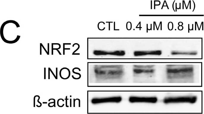


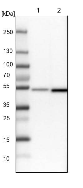


 NL001) and counterstained with DAPI (blue). Specific staining was localized to cytoplasm. View our protocol for
  CTS008) and counterstained with hematoxylin (blue). View our protocol for
  HAF109). This experiment was conducted under reducing conditions and using the 12-230 kDa separation system." title="Simple Western lane view shows lysates of RAW 264.7 mouse monocyte/macrophage cell line untreated (-) or treated (+) with 10 µg/mL LPS for 24 hours, loaded at 0.5 mg/mL. A specific band was detected for IL-1 beta /IL-1F2 at approximately 40 kDa (as indicated) using 2.5 µg/mL of Goat Anti-Mouse IL-1 beta /IL-1F2 Antigen Affinity-purified Polyclonal Antibody (Catalog # AF-401-NA) followed by 1:50 dilution of HRP-conjugated Anti-Goat IgG Secondary Antibody (
 401-ML) stimulates proliferation in the the D10.G4.1 mouse helper T cell line in a dose-dependent manner (orange line) as measured by Resazurin (
 VC006). Before incubation with the primary antibody, tissue was subjected to heat-induced epitope retrieval using VisUCyte Antigen Retrieval Reagent-Basic (


![P. gingivalis and its OMVs differentially induce inflammasome signaling and pyroptosis in murine macrophages. BMM were infected as before (2 h at MOI of 25:1, see Materials and Methods) with viable P. gingivalis (Pg), heat-killed-Pg (HK-Pg), OMVs, or heat-inactivated-OMVs (HI-OMVs) and (A) the activation of inflammasome components in the lysates [or supernatants (sup) where indicated] measured after 24 h by Western blot; beta -actin serves as a loading control throughout. Western blot data are representative of at least three independent experiments. (B) Production of IL-1 beta and IL-18 (by ELISA) from BMM was measured in the supernatant at 24 h. Data are mean ± SD from four independent experiments. Cell viability was determined by measuring extracellular LDH release and by 7-AAD exclusion (by flow-cytometry) at 24 h. 7-AAD is a membrane impermeant dye that is excluded from viable cells. The percent of 7-AAD positive (±SD) cells from three independent experiments was calculated relative to the untreated control. BMM treated with H2O2 (1 mM for 60 min) were included as positive control for 7-AAD. A representative histogram is shown. Cells were also treated where indicated with OMVs in the presence of KYT-1 and KYT-36 (10 μM of each). *p < 0.05. Image collected and cropped by CiteAb from the following publication (//pubmed.ncbi.nlm.nih.gov/28824884), licensed under a CC-BY license. Not internally tested by R&D Systems.](http://images.novusbio.com/fullsize/af-401-na_mouse-il-1-beta-il-1f2-affinity-purified-polyclonal-ab-4120241033831.jpg "P. gingivalis and its OMVs differentially induce inflammasome signaling and pyroptosis in murine macrophages. BMM were infected as before (2 h at MOI of 25:1, see Materials and Methods) with viable P. gingivalis (Pg), heat-killed-Pg (HK-Pg), OMVs, or heat-inactivated-OMVs (HI-OMVs) and (A) the activation of inflammasome components in the lysates [or supernatants (sup) where indicated] measured after 24 h by Western blot; beta -actin serves as a loading control throughout. Western blot data are representative of at least three independent experiments. (B) Production of IL-1 beta and IL-18 (by ELISA) from BMM was measured in the supernatant at 24 h. Data are mean ± SD from four independent experiments. Cell viability was determined by measuring extracellular LDH release and by 7-AAD exclusion (by flow-cytometry) at 24 h. 7-AAD is a membrane impermeant dye that is excluded from viable cells. The percent of 7-AAD positive (±SD) cells from three independent experiments was calculated relative to the untreated control. BMM treated with H2O2 (1 mM for 60 min) were included as positive control for 7-AAD. A representative histogram is shown. Cells were also treated where indicated with OMVs in the presence of KYT-1 and KYT-36 (10 μM of each). *p < 0.05. Image collected and cropped by CiteAb from the following publication (//pubmed.ncbi.nlm.nih.gov/28824884), licensed under a CC-BY license. Not internally tested by R&D Systems.")


 401-ML) was serially diluted and captured by Rat Anti-Mouse IL‑1 beta /IL‑1F2 Monoclonal Antibody (Catalog #
  401-ML) was serially diluted and captured by Rat Anti-Mouse IL‑1 beta /IL‑1F2 Monoclonal Antibody (Catalog #
  401-ML) was serially diluted and captured by Rat Anti-Mouse IL‑1 beta /IL‑1F2 Monoclonal Antibody (Catalog #
  401-ML) was serially diluted and captured by Rat Anti-Mouse IL‑1 beta /IL‑1F2 Monoclonal Antibody (Catalog #
  401-ML) was serially diluted and captured by Rat Anti-Mouse IL‑1 beta /IL‑1F2 Monoclonal Antibody (Catalog #
 ![SDS-Page TNF-alpha [Unconjugated]](https://images.novusbio.com/images/protein/TNF-alpha_210-TA_256.jpg)
![Bioactivity TNF-alpha [Unconjugated]](https://images.novusbio.com/images/protein/TNFalpha_210TA_1658.jpg)
![SEC-MALS TNF-alpha [Unconjugated]](https://images.novusbio.com/images/210-ta_recombinant-human-tnf-alpha-protein-sec-mals-35202312244..jpg)
![N/A IL-6 [HRP]](https://images.novusbio.com/images/elisa/DATA_IL6_M6000_ELISA_936.jpg)
![N/A IL-6 [HRP]](https://images.novusbio.com/images/elisa/IL-6_M6000_ELISA_415.jpg)
![N/A IL-6 [HRP]](https://images.novusbio.com/images/m6000b_mouse-il-6-quantikine-elisa-kit-1752025024034.jpg)
![Bioactivity IL-2 [Unconjugated]](https://images.novusbio.com/images/202-il_recombinant-human-il-2-protein-bioactivity-174202314946.jpg)
![N/A IL-1ra/IL-1F3/IL1RN [HRP]](https://images.novusbio.com/images/elisa/DATA_IL1ra_DRA00_ELISA_802.jpg)
![N/A IL-1ra/IL-1F3/IL1RN [HRP]](https://images.novusbio.com/images/elisa/DATA_IL1ra_DRA00_ELISA_803.jpg)
![N/A IL-1ra/IL-1F3/IL1RN [HRP]](https://images.novusbio.com/images/elisa/IL-1ra_DRA00_ELISA_392.jpg)
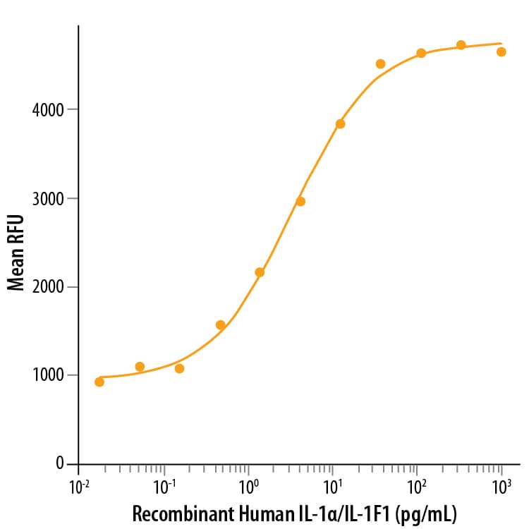
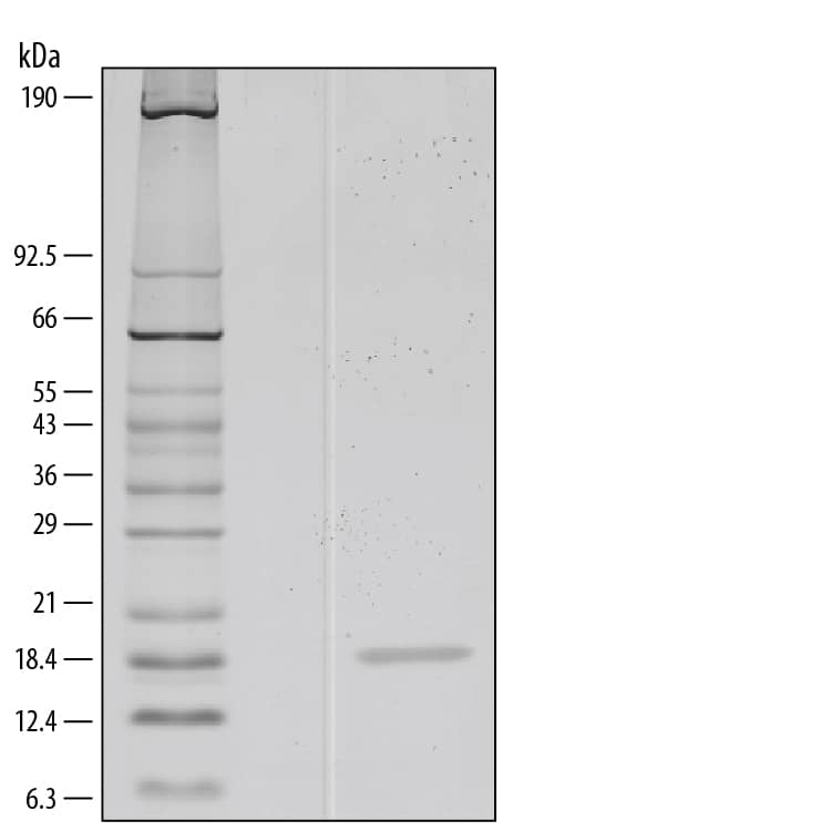
![N/A IL-10 [Biotin]](https://images.novusbio.com/images/elisa/DATA_IL10_DY417_ELISA_2014.jpg)
![SEC-MALS IFN-gamma [Unconjugated]](https://images.novusbio.com/images/485-mi_recombinant-mouse-ifn-gamma-protein-sec-mals-1612202583245.jpg)
![Bioactivity IFN-gamma [Unconjugated]](https://images.novusbio.com/images/protein/IFN-gamma_485-MI_455.jpg)
![SDS-Page IFN-gamma [Unconjugated]](https://images.novusbio.com/images/protein/IFN-gamma_485-MI_407.jpg)
![Bioactivity IL-4 [Unconjugated]](https://images.novusbio.com/images/protein/6507-ilcf_recombinant-human-il-4-cho-expressed-protein-cf-bioactivity-272020133214.jpg)
![Bioactivity GM-CSF [Unconjugated]](https://images.novusbio.com/images/protein/7954-gmcf_recombinant-human-gm-csf-cho-expressed-protein-cf-bioactivity-272020121514.jpg)
![SEC-MALS IL-3 [Unconjugated]](https://images.novusbio.com/images/203-il_recombinant-human-il-3-protein-sec-mals-133202482656..jpg)
![Bioactivity IL-3 [Unconjugated]](https://images.novusbio.com/images/protein/IL-3_203-IL_347.jpg)
![Data IL-3 [Unconjugated]](https://images.novusbio.com/images/203-il_recombinant-human-il-3-protein-data-253202610550.jpg)
![N/A CCL2/JE/MCP-1 [HRP]](https://images.novusbio.com/images/elisa/CCL2_DCP00_ELISA_67.jpg)
![N/A CCL2/JE/MCP-1 [HRP]](https://images.novusbio.com/images/elisa/DATA_CCL2_DCP00_ELISA_633.jpg)
![N/A CCL2/JE/MCP-1 [HRP]](https://images.novusbio.com/images/elisa/DATA_CCL2_DCP00_ELISA_634.jpg)
![Western Blot ICAM-1/CD54 Antibody [Unconjugated]](https://images.novusbio.com/images/af796_mouse-icam-1-cd54-affinity-purified-polyclonal-ab-41202410481192.jpg)
![Western Blot ICAM-1/CD54 Antibody [Unconjugated]](https://images.novusbio.com/images/af796_mouse-icam-1-cd54-affinity-purified-polyclonal-ab-4120241048112.jpg)
![Western Blot ICAM-1/CD54 Antibody [Unconjugated]](https://images.novusbio.com/images/af796_mouse-icam-1-cd54-affinity-purified-polyclonal-ab-4120241049436.jpg)