 A431 cell line. PVDF membrane was probed with 1 µg/mL of Goat Anti-Human EphA2 Antigen Affinity-purified Polyclonal Antibody (Catalog # AF3035) followed by HRP-conjugated Anti-Goat IgG Secondary Antibody (HAF017). A specific band was detected for EphA2 at approximately 110 kDa (as indicated), but not detectable in the knockout A431 cell line. GAPDH (Catalog # MAB5718) is shown as a loading control. This experiment was conducted under reducing conditions and using Immunoblot Buffer Group 1.")
| Reactivity | HuSpecies Glossary |
| Applications | WB, Flow, IHC, CyTOF-ready, KO |
| Clonality | Polyclonal |
| Host | Goat |
| Conjugate | Unconjugated |
| Concentration | LYOPH |
| Immunogen | Mouse myeloma cell line NS0-derived recombinant human EphA2 Gln25-Asn534 Accession # P29317 |
| Specificity | Detects human EphA2 in direct ELISAs and Western blots. In direct ELISAs, less than 45% cross-reactivity with recombinant mouse EphA2 is observed and less than 1% cross-reactivity with recombinant human (rh) EphA1, rhEphA3, rhEphA4, rhEphA5, rhEphA6, rhEphA7 and rhEphA10 is observed. |
| Source | N/A |
| Isotype | IgG |
| Clonality | Polyclonal |
| Host | Goat |
| Gene | EPHA2 |
| Purity Statement | Antigen Affinity-purified |
| Innovator's Reward | Test in a species/application not listed above to receive a full credit towards a future purchase. |
| Dilutions |
|
|
| Publications |
|
| Storage | Use a manual defrost freezer and avoid repeated freeze-thaw cycles.
|
| Buffer | Lyophilized from a 0.2 μm filtered solution in PBS with Trehalose. *Small pack size (SP) is supplied either lyophilized or as a 0.2 µm filtered solution in PBS. |
| Preservative | No Preservative |
| Concentration | LYOPH |
| Reconstitution Instructions | Reconstitute at 0.2 mg/mL in sterile PBS. |
EphA2, also known as Eck, Myk2, and Sek2, is a member of the Eph receptor tyrosine kinase family which binds Ephrins A1, 2, 3, 4, and 5 (1-4). A and B class Eph proteins have a common structural organization. The human EphA2 cDNA encodes a 976 amino acid (aa) precursor including a 24 aa signal sequence, a 510 aa extracellular domain (ECD), a 24 aa transmembrane segment, and a 418 aa cytoplasmic domain. The ECD contains an N-terminal globular domain, a cysteine-rich domain, and two fibronectin type III domains (5). The cytoplasmic domain contains a juxtamembrane motif with two tyrosine residues, which are the major autophosphorylation sites, a kinase domain, and a sterile alpha motif (SAM) (5). The ECD of human EphA2 shares 90-94% aa sequence identity with mouse, bovine, and canine EphA2, and approximately 45% aa sequence identity with human EphA1, 3, 4, 5, 7, and 8. EphA2 becomes autophosphorylated following ligand binding (6, 7) and then interacts with SH2 domain-containing PI3-kinase to activate MAPK pathways (8, 9). Reverse signaling is also propagated through the Ephrin ligand. Transcription of EphA2 is dependent on the expression of E-Cadherin (10), and can be induced by p53 family transcription factors (11). EphA2 is upregulated in breast, prostate, and colon cancer vascular endothelium. Its ligand, EphrinA1, is expressed by the local tumor cells (12, 13). In some cases, EphA2 and EphrinA1 are expressed on the same blood vessels (14). EphA2 signaling cooperates with VEGF receptor signaling in promoting endothelial cell migration (13). The gene encoding human EphA2 maps to a region on chromosome 1 which is frequently deleted in neuroectodermal tumors (15).


![Western Blot FAK [p Tyr397] Antibody](https://images.novusbio.com/images/nbp3-12897_rabbit-fak-p-tyr-397-pab-western-blot-2611202518381522.jpg)
![Western Blot FAK [p Tyr397] Antibody](https://images.novusbio.com/images/FAK-[p-Tyr397]-Antibody-Western-Blot-NBP3-12897-img0006.jpg)
![Western Blot FAK [p Tyr397] Antibody](https://images.novusbio.com/images/nbp3-12897_rabbit-fak-p-tyr-397-pab-western-blot-261120251843458.jpg)
![Immunocytochemistry EGFR Antibody [Unconjugated]](https://images.novusbio.com/images/antibody/EGF_R_AF231_Immunocytochemistry__Immunofluorescence_21143.jpg)
![Flow Cytometry EGFR Antibody [Unconjugated]](https://images.novusbio.com/images/antibody/EGF_R_AF231_Flow_Cytometry_20401.jpg)
![Western Blot EGFR Antibody [Unconjugated]](https://images.novusbio.com/images/antibody/EGF_R_AF231_Western_Blot_19925.jpg)

![Western Blot EphB2 Antibody [Unconjugated]](https://images.novusbio.com/images/af467_human-mouse-ephb2-affinity-purified-polyclonal-ab-41202410485412.jpg)
![Immunohistochemistry EphB2 Antibody [Unconjugated]](https://images.novusbio.com/images/antibody/EphB2_AF467_Immunohistochemistry_20369.jpg)
![Western Blot EphB2 Antibody [Unconjugated]](https://images.novusbio.com/images/antibody/EphB2_AF467_Western_Blot_21511.jpg)
![Intracellular Staining by Flow Cytometry AKT [p Ser473] Antibody [Unconjugated] - Pan Specific](https://images.novusbio.com/images/antibody/Akt3_AF887_Flow_Cytometry_8283.jpg)
![Western Blot AKT [p Ser473] Antibody [Unconjugated] - Pan Specific](https://images.novusbio.com/images/af887_phospho-akt-s473-pan-specific-affinity-purified-pab-41202410485440.jpg)
![Western Blot AKT [p Ser473] Antibody [Unconjugated] - Pan Specific](https://images.novusbio.com/images/af887_phospho-akt-s473-pan-specific-affinity-purified-pab-8120255552843.jpg)


Secondary Antibodies |
Isotype Controls |
|
Repurposing FDA-approved drugs to combat the rise of antibiotic resistance By Beth Melson, MSAntibiotic resistance is a global threat to public health. Widespread, inappropriate use of antibiotics, such as to treat viral infections or promote growth in livestock, has led to increased incid... Read full blog post. |
The concentration calculator allows you to quickly calculate the volume, mass or concentration of your vial. Simply enter your mass, volume, or concentration values for your reagent and the calculator will determine the rest.


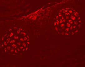
 or isotype control antibody (Catalog # AB-108-C, open histogram), followed by Phycoerythrin-conjugated Anti-Goat IgG Secondary Antibody (Catalog # F0107). View our protocol for Staining Membrane-associated Proteins.")
 at 5 µg/mL overnight at 4 °C. Tissue was stained using the Anti-Goat HRP-DAB Cell & Tissue Staining Kit (brown; Catalog # CTS008) and counterstained with hematoxylin (blue). Specific labeling was localized to the plasma membrane of cancer cells. View our protocol for Chromogenic IHC Staining of Paraffin-embedded Tissue Sections.")
 Heat map for ephrin ligands and Eph receptor mRNAs in selected HGSC cell lines. Data obtained from CCLE and illustrated per cell line. (B, C) Quantification of EFNA1 (B) and EFNA5 (C) mRNAs in OVCAR3 and OVCAR4 with silenced EFNA1 or EFNA5 compared to the controls. N = 3. siScr is set to one. (D–G) EphA2 (total and phosphorylated) in OVCAR3 and OVCAR4 after silencing of EFNA1 or EFNA5 (D) along with EphA2-pY588 (E), EphA2-pS897 (F), and EphA2 (G) quantifications. N = 3. siScr is set to one. Full-length blots are presented in Supplementary Fig. 5. p values (Student’s t-test): *< 0.05; **< 0.01. Image collected and cropped by CiteAb from the following open publication (//pubmed.ncbi.nlm.nih.gov/33893375), licensed under a CC-BY license. Not internally tested by R&D Systems.")
![Immunocytochemistry EphA1 Antibody [Unconjugated]](https://images.novusbio.com/images/antibody/EphA1_AF638_Immunocytochemistry__Immunofluorescence_20119.jpg)
![Western Blot EphA1 Antibody [Unconjugated]](https://images.novusbio.com/images/antibody/EphA1_AF638_Western_Blot_17606.jpg)
![Western Blot Src [p Tyr419] Antibody [Unconjugated]](https://images.novusbio.com/images/antibody/Src_AF2685_Western_Blot_5209.jpg)
![Immunohistochemistry Src [p Tyr419] Antibody [Unconjugated]](https://images.novusbio.com/images/af2685_human-phospho-src-y419-affinity-purified-polyclonal-ab-412024124022.jpg)
![Immunohistochemistry Src [p Tyr419] Antibody [Unconjugated]](https://images.novusbio.com/images/antibody/Src_AF2685_Immunohistochemistry_9943.jpg)
![N/A VEGF [HRP]](https://images.novusbio.com/images/elisa/VEGF_DVE00_ELISA_208.jpg)
![N/A VEGF [HRP]](https://images.novusbio.com/images/elisa/DATA_VEGF_DVE00_ELISA_871.jpg)
![N/A VEGF [HRP]](https://images.novusbio.com/images/elisa/DATA_VEGF_DVE00_ELISA_872.jpg)


![Western Blot ERK2 Antibody [Unconjugated]](https://images.novusbio.com/images/antibody/ERK2_AF1230_Western_Blot_5097.jpg)
![Knockout Validated ERK2 Antibody [Unconjugated]](https://images.novusbio.com/images/antibody/ERK2_AF1230_Knockout_Validated_22864.jpg)
![Immunohistochemistry ERK2 Antibody [Unconjugated]](https://images.novusbio.com/images/antibody/ERK2_AF1230_Immunohistochemistry_20696.jpg)
![Bioactivity ErbB2/Her2 [Unconjugated]](https://images.novusbio.com/images/protein/1129-er_recombinant-human-erbb2-her2-fc-chimera-protein-cf-bioactivity-7122020142841.jpg)
![SDS-Page ErbB2/Her2 [Unconjugated]](https://images.novusbio.com/images/protein/ErbB2_1129-ER_70.jpg)


![Simple Western Ephrin-B1 Antibody [Unconjugated]](https://images.novusbio.com/images/antibody/EphrinB1_AF473_Simple_Western_18962.jpg)
![Western Blot Ephrin-B1 Antibody [Unconjugated]](https://images.novusbio.com/images/antibody/EphrinB1_AF473_Western_Blot_17536.jpg)
![Immunohistochemistry Ephrin-B1 Antibody [Unconjugated]](https://images.novusbio.com/images/antibody/Ephrin-B1_AF473_Immunohistochemistry_7180.jpg)


 or Normal Goat IgG Isotype Control Antibody (Catalog # AB-108-C, open histogram), followed by Phycoerythrin-conjugated Anti-Goat IgG Secondary Antibody (Catalog # F0107).")