![Immunocytochemistry/Immunofluorescence: Aurora A Antibody [NBP1-51843] - HeLa cells were fixed and permeabilized for 10 minutes using -20C MeOH. The cells were incubated with anti- (NBP1-51843) at 2 ug/ml overnight at 4C and detected with an anti-rabbit Dylight 488 (Green) at a 1:1000 dilution for 60 minutes. Alpha tubulin (DM1A) NB100-690 was used as a co-stain at a 1:1000 dilution overnight at 4C and detected with an anti-mouse Dylight 550 (Red) at a 1:1000 dilution for 60 minutes. Nuclei were counterstained with DAPI (Blue). Cells were imaged using a 100X objective and digitally deconvolved.](http://images.novusbio.com/fullsize/Aurora-A-Antibody-Immunocytochemistry-Immunofluorescence-NBP1-51843-img0012.jpg "Immunocytochemistry/Immunofluorescence: Aurora A Antibody [NBP1-51843] - HeLa cells were fixed and permeabilized for 10 minutes using -20C MeOH. The cells were incubated with anti- (NBP1-51843) at 2 ug/ml overnight at 4C and detected with an anti-rabbit Dylight 488 (Green) at a 1:1000 dilution for 60 minutes. Alpha tubulin (DM1A) NB100-690 was used as a co-stain at a 1:1000 dilution overnight at 4C and detected with an anti-mouse Dylight 550 (Red) at a 1:1000 dilution for 60 minutes. Nuclei were counterstained with DAPI (Blue). Cells were imaged using a 100X objective and digitally deconvolved.")
| Reactivity | Hu, MuSpecies Glossary |
| Applications | WB, Simple Western, ICC/IF, IHC |
| Clonality | Polyclonal |
| Host | Rabbit |
| Conjugate | Unconjugated |
| Format | BSA Free |
| Concentration | 1.0 mg/ml |
| Immunogen | A recombinant protein made to an N-terminal region of the human Aurora A protein (within residues 50-200). [Swiss-Prot O14965] |
| Localization | Centrosome, Spindle pole. Note: Detected at the neurite hillock in developing neurons. Localizes on centrosomes in interphase cells and at each spindle pole in mitosis. |
| Marker | Mitosis Marker |
| Isotype | IgG |
| Clonality | Polyclonal |
| Host | Rabbit |
| Gene | AURKA |
| Purity | Immunogen affinity purified |
| Innovator's Reward | Test in a species/application not listed above to receive a full credit towards a future purchase. |
| Dilutions |
|
||
| Application Notes | This AURKA antibody is useful for Immunohistochemistry and Western blot, where a band is seen ~45 kDa. Prior to immunostaining paraffin tissues, antigen retrieval with sodium citrate buffer (pH 6.0) is recommended. In Simple Western only 10 - 15 uL of the recommended dilution is used per data point. See Simple Western Antibody Database for Simple Western validation: Tested in HeLa lysate 0.5 mg/mL, separated by Size, antibody dilution of 1:200, apparent MW was 56 kDa. Separated by Size-Wes, Sally Sue/Peggy Sue. |
||
| Control |
|
||
| Reviewed Applications |
|
||
| Publications |
|
| Storage | Store at 4C short term. Aliquot and store at -20C long term. Avoid freeze-thaw cycles. |
| Buffer | PBS |
| Preservative | 0.05% Sodium Azide |
| Concentration | 1.0 mg/ml |
| Purity | Immunogen affinity purified |


![Immunohistochemistry EN-RAGE/S100A12 Antibody [Unconjugated]](https://images.novusbio.com/images/antibody/EN-RAGE_AF1052_Immunohistochemistry_6671.jpg)
![Simple Western EN-RAGE/S100A12 Antibody [Unconjugated]](https://images.novusbio.com/images/antibody/af1052_human-en-rage-affinity-purified-polyclonal-ab-simple-western-2672021152820.jpg)
![Flow Cytometry EN-RAGE/S100A12 Antibody [Unconjugated]](https://images.novusbio.com/images/antibody/af1052_human-en-rage-affinity-purified-polyclonal-ab-flow-cytometry-572024104928.jpg)


| Images | Ratings | Applications | Species | Date | Details | ||||
|---|---|---|---|---|---|---|---|---|---|
-(01-ml)_NBP1-51843_8651.bmp)
Enlarge |
reviewed by:
Bryan Tinsley |
ICC | Human | 07/01/2014 |
Summary
|
Secondary Antibodies |
Isotype Controls |
Research Areas for Aurora A Antibody (NBP1-51843)Find related products by research area.
|
The concentration calculator allows you to quickly calculate the volume, mass or concentration of your vial. Simply enter your mass, volume, or concentration values for your reagent and the calculator will determine the rest.


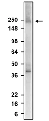
![Simple Western: Aurora A Antibody [NBP1-51843] - Simple Western lane view shows a specific band for Aurora A in 0.5 mg/ml of HeLa lysate. This experiment was performed under reducing conditions using the 12-230kDa separation system.](http://images.novusbio.com/fullsize/Aurora-A-Antibody-Simple-Western-NBP1-51843-img0010.jpg "Simple Western: Aurora A Antibody [NBP1-51843] - Simple Western lane view shows a specific band for Aurora A in 0.5 mg/ml of HeLa lysate. This experiment was performed under reducing conditions using the 12-230kDa separation system.")
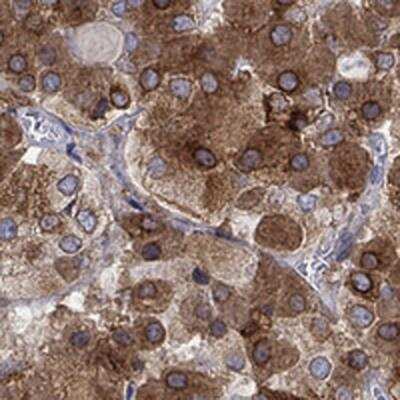
![Immunocytochemistry/Immunofluorescence: Aurora A Antibody [NBP1-51843] - IF Confocal analysis of HeLa cells using Aurora A antibody (NBP1-51843, 1:10). An Alexa Fluor 488-conjugated Goat to rabbit IgG was used as secondary antibody (green). Actin filaments were labeled with Alexa Fluor 568 phalloidin (red). DAPI was used to stain the cell nuclei (blue).](http://images.novusbio.com/fullsize/Aurora-A-Antibody-Immunocytochemistry-Immunofluorescence-NBP1-51843-img0008.jpg "Immunocytochemistry/Immunofluorescence: Aurora A Antibody [NBP1-51843] - IF Confocal analysis of HeLa cells using Aurora A antibody (NBP1-51843, 1:10). An Alexa Fluor 488-conjugated Goat to rabbit IgG was used as secondary antibody (green). Actin filaments were labeled with Alexa Fluor 568 phalloidin (red). DAPI was used to stain the cell nuclei (blue).")
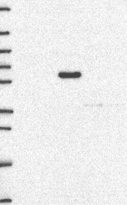
![Western Blot: Aurora A Antibody [NBP1-51843] - WB detection of Aurora A in HeLa whole cell lysate.](http://images.novusbio.com/fullsize/Aurora-A-Antibody-Western-Blot-NBP1-51843-img0011.jpg "Western Blot: Aurora A Antibody [NBP1-51843] - WB detection of Aurora A in HeLa whole cell lysate.")
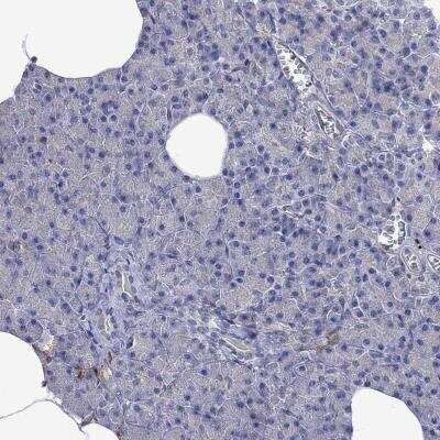
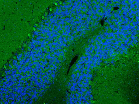
![Immunohistochemistry: Aurora A Antibody [NBP1-51843] - IHC staining of Aurora A in mouse brain.](http://images.novusbio.com/fullsize/Aurora-A-Antibody-Immunohistochemistry-NBP1-51843-img0003.jpg "Immunohistochemistry: Aurora A Antibody [NBP1-51843] - IHC staining of Aurora A in mouse brain.")
![Simple Western: Aurora A Antibody - BSA Free [NBP1-51843] - Analysis of protein expression post-drug treatment. (A) CHLA-10 and (B) TC-71 cells treated with drugs were assessed for changes in protein expression 24 h post-treatment via capillary electrophoresis-based Wes analysis. Increased protein levels of KIF11, p-KIF11Thr926 AURKA, and p-AURKAThr288 were observed for the drug combination group, whereas KIF15 levels were noticeably lower. Similarly, enhanced cleaved-PARP expression was observed with the combination treatment. The uncropped blots are shown in Figures S8 and S9. Image collected and cropped by CiteAb from the following publication (//pubmed.ncbi.nlm.nih.gov/37894278 ), licensed under a CC-BY license. Not internally tested by Novus Biologicals.](http://images.novusbio.com/fullsize/nbp1-51843_rabbit-polyclonal-aurora-a-antibody-308202410445515.jpg "Simple Western: Aurora A Antibody - BSA Free [NBP1-51843] - Analysis of protein expression post-drug treatment. (A) CHLA-10 and (B) TC-71 cells treated with drugs were assessed for changes in protein expression 24 h post-treatment via capillary electrophoresis-based Wes analysis. Increased protein levels of KIF11, p-KIF11Thr926 AURKA, and p-AURKAThr288 were observed for the drug combination group, whereas KIF15 levels were noticeably lower. Similarly, enhanced cleaved-PARP expression was observed with the combination treatment. The uncropped blots are shown in Figures S8 and S9. Image collected and cropped by CiteAb from the following publication (//pubmed.ncbi.nlm.nih.gov/37894278 ), licensed under a CC-BY license. Not internally tested by Novus Biologicals.")
 The DepMap portal was used to access the RNA expression data across different cancer cell lines. Expression in Ewing sarcoma is highlighted in red. Capillary-based analysis of protein lysates from EWS cell lines indicating expression of (B) KIF11 and AURKA and (C) KIF15 and TPX2 protein levels. The uncropped blots are shown in Figure S3. Image collected and cropped by CiteAb from the following open publication (//pubmed.ncbi.nlm.nih.gov/37894278), licensed under a CC-BY license. Not internally tested by Novus Biologicals.")
![SDS-Page TNF-alpha [Unconjugated]](https://images.novusbio.com/images/protein/TNF-alpha_210-TA_256.jpg)
![Bioactivity TNF-alpha [Unconjugated]](https://images.novusbio.com/images/protein/TNFalpha_210TA_1658.jpg)
![SEC-MALS TNF-alpha [Unconjugated]](https://images.novusbio.com/images/210-ta_recombinant-human-tnf-alpha-protein-sec-mals-35202312244..jpg)
![Immunocytochemistry Notch-4 Antibody [Unconjugated]](https://images.novusbio.com/images/antibody/af3847_human-notch-4-intracellular-domain-affinity-purified-pab-immunocytochemistry-3092021102949.jpg)


![Western Blot: Goat anti-Rabbit IgG (H+L) Secondary Antibody [HRP] [NB7160] - Western blot showing vemurafenib treatment in BRAFV600E CRC cells inhibits fission mediator DRP1 with no significant effect on fusion proteins (Mfn1 & 2) using MFN-1 antibody (NBP1-51841) and corresponding secondary antibody, goat anti-rabbit IgG-HRP (NB7160). Image collected and cropped by CiteAb from the following publication (https://pubmed.ncbi.nlm.nih.gov/33738242).](https://images.novusbio.com/images/Goat-anti-Rabbit-IgG-H+L-Secondary-Antibody-HRP-Western-Blot-NB7160-img0001.jpg "Western Blot: Goat anti-Rabbit IgG (H+L) Secondary Antibody [HRP] [NB7160] - Western blot showing vemurafenib treatment in BRAFV600E CRC cells inhibits fission mediator DRP1 with no significant effect on fusion proteins (Mfn1 & 2) using MFN-1 antibody (NBP1-51841) and corresponding secondary antibody, goat anti-rabbit IgG-HRP (NB7160). Image collected and cropped by CiteAb from the following publication (https://pubmed.ncbi.nlm.nih.gov/33738242).")
 followed by 30 min incubation with Goat anti Rabbit HRP conjugated secondary antibodies (Catalog # HAF008) at 1:20 dilution + DAB chromogen (brown). The tissue was counterstained with Hematoxylin (blue). Control was done by omitting primary antibody.")
![Flow Cytometry: Rabbit IgG Isotype Control [NBP2-24891] - An intracellular stain was performed on Raji cells with Adiponectin antibody NB100-65810 (blue) and a matched isotype control NBP2-24893 (orange). Cells were fixed with 4% PFA and then permeablized with 0.1% saponin. Cells were incubated in an antibody dilution of 1 ug/mL for 30 minutes at room temperature, followed by Dylight488-conjugated anti-rabbit secondary antibody. Image using the Azide Free form of this antibody.](https://images.novusbio.com/images/Rabbit--Mouse-IgG-Isotype-Control-Flow-Cytometry-NBP2-24891-img0006.jpg "Flow Cytometry: Rabbit IgG Isotype Control [NBP2-24891] - An intracellular stain was performed on Raji cells with Adiponectin antibody NB100-65810 (blue) and a matched isotype control NBP2-24893 (orange). Cells were fixed with 4% PFA and then permeablized with 0.1% saponin. Cells were incubated in an antibody dilution of 1 ug/mL for 30 minutes at room temperature, followed by Dylight488-conjugated anti-rabbit secondary antibody. Image using the Azide Free form of this antibody.")
-(01-ml)_NBP1-51843_8651.bmp)