HAF018). A specific band was detected for a Tubulin at approximately 52 kDa (as indicated). This experiment was conducted under reducing conditions and using Immunoblot Buffer Group 1." title="Western blot shows lysates of HeLa human cervical epithelial carcinoma cell line, Jurkat human acute T cell leukemia cell line, NIH-3T3 mouse embryonic fibroblast cell line, Neuro-2A mouse neuroblastoma cell line, and Rat-2 rat embryonic fibroblast cell line. PVDF membrane was probed with 0.05 µg/mL of Mouse Anti-Human/Mouse/Rat a Tubulin Monoclonal Antibody (Catalog # MAB9344) followed by HRP-conjugated Anti-Mouse IgG Secondary Antibody (Catalog # HAF018). A specific band was detected for a Tubulin at approximately 52 kDa (as indicated). This experiment was conducted under reducing conditions and using Immunoblot Buffer Group 1." />
| Reactivity | Hu, Mu, RtSpecies Glossary |
| Applications | WB, IHC, ICC/IF |
| Clone | 961216 |
| Clonality | Monoclonal |
| Host | Mouse |
| Conjugate | Unconjugated |
| Immunogen | Human TUBA1A synthetic peptide Accession # Q71U36 |
| Specificity | Detects human alpha Tubulin in direct ELISAs and Western blots. |
| Source | N/A |
| Isotype | IgG2a |
| Clonality | Monoclonal |
| Host | Mouse |
| Purity Statement | Protein A or G purified from hybridoma culture supernatant |
| Innovator's Reward | Test in a species/application not listed above to receive a full credit towards a future purchase. |
| Dilutions |
|
|
| Publications |
|
| Storage | Use a manual defrost freezer and avoid repeated freeze-thaw cycles.
|
| Buffer | Lyophilized from a 0.2 μm filtered solution in PBS with Trehalose. *Small pack size (SP) is supplied either lyophilized or as a 0.2 µm filtered solution in PBS. |
| Reconstitution Instructions | Reconstitute at 0.5 mg/mL in sterile PBS. |
Tubulin alpha-1A chain (TUBA1A), also known as Alpha-tubulin 3, is the principal alpha tubulin in morphologically differentiated neurons. Alpha tubulin dimerizes with beta Tubulin to form microtubules. Microtubules mediate transport of proteins and endosomes within cells, and TUBA1A has been shown to interact with metabotropic glutamate receptor 7 (receptor trafficking), synuclein alpha (synaptic plasticity), and N-syndecan (neurite outgrowth). This gene is mutated in malformations of cortical development, including Lissencephaly resulting in microcephaly, developmental delay and early-onset epileptic seizures.


![Immunohistochemistry beta-III Tubulin Antibody (TuJ-1) [Unconjugated] - Neuron-Specific](https://images.novusbio.com/images/antibody/mab1195_neuron-specific-beta-iii-tubulin-mab-clone-tuj-1-immunohistochemistry-31520231381.jpg)
![Immunocytochemistry beta-III Tubulin Antibody (TuJ-1) [Unconjugated] - Neuron-Specific](https://images.novusbio.com/images/antibody/mab1195_neuron-specific-beta-iii-tubulin-mab-clone-tuj-1-immunocytochemistry-315202313914.jpg)
![Immunocytochemistry/ Immunofluorescence beta-III Tubulin Antibody (TuJ-1) [Unconjugated] - Neuron-Specific](https://images.novusbio.com/images/mab1195_neuron-specific-beta-iii-tubulin-mab-clone-tuj-1-412024103381.jpg)







Secondary Antibodies |
Isotype Controls |
|
Autophagy Research Update: What a difference a year makes! By Christina Towers, PhD Over the last two decades the field of autophagy has exploded! Innovative techniques, comprehensive analysis and disease-relevant models have yielded basic and clinical discoveries of conseque... Read full blog post. |
|
Read full blog post. |
|
Read full blog post. |
|
How to visualize autophagy by microscopy By Christina Towers, PhD Autophagy is a recycling process that relies on the formation of a unique organelle termed an autophagosome. An elegant way to monitor autophagy is through various microscopy techniques to... Read full blog post. |
|
Tips on choosing an ideal loading control antibody for Western Blotting Western blotting is one of the most commonly used antibody assay techniques in cell and molecular biology research since its development over three decades ago, and is considered the gold standard for protein detection and quantification. When p... Read full blog post. |
|
Alpha Tubulin: A Fundamental Cytoskeleton Protein with Many Roles The cytoskeleton consists of three major types of cytosolic fibers: microtubules, microfilaments (actin filaments), and intermediate filaments. Tubulins are the microtubule building block and exist as globular dimeric proteins of alpha/beta chains. Th... Read full blog post. |
|
Time to Start Actin Like a Reliable 'Housekeeper'! A growing body of data and studies using actin antibodies supports a view of the actin cytoskeleton of smooth muscle cells as a dynamic structure that plays an integral role in regulating the development of mechanical tension and the material properti... Read full blog post. |
The concentration calculator allows you to quickly calculate the volume, mass or concentration of your vial. Simply enter your mass, volume, or concentration values for your reagent and the calculator will determine the rest.
| Uniprot |
|


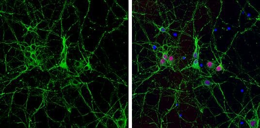
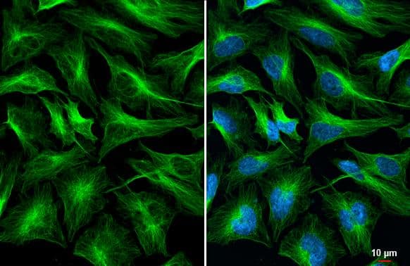
 NL007) and counterstained with DAPI (blue). Specific staining was localized to cytoplasm. View our protocol for
  VC001). Tissue was stained using DAB (brown) and counterstained with hematoxylin (blue). Specific staining was localized to neuronal cytoplasm. View our protocol for


![Immunohistochemistry Reelin Antibody [Unconjugated]](https://images.novusbio.com/images/antibody/Reelin_AF3820_Immunohistochemistry_18505.jpg)
![N/A PLA2G7/PAF-AH/Lp-PLA2 [HRP]](https://images.novusbio.com/images/elisa/PLA2G7_DPLG70_ELISA_161.jpg)
![N/A PLA2G7/PAF-AH/Lp-PLA2 [HRP]](https://images.novusbio.com/images/elisa/DATA_PLA2G7_DPLG70_ELISA_794.jpg)