After August 17, 2026, Novus Biologicals products and services will no longer be available on this website; you will access all products and services on rndsystems.com. Create your R&D Systems online account today.
Recombinant Mouse S100A8/S100A9 Heterodimer Protein (Catalog # 8916-S8) has a molecular weight (MW) of 22-24 kDa as analyzed by SEC-MALS, suggesting that this protein is a heterodimer.
Recombinant Mouse S100A8/S100A9 (Catalog # 8916-S8) induces IL-6 secretion by A375 human melanoma cells. The ED50 for this effect is 1.5-6 μg/mL.
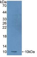
1 μg/lane of Recombinant Mouse S100A8/S100A9 was resolved with SDS-PAGE under reducing (R) and non-reducing (NR) conditions and visualized by silver staining, showing R bands at 6.3, 10.8 kDa and NR band at 6, 9.6 kDa.
Measured by its ability to induce IL-6 secretion by A375 human melanoma cells. Hibino, T. et al. (2013) Cancer Res. 73:172. The ED50 for this effect is 1.5-6 μg/mL.
Source
E. coli-derived mouse S100A8/S100A9 Heterodimer protein
Mouse S100A8 (Pro2-Glu89) Accession # P27005
Mouse S100A9 (Ala2-Lys113) Accession # P31725
N-terminus
C-terminus
N-terminal Sequence
Pro2 (S100A8), Ala2 (S100A9)
Structure / Form
Non-covalent heterodimer
Protein/Peptide Type
Recombinant Proteins
Purity
>95%, by SDS-PAGE visualized with Silver Staining and quantitative densitometry by Coomassie® Blue Staining
Endotoxin Note
<0.10 EU per 1 μg of the protein by the LAL method.
Applications/Dilutions
Dilutions
Bioactivity
Theoretical MW
10 kDa (S100A8), 13 kDa (S100A9). Disclaimer note: The observed molecular weight of the protein may vary from the listed predicted molecular weight due to post translational modifications, post translation cleavages, relative charges, and other experimental factors.
SDS-PAGE
7 kDa and 12 kDa, reducing conditions
Publications
Read Publications using 8916-S8 in the following applications:
Use a manual defrost freezer and avoid repeated freeze-thaw cycles.
12 months from date of receipt, -20 to -70 °C as supplied.
1 month, 2 to 8 °C under sterile conditions after reconstitution.
3 months, -20 to -70 °C under sterile conditions after reconstitution.
Buffer
Lyophilized from a 0.2 μm filtered solution in PBS and DTT.
Purity
>95%, by SDS-PAGE visualized with Silver Staining and quantitative densitometry by Coomassie® Blue Staining
Reconstitution Instructions
Reconstitute at 500 μg/mL in PBS.
Notes
This product is produced by and ships from R&D Systems, Inc., a Bio-Techne brand.
Alternate Names for Recombinant Mouse S100A8/S100A9 Heterodimer Protein, CF
60B8AG
CAGA
calgranulin A
Calprotectin L1L subunit
Calprotectin
CFAG
CGLA
CP-10
Cystic fibrosis antigen
L1AG
Leukocyte L1 complex light chain
MA387
MIF
Migration inhibitory factor-related protein 8
MRP8
NIF
p8
protein S100-A8
S100-A8
S100A8/S100A9 Heterodimer
Urinary stone protein band A
Background
S100A8 (also known as MRP8, Calgranulin A, and CP-10) and S100A9 (also known as MRP14 and Calgranulin B) are pro-inflammatory members of the S100 family of secreted calcium binding proteins (1, 2). The 10 kDa S100A8 and 14 kDa S100A9 each contain two EF-hand calcium binding motifs. Mouse S100A8 shares 57% and 80% amino acid (aa) sequence identity with human and rat S100A8, respectively. Mouse S100A9 shares 57% and 79% amino acid sequence identity with human and rat S100A9, respectively (3, 4). S100A8 and S100A9 can associate into non-covalent homodimers and 34-35 kDa heterodimers known as S100A8/9 or Calprotectin. The presence of calcium and zinc also induces the formation of the tetramer (5, 6). S100A8 and S100A9 are up-regulated in neutrophils, monocytes, Schwann cells, and keratinocytes at sites of inflammation (7-13). The S100A8/9 heterodimer is elevated in rheumatoid arthritis synovial fluid and in the serum of cardiovascular disease, atopic dermatitis, and psoriatic arthritis patients (1, 9, 13-15). It exerts its effects through the receptors RAGE and TLR4 (11, 16). The heterodimer promotes neutrophil infiltration into sites of inflammation and inflammatory cytokine production by monocytes (5, 8, 9, 12). It promotes astrocyte proliferation (16) and the suppression of tumor growth by promoting the influx and activation of NK cells (10, 11). The S100A8/9 heterodimer additionally binds to unsaturated fatty acids such as arachidonic acid (7), and it sequesters manganese, thereby restricting the growth of Mn-dependent bacteria (17).
Averill, M.M. et al. (2012) Arterioscler. Thromb. Vasc. Biol. 32:223.
Kerkhoff, C. et al. (2012) Exp. Dermatol. 21:822.
Lagasse, E. and I.L. Weissman (1992) Blood 79:1907.
Lackmann, M. et al. (1993) J. Immunol. 150:2981.
Ryckman, C. et al. (2003) J. Immunol. 170:3233.
Vogl, T. et al. (2006) Biochim. Biophys. Acta 1763:1298.
Siegenthaler, G. et al. (1997) J. Biol. Chem. 272:9371.
Volz, H.C. et al. (2012) Basic Res. Cardiol. 107:250.
Sunahori, K. et al. (2006) Arthritis Res. Ther. 8:R69.
Zhang, L. et al. (2015) Mol. Med. Rep. 11:4093.
Narumi, K. et al. (2015) J. Immunol. 194:5539.
Chernov, A.V. et al. (2015) J. Biol. Chem. 290:11771.
Jin, S. et al. (2014) Exp. Dermatol. 23:938.
Vogl, T. et al. (2014) Nat. Commun. 5:4593.
Hansson, C. et al. (2014) J. Immunol. Res. 2014:696415.
Ryu, M-J. et al. (2012) J. Biol. Chem. 287:22948.
Damo, S.M. et al. (2013) Proc. Natl. Acad. Sci. USA 110:3841.
The concentration calculator allows you to quickly calculate the volume, mass or concentration of your vial. Simply enter your mass, volume, or concentration values for your reagent and the calculator will determine the rest.
=
÷
Review this Product
Be the first to review our Recombinant Mouse S100A8/S100A9 Heterodimer Protein, CF and receive a gift card or discount.
 has a molecular weight (MW) of 22-24 kDa as analyzed by SEC-MALS, suggesting that this protein is a heterodimer.")
![Immunohistochemistry EN-RAGE/S100A12 Antibody [Unconjugated]](https://images.novusbio.com/images/antibody/EN-RAGE_AF1052_Immunohistochemistry_6671.jpg)
![Simple Western EN-RAGE/S100A12 Antibody [Unconjugated]](https://images.novusbio.com/images/antibody/af1052_human-en-rage-affinity-purified-polyclonal-ab-simple-western-2672021152820.jpg)
![Flow Cytometry EN-RAGE/S100A12 Antibody [Unconjugated]](https://images.novusbio.com/images/antibody/af1052_human-en-rage-affinity-purified-polyclonal-ab-flow-cytometry-572024104928.jpg)


 induces IL-6 secretion by A375 human melanoma cells. The ED<sub>50</sub> for this effect is 1.5-6 μg/mL.")
 and non-reducing (NR) conditions and visualized by silver staining, showing R bands at 6.3, 10.8 kDa and NR band at 6, 9.6 kDa.")
![Immunocytochemistry S100A9 Antibody [Unconjugated]](https://images.novusbio.com/images/antibody/S100A9_AF2065_Immunocytochemistry__Immunofluorescence_22492.jpg)
![Immunohistochemistry S100A9 Antibody [Unconjugated]](https://images.novusbio.com/images/af2065_goat-s100a9-pab-alexa-fluor-594-immunohistochemistry-12122025852812.jpg)
![SDS-Page TNF-alpha [Unconjugated]](https://images.novusbio.com/images/protein/TNF-alpha_210-TA_256.jpg)
![Bioactivity TNF-alpha [Unconjugated]](https://images.novusbio.com/images/protein/TNFalpha_210TA_1658.jpg)
![SEC-MALS TNF-alpha [Unconjugated]](https://images.novusbio.com/images/210-ta_recombinant-human-tnf-alpha-protein-sec-mals-35202312244..jpg)
![N/A IL-6 [HRP]](https://images.novusbio.com/images/elisa/DATA_IL6_M6000_ELISA_936.jpg)
![N/A IL-6 [HRP]](https://images.novusbio.com/images/elisa/IL-6_M6000_ELISA_415.jpg)
![N/A IL-6 [HRP]](https://images.novusbio.com/images/m6000b_mouse-il-6-quantikine-elisa-kit-1752025024034.jpg)


![N/A IL-10 [Biotin]](https://images.novusbio.com/images/elisa/DATA_IL10_DY417_ELISA_2014.jpg)
![N/A C-Reactive Protein/CRP [Biotin]](https://images.novusbio.com/images/elisa/DATA_CReactive_Protein_DY1707_ELISA_1701.jpg)


![N/A CCL2/JE/MCP-1 [HRP]](https://images.novusbio.com/images/elisa/CCL2_DCP00_ELISA_67.jpg)
![N/A CCL2/JE/MCP-1 [HRP]](https://images.novusbio.com/images/elisa/DATA_CCL2_DCP00_ELISA_633.jpg)
![N/A CCL2/JE/MCP-1 [HRP]](https://images.novusbio.com/images/elisa/DATA_CCL2_DCP00_ELISA_634.jpg)

