| Reactivity | MuSpecies Glossary |
| Applications | Bioactivity |
| Format | Carrier-Free |
| Details of Functionality | Measured by its ability to enhance neurite outgrowth of E16-E18 rat embryonic cortical neurons. Recombinant Mouse MOG, immobilized at 1 μg/mL on a 96-well plate, is able to significantly enhance neurite outgrowth. |
| Source | Mouse myeloma cell line, NS0-derived mouse MOG protein Gly29-Gly153, with a C-terminal 10-His tag |
| Accession # | |
| N-terminal Sequence | Gly29 |
| Protein/Peptide Type | Recombinant Proteins |
| Gene | Mog |
| Purity | >90%, by SDS-PAGE with silver staining |
| Endotoxin Note | <0.10 EU per 1 μg of the protein by the LAL method. |
| Dilutions |
|
|
| Theoretical MW | 16 kDa. Disclaimer note: The observed molecular weight of the protein may vary from the listed predicted molecular weight due to post translational modifications, post translation cleavages, relative charges, and other experimental factors. |
|
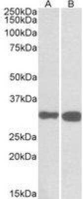
| SDS-PAGE | 19-29 kDa, reducing conditions |
|
| Publications |
|
| Storage | Use a manual defrost freezer and avoid repeated freeze-thaw cycles.
|
| Buffer | Lyophilized from a 0.2 μm filtered solution in PBS. |
| Purity | >90%, by SDS-PAGE with silver staining |
| Reconstitution Instructions | Reconstitute at 100 μg/mL in PBS. |
Myelin oligodendrocyte glycoprotein (MOG) is 28 kDa single-pass transmembrane glycoprotein that is a member of the Ig superfamily (1-4). Mouse MOG is synthesized with a 28 amino acid (aa) signal sequence, a 128 aa extracellular domain (ECD) containing an Ig-like domain, a 21 aa transmembrane domain, and a 69 aa cytosolic fragment featuring a hydrophobic domain that associates with the cytoplasmic face of the plasma membrane. The ECD of mature mouse MOG shares 90% and 95% aa sequence identity with the ECD of human and rat MOG, respectively. Dimerization of MOG occurs via the extracellular Ig-like domain (5-8). MOG is expressed exclusively by oligodendrocytes in the central nervous system (CNS) and is localized to the outer layer of the myelin sheath as well as in the oligodendrocyte plasma membrane (9). MOG expression in the brain can be used as a temporal biomarker for myelin development. MOG is an important antigenic target for autoimmune diseases that mediate demyelination in the CNS (10). In vivo administration of exogenous MOG protein or peptide induces experimental autoimmune encephalomyelitis (EAE) in multiple animal species (11, 12). EAE is used as an animal model for multiple sclerosis and related CNS demyelinating diseases. MOG is thought function as an adhesion molecule as well as a mediator of immune activation in the CNS (2, 9, 13).


| Publication using 8536-MO | Applications | Species |
|---|---|---|
| Scott M. Wemlinger, Chelsea R. Parker Parker Harp, Bo Yu, Ian R. Hardy, Matthew Seefeldt, Jennifer Matsuda et al. Preclinical Analysis of Candidate Anti-Human CD79 Therapeutic Antibodies Using a Humanized CD79 Mouse Model The Journal of Immunology 2022-04-01 [PMID: 35321883] (ELISA Capture, Transgenic Mouse) | ELISA Capture | Transgenic Mouse |
The concentration calculator allows you to quickly calculate the volume, mass or concentration of your vial. Simply enter your mass, volume, or concentration values for your reagent and the calculator will determine the rest.


![SEC-MALS IFN-gamma [Unconjugated]](https://images.novusbio.com/images/485-mi_recombinant-mouse-ifn-gamma-protein-sec-mals-1612202583245.jpg)
![Bioactivity IFN-gamma [Unconjugated]](https://images.novusbio.com/images/protein/IFN-gamma_485-MI_455.jpg)
![SDS-Page IFN-gamma [Unconjugated]](https://images.novusbio.com/images/protein/IFN-gamma_485-MI_407.jpg)
![SDS-Page TNF-alpha [Unconjugated]](https://images.novusbio.com/images/protein/TNF-alpha_210-TA_256.jpg)
![Bioactivity TNF-alpha [Unconjugated]](https://images.novusbio.com/images/protein/TNFalpha_210TA_1658.jpg)
![SEC-MALS TNF-alpha [Unconjugated]](https://images.novusbio.com/images/210-ta_recombinant-human-tnf-alpha-protein-sec-mals-35202312244..jpg)
![N/A IL-10 [Biotin]](https://images.novusbio.com/images/elisa/DATA_IL10_DY417_ELISA_2014.jpg)


![Bioactivity IL-4 [Unconjugated]](https://images.novusbio.com/images/protein/6507-ilcf_recombinant-human-il-4-cho-expressed-protein-cf-bioactivity-272020133214.jpg)
![N/A IL-6 [HRP]](https://images.novusbio.com/images/elisa/DATA_IL6_M6000_ELISA_936.jpg)
![N/A IL-6 [HRP]](https://images.novusbio.com/images/elisa/IL-6_M6000_ELISA_415.jpg)
![N/A IL-6 [HRP]](https://images.novusbio.com/images/m6000b_mouse-il-6-quantikine-elisa-kit-1752025024034.jpg)


![N/A IL-17/IL-17A [Biotin]](https://images.novusbio.com/images/elisa/DATA_IL17_DY421_ELISA_2448.jpg)


![Bioactivity IL-2 [Unconjugated]](https://images.novusbio.com/images/202-il_recombinant-human-il-2-protein-bioactivity-174202314946.jpg)
![Bioactivity CTLA-4 [Unconjugated]](https://images.novusbio.com/images/protein/CTLA4_7268CT_2293.jpg)