| Applications | Bioactivity |
| Format | Carrier-Free |
| Details of Functionality | Measured by its ability to inhibit the M-CSF-induced proliferation of M‑NFS‑60 mouse myelogenous leukemia lymphoblast cells. Halenbeck, R. et al. (1989) Biotechnology 7:710. The ED50 for this effect is 0.004-0.012 µg/mL in the presence of 1 ng/mL of Recombinant Human M-CSF (Catalog # 216-MC). |
||||||||
| Source | Mouse myeloma cell line, NS0-derived human M-CSF R/CD115 protein
|
||||||||
| Accession # | |||||||||
| N-terminal Sequence | Ile20 |
||||||||
| Structure / Form | Disulfide-linked homodimer |
||||||||
| Protein/Peptide Type | Recombinant Proteins |
||||||||
| Gene | CSF1R |
||||||||
| Purity | >97%, by SDS-PAGE under reducing conditions and visualized by silver stain |
||||||||
| Endotoxin Note | <0.10 EU per 1 μg of the protein by the LAL method. |
| Dilutions |
|
|
| Theoretical MW | 81 kDa. Disclaimer note: The observed molecular weight of the protein may vary from the listed predicted molecular weight due to post translational modifications, post translation cleavages, relative charges, and other experimental factors. |
|
| SDS-PAGE | 109-128 kDa, reducing conditions |
|
| Publications |
|
| Storage | Use a manual defrost freezer and avoid repeated freeze-thaw cycles.
|
| Buffer | Lyophilized from a 0.2 μm filtered solution in PBS. |
| Purity | >97%, by SDS-PAGE under reducing conditions and visualized by silver stain |
| Reconstitution Instructions | Reconstitute at 100 μg/mL in sterile PBS. |
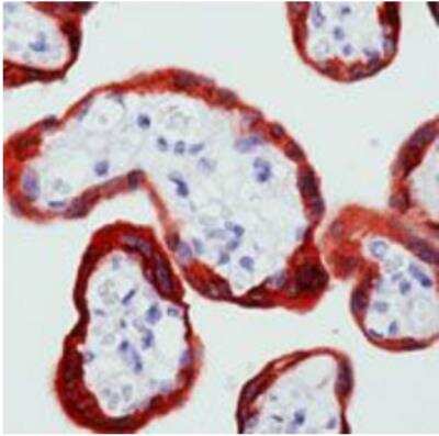
M-CSF receptor, the product of the c-fms proto-oncogene, is a member of the type III subfamily of receptor tyrosine kinases that also includes receptors for SCF and PDGF. These receptors each contain five immunoglobulin-like domains in their extracellular domain (ECD) and a split kinase domain in their intracellular region (1-4). M-CSF receptor is expressed primarily on cells of the monocyte/macrophage lineage, dendritic cells, stem cells and in the developing placenta (1). Human M-CSF receptor cDNA encodes a 972 amino acid (aa) type I membrane protein with a 19 aa signal peptide, a 493 aa extracellular region containing the ligand-binding domain, a 25 aa transmembrane domain and a 435 aa cytoplasmic domain. The human M-CSF R ECD shares 60%, 64%, 72%, 75%, 75% and 76% aa identity with mouse, rat, bovine, canine, feline and equine M-CSF R, respectively. Activators of protein kinase C induce TACE/ADAM17 cleavage of the M-CSF receptor, releasing the functional ligand-binding extracellular domain (5). M-CSF binding induces receptor homodimerization, resulting in transphosphorylation of specific cytoplasmic tyrosine residues and signal transduction (6). The intracellular domain of activated M-CSF R binds more than 150 proteins that affect cell proliferation, survival, differentiation and cytoskeletal reorganization. Among these, PI3Kinase, P42/44 ERK and c-Cbl are key transducers of M-CSF R signals (3, 4). M-CSF R engagement is continuously required for macrophage survival and regulates lineage decisions and maturation of monocytes, macrophages, osteoclasts and DC (3, 4). M-CSF R and integrin alpha v beta 3 share signaling pathways during osteoclastogenesis and deletion of either causes osteopetrosis (7, 8). In the brain, microglia expressing increased
M-CSF R are concentrated with Alzheimers a beta peptide, but their role in pathogenesis is unclear (9, 10).
![Flow Cytometry CD117/c-kit Antibody [Unconjugated]](https://images.novusbio.com/images/af1356_human-mouse-cd117-c-kit-affinity-purified-polyclonal-ab-81202555532.jpg)
![Immunohistochemistry CD117/c-kit Antibody [Unconjugated]](https://images.novusbio.com/images/antibody/af1356_human-mouse-cd117-c-kit-affinity-purified-polyclonal-ab-immunohistochemistry-812202575731.png)
![Immunocytochemistry/ Immunofluorescence CD117/c-kit Antibody [Unconjugated]](https://images.novusbio.com/images/af1356_human-mouse-cd117-c-kit-affinity-purified-polyclonal-ab-41202410481199.jpg)


The concentration calculator allows you to quickly calculate the volume, mass or concentration of your vial. Simply enter your mass, volume, or concentration values for your reagent and the calculator will determine the rest.


![Bioactivity M-CSF [Unconjugated]](https://images.novusbio.com/images/protein/416-ml_recombinant-mouse-m-csf-protein-bioactivity-21102020193736.jpg)
![SEC-MALS M-CSF [Unconjugated]](https://images.novusbio.com/images/416-ml_recombinant-mouse-m-csf-protein-sec-mals-114202410458..jpg)
![SEC-MALS IL-3 [Unconjugated]](https://images.novusbio.com/images/203-il_recombinant-human-il-3-protein-sec-mals-133202482656..jpg)
![Bioactivity IL-3 [Unconjugated]](https://images.novusbio.com/images/protein/IL-3_203-IL_347.jpg)
![Data IL-3 [Unconjugated]](https://images.novusbio.com/images/203-il_recombinant-human-il-3-protein-data-253202610550.jpg)
![SDS-Page TNF-alpha [Unconjugated]](https://images.novusbio.com/images/protein/TNF-alpha_210-TA_256.jpg)
![Bioactivity TNF-alpha [Unconjugated]](https://images.novusbio.com/images/protein/TNFalpha_210TA_1658.jpg)
![SEC-MALS TNF-alpha [Unconjugated]](https://images.novusbio.com/images/210-ta_recombinant-human-tnf-alpha-protein-sec-mals-35202312244..jpg)
![Bioactivity GM-CSF [Unconjugated]](https://images.novusbio.com/images/protein/7954-gmcf_recombinant-human-gm-csf-cho-expressed-protein-cf-bioactivity-272020121514.jpg)


![Western Blot Src [p Tyr419] Antibody [Unconjugated]](https://images.novusbio.com/images/antibody/Src_AF2685_Western_Blot_5209.jpg)
![Immunohistochemistry Src [p Tyr419] Antibody [Unconjugated]](https://images.novusbio.com/images/af2685_human-phospho-src-y419-affinity-purified-polyclonal-ab-412024124022.jpg)
![Immunohistochemistry Src [p Tyr419] Antibody [Unconjugated]](https://images.novusbio.com/images/antibody/Src_AF2685_Immunohistochemistry_9943.jpg)
![SDS-Page TRANCE/TNFSF11/RANK L [Unconjugated]](https://images.novusbio.com/images/protein/TRANCE_462-TEC_442.jpg)
![Bioactivity TRANCE/TNFSF11/RANK L [Unconjugated]](https://images.novusbio.com/images/protein/TRANCE_462-TEC_556.jpg)
![Immunohistochemistry PDGF R beta Antibody [Unconjugated]](https://images.novusbio.com/images/af1042_mouse-pdgf-r-beta-affinity-purified-polyclonal-ab-immunohistochemistry-121220259315424.jpg)
![Immunohistochemistry PDGF R beta Antibody [Unconjugated]](https://images.novusbio.com/images/af1042_mouse-pdgf-r-beta-affinity-purified-polyclonal-ab-immunohistochemistry-121220258463418.jpg)
![Immunohistochemistry PDGF R beta Antibody [Unconjugated]](https://images.novusbio.com/images/af1042_mouse-pdgf-r-beta-affinity-purified-polyclonal-ab-immunohistochemistry-12122025857822.jpg)


![Flow Cytometry: M-CSF R/CD115 Antibody (AFS98) [NBP1-43363] - Staining of thioglycolate-induced peritoneal exudate cells with Anti-Mouse CD115 (c-fms) PE. Appropriate isotype controls were used (open histogram). Cells in the large scatter population were used for analysis.](https://images.novusbio.com/images/M-CSF-R-CD115-Antibody-AFS98-Flow-Cytometry-NBP1-43363-img0002.jpg "Flow Cytometry: M-CSF R/CD115 Antibody (AFS98) [NBP1-43363] - Staining of thioglycolate-induced peritoneal exudate cells with Anti-Mouse CD115 (c-fms) PE. Appropriate isotype controls were used (open histogram). Cells in the large scatter population were used for analysis.")