| Reactivity | HuSpecies Glossary |
| Applications | Bioactivity |
| Format | Carrier-Free |
| Details of Functionality | Measured by its ability of the immobilized protein to support the adhesion of Jurkat human acute T cell leukemia cells. Bechard, D. et al. (2001) J. Immunol. 167:3099. When 1 x 10⁵ cells/well are added to Recombinant Human Endocan/ESM coated plates, adhesion is induced in a dose dependent manner after a 1 hour incubation at 37 °C. The ED50 for this effect is 2-10 μg/mL. Optimal dilutions should be determined by each laboratory for each application. |
| Source | Mouse myeloma cell line, NS0-derived human Endocan/ESM-1 protein Trp20-Arg184, with a C-terminal 10-His tag |
| Accession # | |
| N-terminal Sequence | Trp20 |
| Protein/Peptide Type | Recombinant Proteins |
| Gene | ESM1 |
| Purity | >90%, by SDS-PAGE visualized with Silver Staining and quantitative densitometry by Coomassie® Blue Staining |
| Endotoxin Note | <1.0 EU per 1 μg of the protein by the LAL method. |
| Dilutions |
|
|
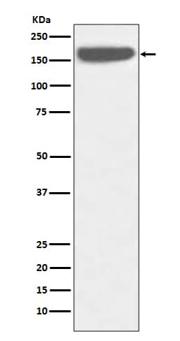
| Theoretical MW | 19.5 kDa. Disclaimer note: The observed molecular weight of the protein may vary from the listed predicted molecular weight due to post translational modifications, post translation cleavages, relative charges, and other experimental factors. |
|
| SDS-PAGE | 24-27 kDa, reducing conditions |
|
| Publications |
|
| Storage | Use a manual defrost freezer and avoid repeated freeze-thaw cycles.
|
| Buffer | Lyophilized from a 0.2 μm filtered solution in PBS. |
| Purity | >90%, by SDS-PAGE visualized with Silver Staining and quantitative densitometry by Coomassie® Blue Staining |
| Reconstitution Instructions | Reconstitute at 100 μg/mL in sterile PBS. |
Endocan, also known as endothelial-cell specific molecule-1 (ESM-1), is a secreted cysteine-rich dermatan sulfate (DS) proteoglycan primarily expressed by endothelial cells within the vascular capillary network in kidney and in the alveolar walls of the lung (1). Endocan expression has also been detected in different epithelia and in adipocytes (2, 3). The expression of endocan is upregulated by TNF alpha , IL-1 beta or lipopolysaccharide and down-regulated by IFN gamma (1). The human Endocan gene encodes a 184 amino acid (aa) residues precursor protein with a 19 aa hydrophobic signal peptide and a 165 aa mature region with 18 Cysteine residues (1). The DS chain is covalently attached to serine 137 (4). Endocan has been shown to bind CD11a/CD18 integrin (also known as lymphocyte function-associated antigen-1, LFA-1) on human lymphocytes, monocytes and Jurkat cells, inhibiting its binding to ICAM-1 and reducing LFA-1-mediated leukocyte activation (5). Endocan binds via its DS chain to hepatocyte growth factor (HGF) to enhance HGF mitogenic activity (3, 6). Genetically engineered cells overexpressing endocan has been shown to induce tumor formation, suggesting that Endocan may be involved in the pathophysiology of tumor growth in vivo (3, 6). Circulating Endocan can be detected in the serum from healthy subjects. In patients with lung cancer or acute and severe sepsis, elevated Endocan concentrations have been reported (2, 6).
![Flow Cytometry CD31/PECAM-1 Antibody [Unconjugated]](https://images.novusbio.com/images/af3628_human-mouse-rat-cd31-pecam-1-affinity-purified-polyclonal-ab-flow-cytometry-30112023144624..jpg)
![Western Blot CD31/PECAM-1 Antibody [Unconjugated]](https://images.novusbio.com/images/af3628_human-mouse-rat-cd31-pecam-1-affinity-purified-polyclonal-ab-41202410325588.jpg)
![Immunohistochemistry CD31/PECAM-1 Antibody [Unconjugated]](https://images.novusbio.com/images/antibody/af3628_mouse-rat-cd31-pecam-1-affinity-purified-polyclonal-ab-immunohistochemistry-2572023161128.jpg)


![Immunohistochemistry VEGFR2/KDR/Flk-1 Antibody [Unconjugated]](https://images.novusbio.com/images/antibody/VEGF_R2_AF644_Immunohistochemistry_6888.jpg)
![Immunohistochemistry VEGFR2/KDR/Flk-1 Antibody [Unconjugated]](https://images.novusbio.com/images/af644_mouse-vegf-r2-flk-1-affinity-purified-polyclonal-ab-4120241048248.jpg)
![In-situ Hybridization VEGFR2/KDR/Flk-1 Antibody [Unconjugated]](https://images.novusbio.com/images/antibody/af644_mouse-vegf-r2-flk-1-affinity-purified-polyclonal-ab-in-situ-hybridization-235202421442..jpg)


The concentration calculator allows you to quickly calculate the volume, mass or concentration of your vial. Simply enter your mass, volume, or concentration values for your reagent and the calculator will determine the rest.


![N/A VEGF [HRP]](https://images.novusbio.com/images/elisa/VEGF_DVE00_ELISA_208.jpg)
![N/A VEGF [HRP]](https://images.novusbio.com/images/elisa/DATA_VEGF_DVE00_ELISA_871.jpg)
![N/A VEGF [HRP]](https://images.novusbio.com/images/elisa/DATA_VEGF_DVE00_ELISA_872.jpg)
![Bioactivity HGF [Unconjugated]](https://images.novusbio.com/images/protein/HGF_294HG_3253.jpg)
![Cell Culture HGF [Unconjugated]](https://images.novusbio.com/images/294-hg_recombinant-human-hgf-protein-cell-culture-252023111456.jpg)
![N/A IL-6 [HRP]](https://images.novusbio.com/images/elisa/DATA_IL6_M6000_ELISA_936.jpg)
![N/A IL-6 [HRP]](https://images.novusbio.com/images/elisa/IL-6_M6000_ELISA_415.jpg)
![N/A IL-6 [HRP]](https://images.novusbio.com/images/m6000b_mouse-il-6-quantikine-elisa-kit-1752025024034.jpg)
![SDS-Page TNF-alpha [Unconjugated]](https://images.novusbio.com/images/protein/TNF-alpha_210-TA_256.jpg)
![Bioactivity TNF-alpha [Unconjugated]](https://images.novusbio.com/images/protein/TNFalpha_210TA_1658.jpg)
![SEC-MALS TNF-alpha [Unconjugated]](https://images.novusbio.com/images/210-ta_recombinant-human-tnf-alpha-protein-sec-mals-35202312244..jpg)
![N/A Angiopoietin-2 [HRP]](https://images.novusbio.com/images/elisa/Angiopoietin-2_DANG20_ELISA_30.jpg)
![N/A Angiopoietin-2 [HRP]](https://images.novusbio.com/images/elisa/DATA_Angiopoietin2_DANG20_ELISA_574.jpg)
![Western Blot ICAM-1/CD54 Antibody [Unconjugated]](https://images.novusbio.com/images/af796_mouse-icam-1-cd54-affinity-purified-polyclonal-ab-41202410481192.jpg)
![Western Blot ICAM-1/CD54 Antibody [Unconjugated]](https://images.novusbio.com/images/af796_mouse-icam-1-cd54-affinity-purified-polyclonal-ab-4120241048112.jpg)
![Western Blot ICAM-1/CD54 Antibody [Unconjugated]](https://images.novusbio.com/images/af796_mouse-icam-1-cd54-affinity-purified-polyclonal-ab-4120241049436.jpg)
![N/A VEGF-C [HRP]](https://images.novusbio.com/images/elisa/DATA_VEGFC_DVEC00_ELISA_874.jpg)
![ELISA VEGF-C [HRP]](https://images.novusbio.com/images/VEGF-C_DVEC00_ELISA_209.png)
![N/A VEGF-C [HRP]](https://images.novusbio.com/images/elisa/DATA_VEGFC_DVEC00_ELISA_873.jpg)
![Bioactivity EGF [Unconjugated]](https://images.novusbio.com/images/protein/EGF_236EG_1570.jpg)
![Cell Culture EGF [Unconjugated]](https://images.novusbio.com/images/236-eg_recombinant-human-egf-protein-cf-25202394526.jpg)
![Cell Culture EGF [Unconjugated]](https://images.novusbio.com/images/236-eg_recombinant-human-egf-protein-cf-bioactivity-25202394053.jpg)
![N/A C-Reactive Protein/CRP [Biotin]](https://images.novusbio.com/images/elisa/DATA_CReactive_Protein_DY1707_ELISA_1701.jpg)
![Bioactivity CXCL12/SDF-1 alpha [Unconjugated]](https://images.novusbio.com/images/protein/CXCL12SDF1_alpha_350NS_1178.jpg)
![SDS-Page CXCL12/SDF-1 alpha [Unconjugated]](https://images.novusbio.com/images/protein/CXCL12_SDF-1_alpha_350-NS_308.jpg)