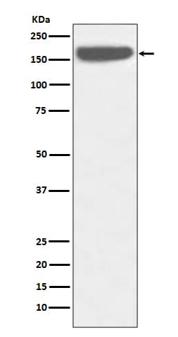
 or treated (+) with 100 µM pervanadate (PV) for 10 minutes. PVDF membrane was probed with 1 µg/mL of Rabbit Anti-Human Phospho-HGF R/c-MET (Y1003) Antigen Affinity-purified Polyclonal Antibody (Catalog # AF4059), followed by HRP-conjugated Anti-Rabbit IgG Secondary Antibody (Catalog # HAF008). A specific band was detected for Phospho-HGF R/ c-MET (Y1003) at approximately 140 kDa (as indicated). This experiment was conducted under reducing conditions and using Immunoblot Buffer Group 1.")
| Reactivity | HuSpecies Glossary |
| Applications | WB, IHC, ICC/IF |
| Clonality | Polyclonal |
| Host | Rabbit |
| Conjugate | Unconjugated |
| Concentration | LYOPH |
| Immunogen | Phosphopeptide containing human HGF R Y1003 site |
| Modification | p Tyr1003 |
| Specificity | Detects human and mouse HGF R/c-MET when phosphorylated at Y1003. |
| Source | N/A |
| Isotype | IgG |
| Clonality | Polyclonal |
| Host | Rabbit |
| Gene | MET |
| Purity Statement | Antigen Affinity-purified |
| Innovator's Reward | Test in a species/application not listed above to receive a full credit towards a future purchase. |
| Dilutions |
|
|
| Publications |
|
| Storage | Use a manual defrost freezer and avoid repeated freeze-thaw cycles.
|
| Buffer | Lyophilized from a 0.2 μm filtered solution in PBS with Trehalose. *Small pack size (SP) is supplied either lyophilized or as a 0.2 µm filtered solution in PBS. |
| Preservative | No Preservative |
| Concentration | LYOPH |
| Reconstitution Instructions | Reconstitute at 0.2 mg/mL in sterile PBS. |
HGF R, also known as Met (from N-methyl-N’-nitro-N-nitrosoguanidine induced), is a glycosylated receptor tyrosine kinase that plays a central role in epithelial morphogenesis and cancer development. HGF R is synthesized as a single chain precursor which undergoes cotranslational proteolytic cleavage. This generates a mature HGF R that is a disulfide-linked dimer composed of a 50 kDa extracellular alpha chain and a 145 kDa transmembrane beta chain (1, 2). The extracellular domain (ECD) contains a seven bladed beta -propeller sema domain, a cysteine-rich PSI/MRS, and four Ig-like E-set domains, while the cytoplasmic region includes the tyrosine kinase domain (3, 4). Proteolysis and alternate splicing generate additional forms of human HGF R which either lack of the kinase domain, consist of secreted extracellular domains, or are deficient in proteolytic separation of the alpha and beta chains (5 - 7). The sema domain, which is formed by both the alpha and beta chains of HGF R, mediates both ligand binding and receptor dimerization (3, 8). Ligand-induced tyrosine phosphorylation in the cytoplasmic region activates the kinase domain and provides docking sites for multiple SH2-containing molecules (9, 10). HGF stimulation induces HGF R downregulation via internalization and proteasome-dependent degradation (11). In the absence of ligand, HGF R forms noncovalent complexes with a variety of membrane proteins including CD44v6, CD151, EGF R, Fas, Integrin alpha 6/ beta 4, Plexins B1, 2, 3, and MSP R/Ron (12 - 19). Ligation of one complex component triggers activation of the other, followed by cooperative signaling effects (12 - 19). Formation of some of these heteromeric complexes is a requirement for epithelial cell morphogenesis and tumor cell invasion (12, 16, 17). Paracrine induction of epithelial cell scattering and branching tubulogenesis results from the stimulation of HGF R on undifferentiated epithelium by HGF released from neighboring mesenchymal cells (20). Genetic polymorphisms, chromosomal translocation, overexpression, and additional splicing and proteolytic cleavage of HGF R have been described in a wide range of cancers (1). Within the ECD, human HGF R shares 86% - 88% aa sequence identity with canine, mouse, and rat HGF R.
![Immunocytochemistry EGFR Antibody [Unconjugated]](https://images.novusbio.com/images/antibody/EGF_R_AF231_Immunocytochemistry__Immunofluorescence_21143.jpg)
![Flow Cytometry EGFR Antibody [Unconjugated]](https://images.novusbio.com/images/antibody/EGF_R_AF231_Flow_Cytometry_20401.jpg)
![Western Blot EGFR Antibody [Unconjugated]](https://images.novusbio.com/images/antibody/EGF_R_AF231_Western_Blot_19925.jpg)
![Intracellular Staining by Flow Cytometry AKT [p Ser473] Antibody [Unconjugated] - Pan Specific](https://images.novusbio.com/images/antibody/Akt3_AF887_Flow_Cytometry_8283.jpg)
![Western Blot AKT [p Ser473] Antibody [Unconjugated] - Pan Specific](https://images.novusbio.com/images/af887_phospho-akt-s473-pan-specific-affinity-purified-pab-41202410485440.jpg)
![Western Blot AKT [p Ser473] Antibody [Unconjugated] - Pan Specific](https://images.novusbio.com/images/af887_phospho-akt-s473-pan-specific-affinity-purified-pab-8120255552843.jpg)
![Immunocytochemistry E-Cadherin Antibody [Unconjugated]](https://images.novusbio.com/images/antibody/ECadherin_AF748_Immunocytochemistry_16573.jpg)
![Western Blot E-Cadherin Antibody [Unconjugated]](https://images.novusbio.com/images/antibody/ECadherin_AF748_Western_Blot_19846.jpg)
![Immunocytochemistry E-Cadherin Antibody [Unconjugated]](https://images.novusbio.com/images/antibody/ECadherin_AF748_Immunocytochemistry_16572.jpg)


![Western Blot EphB2 Antibody [Unconjugated]](https://images.novusbio.com/images/af467_human-mouse-ephb2-affinity-purified-polyclonal-ab-41202410485412.jpg)
![Immunohistochemistry EphB2 Antibody [Unconjugated]](https://images.novusbio.com/images/antibody/EphB2_AF467_Immunohistochemistry_20369.jpg)
![Western Blot EphB2 Antibody [Unconjugated]](https://images.novusbio.com/images/antibody/EphB2_AF467_Western_Blot_21511.jpg)
![HGFR/c-MET [p Tyr1003] Antibody](/sites/all/modules/enterprise-tech/et_datasheets/images/novus_guarantee.png "HGFR/c-MET [p Tyr1003] Antibody")
Secondary Antibodies |
Isotype Controls |
The concentration calculator allows you to quickly calculate the volume, mass or concentration of your vial. Simply enter your mass, volume, or concentration values for your reagent and the calculator will determine the rest.
| Gene Symbol | MET |


 or treated (upper panel) with pervanadate using Rabbit Anti-Human Phospho-HGF R/c-MET (Y1003) Antigen Affinity-purified Polyclonal Antibody (Catalog # AF4059) at 10 µg/mL for 3 hours at room temperature. Cells were stained using the NorthernLights™ 557-conjugated Anti-Rabbit IgG Secondary Antibody (red; Catalog # NL004) and counterstained with DAPI (blue). View our protocol for Fluorescent ICC Staining of Cells on Coverslips.")
![Bioactivity HGF [Unconjugated]](https://images.novusbio.com/images/protein/HGF_294HG_3253.jpg)
![Cell Culture HGF [Unconjugated]](https://images.novusbio.com/images/294-hg_recombinant-human-hgf-protein-cell-culture-252023111456.jpg)
![N/A IL-6 [HRP]](https://images.novusbio.com/images/elisa/DATA_IL6_M6000_ELISA_936.jpg)
![N/A IL-6 [HRP]](https://images.novusbio.com/images/elisa/IL-6_M6000_ELISA_415.jpg)
![N/A IL-6 [HRP]](https://images.novusbio.com/images/m6000b_mouse-il-6-quantikine-elisa-kit-1752025024034.jpg)
![N/A VEGF [HRP]](https://images.novusbio.com/images/elisa/VEGF_DVE00_ELISA_208.jpg)
![N/A VEGF [HRP]](https://images.novusbio.com/images/elisa/DATA_VEGF_DVE00_ELISA_871.jpg)
![N/A VEGF [HRP]](https://images.novusbio.com/images/elisa/DATA_VEGF_DVE00_ELISA_872.jpg)
![Bioactivity EGF [Unconjugated]](https://images.novusbio.com/images/protein/EGF_236EG_1570.jpg)
![Cell Culture EGF [Unconjugated]](https://images.novusbio.com/images/236-eg_recombinant-human-egf-protein-cf-25202394526.jpg)
![Cell Culture EGF [Unconjugated]](https://images.novusbio.com/images/236-eg_recombinant-human-egf-protein-cf-bioactivity-25202394053.jpg)
![Western Blot ERK2 Antibody [Unconjugated]](https://images.novusbio.com/images/antibody/ERK2_AF1230_Western_Blot_5097.jpg)
![Knockout Validated ERK2 Antibody [Unconjugated]](https://images.novusbio.com/images/antibody/ERK2_AF1230_Knockout_Validated_22864.jpg)
![Immunohistochemistry ERK2 Antibody [Unconjugated]](https://images.novusbio.com/images/antibody/ERK2_AF1230_Immunohistochemistry_20696.jpg)


![Bioactivity ErbB2/Her2 [Unconjugated]](https://images.novusbio.com/images/protein/1129-er_recombinant-human-erbb2-her2-fc-chimera-protein-cf-bioactivity-7122020142841.jpg)
![SDS-Page ErbB2/Her2 [Unconjugated]](https://images.novusbio.com/images/protein/ErbB2_1129-ER_70.jpg)
![Western Blot Src [p Tyr419] Antibody [Unconjugated]](https://images.novusbio.com/images/antibody/Src_AF2685_Western_Blot_5209.jpg)
![Immunohistochemistry Src [p Tyr419] Antibody [Unconjugated]](https://images.novusbio.com/images/af2685_human-phospho-src-y419-affinity-purified-polyclonal-ab-412024124022.jpg)
![Immunohistochemistry Src [p Tyr419] Antibody [Unconjugated]](https://images.novusbio.com/images/antibody/Src_AF2685_Immunohistochemistry_9943.jpg)


![Western Blot: Goat anti-Rabbit IgG (H+L) Secondary Antibody [HRP] [NB7160] - Western blot showing vemurafenib treatment in BRAFV600E CRC cells inhibits fission mediator DRP1 with no significant effect on fusion proteins (Mfn1 & 2) using MFN-1 antibody (NBP1-51841) and corresponding secondary antibody, goat anti-rabbit IgG-HRP (NB7160). Image collected and cropped by CiteAb from the following publication (https://pubmed.ncbi.nlm.nih.gov/33738242).](https://images.novusbio.com/images/Goat-anti-Rabbit-IgG-H+L-Secondary-Antibody-HRP-Western-Blot-NB7160-img0001.jpg "Western Blot: Goat anti-Rabbit IgG (H+L) Secondary Antibody [HRP] [NB7160] - Western blot showing vemurafenib treatment in BRAFV600E CRC cells inhibits fission mediator DRP1 with no significant effect on fusion proteins (Mfn1 & 2) using MFN-1 antibody (NBP1-51841) and corresponding secondary antibody, goat anti-rabbit IgG-HRP (NB7160). Image collected and cropped by CiteAb from the following publication (https://pubmed.ncbi.nlm.nih.gov/33738242).")
 followed by 30 min incubation with Goat anti Rabbit HRP conjugated secondary antibodies (Catalog # HAF008) at 1:20 dilution + DAB chromogen (brown). The tissue was counterstained with Hematoxylin (blue). Control was done by omitting primary antibody.")
![Flow Cytometry: Rabbit IgG Isotype Control [NBP2-24891] - An intracellular stain was performed on Raji cells with Adiponectin antibody NB100-65810 (blue) and a matched isotype control NBP2-24893 (orange). Cells were fixed with 4% PFA and then permeablized with 0.1% saponin. Cells were incubated in an antibody dilution of 1 ug/mL for 30 minutes at room temperature, followed by Dylight488-conjugated anti-rabbit secondary antibody. Image using the Azide Free form of this antibody.](https://images.novusbio.com/images/Rabbit--Mouse-IgG-Isotype-Control-Flow-Cytometry-NBP2-24891-img0006.jpg "Flow Cytometry: Rabbit IgG Isotype Control [NBP2-24891] - An intracellular stain was performed on Raji cells with Adiponectin antibody NB100-65810 (blue) and a matched isotype control NBP2-24893 (orange). Cells were fixed with 4% PFA and then permeablized with 0.1% saponin. Cells were incubated in an antibody dilution of 1 ug/mL for 30 minutes at room temperature, followed by Dylight488-conjugated anti-rabbit secondary antibody. Image using the Azide Free form of this antibody.")