
| Reactivity | Hu, Mu, RtSpecies Glossary |
| Applications | WB, ICC/IF, IHC, Mycoplasma |
| Clonality | Polyclonal |
| Host | Rabbit |
| Conjugate | Unconjugated |
| Format | BSA Free |
| Immunogen | This antibody was developed against Recombinant Protein corresponding to amino acids: SESLVVCDVAEDLVEKLRKFRFRKETNNAAIIMKIDKDKRLVVLDEELEGISPDELKDELPERQPRFIVYSYKYQHDDGRVSYPLCFIFSSPVGCKPEQQMMYAGSKN |
| Isotype | IgG |
| Clonality | Polyclonal |
| Host | Rabbit |
| Gene | GMFB |
| Purity | Affinity purified |
| Innovator's Reward | Test in a species/application not listed above to receive a full credit towards a future purchase. |
| Dilutions |
|
||
| Application Notes | For IHC-Paraffin, HIER pH 6 retrieval is recommended. ICC/IF Fixation Permeabilization: Use PFA/Triton X-100. |
||
| Theoretical MW | 17 kDa. Disclaimer note: The observed molecular weight of the protein may vary from the listed predicted molecular weight due to post translational modifications, post translation cleavages, relative charges, and other experimental factors. |
||
| Control Peptide |
|
||
| Publications |
|
| Storage | Store at 4C short term. Aliquot and store at -20C long term. Avoid freeze-thaw cycles. |
| Buffer | PBS (pH 7.2) and 40% Glycerol |
| Preservative | 0.02% Sodium Azide |
| Purity | Affinity purified |


Secondary Antibodies |
Isotype Controls |
Research Areas for GMF-beta Antibody (NBP1-89755)Find related products by research area.
|
The concentration calculator allows you to quickly calculate the volume, mass or concentration of your vial. Simply enter your mass, volume, or concentration values for your reagent and the calculator will determine the rest.


![Immunohistochemistry: GMF-beta Antibody [NBP1-89755] - GMF-beta knockdown suppresses tumor growth & the formation of human-CD31 positive microvessels (hCD31-MVs) in orthotopic U87 xenograft modelsA. The cross-sectional appearance of xenografted gliomas from U87-mock group B. The cross-sectional appearance of xenografted gliomas from U87-shGMF-beta group. Gross tumor boundaries were delineated by white dotlines. C. A hCD31-microvessel & several hCD31-negative vessels in U87-mock tumor. D. No hCD31-MVs, only hCD31-negative vessels in U87-shGMF-beta tumor. Red solid arrows indicate hGMF-beta staining in tumor cells, black solid arrows show hCD31 staining on microvasular endothelia, & open arrows denote vessels negative for hCD31. Scale bar: 100 μm in upper pannels; 50 μm in lower pannels. E. Quantitative comparisons of gross tumor volume between U87-mock & U87-shGMF-beta group. F. Quantitative comparisons of hCD31-microvessel densities (MVDs) between U87-mock & U87-shGMF-beta group. Image collected & cropped by CiteAb from the following publication (//pubmed.ncbi.nlm.nih.gov/26515590), licensed under a CC-BY license. Not internally tested by Novus Biologicals.](http://images.novusbio.com/fullsize/nbp1-89755_rabbit-polyclonal-gmf-beta-antibody-310202416121977.jpg "Immunohistochemistry: GMF-beta Antibody [NBP1-89755] - GMF-beta knockdown suppresses tumor growth & the formation of human-CD31 positive microvessels (hCD31-MVs) in orthotopic U87 xenograft modelsA. The cross-sectional appearance of xenografted gliomas from U87-mock group B. The cross-sectional appearance of xenografted gliomas from U87-shGMF-beta group. Gross tumor boundaries were delineated by white dotlines. C. A hCD31-microvessel & several hCD31-negative vessels in U87-mock tumor. D. No hCD31-MVs, only hCD31-negative vessels in U87-shGMF-beta tumor. Red solid arrows indicate hGMF-beta staining in tumor cells, black solid arrows show hCD31 staining on microvasular endothelia, & open arrows denote vessels negative for hCD31. Scale bar: 100 μm in upper pannels; 50 μm in lower pannels. E. Quantitative comparisons of gross tumor volume between U87-mock & U87-shGMF-beta group. F. Quantitative comparisons of hCD31-microvessel densities (MVDs) between U87-mock & U87-shGMF-beta group. Image collected & cropped by CiteAb from the following publication (//pubmed.ncbi.nlm.nih.gov/26515590), licensed under a CC-BY license. Not internally tested by Novus Biologicals.")


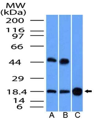
![Western Blot: GMF-beta Antibody [NBP1-89755] - Tubulogenesis of human U87 glioblastoma cells is inhibited by GMF-beta knockdownA. Protein levels of GMF-beta in human glial cell line (HEB) & human glioma cell lines of the different grades (CHG5, SHG44, U87). B. Assessment of GMF-beta knockdown in U87 cells by western blotting. C. Tube formation by U87 mock cells (left panel); impaired tube formation by U87-shGMF-beta cells (right panel). Inspection under a phase contrast fluorescent microscope (× 100). D. Quantified tubulogenesis of U87 mock cells & U87-shGMF-beta cells by pattern recognition system (left panel) & branch point counting system (right panel).*** indicates significant difference with P < 0.001. Image collected & cropped by CiteAb from the following publication (//pubmed.ncbi.nlm.nih.gov/26515590), licensed under a CC-BY license. Not internally tested by Novus Biologicals.](http://images.novusbio.com/fullsize/nbp1-89755_rabbit-polyclonal-gmf-beta-antibody-310202415371963.jpg "Western Blot: GMF-beta Antibody [NBP1-89755] - Tubulogenesis of human U87 glioblastoma cells is inhibited by GMF-beta knockdownA. Protein levels of GMF-beta in human glial cell line (HEB) & human glioma cell lines of the different grades (CHG5, SHG44, U87). B. Assessment of GMF-beta knockdown in U87 cells by western blotting. C. Tube formation by U87 mock cells (left panel); impaired tube formation by U87-shGMF-beta cells (right panel). Inspection under a phase contrast fluorescent microscope (× 100). D. Quantified tubulogenesis of U87 mock cells & U87-shGMF-beta cells by pattern recognition system (left panel) & branch point counting system (right panel).*** indicates significant difference with P < 0.001. Image collected & cropped by CiteAb from the following publication (//pubmed.ncbi.nlm.nih.gov/26515590), licensed under a CC-BY license. Not internally tested by Novus Biologicals.")

![Immunohistochemistry MOG Antibody [Unconjugated]](https://images.novusbio.com/images/antibody/MOG_AF2439_Immunohistochemistry_6749.jpg)
![N/A BDNF [HRP]](https://images.novusbio.com/images/elisa/BDNF_DBD00_ELISA_36.jpg)
![N/A BDNF [HRP]](https://images.novusbio.com/images/elisa/DATA_BDNF_DBD00_ELISA_587.jpg)
![N/A BDNF [HRP]](https://images.novusbio.com/images/elisa/DATA_BDNF_DBD00_ELISA_588.jpg)
![Bioactivity beta-NGF [Unconjugated]](https://images.novusbio.com/images/protein/beta-NGF_256-GF_322.jpg)
![SDS-Page beta-NGF [Unconjugated]](https://images.novusbio.com/images/protein/beta-NGF_256-GF_321.jpg)
![Western Blot ERK2 Antibody [Unconjugated]](https://images.novusbio.com/images/antibody/ERK2_AF1230_Western_Blot_5097.jpg)
![Knockout Validated ERK2 Antibody [Unconjugated]](https://images.novusbio.com/images/antibody/ERK2_AF1230_Knockout_Validated_22864.jpg)
![Immunohistochemistry ERK2 Antibody [Unconjugated]](https://images.novusbio.com/images/antibody/ERK2_AF1230_Immunohistochemistry_20696.jpg)


![Bioactivity GM-CSF [Unconjugated]](https://images.novusbio.com/images/protein/7954-gmcf_recombinant-human-gm-csf-cho-expressed-protein-cf-bioactivity-272020121514.jpg)
![Western Blot p38 alpha Antibody [Unconjugated]](https://images.novusbio.com/images/antibody/p38_alpha_AF8691_Western_Blot_7402.jpg)
![Immunohistochemistry p38 alpha Antibody [Unconjugated]](https://images.novusbio.com/images/antibody/p38_alpha_AF8691_Immunohistochemistry_7087.jpg)
![Western Blot p38 alpha Antibody [Unconjugated]](https://images.novusbio.com/images/antibody/p38_alpha_AF8691_Western_Blot_7401.jpg)
![SDS-Page TNF-alpha [Unconjugated]](https://images.novusbio.com/images/protein/TNF-alpha_210-TA_256.jpg)
![Bioactivity TNF-alpha [Unconjugated]](https://images.novusbio.com/images/protein/TNFalpha_210TA_1658.jpg)
![SEC-MALS TNF-alpha [Unconjugated]](https://images.novusbio.com/images/210-ta_recombinant-human-tnf-alpha-protein-sec-mals-35202312244..jpg)
![N/A CXCL10/IP-10/CRG-2 [HRP]](https://images.novusbio.com/images/elisa/CXCL10_DIP100_ELISA_121.jpg)
![N/A CXCL10/IP-10/CRG-2 [HRP]](https://images.novusbio.com/images/elisa/DATA_CXCL10_DIP100_ELISA_723.jpg)
![N/A CXCL10/IP-10/CRG-2 [HRP]](https://images.novusbio.com/images/elisa/DATA_CXCL10_DIP100_ELISA_724.jpg)
![Neutralization FGF acidic/FGF1 Antibody [Unconjugated]](https://images.novusbio.com/images/antibody/FGF_acidic_AF232_Block_Neutralize_8822.jpg)
![Immunohistochemistry FGF acidic/FGF1 Antibody [Unconjugated]](https://images.novusbio.com/images/antibody/FGF_acidic_AF232_Immunohistochemistry_17183.jpg)
![Western Blot FGF acidic/FGF1 Antibody [Unconjugated]](https://images.novusbio.com/images/antibody/FGF_acidic_AF232_Western_Blot_18640.jpg)


![N/A IL-6 [HRP]](https://images.novusbio.com/images/elisa/DATA_IL6_M6000_ELISA_936.jpg)
![N/A IL-6 [HRP]](https://images.novusbio.com/images/elisa/IL-6_M6000_ELISA_415.jpg)
![N/A IL-6 [HRP]](https://images.novusbio.com/images/m6000b_mouse-il-6-quantikine-elisa-kit-1752025024034.jpg)
![N/A CCL2/JE/MCP-1 [HRP]](https://images.novusbio.com/images/elisa/CCL2_DCP00_ELISA_67.jpg)
![N/A CCL2/JE/MCP-1 [HRP]](https://images.novusbio.com/images/elisa/DATA_CCL2_DCP00_ELISA_633.jpg)
![N/A CCL2/JE/MCP-1 [HRP]](https://images.novusbio.com/images/elisa/DATA_CCL2_DCP00_ELISA_634.jpg)
![Western Blot: Goat anti-Rabbit IgG (H+L) Secondary Antibody [HRP] [NB7160] - Western blot showing vemurafenib treatment in BRAFV600E CRC cells inhibits fission mediator DRP1 with no significant effect on fusion proteins (Mfn1 & 2) using MFN-1 antibody (NBP1-51841) and corresponding secondary antibody, goat anti-rabbit IgG-HRP (NB7160). Image collected and cropped by CiteAb from the following publication (https://pubmed.ncbi.nlm.nih.gov/33738242).](https://images.novusbio.com/images/Goat-anti-Rabbit-IgG-H+L-Secondary-Antibody-HRP-Western-Blot-NB7160-img0001.jpg "Western Blot: Goat anti-Rabbit IgG (H+L) Secondary Antibody [HRP] [NB7160] - Western blot showing vemurafenib treatment in BRAFV600E CRC cells inhibits fission mediator DRP1 with no significant effect on fusion proteins (Mfn1 & 2) using MFN-1 antibody (NBP1-51841) and corresponding secondary antibody, goat anti-rabbit IgG-HRP (NB7160). Image collected and cropped by CiteAb from the following publication (https://pubmed.ncbi.nlm.nih.gov/33738242).")
 followed by 30 min incubation with Goat anti Rabbit HRP conjugated secondary antibodies (Catalog # HAF008) at 1:20 dilution + DAB chromogen (brown). The tissue was counterstained with Hematoxylin (blue). Control was done by omitting primary antibody.")
![Flow Cytometry: Rabbit IgG Isotype Control [NBP2-24891] - An intracellular stain was performed on Raji cells with Adiponectin antibody NB100-65810 (blue) and a matched isotype control NBP2-24893 (orange). Cells were fixed with 4% PFA and then permeablized with 0.1% saponin. Cells were incubated in an antibody dilution of 1 ug/mL for 30 minutes at room temperature, followed by Dylight488-conjugated anti-rabbit secondary antibody. Image using the Azide Free form of this antibody.](https://images.novusbio.com/images/Rabbit--Mouse-IgG-Isotype-Control-Flow-Cytometry-NBP2-24891-img0006.jpg "Flow Cytometry: Rabbit IgG Isotype Control [NBP2-24891] - An intracellular stain was performed on Raji cells with Adiponectin antibody NB100-65810 (blue) and a matched isotype control NBP2-24893 (orange). Cells were fixed with 4% PFA and then permeablized with 0.1% saponin. Cells were incubated in an antibody dilution of 1 ug/mL for 30 minutes at room temperature, followed by Dylight488-conjugated anti-rabbit secondary antibody. Image using the Azide Free form of this antibody.")