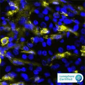
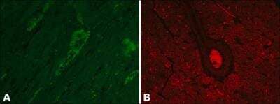
MAB8170) at 20ug/mL at 37 ° Celsius for 4 minutes. Before incubation with the primary antibody, tissue underwent an all-in-one dewaxing and antigen retrieval preprocessing using PreTreatment Module (PT Module) and Dewax and HIER Buffer H (pH 9; Epredia Catalog # TA-999-DHBH).Tissue was stained using the Alexa Fluor™ 647 Goat anti-Mouse IgG Secondary Antibody at 1:200 at 37 ° Celsius for 2 minutes. (Yellow; Lunaphore Catalog # DR647MS) and counterstained with DAPI (blue; Lunaphore Catalog # DR100). Specific staining was localized to the cytoplasm. Protocol available in COMET™ Panel Builder." title="Gelsolin/GSN was detected in immersion fixed paraffin-embedded sections of human Renal Cell Carcinoma using Mouse Anti-Human Gelsolin/GSN, pan Monoclonal Antibody (Catalog # MAB8170) at 20ug/mL at 37 ° Celsius for 4 minutes. Before incubation with the primary antibody, tissue underwent an all-in-one dewaxing and antigen retrieval preprocessing using PreTreatment Module (PT Module) and Dewax and HIER Buffer H (pH 9; Epredia Catalog # TA-999-DHBH).Tissue was stained using the Alexa Fluor™ 647 Goat anti-Mouse IgG Secondary Antibody at 1:200 at 37 ° Celsius for 2 minutes. (Yellow; Lunaphore Catalog # DR647MS) and counterstained with DAPI (blue; Lunaphore Catalog # DR100). Specific staining was localized to the cytoplasm. Protocol available in COMET™ Panel Builder." />
| Applications | WB, Simple Western, IHC, KO, mIF |
| Clone | 893205 |
| Clonality | Monoclonal |
| Host | Mouse |
| Conjugate | Unconjugated |
| Concentration | LYOPH |
| Immunogen | HEK293 human embryonic kidney cell line transfected with human Gelsolin/GSN Met1-Ala782 Accession # P06396 |
| Specificity | Detects human Gelsolin/GSN in ELISAs. Detects human, mouse and rat Gelsolin/GSN in Western Blots. |
| Source | N/A |
| Isotype | IgG1 |
| Clonality | Monoclonal |
| Host | Mouse |
| Gene | GSN |
| Purity Statement | Protein A or G purified from hybridoma culture supernatant |
| Innovator's Reward | Test in a species/application not listed above to receive a full credit towards a future purchase. |
| Dilutions |
|
|
| Publications |
|
| Storage | Use a manual defrost freezer and avoid repeated freeze-thaw cycles.
|
| Buffer | Lyophilized from a 0.2 μm filtered solution in PBS with Trehalose. See Certificate of Analysis for details. *Small pack size (-SP) is supplied either lyophilized or as a 0.2 µm filtered solution in PBS. |
| Preservative | No Preservative |
| Concentration | LYOPH |
| Reconstitution Instructions | Reconstitute at 0.5 mg/mL in sterile PBS. For liquid material, refer to CoA for concentration. |







![Gelsolin/GSN Antibody (893205) [Unconjugated]](/sites/all/modules/enterprise-tech/et_datasheets/images/novus_guarantee.png "Gelsolin/GSN Antibody (893205) [Unconjugated]")
| Publication using MAB8170 | Applications | Species |
|---|---|---|
| Z Chen, K Li, X Yin, H Li, Y Li, Q Zhang, H Wang, Y Qiu Lower Expression of Gelsolin in Colon Cancer and Its Diagnostic Value in Colon Cancer Patients J Cancer, 2019-01-30;10(5):1288-1296. 2019-01-30 [PMID: 30854138] (Neutralization, Human) | Neutralization | Human |
Secondary Antibodies |
Isotype Controls |
The concentration calculator allows you to quickly calculate the volume, mass or concentration of your vial. Simply enter your mass, volume, or concentration values for your reagent and the calculator will determine the rest.


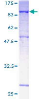
 HAF018). A specific band was detected for Gelsolin/GSN at approximately 95 kDa (as indicated). This experiment was conducted under reducing conditions and using
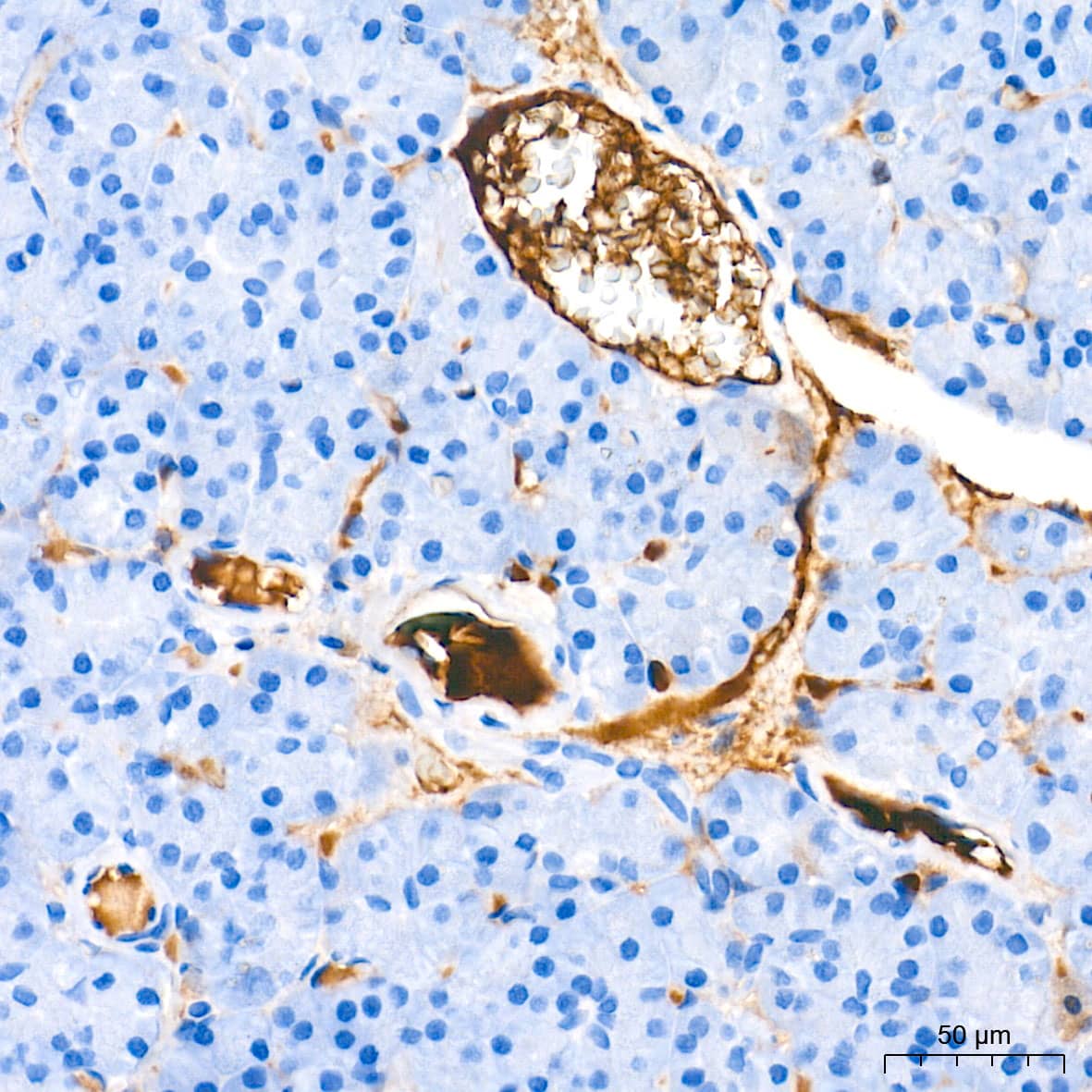
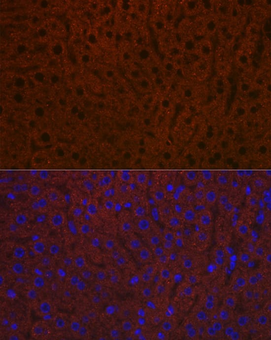
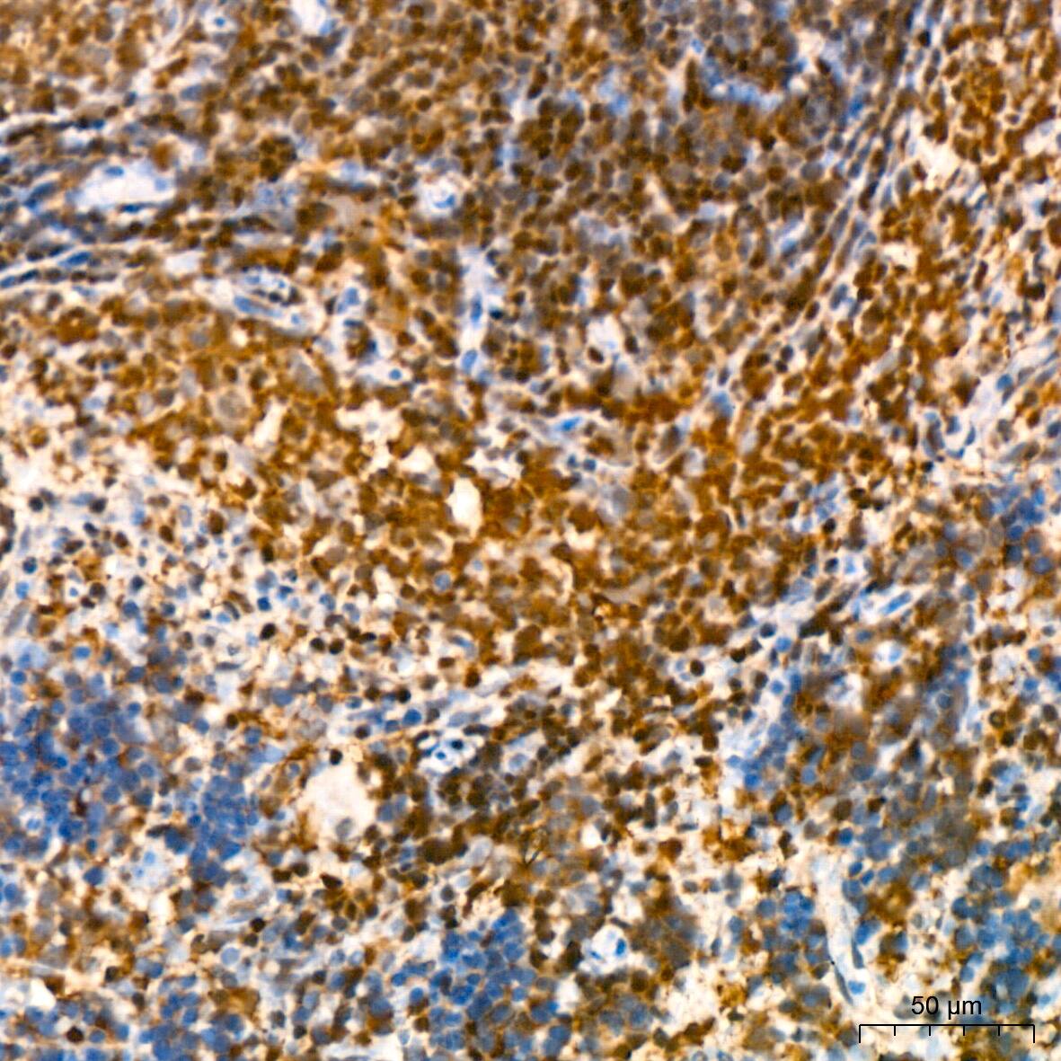
 CTS002) and counterstained with hematoxylin (blue). Specific staining was localized to glomeruli and distal convoluted tubules. View our protocol for
![SDS-Page Neuregulin-1 beta 1/NRG1 beta 1 [Unconjugated]](https://images.novusbio.com/images/protein/Neuregulin-1_beta_1_396-HB_222.jpg)
![SEC-MALS Neuregulin-1 beta 1/NRG1 beta 1 [Unconjugated]](https://images.novusbio.com/images/396-hb_recombinant-human-nrg1-beta-1-hrg1-beta-1-egf-domain-protein-sec-mals-51220258550.jpg)
![Bioactivity Neuregulin-1 beta 1/NRG1 beta 1 [Unconjugated]](https://images.novusbio.com/images/protein/Neuregulin-1_beta_1_396-HB_626.jpg)
![Immunohistochemistry Caspase-3 Antibody [Unconjugated] - Active](https://images.novusbio.com/images/af835_human-mouse-active-caspase-3-affinity-purified-pab-41202410331943.jpg)
![Western Blot Caspase-3 Antibody [Unconjugated] - Active](https://images.novusbio.com/images/af835_human-mouse-active-caspase-3-affinity-purified-pab-812025554170.jpg)
![Western Blot Caspase-3 Antibody [Unconjugated] - Active](https://images.novusbio.com/images/af835_human-mouse-active-caspase-3-affinity-purified-pab-8120255534731.jpg)


![Western Blot Myosin Heavy Chain Antibody (MF20) [Unconjugated]](https://images.novusbio.com/images/mab4470_myosin-heavy-chain-mab-clone-mf20-western-blot-121220259221219.jpg)
![Western Blot Myosin Heavy Chain Antibody (MF20) [Unconjugated]](https://images.novusbio.com/images/mab4470_myosin-heavy-chain-mab-clone-mf20-western-blot-12122025920504.jpg)
![Western Blot Myosin Heavy Chain Antibody (MF20) [Unconjugated]](https://images.novusbio.com/images/mab4470_myosin-heavy-chain-mab-clone-mf20-western-blot-12122025849812.jpg)


![Western Blot Angiopoietin-like Protein 2/ANGPTL2 Antibody [Unconjugated]](https://images.novusbio.com/images/antibody/Angiopoietinlike_Protein_2_AF1444_Western_Blot_19840.jpg)


![Bioactivity IL-2 [Unconjugated]](https://images.novusbio.com/images/202-il_recombinant-human-il-2-protein-bioactivity-174202314946.jpg)