| Immunogen | This affinity purified antibody was prepared by repeated immunizations with a synthetic peptide corresponding to a region near the amino terminal end of human NAG-1 protein. A residue of cysteine was added to facilitate coupling to KLH. |
| Specificity | This antibody specifically reacts with the amino terminal end of human NAG-1 protein from human tissues. A BLAST analysis was used to suggest partial reactivity. |
| Clonality | Polyclonal |
| Host | Rabbit |
| Gene | GDF15 |
| Purity | Immunogen affinity purified |
| Innovator's Reward | Test in a species/application not listed above to receive a full credit towards a future purchase. |
| Dilutions |
|
|
| Application Notes | This affinity purified antibody is suitable for ELISA and western blotting of human and mouse NAG-1 protein. For detection of NAG-1 in human serum, a sandwich ELISA is suggested using this antibody in combination with anti-NAG-1/GDF15 , H variant or D variant specific antibodies. Expect bands in Western blots of approximately 14 and 28 kDa in size corresponding - NAG-1 monomer and dimer, respectively, using the appropriate cell lysate or extract. Although not confirmed, this antibody may work in IHC. |
|
| Readout System | ||
| Reviewed Applications |
|
| Storage | Store at 4C short term. Aliquot and store at -20C long term. Avoid freeze-thaw cycles. |
| Buffer | 0.02 M Potassium Phosphate, 0.15 M Sodium Chloride, pH 7.2, 10 mg/mL Bovine Serum Albumin (BSA) - Immunoglobulin and Protease free |
| Preservative | 0.01% Sodium Azide |
| Concentration | LYOPH |
| Purity | Immunogen affinity purified |
| Reconstitution Instructions | Reconstitute with 50 ul of distilled water to a final concentration of 1.0 mg/ml. |


![Western Blot AKT [p Ser473] Antibody [Unconjugated] - Pan Specific](https://images.novusbio.com/images2/Akt3_AF887_Western_Blot_5930.jpg)
![Simple Western AKT [p Ser473] Antibody [Unconjugated] - Pan Specific](https://images.novusbio.com/images2/16332.jpg)
![Intracellular Staining by Flow Cytometry AKT [p Ser473] Antibody [Unconjugated] - Pan Specific](https://images.novusbio.com/images2/Akt3_AF887_Flow_Cytometry_8283.jpg)
![Immunohistochemistry EGFR Antibody [Unconjugated]](https://images.novusbio.com/images2/EGF_R_AF231_Immunohistochemistry_20892.jpg)
![Immunocytochemistry EGFR Antibody [Unconjugated]](https://images.novusbio.com/images2/EGF_R_AF231_Immunocytochemistry__Immunofluorescence_21143.jpg)
![Western Blot EGFR Antibody [Unconjugated]](https://images.novusbio.com/images2/EGF_R_AF231_Western_Blot_19925.jpg)


| Images | Ratings | Applications | Species | Date | Details | ||||||
|---|---|---|---|---|---|---|---|---|---|---|---|
Enlarge |
reviewed by:
Verified Customer |
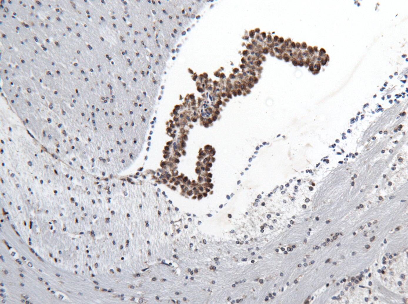
IHC-P | Mouse | 05/22/2014 |
Summary
|
Secondary Antibodies |
Isotype Controls |
Research Areas for GDF-15 Antibody (NBP1-42720)Find related products by research area.
|
|
Taking Biomarker Discovery From 2D to 3D: Increased Biological Activity of EVs Isolated From 3D Prostate Cancer Cultures Jamshed Arslan, Pharm D, PhD Tissues within the human body are made of a three-dimensional (3D) arrangement of cells working together to perform vital functions. The commonly used 2D monolayer cultures have limited ... Read full blog post. |
The concentration calculator allows you to quickly calculate the volume, mass or concentration of your vial. Simply enter your mass, volume, or concentration values for your reagent and the calculator will determine the rest.
| Gene Symbol | GDF15 |


![Immunocytochemistry EGR1 Antibody [Unconjugated]](https://images.novusbio.com/images/antibody/af2818_human-egr1-affinity-purified-polyclonal-ab-immunocytochemistry-3102024831..jpg)
![N/A IL-6 [HRP]](https://images.novusbio.com/images2/DATA_IL6_M6000_ELISA_936.jpg)
![N/A IL-6 [HRP]](https://images.novusbio.com/images2/IL-6_M6000_ELISA_415.jpg)
![N/A IL-6 [HRP]](https://images.novusbio.com/images/m6000b_mouse-il-6-quantikine-elisa-kit-44202415433118.jpg)


![SDS-PAGE TNF-alpha [Unconjugated]](https://images.novusbio.com/images2/TNF-alpha_210-TA_256.jpg)
![Bioactivity TNF-alpha [Unconjugated]](https://images.novusbio.com/images2/TNFalpha_210TA_1658.jpg)
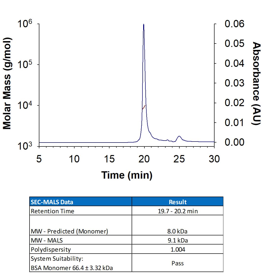
![SEC-MALS TNF-alpha [Unconjugated]](https://images.novusbio.com/images/210-ta_recombinant-human-tnf-alpha-protein-sec-mals-35202312244..jpg)
![Western Blot ERK2 Antibody [Unconjugated]](https://images.novusbio.com/images2/ERK2_AF1230_Western_Blot_5097.jpg)
![Immunohistochemistry ERK2 Antibody [Unconjugated]](https://images.novusbio.com/images2/ERK2_AF1230_Immunohistochemistry_20696.jpg)
![Knockout Validated ERK2 Antibody [Unconjugated]](https://images.novusbio.com/images2/ERK2_AF1230_Knockout_Validated_22864.jpg)


![N/A C-Reactive Protein/CRP [Biotin]](https://images.novusbio.com/images2/DATA_CReactive_Protein_DY1707_ELISA_1701.jpg)
![Immunocytochemistry ErbB4/Her4 Antibody (182803) [Unconjugated]](https://images.novusbio.com/images2/ErbB4_MAB1131_Immunocytochemistry__Immunofluorescence_21145.jpg)
![Immunohistochemistry ErbB4/Her4 Antibody (182803) [Unconjugated]](https://images.novusbio.com/images2/ErbB4_MAB1131_Immunohistochemistry_12968.jpg)


![N/A Hepcidin Antimicrobial Peptide [HRP]](https://images.novusbio.com/images2/Hepcidin_DHP250_ELISA_1121.jpg)
![N/A Hepcidin Antimicrobial Peptide [HRP]](https://images.novusbio.com/images2/Hepcidin_DHP250_ELISA_1122.jpg)
![SDS-PAGE ErbB2/Her2 [Unconjugated]](https://images.novusbio.com/images2/ErbB2_1129-ER_70.jpg)
![Bioactivity ErbB2/Her2 [Unconjugated]](https://images.novusbio.com/images2/1129-er_recombinant-human-erbb2-her2-fc-chimera-protein-cf-bioactivity-7122020142841.jpg)


![Immunocytochemistry Caspase-3 Antibody [Unconjugated] - Active](https://images.novusbio.com/images2/Caspase-3_AF835_Immunocytochemistry_6532.jpg)
![Immunohistochemistry Caspase-3 Antibody [Unconjugated] - Active](https://images.novusbio.com/images2/Caspase3_AF835_Immunohistochemistry_22976.jpg)
![Immunocytochemistry Caspase-3 Antibody [Unconjugated] - Active](https://images.novusbio.com/images2/Caspase-3_AF835_Immunocytochemistry_9340.jpg)
