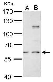
 followed by HRP-conjugated Anti-Goat IgG Secondary Antibody (HAF017). A specific band was detected for Decorin at approximately 100 kDa (as indicated). This experiment was conducted under reducing conditions and using Immunoblot Buffer Group 1.")
| Reactivity | HuSpecies Glossary |
| Applications | WB, Simple Western, IHC |
| Clonality | Polyclonal |
| Host | Goat |
| Conjugate | Unconjugated |
| Concentration | LYOPH |
| Immunogen | S. frugiperda insect ovarian cell line Sf 21-derived recombinant human Decorin Gly17-Lys359 Accession # P07585 |
| Specificity | Detects human Decorin in direct ELISAs and Western blots. |
| Source | N/A |
| Isotype | IgG |
| Clonality | Polyclonal |
| Host | Goat |
| Gene | DCN |
| Purity Statement | Antigen Affinity-purified |
| Innovator's Reward | Test in a species/application not listed above to receive a full credit towards a future purchase. |
| Dilutions |
|
|
| Publications |
|
| Storage | Use a manual defrost freezer and avoid repeated freeze-thaw cycles.
|
| Buffer | Lyophilized from a 0.2 μm filtered solution in PBS with Trehalose. *Small pack size (SP) is supplied either lyophilized or as a 0.2 µm filtered solution in PBS. |
| Preservative | No Preservative |
| Concentration | LYOPH |
| Reconstitution Instructions | Reconstitute at 0.2 mg/mL in sterile PBS. |
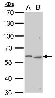
Decorin is a small secreted chondroitin/dermatan sulfate proteoglycan in the family of small leucine-rich proteoglycans (SLRPs). SLRP family members are characterized by N-terminal and C-terminal cysteine-rich regions which flank the central region containing 10‑12 tandem leucine-rich repeats (LRR) (1, 2). The human Decorin cDNA encodes a 359 amino acid (aa) precursor that includes a 16 aa signal sequence and a 14 aa propeptide. The 329 aa mature protein contains twelve LRR. Alternate splicing generates five isoforms with variable length deletions (3). Mature human and mouse Decorin share 80% aa sequence identity. In Decorin, serine 34 in the N-terminal domain is O-glycosylated. Naturally occurring Decorin proteoglycan has a molecular mass of approximately 100 kDa, and the deglycosylated Decorin core protein has a mass of approximately 40 kDa. Decorin binds to fibronectin, TGF-beta , and type I and type II collagens. The binding of Decorin to various molecules was reported to be mediated via the core protein. Decorin has been implicated in matrix assembly and has also been reported to suppress the growth of various tumor cell lines by activating the epidermal growth factor receptor.


![Western Blot Osteopontin/OPN Antibody [Unconjugated]](https://images.novusbio.com/images/af808_mouse-osteopontin-opn-affinity-purified-polyclonal-ab-western-blot-121220259133019.jpg)
![Western Blot Osteopontin/OPN Antibody [Unconjugated]](https://images.novusbio.com/images/af808_mouse-osteopontin-opn-affinity-purified-polyclonal-ab-western-blot-12122025932150.jpg)
![Western Blot Osteopontin/OPN Antibody [Unconjugated]](https://images.novusbio.com/images/af808_mouse-osteopontin-opn-affinity-purified-polyclonal-ab-western-blot-121220259224819.jpg)
![Immunocytochemistry EGFR Antibody [Unconjugated]](https://images.novusbio.com/images/antibody/EGF_R_AF231_Immunocytochemistry__Immunofluorescence_21143.jpg)
![Flow Cytometry EGFR Antibody [Unconjugated]](https://images.novusbio.com/images/antibody/EGF_R_AF231_Flow_Cytometry_20401.jpg)
![Western Blot EGFR Antibody [Unconjugated]](https://images.novusbio.com/images/antibody/EGF_R_AF231_Western_Blot_19925.jpg)


Secondary Antibodies |
Isotype Controls |
The concentration calculator allows you to quickly calculate the volume, mass or concentration of your vial. Simply enter your mass, volume, or concentration values for your reagent and the calculator will determine the rest.


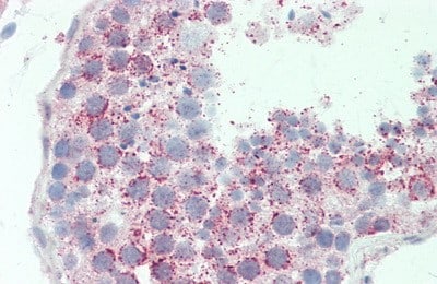
 at 15 µg/mL overnight at 4 °C. Tissue was stained using the Anti-Goat HRP-DAB Cell & Tissue Staining Kit (brown; (CTS008) and counterstained with hematoxylin (blue). Specific staining was localized to intralobular stromal tissue. View our protocol for Chromogenic IHC Staining of Paraffin-embedded Tissue Sections.")
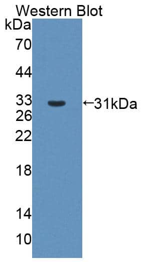
 using 5 µg/mL of Goat Anti-Human Decorin Antigen Affinity-purified Polyclonal Antibody (Catalog # AF143) . This experiment was conducted under reducing conditions and using the 12-230 kDa separation system.")
 Immunohistochemistry using a polyclonal goat anti-human DCN antibody and Alexa Fluor 488 secondary antibody (green fluorescence), and counterstained with DAPI (blue fluorescence) in representative site-matched sections of NS and HSc (scale bar = 50 μm). (b) Relative expression of DCN in matched superficial and deep NS and HSc sections was calculated from fluorescence using ImageJ (mean ± SEM, n = 4 patients, * P < 0.001). Image collected and cropped by CiteAb from the following open publication (//dx.plos.org/10.1371/journal.pone.0123054), licensed under a CC-BY license. Not internally tested by R&D Systems.")
 Immunohistochemistry using a polyclonal goat anti-human DCN antibody and Alexa Fluor 488 secondary antibody (green fluorescence), and counterstained with DAPI (blue fluorescence) in representative site-matched sections of NS and HSc (scale bar = 50 μm). (b) Relative expression of DCN in matched superficial and deep NS and HSc sections was calculated from fluorescence using ImageJ (mean ± SEM, n = 4 patients, * P < 0.001). Image collected and cropped by CiteAb from the following open publication (//dx.plos.org/10.1371/journal.pone.0123054), licensed under a CC-BY license. Not internally tested by R&D Systems.")


![Immunohistochemistry Lumican Antibody [Unconjugated]](https://images.novusbio.com/images/antibody/Lumican_AF2846_Immunohistochemistry_17809.jpg)
![Western Blot Lumican Antibody [Unconjugated]](https://images.novusbio.com/images/antibody/Lumican_AF2846_Western_Blot_17014.jpg)
![Simple Western Lumican Antibody [Unconjugated]](https://images.novusbio.com/images/antibody/af2846_human-lumican-affinity-purified-polyclonal-ab-simple-western-332025105038..jpg)

![Immunocytochemistry SPARC Antibody [Unconjugated]](https://images.novusbio.com/images/antibody/SPARC_AF942_Immunocytochemistry__Immunofluorescence_23321.jpg)
![Intracellular Staining by Flow Cytometry SPARC Antibody [Unconjugated]](https://images.novusbio.com/images/antibody/af942_mouse-sparc-affinity-purified-polyclonal-ab-intracellular-staining-by-flow-cytometry-22122020141853.jpg)
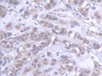
![Immunohistochemistry SPARC Antibody [Unconjugated]](https://images.novusbio.com/images/antibody/SPARC_AF942_Immunohistochemistry_10419.jpg)


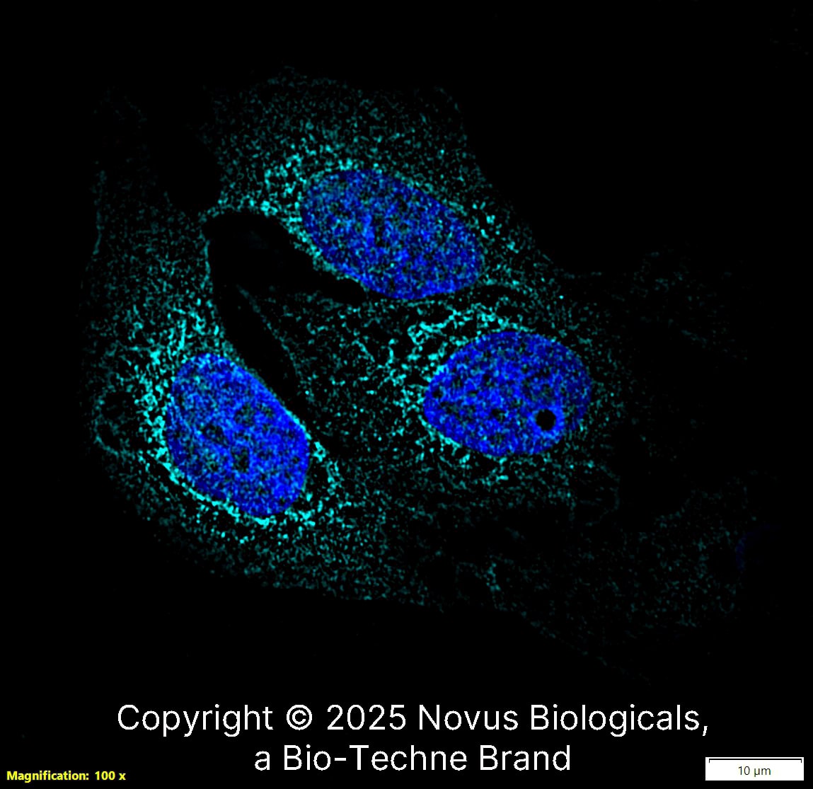
![Immunocytochemistry Catalase Antibody [Unconjugated]](https://images.novusbio.com/images/antibody/Catalase_AF3398_Immunocytochemistry__Immunofluorescence_19451.jpg)
![Simple Western Catalase Antibody [Unconjugated]](https://images.novusbio.com/images/antibody/Catalase_AF3398_Simple_Western_16990.jpg)


 or Normal Goat IgG Isotype Control Antibody (Catalog # AB-108-C, open histogram), followed by Phycoerythrin-conjugated Anti-Goat IgG Secondary Antibody (Catalog # F0107).")