HAF007). A specific band was detected for Calponin 1 at approximately 40 kDa (as indicated). This experiment was conducted under reducing conditions and using Immunoblot Buffer Group 1." title="Western blot shows lysates of MDA-MB-231 human breast cancer cell line, MCF-7 human breast cancer cell line, NMuMG mouse mammary gland epithelial cell line, and A7r5 rat thoracic aortic smooth muscle cell line. PVDF membrane was probed with 1 µg/mL of Mouse Anti-Human Calponin 1 Monoclonal Antibody (Catalog # MAB7900) followed by HRP-conjugated Anti-Mouse IgG Secondary Antibody (Catalog # HAF007). A specific band was detected for Calponin 1 at approximately 40 kDa (as indicated). This experiment was conducted under reducing conditions and using Immunoblot Buffer Group 1." />
| Reactivity | HuSpecies Glossary |
| Applications | WB, IHC, ICC/IF |
| Clone | 836701 |
| Clonality | Monoclonal |
| Host | Mouse |
| Conjugate | Unconjugated |
| Concentration | LYOPH |
| Immunogen | E. coli-derived recombinant human Calponin 1 Ser2-Ala297 Accession # P51911 |
| Specificity | Detects human Calponin 1 in ELISAs. Detects human, mouse and rat Calponin 1 in Western Blots. In direct ELISAs, no cross-reactivity with recombinant human Calponin 3 is observed. |
| Source | N/A |
| Isotype | IgG3 |
| Clonality | Monoclonal |
| Host | Mouse |
| Gene | CNN1 |
| Purity Statement | Protein A or G purified from hybridoma culture supernatant |
| Innovator's Reward | Test in a species/application not listed above to receive a full credit towards a future purchase. |
| Storage | Use a manual defrost freezer and avoid repeated freeze-thaw cycles.
|
| Buffer | Lyophilized from a 0.2 μm filtered solution in TBS with Trehalose. *Small pack size (SP) is supplied either lyophilized or as a 0.2 µm filtered solution in PBS. |
| Preservative | No Preservative |
| Concentration | LYOPH |
| Reconstitution Instructions | Sterile PBS to a final concentration of 0.5 mg/mL. |
CNN-1 (Calponin 1 [calcium and calmodulin-binding troponin T-like protein]; also Calponin basic, CaP and Calponin H1) is a 32-36 kDa cytoplasmic member of the calponin family of proteins. Although reportedly expressed in fibroblasts and endothelial cells, it actually appears to be restricted to smooth muscle and smooth muscle-like cells such as myoepithelium and myofibroblasts in the adult. CNN-1 interacts with F-actin in a phosphorylation-dependent manner. When nonphosphorylated, CNN-1 blocks actomyosin ATPase activity, contributing to the stabilization of actin stress fiber bundles. Thus, CNN-1 expression inhibits cell motility and the formation of podosomes. Human CNN-1 is 297 amino acids (aa) in length. It contains one CH/calponin homology domain (aa 30-127), and three consecutive calponin-like repeats (aa 164-268). The repeats are suggested to mediate actin binding. There are five potential Ser/Thr phosphorylation sites. Full-length human CNN-1 shares 97% aa sequence identity with mouse CNN-1.




![Flow Cytometry CD31/PECAM-1 Antibody [Unconjugated]](https://images.novusbio.com/images/af3628_human-mouse-rat-cd31-pecam-1-affinity-purified-polyclonal-ab-flow-cytometry-30112023144624..jpg)
![Western Blot CD31/PECAM-1 Antibody [Unconjugated]](https://images.novusbio.com/images/af3628_human-mouse-rat-cd31-pecam-1-affinity-purified-polyclonal-ab-41202410325588.jpg)
![Immunohistochemistry CD31/PECAM-1 Antibody [Unconjugated]](https://images.novusbio.com/images/antibody/af3628_mouse-rat-cd31-pecam-1-affinity-purified-polyclonal-ab-immunohistochemistry-2572023161128.jpg)




![Immunohistochemistry Albumin Antibody (188835) [Unconjugated] - Serum](https://images.novusbio.com/images/antibody/Albumin_MAB1455_Immunohistochemistry_23317.jpg)
![Immunocytochemistry Albumin Antibody (188835) [Unconjugated] - Serum](https://images.novusbio.com/images/antibody/Albumin_MAB1455_Immunocytochemistry__Immunofluorescence_17777.jpg)
![Intracellular Staining by Flow Cytometry Albumin Antibody (188835) [Unconjugated] - Serum](https://images.novusbio.com/images/antibody/Albumin_MAB1455_Flow_Cytometry_15110.jpg)
Secondary Antibodies |
Isotype Controls |
The concentration calculator allows you to quickly calculate the volume, mass or concentration of your vial. Simply enter your mass, volume, or concentration values for your reagent and the calculator will determine the rest.


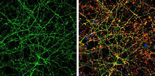
 NL007) and counterstained with DAPI (blue). Specific staining was localized to cytoplasm. View our protocol for
  CTS013). Tissue was stained using the Anti-Mouse HRP-DAB Cell & Tissue Staining Kit (brown; Catalog #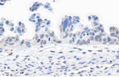


![N/A CD40 Ligand/TNFSF5 [HRP]](https://images.novusbio.com/images/elisa/CD40_Ligand_DCDL40_ELISA_60.jpg)
![N/A CD40 Ligand/TNFSF5 [HRP]](https://images.novusbio.com/images/elisa/DATA_CD40_Ligand_DCDL40_ELISA_624.jpg)

![Bioactivity ErbB2/Her2 [Unconjugated]](https://images.novusbio.com/images/protein/1129-er_recombinant-human-erbb2-her2-fc-chimera-protein-cf-bioactivity-7122020142841.jpg)
![SDS-Page ErbB2/Her2 [Unconjugated]](https://images.novusbio.com/images/protein/ErbB2_1129-ER_70.jpg)
![Western Blot TRAF-2 Antibody (324522) [Unconjugated]](https://images.novusbio.com/images/antibody/TRAF-2_MAB3277_Western_Blot_6081.jpg)