
| Reactivity | Hu, Mu, Po, Ca, Pm, Rt(-)Species Glossary |
| Applications | WB, Func, ICC/IF, IHC, IP, Dual ISH-IHC, KO |
| Clone | E6-6 |
| Clonality | Monoclonal |
| Host | Mouse |
| Conjugate | Unconjugated |
| Format | BSA Free |
| Concentration | 1.0 mg/ml |
| Immunogen | Synthetic peptide conjugated to KLH corresponding to the C-terminus of human Bestrophin 1 (KDHMDPYWALENRDEAHS) [Uniprot: O76090] |
| Isotype | IgG1 Kappa |
| Clonality | Monoclonal |
| Host | Mouse |
| Gene | BEST1 |
| Purity | Protein A or G purified |
| Innovator's Reward | Test in a species/application not listed above to receive a full credit towards a future purchase. |
| Dilutions |
|
|
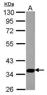
| Application Notes | In Western blot, this antibody recognizes a band at ~68 kDa representing Bestrophin. Please see protocol for treatment of cell extracts. The observed molecular weight of the protein may vary from the listed predicted molecular weight due to post translational modifications, post translation cleavages, relative charges, and other experimental factors. |
|
| Reviewed Applications |
|
|
| Publications |
|
| Storage | Aliquot and store at -20C or -80C. Avoid freeze-thaw cycles. |
| Buffer | PBS |
| Preservative | 0.02% Sodium Azide |
| Concentration | 1.0 mg/ml |
| Purity | Protein A or G purified |
![Immunohistochemistry ACE/CD143 Antibody [Unconjugated]](https://images.novusbio.com/images/antibody/ACE_AF1513_Immunohistochemistry_6703.jpg)
![Western Blot ACE/CD143 Antibody [Unconjugated]](https://images.novusbio.com/images/antibody/ACE_AF1513_Flow_Cytometry_20028.jpg)
![Simple Western ACE/CD143 Antibody [Unconjugated]](https://images.novusbio.com/images/antibody/ACE_AF1513_Simple_Western_20109.jpg)


| Images | Ratings | Applications | Species | Date | Details | ||||||||
|---|---|---|---|---|---|---|---|---|---|---|---|---|---|

Enlarge |
reviewed by:
Verified Customer |
WB | Human and Rat | 12/29/2016 |
Summary
Comments
|
Secondary Antibodies |
Isotype Controls |
Research Areas for Bestrophin 1 Antibody (NB300-164)Find related products by research area.
|
|
Vision Infographic: Do you see how I see? Vision involves several parts of the eye processing light which send signals to the brain via the optic nerve to process information. Learn more about the vision process and related ocular proteins in the infographic below. Novus Biological... Read full blog post. |
|
Bestrophin 1: Implications in Progressive Vision Loss The human Bestrophin family has four members, Best1, Best2, Best3 and Best4. These transmembrane proteins can function as chloride channels, and can also regulate calcium channels (1). The Bestrophins all have a conserved domain which begins at the N-... Read full blog post. |
|
"I can see clearly now": Targeting Bestrophin 1 to treat Bestrophinopathies Encoded by the VMD2 gene on chromosome 11q13 Bestrophin 1 is the prototypic member of the RFP family of proteins which are more commonly called "bestrophins". The protein family was originally identified in Caenorhabditis elegans based on a conserved ... Read full blog post. |
The concentration calculator allows you to quickly calculate the volume, mass or concentration of your vial. Simply enter your mass, volume, or concentration values for your reagent and the calculator will determine the rest.


![Immunohistochemistry-Paraffin: Bestrophin 1 Antibody (E6-6) - BSA Free [NB300-164] - Bestrophin 1 was detected in immersion fixed paraffin sections of human small intestine using t Mouse Anti-Human Bestrophin 1 Monoclonal Antibody (Catalog # NB300-164) at 5 ug/mL for 1 hour at room temperature followed by incubation with the Anti-Mouse IgG VisUCyte™ HRP Polymer Antibody (Catalog # VC001). Tissue was stained using DAB (brown) and counterstained with hematoxylin (blue). Specific staining was localized to the cell surface and extracellular.](http://images.novusbio.com/fullsize/Bestrophin-1-Antibody-E6-6-BSA-Free-Immunohistochemistry-Paraffin-NB300-164-img0005.jpg "Immunohistochemistry-Paraffin: Bestrophin 1 Antibody (E6-6) - BSA Free [NB300-164] - Bestrophin 1 was detected in immersion fixed paraffin sections of human small intestine using t Mouse Anti-Human Bestrophin 1 Monoclonal Antibody (Catalog # NB300-164) at 5 ug/mL for 1 hour at room temperature followed by incubation with the Anti-Mouse IgG VisUCyte™ HRP Polymer Antibody (Catalog # VC001). Tissue was stained using DAB (brown) and counterstained with hematoxylin (blue). Specific staining was localized to the cell surface and extracellular.")
![Western Blot: Bestrophin 1 Antibody (E6-6) - BSA Free [NB300-164] - Detection of Bestrophin (68 kDa) from human RPE cell lysate.](http://images.novusbio.com/fullsize/Bestrophin-1-Antibody-E6-6-BSA-Free-Western-Blot-NB300-164-img0003.jpg "Western Blot: Bestrophin 1 Antibody (E6-6) - BSA Free [NB300-164] - Detection of Bestrophin (68 kDa) from human RPE cell lysate.")
![Immunohistochemistry-Paraffin: Bestrophin 1 Antibody (E6-6) - BSA Free [NB300-164] - Bestrophin 1 was detected in immersion fixed paraffin-embedded sections of human brain using Mouse Anti-Human Bestrophin 1 (E6-6) Monoclonal Antibody (Catalog # NB300-164) at 1:300 for 1 hour at room temperature followed by incubation with the Anti-Mouse IgG VisUCyte™ HRP Polymer Antibody (Catalog # VC001). Tissue was stained using DAB (brown) and counterstained with hematoxylin (blue). Specific staining was localized to the cytoplasm in neurons.](http://images.novusbio.com/fullsize/Bestrophin-1-Antibody-E6-6-BSA-Free-Immunohistochemistry-Paraffin-NB300-164-img0004.jpg "Immunohistochemistry-Paraffin: Bestrophin 1 Antibody (E6-6) - BSA Free [NB300-164] - Bestrophin 1 was detected in immersion fixed paraffin-embedded sections of human brain using Mouse Anti-Human Bestrophin 1 (E6-6) Monoclonal Antibody (Catalog # NB300-164) at 1:300 for 1 hour at room temperature followed by incubation with the Anti-Mouse IgG VisUCyte™ HRP Polymer Antibody (Catalog # VC001). Tissue was stained using DAB (brown) and counterstained with hematoxylin (blue). Specific staining was localized to the cytoplasm in neurons.")

![Dual RNAscope ISH-IHC: Bestrophin 1 Antibody (E6-6) - BSA Free [NB300-164] - Formalin-fixed paraffin-embedded tissue sections of human duodenum were probed for Bestrophin 1 mRNA (ACD RNAScope Probe, catalog # 433181; Fast Red chromogen, ACD catalog # 322360). Adjacent tissue section was processed for immunohistochemistry using mouse anti-human (Novus Biologicals catalog # NB300-164) at 0.3ug/mL with overnight incubation at 4 degrees Celsius followed by incubation with anti-mouse IgG VisUCyte HRP Polymer Antibody (Catalog # VC001) and DAB chromogen (yellow-brown). Tissue was counterstained with hematoxylin (blue). IHC signal is confined to cytoplasm.](http://images.novusbio.com/fullsize/Bestrophin-1-Antibody-E6-6-BSA-Free-Dual-RNAscope-ISH-IHC-NB300-164-img0006.jpg "Dual RNAscope ISH-IHC: Bestrophin 1 Antibody (E6-6) - BSA Free [NB300-164] - Formalin-fixed paraffin-embedded tissue sections of human duodenum were probed for Bestrophin 1 mRNA (ACD RNAScope Probe, catalog # 433181; Fast Red chromogen, ACD catalog # 322360). Adjacent tissue section was processed for immunohistochemistry using mouse anti-human (Novus Biologicals catalog # NB300-164) at 0.3ug/mL with overnight incubation at 4 degrees Celsius followed by incubation with anti-mouse IgG VisUCyte HRP Polymer Antibody (Catalog # VC001) and DAB chromogen (yellow-brown). Tissue was counterstained with hematoxylin (blue). IHC signal is confined to cytoplasm.")
 Immunoblotting showing the expression of RPE-specific proteins BEST1, RPE65, CRALBP, & the loading control beta -Actin in hPSC-RPE (a) & iPSC-RPE (b) cells. Two gels/blots in the same panel were prepared from the same cell lysate of each PSC-RPE to detect BEST1 + beta -Actin, & RPE65 + CRALBP, respectively. Image collected & cropped by CiteAb from the following publication (//pubmed.ncbi.nlm.nih.gov/34061021), licensed under a CC-BY license. Not internally tested by Novus Biologicals.")
![Western Blot: Bestrophin 1 Antibody (E6-6) - BSA Free [NB300-164] - (a) Schematic drawing showing localization of the different mutations (black diamonds) tested in our study (modified from [12]); (b) Western blot analysis of the normal & mutant human Best1 protein in transiently transfected MDCK cells. Best1 proteins are detectable as a 68 kDa band in all transfected cells, but not in non-transfected controls (MDCK lane). Actin bands are shown to indicate equal loading of cell lysates. Image collected & cropped by CiteAb from the following publication (//pubmed.ncbi.nlm.nih.gov/23880862), licensed under a CC-BY license. Not internally tested by Novus Biologicals.](http://images.novusbio.com/fullsize/nb300-164_mouse-monoclonal-bestrophin-1-antibody-e6-6-31020241534336.jpg "Western Blot: Bestrophin 1 Antibody (E6-6) - BSA Free [NB300-164] - (a) Schematic drawing showing localization of the different mutations (black diamonds) tested in our study (modified from [12]); (b) Western blot analysis of the normal & mutant human Best1 protein in transiently transfected MDCK cells. Best1 proteins are detectable as a 68 kDa band in all transfected cells, but not in non-transfected controls (MDCK lane). Actin bands are shown to indicate equal loading of cell lysates. Image collected & cropped by CiteAb from the following publication (//pubmed.ncbi.nlm.nih.gov/23880862), licensed under a CC-BY license. Not internally tested by Novus Biologicals.")
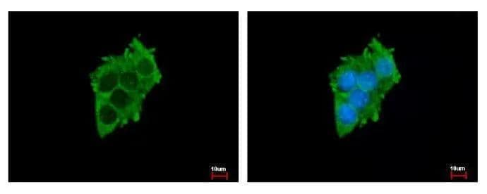
![Immunocytochemistry/ Immunofluorescence: Bestrophin 1 Antibody (E6-6) - BSA Free [NB300-164] - Subcellular localization of WT & mutant BEST1 in iPSC-RPEs. Confocal images showing the co-staining of BEST1, Collagen IV & Hoechst in iPSC-RPEs derived from a WT donor or patients. Image collected & cropped by CiteAb from the following publication (//pubmed.ncbi.nlm.nih.gov/31836750), licensed under a CC-BY license. Not internally tested by Novus Biologicals.](http://images.novusbio.com/fullsize/nb300-164_mouse-monoclonal-bestrophin-1-antibody-e6-6-310202415363746.jpg "Immunocytochemistry/ Immunofluorescence: Bestrophin 1 Antibody (E6-6) - BSA Free [NB300-164] - Subcellular localization of WT & mutant BEST1 in iPSC-RPEs. Confocal images showing the co-staining of BEST1, Collagen IV & Hoechst in iPSC-RPEs derived from a WT donor or patients. Image collected & cropped by CiteAb from the following publication (//pubmed.ncbi.nlm.nih.gov/31836750), licensed under a CC-BY license. Not internally tested by Novus Biologicals.")
![Western Blot: Bestrophin 1 Antibody (E6-6) - BSA Free [NB300-164] - (a) HPRT & BEST1 mRNAs are expressed in MDCK & RPE-J cells. M–100 bp ladder, N–negative control; (b) Quantification of BEST1 expression levels between MDCK & RPE-J cells using quantitative Real-Time PCR. Fold change variation in BEST1 expression levels is reported as 2^-delta delta Ct value, the reference mRNA being HPRT (mean ± SEM., n = 2); (c) Western blot analysis—Best1 protein is not synthesized by RPE-J or MDCK cells. After transfection, MDCK produce human Best1 at 68 kDa. Image collected & cropped by CiteAb from the following publication (//pubmed.ncbi.nlm.nih.gov/23880862), licensed under a CC-BY license. Not internally tested by Novus Biologicals.](http://images.novusbio.com/fullsize/nb300-164_mouse-monoclonal-bestrophin-1-antibody-e6-6-310202415284544.jpg "Western Blot: Bestrophin 1 Antibody (E6-6) - BSA Free [NB300-164] - (a) HPRT & BEST1 mRNAs are expressed in MDCK & RPE-J cells. M–100 bp ladder, N–negative control; (b) Quantification of BEST1 expression levels between MDCK & RPE-J cells using quantitative Real-Time PCR. Fold change variation in BEST1 expression levels is reported as 2^-delta delta Ct value, the reference mRNA being HPRT (mean ± SEM., n = 2); (c) Western blot analysis—Best1 protein is not synthesized by RPE-J or MDCK cells. After transfection, MDCK produce human Best1 at 68 kDa. Image collected & cropped by CiteAb from the following publication (//pubmed.ncbi.nlm.nih.gov/23880862), licensed under a CC-BY license. Not internally tested by Novus Biologicals.")
![Immunocytochemistry/ Immunofluorescence: Bestrophin 1 Antibody (E6-6) - BSA Free [NB300-164] - Characterization of WT iPSC & iPSC-RPE.(A) Phase picture of established WT iPSC line before differentiation. Scale bar, 400 μm. (B) Immunocytofluorescence images of pluripotency markers in established iPSC. Scale bar, 200 μm. (C) Confocal images showing plasma membrane localization of BEST1. Scale bar, 10 μm. (D) Comparison of current amplitudes in iPSC-RPEs from two BEST1 WT donors. Bar chart showing the steady-state current amplitudes at 0 [Ca2+]i, 1.2 μM [Ca2+]i, & 1.2 μM [Ca2+]i + 100 μM NFA in RPEs from two distinct BEST1 WT human donors; n = 5–6. ∗$p<0.05 compared to current amplitudes at 1.2 μM [Ca2+]i from donor #1 & #2, respectively, using two-tailed unpaired Student t test. Image collected & cropped by CiteAb from the following publication (//pubmed.ncbi.nlm.nih.gov/29063836), licensed under a CC-BY license. Not internally tested by Novus Biologicals.](http://images.novusbio.com/fullsize/nb300-164_mouse-monoclonal-bestrophin-1-antibody-e6-6-31020241535684.jpg "Immunocytochemistry/ Immunofluorescence: Bestrophin 1 Antibody (E6-6) - BSA Free [NB300-164] - Characterization of WT iPSC & iPSC-RPE.(A) Phase picture of established WT iPSC line before differentiation. Scale bar, 400 μm. (B) Immunocytofluorescence images of pluripotency markers in established iPSC. Scale bar, 200 μm. (C) Confocal images showing plasma membrane localization of BEST1. Scale bar, 10 μm. (D) Comparison of current amplitudes in iPSC-RPEs from two BEST1 WT donors. Bar chart showing the steady-state current amplitudes at 0 [Ca2+]i, 1.2 μM [Ca2+]i, & 1.2 μM [Ca2+]i + 100 μM NFA in RPEs from two distinct BEST1 WT human donors; n = 5–6. ∗$p<0.05 compared to current amplitudes at 1.2 μM [Ca2+]i from donor #1 & #2, respectively, using two-tailed unpaired Student t test. Image collected & cropped by CiteAb from the following publication (//pubmed.ncbi.nlm.nih.gov/29063836), licensed under a CC-BY license. Not internally tested by Novus Biologicals.")
![Western Blot: Bestrophin 1 Antibody (E6-6) - BSA Free [NB300-164] - CRISPR/Cas9-mediated gene silencing in combination with augmentation.(a) Augmented BEST1-GFP & endogenous BEST1 were detected by immunoblotting in hPSC-RPE cells. (b) Schematic of the baculovirus-based silencing (BVSi) vector. (c) Immunoblotting showing the knockdown of endogenous BEST1 expression with BVSi vectors & augmentation of wobble BEST1-mCherry in WT hPSC-RPE cells. (d) Immunoblotting showing the knockdown of endogenous BEST1 expression with BVSi 3–8 & augmentation of wobble BEST1-mCherry in hPSC-RPE cells carrying BEST1 gain-of-function mutations. Image collected & cropped by CiteAb from the following publication (//pubmed.ncbi.nlm.nih.gov/34061021), licensed under a CC-BY license. Not internally tested by Novus Biologicals.](http://images.novusbio.com/fullsize/nb300-164_mouse-monoclonal-bestrophin-1-antibody-e6-6-31020241537190.jpg "Western Blot: Bestrophin 1 Antibody (E6-6) - BSA Free [NB300-164] - CRISPR/Cas9-mediated gene silencing in combination with augmentation.(a) Augmented BEST1-GFP & endogenous BEST1 were detected by immunoblotting in hPSC-RPE cells. (b) Schematic of the baculovirus-based silencing (BVSi) vector. (c) Immunoblotting showing the knockdown of endogenous BEST1 expression with BVSi vectors & augmentation of wobble BEST1-mCherry in WT hPSC-RPE cells. (d) Immunoblotting showing the knockdown of endogenous BEST1 expression with BVSi 3–8 & augmentation of wobble BEST1-mCherry in hPSC-RPE cells carrying BEST1 gain-of-function mutations. Image collected & cropped by CiteAb from the following publication (//pubmed.ncbi.nlm.nih.gov/34061021), licensed under a CC-BY license. Not internally tested by Novus Biologicals.")
![Western Blot: Bestrophin 1 Antibody (E6-6) - BSA Free [NB300-164] - CRISPR/Cas9-mediated gene silencing in combination with augmentation.(a) Augmented BEST1-GFP & endogenous BEST1 were detected by immunoblotting in hPSC-RPE cells. (b) Schematic of the baculovirus-based silencing (BVSi) vector. (c) Immunoblotting showing the knockdown of endogenous BEST1 expression with BVSi vectors & augmentation of wobble BEST1-mCherry in WT hPSC-RPE cells. (d) Immunoblotting showing the knockdown of endogenous BEST1 expression with BVSi 3–8 & augmentation of wobble BEST1-mCherry in hPSC-RPE cells carrying BEST1 gain-of-function mutations. Image collected & cropped by CiteAb from the following publication (//pubmed.ncbi.nlm.nih.gov/34061021), licensed under a CC-BY license. Not internally tested by Novus Biologicals.](http://images.novusbio.com/fullsize/nb300-164_mouse-monoclonal-bestrophin-1-antibody-e6-6-31020241539730.jpg "Western Blot: Bestrophin 1 Antibody (E6-6) - BSA Free [NB300-164] - CRISPR/Cas9-mediated gene silencing in combination with augmentation.(a) Augmented BEST1-GFP & endogenous BEST1 were detected by immunoblotting in hPSC-RPE cells. (b) Schematic of the baculovirus-based silencing (BVSi) vector. (c) Immunoblotting showing the knockdown of endogenous BEST1 expression with BVSi vectors & augmentation of wobble BEST1-mCherry in WT hPSC-RPE cells. (d) Immunoblotting showing the knockdown of endogenous BEST1 expression with BVSi 3–8 & augmentation of wobble BEST1-mCherry in hPSC-RPE cells carrying BEST1 gain-of-function mutations. Image collected & cropped by CiteAb from the following publication (//pubmed.ncbi.nlm.nih.gov/34061021), licensed under a CC-BY license. Not internally tested by Novus Biologicals.")
![Western Blot: Bestrophin 1 Antibody (E6-6) - BSA Free [NB300-164] - Expression of RPE-specific marker proteins in hPSC-RPE & iPSC-RPE cells.(a–b) Immunoblotting showing the expression of RPE-specific proteins BEST1, RPE65, CRALBP, & the loading control beta -Actin in hPSC-RPE (a) & iPSC-RPE (b) cells. Two gels/blots in the same panel were prepared from the same cell lysate of each PSC-RPE to detect BEST1 + beta -Actin, & RPE65 + CRALBP, respectively. Image collected & cropped by CiteAb from the following publication (//pubmed.ncbi.nlm.nih.gov/34061021), licensed under a CC-BY license. Not internally tested by Novus Biologicals.](http://images.novusbio.com/fullsize/nb300-164_mouse-monoclonal-bestrophin-1-antibody-e6-6-31020241535628.jpg "Western Blot: Bestrophin 1 Antibody (E6-6) - BSA Free [NB300-164] - Expression of RPE-specific marker proteins in hPSC-RPE & iPSC-RPE cells.(a–b) Immunoblotting showing the expression of RPE-specific proteins BEST1, RPE65, CRALBP, & the loading control beta -Actin in hPSC-RPE (a) & iPSC-RPE (b) cells. Two gels/blots in the same panel were prepared from the same cell lysate of each PSC-RPE to detect BEST1 + beta -Actin, & RPE65 + CRALBP, respectively. Image collected & cropped by CiteAb from the following publication (//pubmed.ncbi.nlm.nih.gov/34061021), licensed under a CC-BY license. Not internally tested by Novus Biologicals.")
![Immunocytochemistry/ Immunofluorescence: Bestrophin 1 Antibody (E6-6) - BSA Free [NB300-164] - Impact of BEST1 pathogenic variants in bestrophin-1 RPE localization. Cryosections obtained from the BD donors & an 88-year-old control were labeled with antibodies specific to bestrophin-1 (green), while cell nuclei have been labeled with TO-PRO-3 (blue). Bruch’s membrane is indicated by the hashed white line. Arrow = mislocalized apical RPE distribution of bestrophin-1; arrowheads = basolateral RPE distribution of bestrophin-1; double arrowheads = intracellular bestrophin-1. Scale bar = 40 μm (all images). Image collected & cropped by CiteAb from the following publication (//pubmed.ncbi.nlm.nih.gov/33154968), licensed under a CC-BY license. Not internally tested by Novus Biologicals.](http://images.novusbio.com/fullsize/nb300-164_mouse-monoclonal-bestrophin-1-antibody-e6-6-310202415395959.jpg "Immunocytochemistry/ Immunofluorescence: Bestrophin 1 Antibody (E6-6) - BSA Free [NB300-164] - Impact of BEST1 pathogenic variants in bestrophin-1 RPE localization. Cryosections obtained from the BD donors & an 88-year-old control were labeled with antibodies specific to bestrophin-1 (green), while cell nuclei have been labeled with TO-PRO-3 (blue). Bruch’s membrane is indicated by the hashed white line. Arrow = mislocalized apical RPE distribution of bestrophin-1; arrowheads = basolateral RPE distribution of bestrophin-1; double arrowheads = intracellular bestrophin-1. Scale bar = 40 μm (all images). Image collected & cropped by CiteAb from the following publication (//pubmed.ncbi.nlm.nih.gov/33154968), licensed under a CC-BY license. Not internally tested by Novus Biologicals.")
![Immunocytochemistry/ Immunofluorescence: Bestrophin 1 Antibody (E6-6) - BSA Free [NB300-164] - Subcellular localization of WT & mutant BEST1 in iPSC-RPEs. Confocal images showing the co-staining of BEST1, Collagen IV & Hoechst in iPSC-RPEs derived from a WT donor or patients. Image collected & cropped by CiteAb from the following publication (//pubmed.ncbi.nlm.nih.gov/31836750), licensed under a CC-BY license. Not internally tested by Novus Biologicals.](http://images.novusbio.com/fullsize/nb300-164_mouse-monoclonal-bestrophin-1-antibody-e6-6-310202415175245.jpg "Immunocytochemistry/ Immunofluorescence: Bestrophin 1 Antibody (E6-6) - BSA Free [NB300-164] - Subcellular localization of WT & mutant BEST1 in iPSC-RPEs. Confocal images showing the co-staining of BEST1, Collagen IV & Hoechst in iPSC-RPEs derived from a WT donor or patients. Image collected & cropped by CiteAb from the following publication (//pubmed.ncbi.nlm.nih.gov/31836750), licensed under a CC-BY license. Not internally tested by Novus Biologicals.")
![Immunocytochemistry/ Immunofluorescence: Bestrophin 1 Antibody (E6-6) - BSA Free [NB300-164] - (a) X-Z confocal single image scan of transiently transfected cells with different BEST1 cDNA constructs showing mislocalization of mutants Y85H, Q96R, L100R & Y227N. Cells were stained for Best1 (green), beta -catenin (red) & nuclei (blue). Scale bar = 10 μm; (b) Z-series confocal stack signals corresponding to each labeling were quantified. Curves indicate the pixel intensity of each section along the Z-axis for each cell (Best1, green; beta -catenin, red; nuclei, blue). The black vertical line indicates the Z-focal plane chosen as threshold for apical & basolateral domains separation. Basolateral & apical sides are as indicated. Horizontal axis represents μm distance & vertical axis shows pixel intensities; (c) Bar graph illustrating quantification of Best1 mutants distribution in the basolateral & apical domains of the cells compared with normal protein (mean ± SEM., n = 10, *p < 0.01, ***p < 0.0001). Image collected & cropped by CiteAb from the following publication (//pubmed.ncbi.nlm.nih.gov/23880862), licensed under a CC-BY license. Not internally tested by Novus Biologicals.](http://images.novusbio.com/fullsize/nb300-164_mouse-monoclonal-bestrophin-1-antibody-e6-6-31020241621835.jpg "Immunocytochemistry/ Immunofluorescence: Bestrophin 1 Antibody (E6-6) - BSA Free [NB300-164] - (a) X-Z confocal single image scan of transiently transfected cells with different BEST1 cDNA constructs showing mislocalization of mutants Y85H, Q96R, L100R & Y227N. Cells were stained for Best1 (green), beta -catenin (red) & nuclei (blue). Scale bar = 10 μm; (b) Z-series confocal stack signals corresponding to each labeling were quantified. Curves indicate the pixel intensity of each section along the Z-axis for each cell (Best1, green; beta -catenin, red; nuclei, blue). The black vertical line indicates the Z-focal plane chosen as threshold for apical & basolateral domains separation. Basolateral & apical sides are as indicated. Horizontal axis represents μm distance & vertical axis shows pixel intensities; (c) Bar graph illustrating quantification of Best1 mutants distribution in the basolateral & apical domains of the cells compared with normal protein (mean ± SEM., n = 10, *p < 0.01, ***p < 0.0001). Image collected & cropped by CiteAb from the following publication (//pubmed.ncbi.nlm.nih.gov/23880862), licensed under a CC-BY license. Not internally tested by Novus Biologicals.")


 - BSA Free")


![Bioactivity IGF-I/IGF-1 [Unconjugated]](https://images.novusbio.com/images/protein/IGF-I_291-G1_41.jpg)
![Mass Spectrometry IGF-I/IGF-1 [Unconjugated]](https://images.novusbio.com/images/protein/IGF-I_291-G1_42.jpg)
![SEC-MALS IGF-I/IGF-1 [Unconjugated]](https://images.novusbio.com/images/291-g1_recombinant-human-igf-i-igf-1-protein-cf-sec-mals-224202691859.jpg)


![N/A Osteoprotegerin/TNFRSF11B [Biotin]](https://images.novusbio.com/images/elisa/DATA_Osteoprotegerin_DY805_ELISA_2238.jpg)


![Immunohistochemistry Insulin Antibody (182410) [Unconjugated]](https://images.novusbio.com/images/antibody/mab1417_human-bovine-mouse-insulin-mab-clone-182410-immunohistochemistry-308202115145.jpg)
![Immunocytochemistry Insulin Antibody (182410) [Unconjugated]](https://images.novusbio.com/images/antibody/Insulin_MAB1417_Immunocytochemistry_9376.jpg)
![SDS-Page TRANCE/TNFSF11/RANK L [Unconjugated]](https://images.novusbio.com/images/protein/TRANCE_462-TEC_442.jpg)
![Bioactivity TRANCE/TNFSF11/RANK L [Unconjugated]](https://images.novusbio.com/images/protein/TRANCE_462-TEC_556.jpg)
![N/A IL-6 [HRP]](https://images.novusbio.com/images/elisa/DATA_IL6_M6000_ELISA_936.jpg)
![N/A IL-6 [HRP]](https://images.novusbio.com/images/elisa/IL-6_M6000_ELISA_415.jpg)
![N/A IL-6 [HRP]](https://images.novusbio.com/images/m6000b_mouse-il-6-quantikine-elisa-kit-1752025024034.jpg)
![N/A SLPI [HRP]](https://images.novusbio.com/images/elisa/SLPI_DPI00_ELISA_160.jpg)
![N/A SLPI [HRP]](https://images.novusbio.com/images/elisa/DATA_SLPI_DPI00_ELISA_792.jpg)

 followed by 30 min incubation with Goat anti Mouse HRP conjugated secondary antibodies (Catalog # HAF007) at 1:20 dilution + DAB chromogen (brown). The tissue was counterstained with Hematoxylin (blue). Control was done by omitting primary antibody.")
![Flow Cytometry: Mouse IgG1 Kappa Isotype Control (P3.6.2.8.1) [NBP1-43319] - Analysis of Alexa Fluor (R) 647 conjugate of NBP1-43319. Mouse IgG1 isotype control was used as a negative control. Flow cytometry image submitted by a verified customer review.](https://images.novusbio.com/images/Mouse-IgG1-Kappa-Light-Chain-Isotype-Control-P3-6-2-8-1-Flow-Cytometry-NBP1-43319-img0001.jpg "Flow Cytometry: Mouse IgG1 Kappa Isotype Control (P3.6.2.8.1) [NBP1-43319] - Analysis of Alexa Fluor (R) 647 conjugate of NBP1-43319. Mouse IgG1 isotype control was used as a negative control. Flow cytometry image submitted by a verified customer review.")
